Embed Size (px)
Citation preview
RESEARCH ARTICLE
Impaired anti-fibrotic effect of bone marrow-
derived mesenchymal stem cell in a mouse
model of pulmonary paracoccidioidomycosis
Julian Camilo Arango1,2, Juan David Puerta-Arias1, Paula Andrea Pino-Tamayo1,3, Lina
Marıa Salazar-Pelaez4, Mauricio Rojas5, Angel Gonzalez2,6*
1 Medical and Experimental Mycology Group, Corporacion para Investigaciones Biologicas (CIB)–
Universidad de Antioquia, Medellın, Colombia, 2 School of Microbiology, Universidad de Antioquia, Medellın,
Colombia, 3 Department of Microbiology and Immunology, Weill Cornell Medical College, New York, New
York, Unites States of America, 4 Basic Sciences Group, Universidad CES, Medellın, Colombia, 5 Dorothy
P. & Richard P. Simmons Center for Interstitial Lung Disease, School of Medicine, University of Pittsburgh,
Pittsburgh, Pennsylvania, Unites States of America, 6 Basic and Applied Microbiology Research Group
(MICROBA), Universidad de Antioquia, Medellın, Colombia
Abstract
Bone marrow-derived mesenchymal stem cells (BMMSCs) have been consider as a promis-
ing therapy in fibrotic diseases. Experimental models suggest that BMMSCs may be used
as an alternative therapy to treat chemical- or physical-induced pulmonary fibrosis. We
investigated the anti-fibrotic potential of BMMSCs in an experimental model of lung fibrosis
by infection with Paracoccidioides brasiliensis. BMMSCs were isolated and purified from
BALB/c mice using standardized methods. BALB/c male mice were inoculated by intranasal
infection of 1.5x10⁶ P. brasiliensis yeasts. Then, 1x10⁶ BMMSCs were administered intra
venous at 8th week post-infection (p.i.). An additional group of mice was treated with itraco-
nazole (ITC) two weeks before BMMSCs administration. Animals were sacrificed at 12th
week p.i. Histopathological examination, fibrocytes counts, soluble collagen and fibrosis-
related genes expression in lungs were evaluated. Additionally, human fibroblasts were
treated with homogenized lung supernatants (HLS) to determine induction of collagen
expression. Histological analysis showed an increase of granulomatous inflammatory areas
in BMMSCs-treated mice. A significant increase of fibrocytes count, soluble collagen and
collagen-3α1, TGF-β3, MMP-8 and MMP-15 genes expression were also observed in those
mice. Interestingly, when combined therapy BMMSCs/ITC was used there is a decrease of
TIMP-1 and MMP-13 gene expression in infected mice. Finally, human fibroblasts stimu-
lated with HLS from infected and BMMSCs-transplanted mice showed a higher expression
of collagen I. In conclusion, our findings indicate that late infusion of BMMSCs into mice
infected with P. brasiliensis does not have any anti-fibrotic effect; possibly because their
interaction with the fungus promotes collagen expression and tissue remodeling.
PLOS Neglected Tropical Diseases | https://doi.org/10.1371/journal.pntd.0006006 October 17, 2017 1 / 16
a1111111111
a1111111111
a1111111111
a1111111111
a1111111111
OPENACCESS
Citation: Arango JC, Puerta-Arias JD, Pino-Tamayo
PA, Salazar-Pelaez LM, Rojas M, Gonzalez A (2017)
Impaired anti-fibrotic effect of bone marrow-
derived mesenchymal stem cell in a mouse model
of pulmonary paracoccidioidomycosis. PLoS Negl
Trop Dis 11(10): e0006006. https://doi.org/
10.1371/journal.pntd.0006006
Editor: Chaoyang Xue, Rutgers University, UNITED
STATES
Received: July 12, 2017
Accepted: October 2, 2017
Published: October 17, 2017
Copyright: © 2017 Arango et al. This is an open
access article distributed under the terms of the
Creative Commons Attribution License, which
permits unrestricted use, distribution, and
reproduction in any medium, provided the original
author and source are credited.
Data Availability Statement: All relevant data are
within the paper.
Funding: This work was supported by
Departamento Administrativo de Ciencia,
Tecnologıa e Innovacion (COLCIENCIAS), Bogota,
Colombia, Project 358-2011 (Code number 2213-
54531-595), and Universidad de Antioquia. MR is
funded by NIH R01 HL123766-01A1. The funders
had no role in study design, data collection and

Author summary
This is the first study that evaluates the effect of BMMSCs therapy for lung fibrosis
induced by the fungal pathogen Paracoccidioides brasiliensis, the causative agent of para-
coccidioidomycosis, one of the most important systemic endemic mycosis diagnosed in
South America and Central America. Our findings showed an impaired anti-fibrotic effect
of BMMSCs transplantation. This effect could be triggered by either the chronic inflam-
matory microenvironment induced by P. brasiliensis or by a direct interaction between
BMMSCs and the fungus, resulting in an exacerbation of the pulmonary fibrosis. In fact,
the pro-fibrotic effect exerted by BMMSCs was toned-down by the usage of the antifungal
ITC.
Introduction
Bone marrow-derived mesenchymal stem cells (BMMSCs) are adult stem cells capable of both
renew themselves and differentiate in vitro into multiple cell lineages [1]. These cells can also
modulate the inflammatory response and induce tissue regeneration through release of cyto-
kines, chemokines, growth factors and genetic material (e.g. miRNAs) [2–6]. Likewise, their
microbicidal properties have been already described [7–10]. Cell-based therapies in regenera-
tive medicine using syngeneic or autologous BMMSCs are considered a promising approach,
because they do not induce tissue rejection and exert a localized effect than the systemic classi-
cal pharmacological strategies [11].
BMMSCs have also been evaluated in animal models of acute lung injury induced by chem-
icals, such as bleomycin [12, 13] and hydrochloric acid (HCl) [14]. These studies have shown
that BMMSCs secrete cytokines, chemokines, growth factors and extracellular matrix proteins,
and that can influence the magnitude and quality of the immune response (e.g. modulating the
inflammatory response), and promote tissue repair. Likewise, BMMSCs can differentiate into
pulmonary stromal cells (e.g. lung fibroblasts and myofibroblasts) [12, 14, 15].
Pulmonary fibrosis (PF) is a process characterized by excessive deposition of collagen and
extracellular matrix components that results in a pathological remodeling of the pulmonary
architecture. Thus, patients with PF exhibit radiographic, but also functional and clinical alter-
ations in the lung [16]. From the pathological perspective, PF is a dynamic process involving
immune system cells and soluble factors including leukotrienes, cytokines (IFNγ, TNFα, IL1,
IL4, IL6, IL17), chemokines (CCL2, CCL3, CXCL12), reactive oxygen species (ROS), growth
factors [platelet-derived growth factor (PDGF), vascular endothelial growth factor (VEGF),
insulin-like growth factor (IGF)], and membrane-bounded and soluble molecules such as
prostaglandins, metalloproteinases (and their tissue inhibitors), among others [17]. An imbal-
ance between pro-fibrotic responses and anti-inflammatory and pro-tissue repair agents,
results in the differentiation and activation of myofibroblasts which once activated produce
abundant amounts of collagen, thus inducing fibrosis of the pulmonary parenchyma [18]. The
participation of all the previous components in the PF has been extensively studied in animal
models [16, 18, 19].
Pulmonary fibrosis can be induced by microbial agents including the dimorphic fungal
pathogen Paracoccidioides brasiliensis, the causative agent of paracoccidioidomycosis (PCM),
disease that is considered one of the most important endemic systemic mycosis in South
America and Central America [20–23]. Brazil, Colombia and Venezuela are the countries with
the highest number of cases reported so far, with an estimated of 10 million people infected
[20, 21]. The chronic form of PCM is the most frequent clinical presentation (90% of the
BMMSCs fail to attenuate pulmonary fibrosis in PCM
PLOS Neglected Tropical Diseases | https://doi.org/10.1371/journal.pntd.0006006 October 17, 2017 2 / 16
analysis, decision to publish, or preparation of the
manuscript.
Competing interests: The authors have declared
that no competing interests exist.

cases), and it is characterized by a granulomatous inflammatory response with fibrosis devel-
opment and loss of respiratory function, which is observed in 60% of the patients [22]. Itraco-
nazole (ITC) is the treatment of choice in PCM [23]. Nonetheless, it exerts a fungistatic effect
against P. brasiliensis in vivo, and it does not attenuate the pulmonary alterations induced by
the fungal infection, including fibrosis [24, 25]. Animal models of PCM have allowed charac-
terization of the mechanisms involved in the development of pulmonary fibrosis, and evaluate
diverse strategies to treat it. Thus, combined therapies including pentoxifylline plus ITC [26],
or an anti- neutrophil monoclonal antibody alone or in combination with ITC [27, 28], have
showed that such treatment strategies reduced substantially the PF. However, the potential
risk for using immunosuppressive drugs or biological agents, mainly in host with other
unknown latent infections, should be considered [29].
The objective of this study was to investigate the regenerative effect of BMMSC on PF
induced by the fungal pathogen P. brasiliensis in an in vivo experimental model of PCM; none-
theless, our findings indicated that these cells impaired the anti-fibrotic effect and by the con-
trary, an exacerbated fibrotic process was observed.
Materials and methods
BMMSCs isolation, purification and characterization
BMMSCs were obtained from four weeks-old BALB/c mice from the breeding colony maintained
at the Corporación para Investigaciones Biológicas (CIB, Medellın-Colombia). The BMMSCs isola-
tion and purification protocols were adapted from a protocol previously described by Rojas et al[12]. Briefly, mice were anesthetized with a solution of ketamine (80 mg/kg) and Xylazine (8 mg/
kg) via intramuscular. Femurs and tibias were removed and bone marrow cells were isolated by
flushing with Dulbecco’s Modified Eagle Medium (DMEM)-low glucose (GIBCO, Invitrogen Cor-
poration, Carlsbad, CA, USA) containing penicillin/streptomycin1% (vol/vol) (GIBCO). Cells were
transferred to cell culture flasks (Eppendorf, Hamburg, Germany) with DMEM-low glucose supple-
mented with 10% (vol/vol) fetal bovine serum (FBS) (GIBCO) and nonessential amino acids 1%
(vol/vol) (GIBCO), followed by incubation at 37˚C in 5% CO2. Non-adherent cells were removed
after 48 hours, and maintained in standard culture media for 7 days.
In order to exclude hematopoietic stem cells and leucocytes, a magnetic bead-based mouse
cell depletion kit (Miltenyi Biotec, Bergisch Gladbach, Germany) containing anti-CD45, anti-
CD11b, anti-CD5, anti-Gr1 (Ly-6/C), and anti-Ter 119 monoclonal antibodies was used. The
BMMSC surface markers expression profile was determined by flow cytometry. The following
antibodies were used: isothiocyanate (FITC) anti-CD45 (BD Pharmingen, San Diego, CA,
USA), phycoerythrin (PE)-Cy5-anti-CD44, allophycocyanin (APC) anti-CD105, PE-Cy7-anti-
CD106, APC-anti-TER-119, Pacific blue-anti-SCA-1, and PE-anti-CD73 (Biolegend, San
Diego, CA, USA). Cells were analyzed using a FACS Canto II system (BD Biosciences, San Jose,
CA, USA) and FlowJo V10 software (FlowJo, LLC, Data Analysis software, Ashland, OR, USA).
In addition, a differentiation assay to demonstrated the BMMSCs plasticity (differentiation to
chondrogenic, adipogenic and osteogenic lineages) was performed using a differentiation com-
mercial kit, and following the manufacturer’s instructions [StemPro (Waltham, MA, USA)].
Finally, the purified cells were kept in standard culture media until the day of transplant.
Ethical statement
This study was carried out following the Colombian (Law 84/1989, Resolution No. 8430/
1993), European Union, and Canadian Council on Animal Care regulations. The protocol was
approved by the Institutional Ethics Committee of the CIB (Acta No.95).
BMMSCs fail to attenuate pulmonary fibrosis in PCM
PLOS Neglected Tropical Diseases | https://doi.org/10.1371/journal.pntd.0006006 October 17, 2017 3 / 16
Mouse model of chronic pulmonary paracoccidioidomycosis
A highly virulent strain of P. brasiliensis (Pb18) was used in order to develop the experimental
pulmonary fibrosis model as described previously [28]. Briefly, BALB/c male mice (8 weeks
old) were intranasally infected with 1.5 x 10⁶ P. brasiliensis yeast cells contained in 60 μl of
phosphate-buffered saline (PBS). The total inoculum was split into two equal doses, which
were instilled within a 5–10 minutes (min) period. Non-infected (control) mice were inocu-
lated with 60 μl of PBS.
BMMSCs transplant
Infected and non-infected mice were intravenously injected with 1x10⁶ BMMSCs at 8th week
post-challenged given in a single dose. Six week post-inoculation, an additional group of
infected animals was treated with 100 μl of Itraconazole (ITC) oral solution (Sporanox, Jans-
sen-Cilag S.A., Mexico) administered at a dose of 1mg/day in order to achieve serum levels
equal to 1 μg/mL. The above treatment was administrated daily and uninterruptedly for 6
weeks by gavage. All animals included in the various experimental groups were sacrificed at
week 12th p.i. and their lungs harvest for further studies.
Fibrocytes count by flow cytometry
Lungs of mice were removed, homogenized and sequentially filtered through 70 and 40μm
sterile cell strainers (Thermo Fisher Scientific Inc, Waltham, MA, USA) in RPMI cell culture
medium plus 1% (vol/vol) FBS (Sigma-Aldrich, Saint Louis, MO, USA). Cells suspension were
centrifuged at 500 G, 10˚C for 10 min, and red blood cells were lysed using the ACK Lysing
Buffer (GIBCO). Viability of the cells was determined by trypan blue exclusion test with sam-
ples being used if they were 95% of viable. Cells were resuspended in RPMI plus 10% FBS and
counted using a hemocytometer. Fc receptors were blocked using a purified rat anti-mouse
CD16/CD32 (BD Pharmigen, San Diego, CA, USA). Then, cells were treated with Cytofix/
Cytoperm and Perm/Wash solution (BD Pharmigen, San Diego, CA, USA) [28]. Fibrocytes
were determined using FITC anti-collagen I (Rocklad inc Limerick USA), PE anti-CD45 (Bio-
legend San Diego USA), and APC anti-CD34 (BD Pharmingen, San Diego USA). Anti-mouse
IgG-FITC (Rocklad), anti-mouse IgG2aκ-PE (Biolegend) and anti-mouse IgG1κ-APC (BD)
were used as isotype controls. The stained cell suspensions were fixed with FACS buffer/1%
(vol/vol) PFA (Carlo Erba, Barcelona, Spain). Assays were performed using a FACS Canto II
system (BD Biosciences, San Jose, CA, USA), while information analysis were done using
FlowJo V10 (FlowJo, LLC, Data Analysis software, Ashland, OR, USA). Fibrocyte population
was analyzed as follows: (a) cell events in region 1 (R1) were gated by forward scatter versus
side scatter areas; (b) CD45+ events were gated from R1 by side scatter area versus CD45 stain-
ing to establish the R2 region, from which (c) cell events were gated to determine fibrocytes by
collagen 1+ (intracellular) and CD34+ (surface). The number of fibrocytes was determined by
multiplying the percentage of the gated population by the total number of leukocytes (CD45+
population).
Histopathological analysis
Lungs were processed and analyzed as described by Puerta-Arias et al [28]. Briefly, lungs were
perfused with 1X PBS to wash out red blood cells. Tissue fixation was completed in a 4% buff-
ered formalin solution. Then, fixed tissues were embedded in paraffin and sections stained
with Masson trichrome, and examined using a Nikon Eclipse Ci-L microscope—Nikon
DS-Fi2 digital camera. A morphometric analysis was performed using NIS Elements 4.30.02
BMMSCs fail to attenuate pulmonary fibrosis in PCM
PLOS Neglected Tropical Diseases | https://doi.org/10.1371/journal.pntd.0006006 October 17, 2017 4 / 16
Laboratory Image Software (Nikon Instruments Inc., Melville, USA). The percentage of occu-
pied area by the inflammatory response was calculated by dividing the total inflamed area,
which includes cellular infiltrates and granulomatous lesions by the total area of the lung.
Soluble collagen determination
Homogenized lung suspensions were treated with acid neutralizing reagent (0.5M acetic acid,
0.1 mg/ml pepsin) (Sigma-Aldrich, Saint Louis, MO, USA). Then, colorimetric detection of
soluble collagen content was performed according to the manufacturer’s protocol of a sircol
collagen assay kit (Biocolor, Northern Ireland, U.K.). A calibration curve was constructed
using bovine collagen-I in the range of 1–10 μg.
Determination of collagen expression by human fibroblasts stimulated
with homogenized lung supernatants from experimental animals
Human lung fibroblasts were obtained from Rojas’ Lab repository, collected under an estab-
lished protocol from the University of Pittsburgh Center for Organ Research Involving Dece-
dents (CORID). Cultures of human fibroblasts (2x10⁴ cells/200uL, pass 4) were treated with
soluble lungs supernatants (protein concentration 10ug/mL) from all experimental groups, for
24h at 37˚C. Then, fibroblast activity was determined by measuring the expression of collagen
type-I gen using reverse transcriptase real-time-PCR (RT-qPCR) assays, as previously
described [29]. As controls, we used PBS and TGF-β [(5ng/ml final concentration) Peprotech
Rocky Hill, United States].
Real time PCR analysis
All real time PCR assays were performed as previously described [28]. Briefly, RNA was
obtained from lungs of mice using Trizol (Invitrogen, Carlsbad, CA, USA). Samples were
treated with DNase I (Thermo Fisher Scientific Inc, Waltham, MA, USA), and cDNA was syn-
thesized using 500ng of total RNA using cDNA synthesis kit for RT-qPCR according to the
manufacturer’s instructions (Thermo Fisher Scientific Inc, Waltham, MA, USA). Real-time
PCR was done using Maxima EVAGreen/Fluorescein qPCR Master Mix according to the
manufacturer’s instructions (Applied Biological Materials ABM Inc, Richmond, Canada). The
CFX96 Real-Time PCR Detection System (Bio-Rad, Headquarters Hercules, California, USA)
was employed to measure gene expression levels. Melting curve analysis was performed after
the amplification phase of real time PCR assays to eliminate the possibility of non-specific
amplification or primer-dimer formation. Validation of housekeeping genes for normalization
mRNA expression was performed before gene expression analysis. Expression of fibrosis-
related genes encoding for collagen, transforming growth factor beta (TGF-β), matrix metallo-
proteinases (MMP) and tissue inhibitor of metalloproteinases (TIMP) were evaluated. Fold
changes in the target gene mRNA expression were quantified relative to glycer-aldehyde-
3-phosphate dehydrogenase (GAPDH the housekeeping gene previously defined) [28]. Each
experiment was repeated twice using 5 mice per each one of the groups with gene expression
analysis being conducted by triplicate.
Statistical analysis
Data analysis was performed using Graph Pad Prism software version 7 (GraphPad Software,
Inc., La Jolla, CA, USA). Normality for all values was calculated by the Shapiro-Wilk test and
when comparisons between three or more groups were required, the ANOVA test was
employed. On the other hand, comparisons between two specific groups were determined by
BMMSCs fail to attenuate pulmonary fibrosis in PCM
PLOS Neglected Tropical Diseases | https://doi.org/10.1371/journal.pntd.0006006 October 17, 2017 5 / 16
Student-t test. Mean and standard error of the mean (SEM) were calculated for all analyses.
We considerate P<0.05 values as significant.
Results
BMMSCs therapy induced an increase in pulmonary inflammation and
fibrosis in the experimental model of paracoccidioidomycosis
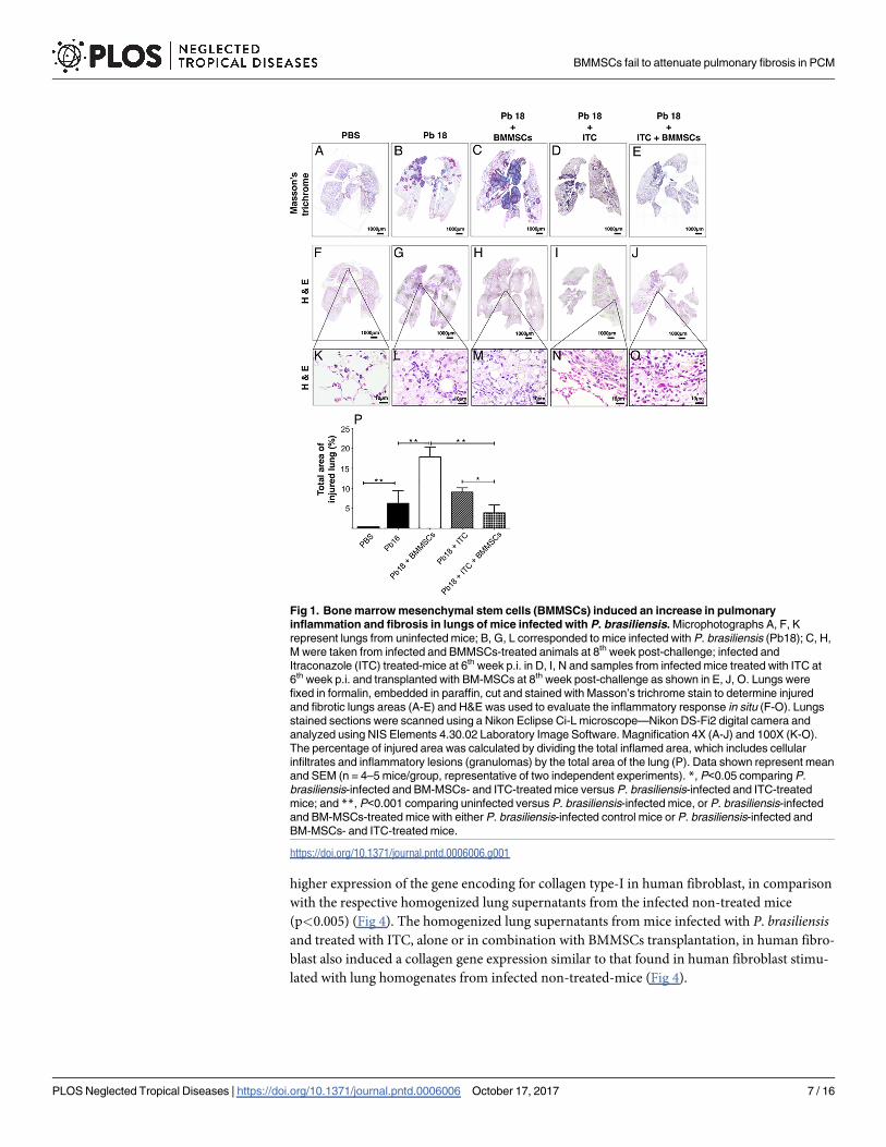
We determined the granulomatous inflammatory areas through histopathological analysis in
lung of experimental mice. We observed that lungs of P. brasiliensis infected-mice developed a
granulomatous inflammatory response with collagen fibers surrounding granulomas (Fig 1A).
Interestingly, administration of BMMSCs in infected mice showed an exacerbation of the
inflammatory process, with a higher granulomatous inflammation and fibrosis areas with loss
of parenchyma (Fig 1B). In contrast, the infected animals treated with ITC alone showed
inflammatory and fibrotic responses similar to those infected and non-transplanted mice (Fig
1C). Moreover, the combined administration of BMMSCs/ITC in P. brasiliensis-infected mice
considerably decreased the inflammatory response and fibrosis (Fig 1D), in comparison with
those infected animals that only received cell-based therapy. A morphometric analysis revealed
that occupied area by granulomatous inflammation in infected and transplanted mice was
twice higher when compared with infected non-treated mice (p<0.001), or three time that
infected and BMMSCs/ITC-treated animals (p<0.001) (Fig 1E). There was statistically signifi-
cant difference in the average of occupied area by granulomas between infected and ITC-
treated mice and those that received combined therapy (p<0.005).
BMMSCs administration increases the number of fibrocytes in lungs
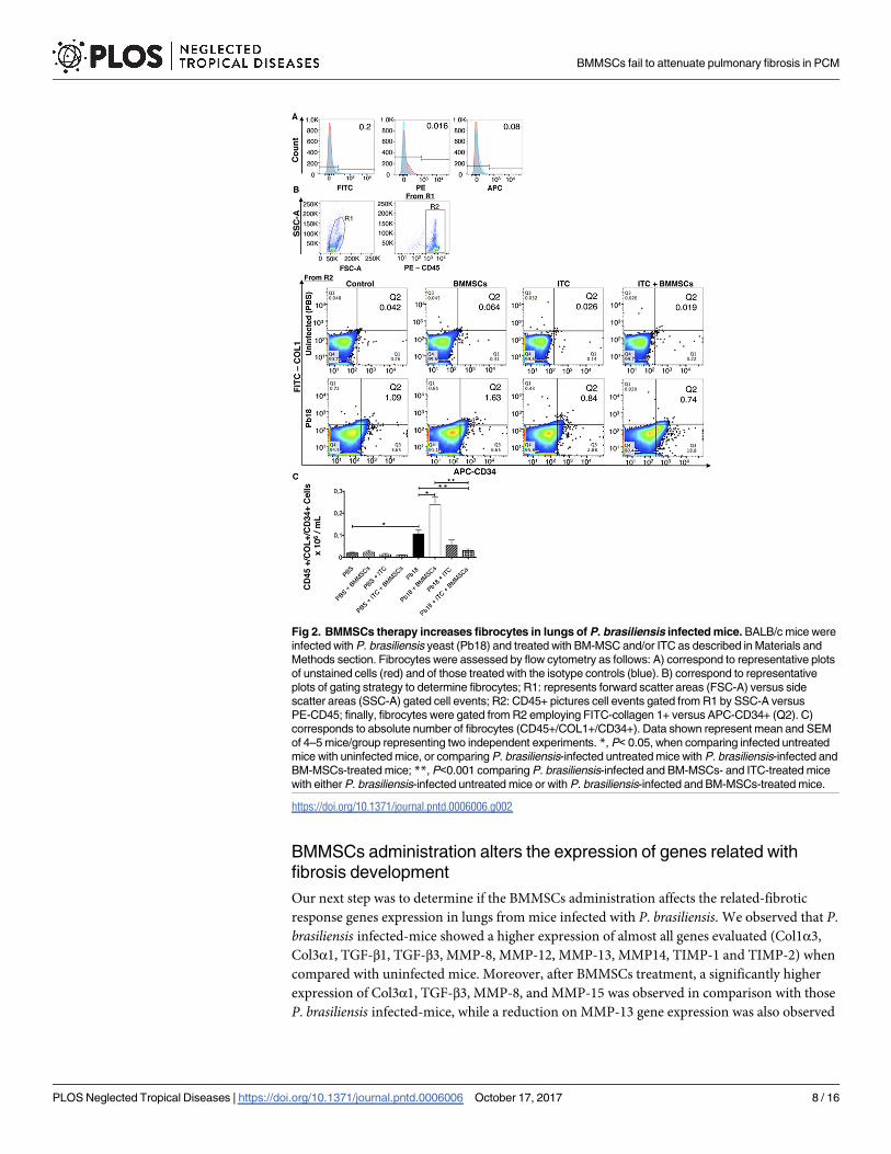
Fibrocytes are bone marrow-derived fibroblast progenitor cells that have been implicated in
tissue remodeling or repairing process, including the development of fibrosis. Following flow
cytometry analysis we found a significantly increased number of fibrocytes (CD45+/CD34+/
Collagen1+) in lungs from infected mice (p<0.005) (Fig 2) relative to PBS instilled controls.
Moreover, infected and BMMSCs-treated animals showed almost twice the number of fibro-
cytes when compared with infected non-treated mice (p<0.005) (Fig 2). Interestingly, ITC
treatment, in combination with BMMSCs, reduced the fibrocytes counts, versus P. brasiliensisinfected mice (p<0.001) or infected BMMSCs-treated animals (p<0.001) (Fig 2).
BMMSC therapy increases lung soluble collagen content
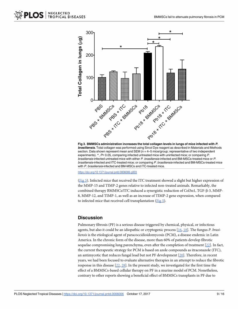
Collagen is considered the most important extracellular matrix protein involved in fibrosis.
Accordingly, we determined the effect of BMMSCs therapy on lung soluble collagen content.
Significantly increased levels of collagen in lungs from mice infected with P. brasiliensis were
observed when compared with PBS instilled animals (p<0.005) (Fig 3). Infected and
BMMSCs-treated mice exhibited an increased significantly in collagen content relative to
infected non-treated animals (p<0.001) (Fig 3). Remarkably, ITC treatment reduced the
amount of soluble collagen in the lungs from both, P. brasiliensis infected-mice (p<0.005), or
those with BMMSCs transplantation (p<0.005) (Fig 3).
Human fibroblasts stimulated with homogenized lung supernatants from
experimental animals show increased collagen gene expression
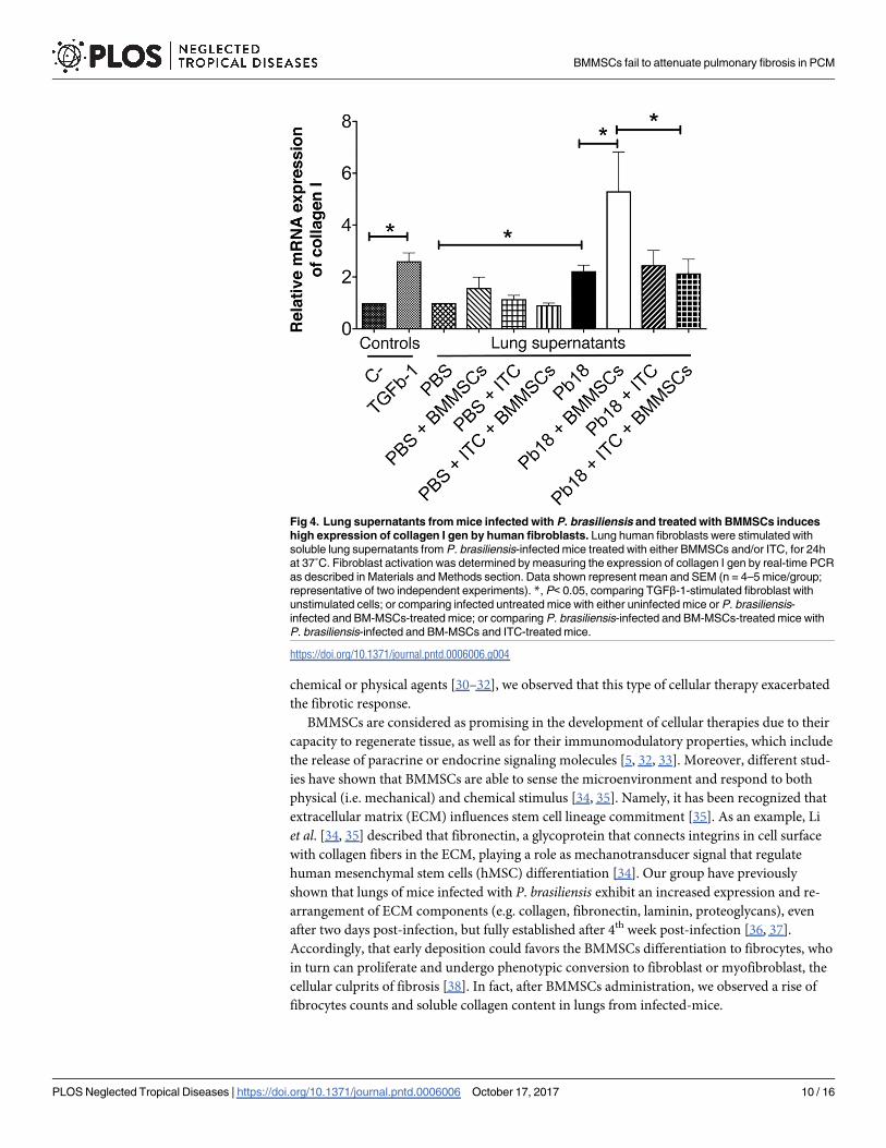
To assessment the capability of lung homogenized to activate human lung fibroblasts, we per-
formed in vitro assays stimulating fibroblasts with lung supernatants from experimental ani-
mals. We observed that lung supernatants from infected and BMMSCs-treated mice induced a
BMMSCs fail to attenuate pulmonary fibrosis in PCM
PLOS Neglected Tropical Diseases | https://doi.org/10.1371/journal.pntd.0006006 October 17, 2017 6 / 16
higher expression of the gene encoding for collagen type-I in human fibroblast, in comparison
with the respective homogenized lung supernatants from the infected non-treated mice
(p<0.005) (Fig 4). The homogenized lung supernatants from mice infected with P. brasiliensisand treated with ITC, alone or in combination with BMMSCs transplantation, in human fibro-
blast also induced a collagen gene expression similar to that found in human fibroblast stimu-
lated with lung homogenates from infected non-treated-mice (Fig 4).
Fig 1. Bone marrow mesenchymal stem cells (BMMSCs) induced an increase in pulmonary
inflammation and fibrosis in lungs of mice infected with P. brasiliensis. Microphotographs A, F, K
represent lungs from uninfected mice; B, G, L corresponded to mice infected with P. brasiliensis (Pb18); C, H,
M were taken from infected and BMMSCs-treated animals at 8th week post-challenge; infected and
Itraconazole (ITC) treated-mice at 6th week p.i. in D, I, N and samples from infected mice treated with ITC at
6th week p.i. and transplanted with BM-MSCs at 8th week post-challenge as shown in E, J, O. Lungs were
fixed in formalin, embedded in paraffin, cut and stained with Masson’s trichrome stain to determine injured
and fibrotic lungs areas (A-E) and H&E was used to evaluate the inflammatory response in situ (F-O). Lungs
stained sections were scanned using a Nikon Eclipse Ci-L microscope—Nikon DS-Fi2 digital camera and
analyzed using NIS Elements 4.30.02 Laboratory Image Software. Magnification 4X (A-J) and 100X (K-O).
The percentage of injured area was calculated by dividing the total inflamed area, which includes cellular
infiltrates and inflammatory lesions (granulomas) by the total area of the lung (P). Data shown represent mean
and SEM (n = 4–5 mice/group, representative of two independent experiments). *, P<0.05 comparing P.
brasiliensis-infected and BM-MSCs- and ITC-treated mice versus P. brasiliensis-infected and ITC-treated
mice; and **, P<0.001 comparing uninfected versus P. brasiliensis-infected mice, or P. brasiliensis-infected
and BM-MSCs-treated mice with either P. brasiliensis-infected control mice or P. brasiliensis-infected and
BM-MSCs- and ITC-treated mice.
https://doi.org/10.1371/journal.pntd.0006006.g001
BMMSCs fail to attenuate pulmonary fibrosis in PCM
PLOS Neglected Tropical Diseases | https://doi.org/10.1371/journal.pntd.0006006 October 17, 2017 7 / 16
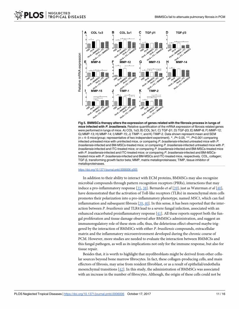
BMMSCs administration alters the expression of genes related with
fibrosis development
Our next step was to determine if the BMMSCs administration affects the related-fibrotic
response genes expression in lungs from mice infected with P. brasiliensis. We observed that P.
brasiliensis infected-mice showed a higher expression of almost all genes evaluated (Col1α3,
Col3α1, TGF-β1, TGF-β3, MMP-8, MMP-12, MMP-13, MMP14, TIMP-1 and TIMP-2) when
compared with uninfected mice. Moreover, after BMMSCs treatment, a significantly higher
expression of Col3α1, TGF-β3, MMP-8, and MMP-15 was observed in comparison with those
P. brasiliensis infected-mice, while a reduction on MMP-13 gene expression was also observed
Fig 2. BMMSCs therapy increases fibrocytes in lungs of P. brasiliensis infected mice. BALB/c mice were
infected with P. brasiliensis yeast (Pb18) and treated with BM-MSC and/or ITC as described in Materials and
Methods section. Fibrocytes were assessed by flow cytometry as follows: A) correspond to representative plots
of unstained cells (red) and of those treated with the isotype controls (blue). B) correspond to representative
plots of gating strategy to determine fibrocytes; R1: represents forward scatter areas (FSC-A) versus side
scatter areas (SSC-A) gated cell events; R2: CD45+ pictures cell events gated from R1 by SSC-A versus
PE-CD45; finally, fibrocytes were gated from R2 employing FITC-collagen 1+ versus APC-CD34+ (Q2). C)
corresponds to absolute number of fibrocytes (CD45+/COL1+/CD34+). Data shown represent mean and SEM
of 4–5 mice/group representing two independent experiments. *, P< 0.05, when comparing infected untreated
mice with uninfected mice, or comparing P. brasiliensis-infected untreated mice with P. brasiliensis-infected and
BM-MSCs-treated mice; **, P<0.001 comparing P. brasiliensis-infected and BM-MSCs- and ITC-treated mice
with either P. brasiliensis-infected untreated mice or with P. brasiliensis-infected and BM-MSCs-treated mice.
https://doi.org/10.1371/journal.pntd.0006006.g002
BMMSCs fail to attenuate pulmonary fibrosis in PCM
PLOS Neglected Tropical Diseases | https://doi.org/10.1371/journal.pntd.0006006 October 17, 2017 8 / 16
(Fig 5). Infected mice that received the ITC treatment showed a slight but higher expression of
the MMP-15 and TIMP-2 genes relative to infected non-treated animals. Remarkably, the
combined therapy BMMSCs/ITC induced a synergistic reduction of Col3α1, TGF-β-3, MMP-
8, MMP-12, and TIMP-1, as well as an increase of TIMP-2 gene expression, when compared
to infected mice that received cell transplantation (Fig 5).
Discussion
Pulmonary fibrosis (PF) is a serious disease triggered by chemical, physical, or infectious
agents, but also it could be an idiopathic or cryptogenic process [16, 18]. The fungus P. brasi-liensis is the etiological agent of paracoccidioidomycosis (PCM), a disease endemic in Latin
America. In the chronic form of the disease, more than 60% of patients develop fibrotic
sequelae compromising lung parenchyma, even after the completion of treatment [22]. In fact,
the current therapeutic strategy for PCM is based on azole compounds as itraconazole (ITC),
an antimycotic that reduces fungal load but not PF development [20]. Therefore, in recent
years, we had been focused to evaluate alternative therapies in an attempt to reduce the fibrotic
response in this disease [22, 28]. In the present study, we investigated for the first time the
effect of a BMMSCs-based cellular therapy on PF in a murine model of PCM. Nonetheless,
contrary to other reports showing a beneficial effect of BMMSCs transplants in PF due to
Fig 3. BMMSCs administration increases the total collagen levels in lungs of mice infected with P.
brasiliensis. Total collagen was performed using Sircol Dye reagent as described in Materials and Methods
section. Data shown represent mean and SEM (n = 4–5 mice/group; representative of two independent
experiments). *, P< 0.05, comparing infected untreated mice with uninfected mice; or comparing P.
brasiliensis-infected untreated mice with either P. brasiliensis-infected and BM-MSCs-treated mice or P.
brasiliensis-infected and ITC-treated mice; or comparing P. brasiliensis-infected and BM-MSCs-treated mice
with P. brasiliensis-infected and BM-MSCs and ITC-treated mice.
https://doi.org/10.1371/journal.pntd.0006006.g003
BMMSCs fail to attenuate pulmonary fibrosis in PCM
PLOS Neglected Tropical Diseases | https://doi.org/10.1371/journal.pntd.0006006 October 17, 2017 9 / 16
chemical or physical agents [30–32], we observed that this type of cellular therapy exacerbated
the fibrotic response.
BMMSCs are considered as promising in the development of cellular therapies due to their
capacity to regenerate tissue, as well as for their immunomodulatory properties, which include
the release of paracrine or endocrine signaling molecules [5, 32, 33]. Moreover, different stud-
ies have shown that BMMSCs are able to sense the microenvironment and respond to both
physical (i.e. mechanical) and chemical stimulus [34, 35]. Namely, it has been recognized that
extracellular matrix (ECM) influences stem cell lineage commitment [35]. As an example, Li
et al. [34, 35] described that fibronectin, a glycoprotein that connects integrins in cell surface
with collagen fibers in the ECM, playing a role as mechanotransducer signal that regulate
human mesenchymal stem cells (hMSC) differentiation [34]. Our group have previously
shown that lungs of mice infected with P. brasiliensis exhibit an increased expression and re-
arrangement of ECM components (e.g. collagen, fibronectin, laminin, proteoglycans), even
after two days post-infection, but fully established after 4th week post-infection [36, 37].
Accordingly, that early deposition could favors the BMMSCs differentiation to fibrocytes, who
in turn can proliferate and undergo phenotypic conversion to fibroblast or myofibroblast, the
cellular culprits of fibrosis [38]. In fact, after BMMSCs administration, we observed a rise of
fibrocytes counts and soluble collagen content in lungs from infected-mice.
Fig 4. Lung supernatants from mice infected with P. brasiliensis and treated with BMMSCs induces
high expression of collagen I gen by human fibroblasts. Lung human fibroblasts were stimulated with
soluble lung supernatants from P. brasiliensis-infected mice treated with either BMMSCs and/or ITC, for 24h
at 37˚C. Fibroblast activation was determined by measuring the expression of collagen I gen by real-time PCR
as described in Materials and Methods section. Data shown represent mean and SEM (n = 4–5 mice/group;
representative of two independent experiments). *, P< 0.05, comparing TGFβ-1-stimulated fibroblast with
unstimulated cells; or comparing infected untreated mice with either uninfected mice or P. brasiliensis-
infected and BM-MSCs-treated mice; or comparing P. brasiliensis-infected and BM-MSCs-treated mice with
P. brasiliensis-infected and BM-MSCs and ITC-treated mice.
https://doi.org/10.1371/journal.pntd.0006006.g004
BMMSCs fail to attenuate pulmonary fibrosis in PCM
PLOS Neglected Tropical Diseases | https://doi.org/10.1371/journal.pntd.0006006 October 17, 2017 10 / 16
In addition to their ability to interact with ECM proteins, BMMSCs may also recognize
microbial compounds through pattern recognition receptors (PRRs), interactions that may
induce a pro-inflammatory response [35, 38]. Bernardo et al [39], just as Waterman et al [40],
have demonstrated that the activation of Toll-like receptors (TLRs) in mesenchymal stem cells
promotes their polarization into a pro-inflammatory phenotype, named MSC1, which can fuel
inflammation and subsequent fibrosis [39, 40]. In this sense, it has been reported that the inter-
action between P. brasiliensis and TLR4 lead to a severe fungal infection, associated with an
enhanced exacerbated proinflammatory response [41]. All these reports support both the fun-
gal proliferation and tissue damage observed after BMMSCs administration, and suggest an
immunoregulatory role of these stem cells; thus, the deleterious effect observed maybe trig-
gered by the interaction of BMMSCs with either P. brasiliensis compounds, extracellular
matrix and the inflammatory microenvironment developed during the chronic course of
PCM. However, more studies are needed to evaluate the interaction between BMMCSs and
this fungal pathogen, as well as its implications not only for the immune response, but also for
tissue repair.
Besides that, it is worth to highlight that myofibroblasts might be derived from other cellu-
lar sources beyond bone marrow fibrocytes. In fact, these collagen-producing cells, and main
effectors of fibrosis, may arise from resident fibroblast, or as a result of epithelial/endothelia
mesenchymal transitions [42]. In this study, the administration of BMMSCs was associated
with an increase in the number of fibrocytes. Although, the origin of these cells could not be
Fig 5. BMMSCs therapy alters the expression of genes related with the fibrosis process in lungs of
mice infected with P. brasiliensis. Relative quantification of the mRNA expression of fibrosis related genes
were performed in lungs of mice. A) COL 1α3; B) COL 3α1; C) TGF-β1; D) TGF-β3; E) MMP-8; F) MMP-12;
G) MMP-13; H) MMP-14; I) MMP-15; J) TIMP-1; and K) TIMP-2. Data shown represent mean and SEM
(n = 4–5 mice/group; representative of two independent experiment). *, P< 0.05, **, P<0.001 comparing
infected untreated mice with uninfected mice, or comparing P. brasiliensis-infected untreated mice with P.
brasiliensis-infected and BM-MSCs-treated mice; or comparing P. brasiliensis-infected untreated mice with P.
brasiliensis-infected and ITC-treated mice; or comparing P. brasiliensis-infected and BM-MSCs-treated mice
with P. brasiliensis-infected and ITC-treated mice; or comparing P. brasiliensis-infected and BM-MSCs-
treated mice with P. brasiliensis-infected and BM-MSCs and ITC-treated mice, respectively. COL, collagen;
TGF-β, transforming growth factor beta; MMP, matrix metalloproteinases; TIMP, tissue inhibitor of
metalloproteinases.
https://doi.org/10.1371/journal.pntd.0006006.g005
BMMSCs fail to attenuate pulmonary fibrosis in PCM
PLOS Neglected Tropical Diseases | https://doi.org/10.1371/journal.pntd.0006006 October 17, 2017 11 / 16
confirmed, we may suppose that they could come from bone marrow or pericytes, which sub-
sequently differentiate into fibroblasts and then into myofibroblasts collagen-producer cells,
thus contributing to the increase of PF in those P. brasiliensis infected- and BMMSCs treated-
mice. Accordingly, we evaluated the collagen I gene expression in human fibroblast stimulated
with homogenized lung supernatants from P. brasiliensis infected mice. We found that super-
natants from infected and BMMSCs-transplanted mice induced a higher production of type I
collagen transcript in human fibroblast, in comparison with those cells stimulated with
infected non-treated animals. These results clearly indicate the presence of molecules from the
lung microenvironment able to stimulate the collagen production in human fibroblast.
Among the major stimuli to activate fibroblasts, IL6, TGFβ, IL13 and FGF are found, and once
activated, these cells differentiate into myofibroblasts or could produce pro-fibrotic molecules
such as IL1, VEGF, Insulin-like growth factor 2 (IGFII), Insulin-like growth factor-binding
protein (IGFBP), IL6 and IL33 [42]. In fact, we observed an increased expression of Col 3α1
and TGFβ-3 genes in lungs of P. brasiliensis–infected mice treated with BMMSCs. In concur-
rence with these reports, more recently we have observed a significant increase of IL-1α, IL-1β,
IL-6, TNF-α and IL17 levels in homogenized lung supernatants from mice infected with P.
brasiliensis [28]. All these cytokines have also been implicated in the pathogenesis of PF. None-
theless, a direct activation of fibroblast by P. brasiliensis compounds could not be ruled out.
Matrix metalloproteinases (MMPs) are a family of zinc- and calcium-dependent endopepti-
dases (around 25 members are know so far) that are either secreted or membrane-bound
enzymes [43]. MMPs have long been considered to be essentials for ECM remodeling, which
is critical in embryonic development and tissue homeostasis, including inflammatory response
and tissue repair [43]. In this context, as stated by Pardo et al, MMPs not only degrade ECM
components, but also release, cleave and active a wide range of growth factors, cytokines, che-
mokines and cell surface receptors affecting numerous cell functions (e.g. adhesion, prolifera-
tion, differentiation, migration, cell death) [44]. Thus, MMPs and their tissue inhibitors
(TIMPs) play a central role in the extracellular pathways of ECM degradation and, therefore,
in fibrosis development or resolution [44]. Namely, MMP8, a collagenase, can also cleave the
chemokines CXCL8 and LIX, resulting in enhanced chemoattractant activities, which could be
associated with fibrosis development [43, 44]. These results show that after BMMSC adminis-
tration there was not only an increase in the expression MMP8 gene but also elevated neutro-
phils counts, findings noticed before in association with development of fibrosis as observed in
our PCM model previously reported [28]. BMMSCs transplantation also induced a decrease in
gene coding for MMP-13, another collagenase, but not changes in the expression of TIMP-1
and TIMP2 genes were observed. Meanwhile, the ITC/BMMSC transplantation therapy
decreased synergistically the expression of TIMP-1 and MMP-13. Over expression of TIMP-1
is associated whit liver fibrosis [45], while MMP-13 cleaves and inactivates CCL2, CCL7 and
CXCL12 leading to reduction in chemotaxis, as well as to a decrease in the fibrosis process [43,
45]. However, our data interpretation relative to MMPs or TIMPs gene expression is limited,
as the current knowledge concerning pathological tissue repair in PMC is scarce. In addition,
although most of the fibrosis-related genes analyzed showed small fold-changes with statistical
significant, a possible meaningful biological effect cannot be ruled-out.
ITC is the antifungal treatment of choice in PCM. Of note, additionally to its antifungal
effect, it has been recently documented that this antifungal medication also exhibits immuno-
modulatory properties [46]. Moreover, in a previous work, we found that ITC reduces the
expression of certain genes encoding for pro-inflammatory cytokines (IFN-γ, IL-6, IL-17,
TGF-β1, TNF-α), transcriptional factors (T-bet, GATA-3, Spi-1, RoRc, Ahr, FoxP3), and fibro-
sis development (MMP-1, MMP-8, MMP-13, and Col3α1). Additionally, it also diminished
the number of inflammatory cells—including neutrophils—in the lungs of mice infected with
BMMSCs fail to attenuate pulmonary fibrosis in PCM
PLOS Neglected Tropical Diseases | https://doi.org/10.1371/journal.pntd.0006006 October 17, 2017 12 / 16
P. brasiliensis [27]. In the present study, it was observed that the ITC regulates the expression
of the MMP-15 and TIMP-2 genes that have been recognized as inducers of pulmonary fibro-
sis [27].
Conclusions
Overall, our results demonstrated an exacerbating effect of the BMMSCs therapy on pulmo-
nary fibrosis induced by P. brasiliensis infection. We hypothesized that this outcome could be
triggered by either the interaction with P. brasiliensis compounds or by the inflammatory
microenvironment induced by this process. Nonetheless, the combined therapy ITC/BMMSCs
showed promising results since synergistically it reduced TIMP-1 and MMP-13. Thus, the use
of BMMSC under different conditions or combined with other treatments (e.g. ITC) opens the
possibility to new therapeutic approaches for this type of fibrosis resulting from an infectious
disease.
PCM is considered a neglected tropical disease mostly affecting low income individuals
who live in underdeveloped Latin American rural regions where the technology and the
resources needed to administer the immunotherapeutic measures here suggested would prob-
ably not be available. Nonetheless, the implementation of cellular therapies is progressing and
the prospects are to arrive in a few years to the administration of autologous bone marrow or
stem cells obtained from adipose tissues even in these regions.
Acknowledgments
We thank Dr. David Arboleda for his support in providing in BMMSC isolation and character-
ization methodologies.
Author Contributions
Conceptualization: Lina Marıa Salazar-Pelaez, Angel Gonzalez.
Data curation: Julian Camilo Arango, Juan David Puerta-Arias, Paula Andrea Pino-Tamayo.
Formal analysis: Julian Camilo Arango, Juan David Puerta-Arias, Paula Andrea Pino-
Tamayo, Lina Marıa Salazar-Pelaez, Mauricio Rojas, Angel Gonzalez.
Funding acquisition: Angel Gonzalez.
Investigation: Julian Camilo Arango, Juan David Puerta-Arias, Paula Andrea Pino-Tamayo,
Lina Marıa Salazar-Pelaez, Mauricio Rojas, Angel Gonzalez.
Methodology: Julian Camilo Arango, Juan David Puerta-Arias, Paula Andrea Pino-Tamayo,
Mauricio Rojas.
Project administration: Angel Gonzalez.
Resources: Mauricio Rojas, Angel Gonzalez.
Supervision: Angel Gonzalez.
Visualization: Julian Camilo Arango, Juan David Puerta-Arias, Paula Andrea Pino-Tamayo,
Mauricio Rojas, Angel Gonzalez.
Writing – original draft: Julian Camilo Arango, Lina Marıa Salazar-Pelaez, Mauricio Rojas,
Angel Gonzalez.
BMMSCs fail to attenuate pulmonary fibrosis in PCM
PLOS Neglected Tropical Diseases | https://doi.org/10.1371/journal.pntd.0006006 October 17, 2017 13 / 16
References1. Ullah I, Subbarao RB, Rho GJ. Human mesenchymal stem cells—current trends and future prospec-
tive. Biosci Rep. 2015; 35(2).
2. Bianco P. "Mesenchymal" stem cells. Annu Rev Cell Dev Biol. 2014; 30:677–704. https://doi.org/10.
1146/annurev-cellbio-100913-013132 PMID: 25150008
3. Burrello J, Monticone S, Gai C, Gomez Y, Kholia S, Camussi G. Stem Cell-Derived Extracellular Vesi-
cles and Immune-Modulation. Front Cell Dev Biol. 2016; 4:83. https://doi.org/10.3389/fcell.2016.00083
PMID: 27597941
4. Farini A, Sitzia C, Erratico S, Meregalli M, Torrente Y. Clinical applications of mesenchymal stem cells
in chronic diseases. Stem Cells Int. 2014; 2014:306573. https://doi.org/10.1155/2014/306573 PMID:
24876848
5. Gao F, Chiu SM, Motan DA, Zhang Z, Chen L, Ji HL, et al. Mesenchymal stem cells and immunomodu-
lation: current status and future prospects. Cell Death Dis. 2016; 7:e2062. https://doi.org/10.1038/
cddis.2015.327 PMID: 26794657
6. Hoch AI, Leach JK. Concise review: optimizing expansion of bone marrow mesenchymal stem/stromal
cells for clinical applications. Stem Cells Transl Med. 2015; 4(4):412. https://doi.org/10.5966/sctm.
2013-0196erratum PMID: 25795657
7. Krasnodembskaya A, Song Y, Fang X, Gupta N, Serikov V, Lee JW, et al. Antibacterial effect of human
mesenchymal stem cells is mediated in part from secretion of the antimicrobial peptide LL-37. Stem
Cells. 2010; 28(12):2229–38. https://doi.org/10.1002/stem.544 PMID: 20945332
8. Lathrop MJ, Brooks EM, Bonenfant NR, Sokocevic D, Borg ZD, Goodwin M, et al. Mesenchymal stro-
mal cells mediate Aspergillus hyphal extract-induced allergic airway inflammation by inhibition of the
Th17 signaling pathway. Stem Cells Transl Med. 2014; 3(2):194–205. https://doi.org/10.5966/sctm.
2013-0061 PMID: 24436442
9. Nemeth K, Mayer B, Mezey E. Modulation of bone marrow stromal cell functions in infectious diseases
by toll-like receptor ligands. J Mol Med (Berl). 2010; 88(1):5–10.
10. Tang J, Wu T, Xiong J, Su Y, Zhang C, Wang S, et al. Porphyromonas gingivalis lipopolysaccharides
regulate functions of bone marrow mesenchymal stem cells. Cell Prolif. 2015; 48(2):239–48. https://doi.
org/10.1111/cpr.12173 PMID: 25676907
11. Alagesan S, Griffin MD. Autologous and allogeneic mesenchymal stem cells in organ transplantation:
what do we know about their safety and efficacy? Curr Opin Organ Transplant. 2014; 19(1):65–72.
https://doi.org/10.1097/MOT.0000000000000043 PMID: 24370985
12. Rojas M, Xu J, Woods CR, Mora AL, Spears W, Roman J, et al. Bone marrow-derived mesenchymal
stem cells in repair of the injured lung. Am J Respir Cell Mol Biol. 2005; 33(2):145–52. https://doi.org/10.
1165/rcmb.2004-0330OC PMID: 15891110
13. Srour N, Thebaud B. Mesenchymal Stromal Cells in Animal Bleomycin Pulmonary Fibrosis Models: A
Systematic Review. Stem Cells Transl Med. 2015; 4(12):1500–10. https://doi.org/10.5966/sctm.2015-
0121 PMID: 26494779
14. Sun Z, Gong X, Zhu H, Wang C, Xu X, Cui D, et al. Inhibition of Wnt/beta-catenin signaling promotes
engraftment of mesenchymal stem cells to repair lung injury. J Cell Physiol. 2014; 229(2):213–24.
https://doi.org/10.1002/jcp.24436 PMID: 23881674
15. Sordi V, Malosio ML, Marchesi F, Mercalli A, Melzi R, Giordano T, et al. Bone marrow mesenchymal
stem cells express a restricted set of functionally active chemokine receptors capable of promoting
migration to pancreatic islets. Blood. 2005; 106(2):419–27. https://doi.org/10.1182/blood-2004-09-3507
PMID: 15784733
16. Todd NW, Luzina IG, Atamas SP. Molecular and cellular mechanisms of pulmonary fibrosis. Fibrogen-
esis Tissue Repair. 2012; 5(1):11. https://doi.org/10.1186/1755-1536-5-11 PMID: 22824096
17. Wynn TA, Ramalingam TR. Mechanisms of fibrosis: therapeutic translation for fibrotic disease. Nat
Med. 2012; 18(7):1028–40. https://doi.org/10.1038/nm.2807 PMID: 22772564
18. Wick G, Grundtman C, Mayerl C, Wimpissinger TF, Feichtinger J, Zelger B, et al. The immunology of
fibrosis. Annu Rev Immunol. 2013; 31:107–35. https://doi.org/10.1146/annurev-immunol-032712-
095937 PMID: 23516981
19. Abreu SC, Antunes MA, Pelosi P, Morales MM, Rocco PR. Mechanisms of cellular therapy in respira-
tory diseases. Intensive Care Med. 2011; 37(9):1421–31. https://doi.org/10.1007/s00134-011-2268-3
PMID: 21656291
20. de Oliveira HC, Assato PA, Marcos CM, Scorzoni L, de Paula ESAC, Da Silva Jde F, et al. Paracocci-
dioides-host Interaction: An Overview on Recent Advances in the Paracoccidioidomycosis. Front Micro-
biol. 2015; 6:1319. https://doi.org/10.3389/fmicb.2015.01319 PMID: 26635779
BMMSCs fail to attenuate pulmonary fibrosis in PCM
PLOS Neglected Tropical Diseases | https://doi.org/10.1371/journal.pntd.0006006 October 17, 2017 14 / 16
21. Vallabhaneni S, Mody RK, Walker T, Chiller T. The Global Burden of Fungal Diseases. Infect Dis Clin
North Am. 2016; 30(1):1–11. https://doi.org/10.1016/j.idc.2015.10.004 PMID: 26739604
22. Cano LE, Gonzalez A, Lopera D, Naranjo T, Restrepo A. Pulmonary Paracoccidioidomycosis: Clinical,
Immunological and Histopathological Aspects, Lung Diseases In: Irusen EM, editor. Lung Diseases—
Selected State of the Art Reviews: InTech; 2012. p. 359–92.
23. Colombo AL, Tobon A, Restrepo A, Queiroz-Telles F, Nucci M. Epidemiology of endemic systemic fun-
gal infections in Latin America. Med Mycol. 2011; 49(8):785–98. https://doi.org/10.3109/13693786.
2011.577821 PMID: 21539506
24. Shikanai-Yasuda MA, Telles Filho Fde Q, Mendes RP, Colombo AL, Moretti ML. [Guidelines in para-
coccidioidomycosis]. Rev Soc Bras Med Trop. 2006; 39(3):297–310. PMID: 16906260
25. Tobon AM, Agudelo CA, Osorio ML, Alvarez DL, Arango M, Cano LE, et al. Residual pulmonary abnor-
malities in adult patients with chronic paracoccidioidomycosis: prolonged follow-up after itraconazole
therapy. Clin Infect Dis. 2003; 37(7):898–904. https://doi.org/10.1086/377538 PMID: 13130400
26. Naranjo TW, Lopera DE, Diaz-Granados LR, Duque JJ, Restrepo AM, Cano LE. Combined itracona-
zole-pentoxifylline treatment promptly reduces lung fibrosis induced by chronic pulmonary paracocci-
dioidomycosis in mice. Pulm Pharmacol Ther. 2011; 24(1):81–91. https://doi.org/10.1016/j.pupt.2010.
09.005 PMID: 20851204
27. Puerta-Arias JD, Pino-Tamayo PA, Arango JC, Salazar-Pelaez LM, Gonzalez A. Itraconazole in combi-
nation with neutrophil depletion reduces the expression of genes related to pulmonary fibrosis in an
experimental model of paracoccidioidomycosis. Med Mycol. 2017; In press.
28. Puerta-Arias JD, Pino-Tamayo PA, Arango JC, Gonzalez A. Depletion of Neutrophils Promotes the
Resolution of Pulmonary Inflammation and Fibrosis in Mice Infected with Paracoccidioides brasiliensis.
PLoS One. 2016; 11(9):e0163985. https://doi.org/10.1371/journal.pone.0163985 PMID: 27690127
29. Fica A. [Infections in patients affected by rheumatologic diseases associated to glucocorticoid use or
tumor necrosis factor-alpha inhibitors]. Rev Chilena Infectol. 2014; 31(2):181–95. https://doi.org/10.
4067/S0716-10182014000200009 PMID: 24878907
30. Antunes MA, Laffey JG, Pelosi P, Rocco PR. Mesenchymal stem cell trials for pulmonary diseases. J
Cell Biochem. 2014; 115(6):1023–32. https://doi.org/10.1002/jcb.24783 PMID: 24515922
31. Lee EJ. Mesenchymal Stem Cell Therapy in Pulmonary Disease. Korean J Med. 2015; 89(5):522–6.
32. Wecht S, Rojas M. Mesenchymal stem cells in the treatment of chronic lung disease. Respirology.
2016; 21(8):1366–75. https://doi.org/10.1111/resp.12911 PMID: 27688156
33. Zhao F, Zhang YF, Liu YG, Zhou JJ, Li ZK, Wu CG, et al. Therapeutic effects of bone marrow-derived
mesenchymal stem cells engraftment on bleomycin-induced lung injury in rats. Transplant Proc. 2008;
40(5):1700–5. https://doi.org/10.1016/j.transproceed.2008.01.080 PMID: 18589176
34. Li B, Moshfegh C, Lin Z, Albuschies J, Vogel V. Mesenchymal stem cells exploit extracellular matrix as
mechanotransducer. Sci Rep. 2013; 3:2425. https://doi.org/10.1038/srep02425 PMID: 23939587
35. Prockop DJ. Inflammation, fibrosis, and modulation of the process by mesenchymal stem/stromal cells.
Matrix Biol. 2016; 51:7–13. https://doi.org/10.1016/j.matbio.2016.01.010 PMID: 26807758
36. Gonzalez A, Lenzi HL, Motta EM, Caputo L, Restrepo A, Cano LE. Expression and arrangement of
extracellular matrix proteins in the lungs of mice infected with Paracoccidioides brasiliensis conidia. Int J
Exp Pathol. 2008; 89(2):106–16. https://doi.org/10.1111/j.1365-2613.2008.00573.x PMID: 18336528
37. Gonzalez A, Restrepo A, Cano LE. Pulmonary immune responses induced in BALB/c mice by Paracoc-
cidioides brasiliensis conidia. Mycopathologia. 2008; 165(4–5):313–30. https://doi.org/10.1007/s11046-
007-9072-1 PMID: 18777636
38. Somaiah C, Kumar A, Mawrie D, Sharma A, Patil SD, Bhattacharyya J, et al. Collagen Promotes Higher
Adhesion, Survival and Proliferation of Mesenchymal Stem Cells. PLoS One. 2015; 10(12):e0145068.
https://doi.org/10.1371/journal.pone.0145068 PMID: 26661657
39. Bernardo ME, Fibbe WE. Mesenchymal stromal cells: sensors and switchers of inflammation. Cell
Stem Cell. 2013; 13(4):392–402. https://doi.org/10.1016/j.stem.2013.09.006 PMID: 24094322
40. Waterman RS, Tomchuck SL, Henkle SL, Betancourt AM. A new mesenchymal stem cell (MSC) para-
digm: polarization into a pro-inflammatory MSC1 or an Immunosuppressive MSC2 phenotype. PLoS
One. 2010; 5(4):e10088. https://doi.org/10.1371/journal.pone.0010088 PMID: 20436665
41. Loures FV, Pina A, Felonato M, Araujo EF, Leite KR, Calich VL. Toll-like receptor 4 signaling leads to
severe fungal infection associated with enhanced proinflammatory immunity and impaired expansion of
regulatory T cells. Infect Immun. 2010; 78(3):1078–88. https://doi.org/10.1128/IAI.01198-09 PMID:
20008536
42. Kendall RT, Feghali-Bostwick CA. Fibroblasts in fibrosis: novel roles and mediators. Front Pharmacol.
2014; 5:123. https://doi.org/10.3389/fphar.2014.00123 PMID: 24904424
BMMSCs fail to attenuate pulmonary fibrosis in PCM
PLOS Neglected Tropical Diseases | https://doi.org/10.1371/journal.pntd.0006006 October 17, 2017 15 / 16
43. Matthew G, Parks WC. Diverse Functions of Matrix Metalloproteinases during Fibrosis. Disease Models
& Mechanisms. 2017; 7(2):193–203.
44. Pardo A, Cabrera S, Maldonado M, Selman M. Role of matrix metalloproteinases in the pathogenesis of
idiopathic pulmonary fibrosis. Respir Res. 2016; 17:23. https://doi.org/10.1186/s12931-016-0343-6
PMID: 26944412
45. Guo Y, Chen B, Chen LJ, Zhang CF, Xiang C. Current status and future prospects of mesenchymal
stem cell therapy for liver fibrosis. J Zhejiang Univ Sci B. 2016; 17(11):831–41. https://doi.org/10.1631/
jzus.B1600101 PMID: 27819130
46. Muenster S, Bode C, Diedrich B, Jahnert S, Weisheit C, Steinhagen F, et al. Antifungal antibiotics mod-
ulate the pro-inflammatory cytokine production and phagocytic activity of human monocytes in an in
vitro sepsis model. Life Sci. 2015; 141:128–36. https://doi.org/10.1016/j.lfs.2015.09.004 PMID:
26382596
BMMSCs fail to attenuate pulmonary fibrosis in PCM
PLOS Neglected Tropical Diseases | https://doi.org/10.1371/journal.pntd.0006006 October 17, 2017 16 / 16