Embed Size (px)
Citation preview
181
Mycobiology 40(3) : 181-188 (2012) http://dx.doi.org/10.5941/MYCO.2012.40.3.181© The Korean Society of Mycology pISSN 1229-8093
eISSN 2092-9323
RESEARCH ARTICLE
Immunomodulating and Antitumor Activities of Panellus serotinus Polysaccharides
Jeong Hwa Kim1, Jae Seong Lee1, Kyung Rim Lee1, Mi Ja Shim2, Min Woong Lee3, Pyung Gyun Shin4,Jong Chun Cheong4, Young Bok Yoo4 and Tae Soo Lee1,5
*
1Division of Life Sciences, University of Incheon, Incheon 406-840, Korea2Department of Life Science, University of Seoul, Seoul 130-743, Korea3Department of Life Science, Dongguk University, Seoul 100-715, Korea4Mushroom Division, National Institute of Horticultural and Herbal Science, Rural Development Administration, Eumseong 369-873, Korea5Bio-Resource and Environmental Center, University of Incheon, Incheon 406-840, Korea
(Received August 7, 2012. Revised August 19, 2012. Accepted August 28, 2012)
This study was initiated in order to investigate the anticancer and immunomodulating activities of crude polysaccharidesextracted in methanol, neutral saline, and hot water (hereinafter referred to as Fr. MeOH, Fr. NaCl, and Fr. HW, respec-tively) from the fruiting bodies of Panellus serotinus. Content of β-glucan and protein in Fr. MeOH, Fr. NaCl, and Fr. HWextracts of P. serotinus ranged from 22.92~28.52 g/100 g and 3.24~3.68 g/100 g, respectively. In vitro cytotoxicity tests, noneof the various fractions of crude polysaccharides were cytotoxic against sarcoma 180, HT-29, NIH3T3, and RAW 264.7 celllines at the tested concentration. Intraperitoneal injection with crude polysaccharides resulted in a life prolongation effectof 23.53~44.71% in mice previously inoculated with sarcoma 180. Treatment with Fr. HW resulted in an increase in thenumbers of spleen cells by 1.3 fold at the concentration of 50 µg/mL compared with control. Treatment with Fr. NaClresulted in improvement of the immuno-potentiating activity of B lymphocytes by increasing the alkaline phosphatase activityby 1.4 fold, compared with control, at the concentration of 200 µg/mL. Among the three fractions, maximum nitric oxide(13.48 µM) was recorded at 500 µg/mL in Fr. HW. Production of tumor necrosis factor alpha, interleukin-1β, and interleukin-6 was significantly higher, compared to the positive control, concanavalin A, at the tested concentration. Therefore, treatmentwith crude polysaccharides extracted from the fruiting body of P. serotinus could result in improvement of antitumor activity.
KEYWORDS : Antitumor activities, Crude polysaccharides, Immunomodulating, Mouse sarcoma 180, Panellus serotinus
Introduction
Panellus serotinus, known as late fall oyster mushroom,has been valued by humankind as an edible and therapeuticresource. A number of bioactive molecules, includingantitumor substances, have been identified in manymushroom species. Polysaccharides, with antitumor andimmunomodulating properties, are the best known andmost potent mushroom derived substances [1, 2].
Polysaccharides are a structurally diverse group ofbiological macromolecules composed of repetitive structuralfeatures, which are polymers of monosaccharide residuesjoined to each other by glycosidic linkages [3]. Antitumorpolysaccharides isolated from mushrooms are either watersoluble β-glucans, β-glucans with heterosaccharide chainsof xylose, mannose, galactose, and uronic acid or β-glucanprotein complexes. As a general rule, immunopotentialactivity of the protein linked glucans is greater than thatof the corresponding glucans [4]. The mode of action of
β-glucan differs from that of conventional chemotherapeuticagents in that it is immunotherapeutic. Global awarenessof cancer as the second largest cause of death in people ofvarious ages and racial backgrounds has led to significantresearch efforts and clinical studies in the fight against thedisease [2, 5].
Despite the therapeutic potential and the clinicalimportance of P. serotinus, no studies on its antitumor andimmunomodulating activities have been reported. In thepresent study, crude polysaccharides were extracted fromfruiting bodies of P. serotinus using methanol, neutralsaline, and hot water for investigation of antitumor andimmunomodulating activities. The in vitro cytotoxicactivities of four cell lines and in vivo antitumor effectson sarcoma 180 tumor bearing mice were studied. Inaddition, proliferation of murine spleen cells, alkalinephosphatase (APase) activity, nitric oxide (NO), andcytokine production in murine peritoneal macrophageswere also investigated.
*Corresponding author <E-mail : [email protected]>
This is an Open Access article distributed under the terms of the Creative Commons Attribution Non-Commercial License (http://creativecommons.org/licenses/by-nc/3.0/) which permits unrestricted non-commercial use, distribution, and reproduction in any medium,provided the original work is properly cited.

182 Kim et al.
Materials and Methods
This study was conducted from November 2010 toDecember 2011 at the Animal House and Laboratory ofApplied Microbiology, Division of Life Sciences. Theexperimental protocols were approved by the AnimalCare Ethics Committee at the University of Incheon,Republic of Korea. All experimental procedures wereperformed in accordance with the guide for care and useof experimental animals.
Mushroom and extraction. Fresh fruiting bodies of P.serotinus were collected from Deogyusan National Park,Korea. A pure culture was deposited in the CultureCollection and DNA Bank of Mushroom (CCDBM),Division of Life Sciences, University of Incheon, Korea,and an accession number was acquired, IUM-3346. Freshfruiting bodies were dried with hot air at 40oC for 48 hrand pulverized.
One hundred grams of pulverized fruiting bodies of P.serotinus were extracted with 3,000 mL of 80% methanoland neutral saline (0.9% NaCl) with stirring at 150 rpmfor 24 hr at 25oC to obtain methanol and NaCl extracts.The mixture was filtered through two layers of WhatmanNo. 1 filter paper (Whatman, Maidstone, UK). The samequantity of sample was boiled at 100oC for 3 hr with3,000 mL deionized distilled water to obtain a hot waterextract. The mixture was cooled at room temperature andfiltered through a Whatman No. 1 filter paper. The residuesof methanol, NaCl, and hot water extraction were thentreated two more times in the same manner. All supernatantsobtained of each extract were combined and mixed withfour volumes of ethanol and allowed to stand overnight at4oC. The precipitate was collected by centrifugation,dissolved in distilled water, dialyzed for 48 hr at 4oC, andlyophilized. This fraction was referred to as the methanolextract (Fr. MeOH), neutral saline extract (Fr. NaCl), andhot water extract (Fr. HW). The yields from the methanol,NaCl, and hot water extracts of P. serotinus were 20.54,18.81, and 19.25% (w/w), respectively.
Animals. Five-wk-old inbred male ICR mice (22 ± 3 g)were purchased from Central Lab. Animal Inc., Seoul,Korea. All mice were acclimated to the animal house fora period of one week. Mice were housed in an animalroom at 23 ± 2oC under a 12-hr dark-light cycle (17:00~05:00) and a relative humidity of 50~60%. During theexperimental period, mice received the standard basal diet,purchased from Central Lab Animal Inc. (Seoul, Korea).
Cell lines. Mouse sarcoma 180, colon cancer (HT-29),mouse embryonic fibroblast cells (NIH3T3), and murinemacrophage cell (RAW 264.7) lines were purchased fromKorean Cell Line Bank of Seoul National University,
Seoul, Korea. HT-29, NIH3T3, and RAW 264.7 cell lineswere cultured in Dulbecco’s modified Eagle’s mediumsupplemented with penicillin (100 U/mL), streptomycin(100 mg/mL), and 10% fetal bovine serum at 37oC with5% atmospheric CO2 in a humidified incubator. Sarcoma180 cells were maintained in ascitic form by serialtransplantation every seven days in an ICR male mouse.
Determination of β-glucan and total protein. Amushroom and yeast β-glucan assay kit (MegazymeInternational Ireland Ltd., Wicklow, Ireland) was used forquantitative determination of the β-glucan contents of threedifferent fractions extracted from fruiting bodies of P.serotinus. In brief, for determination of total glucan (α-and β-), 5 mg of each fraction was suspended in 75 µL ofconcentrated HCl and incubated at 45oC for 30 min;500 µL distilled water was then added, and it was placedin a boiling water bath for 2 hr. The pH was neutralizedwith 500 µL of 2 N KOH, followed by centrifugation for10 min at 1,500 ×g. Fifty microliters of the supernatantwas digested with an aliquot of exo-1,3-β-glucanase (20 U/mL) plus β-glucosidase (4 U/mL) in 200 mM sodiumacetate buffer (pH 5.0). The hydrolysates were incubatedwith 1.5 mL of glucose oxidase/peroxidase mixture (GO/POD), followed by incubation at 40oC for 1 hr. Theabsorbance of the solution was measured at 510 nm. Formeasurement of α-glucan, 5 mg of each fraction wassuspended in 100 µL of 2 N KOH for 20 min andneutralized with 400 µL of 1.2 M sodium acetate buffer(pH 3.8). Then, the solution was centrifuged for 10 min at1,500 ×g and aliquots of amyloglucosidase (1,630 U/mL)plus invertase (500 U/mL) were added to the 50 µL ofsupernatant, followed by incubation at 40oC for 30 min.The solution was incubated with 1.5 mL of mixture ofGO/POD at 40oC for 20 min and absorbance was measuredat 510 nm. β-Glucan was determined by subtracting α-glucan from total glucan content.
Protein content of each fraction was quantified byBradford method [6], using bovine serum albumin (BSA)as a standard. Total protein content of the fractions isexpressed as g of BSA equivalent per 100 g of dry weight.
Cytotoxicity. A rapid colorimetric method previouslydescribed by Mosmann [7] was used in the 3-(4,5-dimethyl-1-2-thiazolyl)-2,5-diphenyl-2H-tetrazolium bromide(MTT) assay, for measurement of cell viability andproliferation. Briefly, for the MTT assay, 100 µL of HT-29, NIH3T3, and RAW 264.7 cells (1 × 10⁵ cells/well) weretreated with 10, 100, 1,000, and 2,000µg/mL concentrationsof three different fractions (Fr. MeOH, Fr. NaCl, and Fr.HW) of P. serotinus and cultured for 24 hr in 96-wellmicroplates at 37oC with 5% atmospheric CO2. Then,10 µL of 5 mg/mL of MTT solution was added, followedby incubation at 37oC with 5% atmospheric CO2 for 4 hr
Antitumor Effect of Panellus serotinus Polysaccharides 183
under dark conditions. Following removal of the supernatant,purple formazan crystals produced were dissolved in 100 µLof dimethylsulfoxide, and quantified by measurement ofoptical density (OD) at 570 nm using a microplate reader.For the cytotoxicity assay of sarcoma 180, 50 µL ofsarcoma 180 cells (2 × 10⁵ cells/well) were treated with10, 100, 1,000, and 2,000 µg/mL concentrations of threedifferent extracts of P. serotinus and cultured for 24 hr in96-well microplates at 37oC with 5% atmospheric CO2.Then, 1 mg/mL of 2,3-bis(2-methoxyl-4nitro-5-sulfophenyl)-2H-tetrazolium-5-carboxanilide (XTT) solution was mixedwith 30 µL of 25 µM phenazine methosulfate, followedby incubation at 37oC with 5% atmospheric CO2 for 2 hrunder dark conditions. OD was then measured at 450 nmusing a microplate reader. Viability was defined as theratio (expressed as a percentage) of absorbance of treatedcells to untreated cells that served as control.
In vivo assay of antitumor activity. Antitumor activitiesof three different extracts of P. serotinus were assayedagainst mouse sarcoma 180 cells (ascitic type, 5 × 10⁵
cells) implanted in a six-wk-old ICR mouse. The testsample was dissolved in phosphate buffered saline (PBS,pH 7.4; Gibco BRL, Gaitherburg, MD, USA) and filteredthrough a 0.22 µm membrane filter (Millipore Co., Bedford,MA, USA), followed by intraperitoneal injection in micefor 10 consecutive days at a dose of 20 mg/kg, startingfrom 24 hr after tumor implantation. Antitumor activitiesof P. serotinus against sarcoma 180 tumor bearing ICRmice were evaluated according to the increase in life span(ILS). The method previously described by Geran et al.[8] was used for calculation of ILS.
ILS (%) = [(T − C)/C)] × 100
Where, T is the mean of survival day (MSD) of thetreated groups and C is the MSD of the control group.
Proliferation of murine spleen cells. The WST-1 assaywas performed to test for proliferation of murine spleencells [9]. Six-wk-old ICR male mice were sacrificed bycervical dislocation, followed by aseptic removal of thespleen and grinding of the spleen using a 100-mesh sieve(Bellco Glass Inc., Vineland, NJ, USA). Two volumes oflymphocyte separation medium (PAA Laboratory Gmbh,Pasching, Austria) were added to the extracted solution,which was then centrifuged for 20 min at 400 ×g. Monocytecells of spleen were selectively separated and centrifugedthree times for 5 min at 300 ×g. The spleen cells (2 × 10⁵
cells/mL) were then added to RPMI 1640 mediumsupplemented with heat inactivated fetal bovine serum,followed by treatment with 50, 200, and 500 µg/mLconcentrations of three different extracts of P. serotinus andincubated for 48 hr in 96-well microplates at 37oC with 5%atmospheric CO2 under dark conditions. In the same manner,
lipopolysaccharide (LPS) used as a positive control wasincubated with 5 and 10 µg/mL concentrations. Thereafter,10 µL of a 5 mg/mL concentration of WST-1 assay solutionwas added to each well, followed by incubation for 4 hr at37oC with 5% atmospheric CO2 under dark conditions.OD was measured at 440 nm using a microplate reader.
APase activity in murine spleen cells. A methodpreviously described by Ohno et al. [10] was used formeasurement of APase activity of murine spleen cells.Six-wk-old male ICR mice were sacrificed by cervicaldislocation and cell suspensions of the spleen were preparedaseptically. Fifty, 100, and 200 µg/mL concentrations ofthree different extracts of P. serotinus were applied to100 µL of spleen cells (1 × 10⁶ cells/well), followed byincubation for 48 hr in 96-well microplates at 37oC with5% atmospheric CO2. LPS at 5 and 50 µg/mL was appliedto 100 µL of spleen cells (1 × 10⁶ cells/well), followed byincubation for 48 hr in 96-well microplates at 37oC with5% atmospheric CO2. Cell suspensions were collected andfreezed-thawed, followed by addition of 50 mM of sodiumcarbonate buffer containing p-nitrophenyl-phosphate (0.1 mg/mL) and MgCl2 (1 mM) to 10 µL of the cell lysate. Thereaction mixture was incubated for 1 hr at 37oC with 5%atmospheric CO2 and the reaction was terminated byaddition of 50 µL of 0.3 N ice cold NaOH. Absorbancewas measured at 405 nm. APase activity of spleen cellswas expressed as the stimulation index (SI).
SI = mean OD in the treated group/mean OD in thecontrol group.
NO production by RAW 264.7 macrophages. A methoddescribed previously by Choi et al. [11] was used forassessment of NO production in the culture supernatantsof RAW 264.7. Briefly, 100 µL of RAW 264.7 cells (1 ×10⁵ cells/well) were treated with 50, 100, and 200 µg/mLconcentrations of three different extracts of P. serotinusand incubated for 48 hr in 96-well microplates at 37oCwith 5% atmospheric CO2. LPS, the positive control, wasapplied to 100 µL of RAW 264.7 cells (1 × 10⁵ cells/well)at concentrations of 1, 10, and 50 µg/mL, followedby culture for 48 hr in 96-well microplates at 37oC with5% atmospheric CO2. Then, an equal volume of Griessreagent (1% sulfanilamide, 0.1% naphthlethylenediaminedihydrochloride in 2.5% phosphoric acid) was mixed withthe culture supernatants and allowed to stand for 10 min.OD was measured at 540 nm using a microplate reader.Nitrite concentration was calculated from a standard curveprepared with known concentrations of sodium nitrite.
Determination of cytokine production in murineperitoneal macrophages. Six-wk-old male ICR micewere sacrificed by cervical dislocation, followed by
184 Kim et al.
washing the peritoneal cavity with 5.0 mL of sterile coldPBS, and passed through a 100-mesh sieve for removal ofdebris. Then, the exudate cells were centrifuged 400 ×gfor 30 min, followed by suspending pelleted cells in RPMI1640 medium supplemented with 10% heat-inactivatedfetal bovine serum, 2 mM glutamine, 100 U/mL penicillin,and 100 µg streptomycin at 37oC with 5% atmosphericCO2 for 1 hr. Nonadherent cells were removed with warmPBS and adherent cells were then trypsinized and viablecell counts (Trypan blue test) were performed using ahemacytometer. Macrophages were cultured at a densityof 5 × 10⁵ cells/mL using three different concentrations(10, 100, and 1,000 µg/mL) of each extract of P. serotinusand incubated for 48 hr in 24-well microplates at 37oCwith 5% atmospheric CO2 under dark conditions. In thesame manner, positive controls, LPS and concanavalin A(Con A), were incubated with different concentrations of1, 5, and 10 µg/mL and supernatants were collected andused for ELISA cytokine assay. A commercially availableELISA kit (Koma Biotech, Seoul, Korea) for tumornecrosis factor α (TNF-α), interleukin-1β (IL-1β), andinterleukin-6 (IL-6) was used according to the manufacturer’sinstructions for measurement of cytokine levels. Standardcurves were used for calculation of cytokine concentration.
Table 1. β-Glucan and protein contents of various fractionsextracted from fruiting bodies of Panellus serotinus
Fractions β-Glucan Protein
Fr. MeOH 28.52 ± 2.61a 3.24 ± 0.52Fr. NaCl 22.92 ± 0.68b 3.68 ± 0.87Fr. HW 27.03 ± 1.27a 3.43 ± 1.09
Values are expressed as mean ± SD (n = 3).Values in the second column that do not share a common superscriptare significantly different at p ≤ 0.05.Fr. MeOH, fractions extracted with 80% methanol; Fr. NaCl, fractionsextracted with 0.9% NaCl solution; Fr. HW, fractions extracted withhot water.
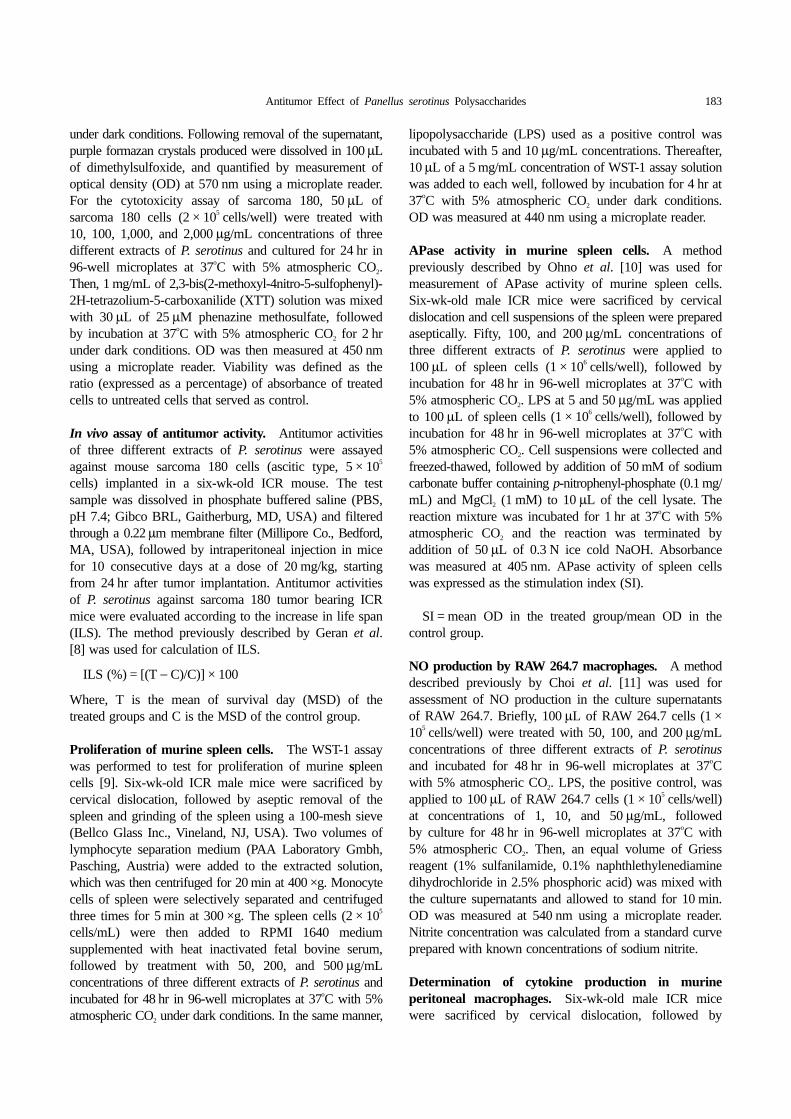
Fig. 1. In vitro cytotoxicity activity against sarcoma 180 (A), HT-29 (B), NIH3T3 (C), and RAW 264.7 (D) of differentconcentrations of various fractions extracted from fruiting bodies of Panellus serotinus. Values are expressed asmeans ± SD (n = 5). Fr. MeOH, fractions extracted with 80% methanol; Fr. NaCl, fractions extracted with 0.9% NaClsolution; Fr. HW, fractions extracted with hot water.
Statistical analysis. Data were expressed as mean ± SD.One-way analysis of variance, followed by Duncan’s newmultiple-range test, was performed for analysis of intergroupdifferences. SPSS ver. 11.5 (SPSS Inc., Chicago, IL,USA) was used for the analysis. A p ≤ 0.05 was consideredstatistically significant.
Results and Discussion
β-Glucan and protein content. Measurements of β-glucan and protein contents of various fractions, extracted
Antitumor Effect of Panellus serotinus Polysaccharides 185
from fruiting bodies of P. serotinus, were performed(Table 1). The highest amount of β-glucan was recordedin Fr. MeOH (28.52 g/100 g), followed by Fr. HW(27.03 g/100 g) and Fr. NaCl (22.92 g/100 g), respectively.Similar protein levels were recorded in Fr. FeOH, NaCl,and Fr. HW fractions of P. serotinus.
The immunomodulating effects of β-glucans duringdevelopment of immune reactions are well established.Due to their immunomodulatory and antitumor effects, β-glucans and β-glucan-protein complexes isolated frommushrooms have been used as a source of therapeuticagents [12]. Several investigators have isolated and purifiedimmunomodulating polysaccharides from mushrooms asa biological response modifier [13]. The anti-tumoractivities of polysaccharides are mainly the result of theirimmunopotentiation effects [14, 15].
In vitro assay of cytotoxicity. The results of the in vitrocytotoxicity activities of three different fractions of P.serotinus against sarcoma 180, HT-29, NIH3T3, andRAW 264.7 cell lines are shown in Fig. 1. At 10~2,000µg/mL, cell viability of Fr. MeOH, Fr. NaCl, and Fr. HWfractions against sarcoma 180, HT-29, NIH3T3, and RAW264.7 cell lines ranged from 70~122, 74~101, and 96~129%, respectively (Fig. 1A), 103~112, 67~101, and 73~106%, respectively (Fig. 1B), 78~90, 60~77, and 68~79%, respectively (Fig. 1C), and 97~110, 98~110, and110~114%, respectively (Fig. 1D).
The results indicated that Fr. MeOH, Fr. NaCl, and Fr.HW fractions extracted from fruiting bodies of P.serotinus had no significant cytotoxic effects on four celllines at the tested concentrations. In vitro cytotoxicityassays can be used for general screening of chemicals forprediction of toxicity [16]. In our earlier study, hot waterextract from Elfvingia applanata did not inhibit proliferationof HT-29, Hep G2, TR, and sarcoma 180 cancer cells[17]. In another study, Lee et al. [18] reported that a hot-water extract of Inonotus obliquus exerted little inhibitoryactivity against proliferation of human colon cancer cells,in good agreement with our results.
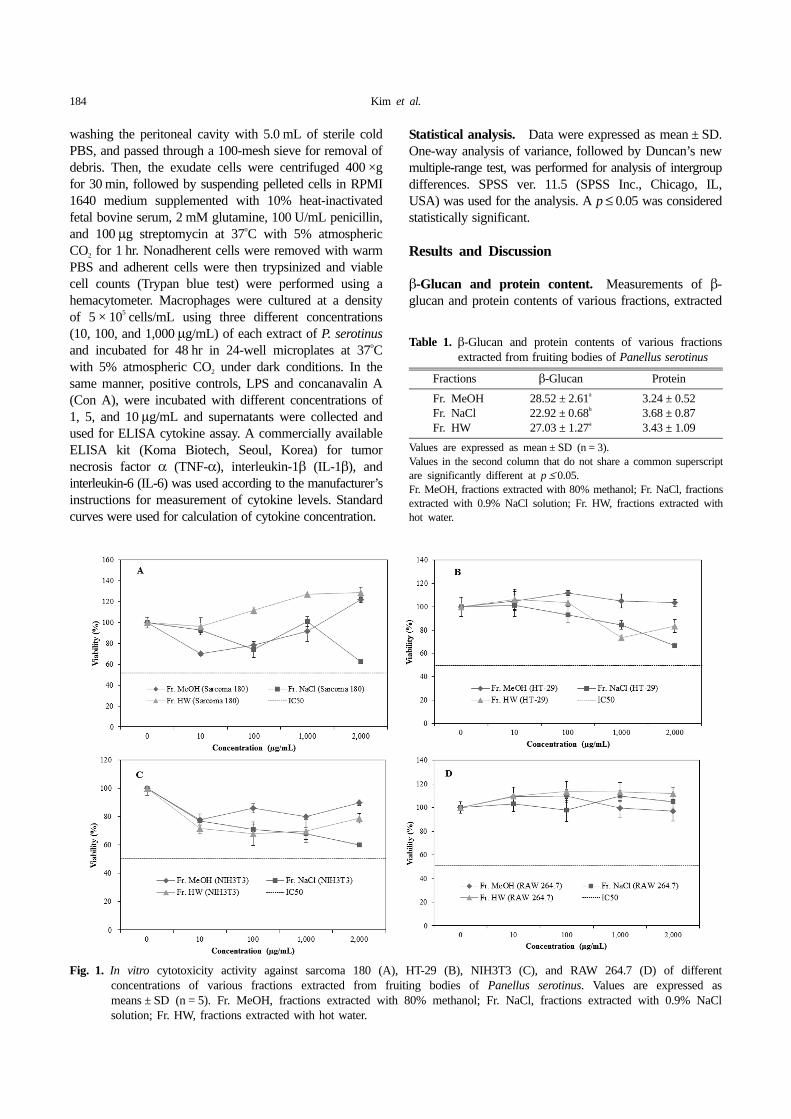
In vivo assay of antitumor activity. Antitumor activitiesof Fr. MeOH, Fr. NaCl, and Fr. HW fractions of P.serotinus were tested against sarcoma 180 tumor bearingmice. According to the results, the highest ILS wasrecorded in Fr. MeOH (44.71%), followed by Fr. NaCl(43.53%), and Fr. HW (23.53%), respectively comparedto the control group (Fig. 2). According to the results, themean life span of the group treated with Fr. MeOHshowed a significant increase and fruiting bodies of P.serotinus might contain effective antitumor substances againstsarcoma 180.
Shim et al. [19] reported that treatment of Paecilomycessinclairii with methanol extract resulted in inhibited growth
of sarcoma 180 tumor cells and prolongation of the lifespan of mice by 32.3%. In general, the criteria for judgingthe antitumor effect of any substance include prolongationof the life span by more than 25% [20]. This observationis consistent with our observations; the mean life span ofthe group treated with Fr. NaCl showed a significantincrease. It might be concluded that polysaccharides of P.serotinus have a strong anticancer effect.
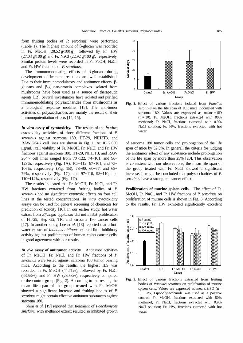
Proliferation of murine spleen cells. The effect of Fr.MeOH, Fr. NaCl, and Fr. HW fractions of P. serotinus onproliferation of murine cells is shown in Fig. 3. Accordingto the results, Fr. HW exhibited significantly excellent
Fig. 2. Effect of various fractions isolated from Panellusserotinus on the life span of ICR mice inoculated withsarcoma 180. Values are expressed as means ± SD(n = 10). Fr. MeOH, fractions extracted with 80%methanol; Fr. NaCl, fractions extracted with 0.9%NaCl solution; Fr. HW, fractions extracted with hotwater.
Fig. 3. Effect of various fractions extracted from fruitingbodies of Panellus serotinus on proliferation of murinespleen cells. Values are expressed as means ± SD (n =5). LPS, Lipopolysaccharide was used as a positivecontrol; Fr. MeOH, fractions extracted with 80%methanol; Fr. NaCl, fractions extracted with 0.9%NaCl solution; Fr. HW, fractions extracted with hotwater.
186 Kim et al.
activities, while good and moderate effects were observedfor Fr. MeOH and Fr. NaCl, respectively, compared tocontrol. However, at the concentration of 50 µg/mL, LPSalso exhibited excellent activities.
The results suggested that β-glucan of P. serotinus canimprove the immune response of the host by stimulatingproliferation of immune-organ, murine spleen cells. Liet al. [21] reported that treatment with proteoglycanextracted from crude liquid culture medium and myceliaof Phellinus nigricans resulted in stimulated proliferationof lymphocytes of spleen cells and increased productionof TNF-α. Murine spleen cells are the main residence ofvarious immune cells and are also important for hostimmune response.
APase activity in murine spleen cells. Stimulation ofsplenic lymphocytes with LPS, Fr. MeOH, Fr. NaCl, andFr. HW at 50 µg/mL resulted in an increase in APaseactivities of 1.63-, 1.22-, 1.06-, and 1.08-fold, respectively,compared to control (Fig. 4). APase activity in murinespleen cells showed a significant increase in Fr. NaCl at200 µg/mL, compared with positive control.
Cha et al. [22] reported an increase in APase activity of1.2~1.6-fold upon stimulation with crude polysaccharidesof Agaricus brasiliensis at concentrations of 50~200 µg/mL. Therefore, it is concluded that treatment with Fr.NaCl could result in improved immunostimulating activityof the host via increasing APase activity.
NO production by RAW 264.7 macrophages. NOproduction activity in the culture supernatants of RAW264.7 macrophages with various concentrations (50, 200,and 500 µg/mL) of Fr. MeOH, Fr. NaCl, and Fr. HW
fractions of P. serotinus ranged from 4.61~6.59, 8.89~12.85, and 6.68~13.48 µM, respectively. In the controlgroup, 4.47 µM of NO was released, while 12.04, 9.97,and 11.37 µM of NO were produced by treatment withLPS at the concentrations of 1, 5, and 50 µg/mL (Fig. 5).
The results demonstrated that stimulation with β-glucanof P. serotinus can result in an increase in production ofNO and improvement of the immune response in ICRmice. Our results were similar to those of Kim et al. [23],who observed that RAW 264.7 macrophages stimulatedby polysaccharides extracted from Phellinus linteus showedan increase in production of NO in a dose-dependentmanner. Ooi and Liu [12] reported that polysaccharidesextracted from mushrooms exert anti-tumor effects throughactivation of different immune responses in the host ratherthan by direct killing of tumor cells.
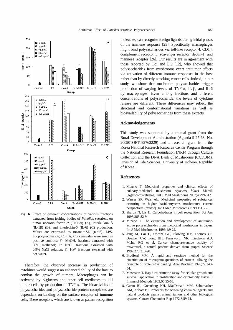
Cytokine production in murine peritoneal macrophages.The results on production of cytokines of three differentfractions of P. serotinus are shown in Fig. 6. At 10~1,000 µg/mL of Fr. MeOH, Fr. NaCl, and Fr. HW,production of TNF-α, IL-1β, and IL-6 ranged from 60.04~89.45, 179.38~208.31, and 75.36~130.50 pg/mL, respectively(Fig. 6A), 32.68~59.71, 78.00~103.20, and 81.45~155.60 pg/mL, respectively (Fig. 6B), and 148.08~191.63, 202.78~254.78, and 214.93~239.92 pg/mL, respectively (Fig. 6C).The results indicated that production of TNF-α, IL-1β,and IL-6 was significantly higher, compared to control,and IL-6 production was excellent, compared to TNF-α,IL-1β, and Con A at the tested concentrations of thevarious fractions of P. serotinus.
TNF-α, Il-1β, and IL-6 are important regulators of hostdefense against tumor cells [24].
Fig. 5. Effect of various fractions extracted from fruitingbodies of Panellus serotinus on nitric oxide productionin RAW 264.7. Values expressed as means ± SD (n =5). LPS, Lipopolysaccharide was used as a positivecontrol; Fr. MeOH, fractions extracted with 80%methanol; Fr. NaCl, fractions extracted with 0.9%NaCl solution; Fr. HW, fractions extracted with hotwater.
Fig. 4. Effect of various fractions extracted from fruiting bodiesof Panellus serotinus on alkaline phosphatase activityin murine spleen cells. Values are expressed as means± SD (n = 5). LPS, Lipopolysaccharide was used as apositive control; Fr. MeOH, fractions extracted with80% methanol; Fr. NaCl, fractions extracted with0.9% NaCl solution; Fr. HW, fractions extracted withhot water.
Antitumor Effect of Panellus serotinus Polysaccharides 187
Therefore, the observed increase in production ofcytokines would suggest an enhanced ability of the host tocombat the growth of tumors. Macrophages can beactivated by β-glucans and other cell mediators to killtumor cells by production of TNF-α. The bioactivities ofpolysaccharides and polysaccharide-protein complexes aredependent on binding on the surface receptor of immunecells. These receptors, which are known as pattern recognition
molecules, can recognize foreign ligands during initial phasesof the immune response [25]. Specifically, macrophagesmight bind polysaccharides via toll-like receptor 4, CD14,complement receptor 3, scavenger receptor, dectin-1, andmannose receptor [26]. Our results are in agreement withthose reported by Ooi and Liu [12], who showed thatpolysaccharides from mushrooms exert antitumor effectsvia activation of different immune responses in the hostrather than by directly attacking cancer cells. Indeed, in ourstudy, we show that mushroom polysaccharides triggerproduction of varying levels of TNF-α, IL-β, and IL-6by macrophages. Even among fractions and differentconcentrations of polysaccharide, the levels of cytokinerelease are different. These differences may reflect thestructural and conformational variations as well asbioavailability of polysaccharides from these extracts.
Acknowledgements
This study was supported by a mutual grant from theRural Development Administration (Agenda 9-27-63; No.200901OFT092763229) and a research grant from theKorea National Research Resource Center Program throughthe National Research Foundation (NRF) through CultureCollection and the DNA Bank of Mushrooms (CCDBM),Division of Life Sciences, University of Incheon, Republicof Korea.
References
1. Mizuno T. Medicinal properties and clinical effects ofculinary-medicinal mushroom Agaricus blazei Murrill(Agaricomycetideae). Int J Med Mushrooms 2002;4:299-312.
2. Wasser SP, Weis AL. Medicinal properties of substancesoccurring in higher basidiomycetes mushrooms: currentperspectives (review). Int J Med Mushrooms 1999;1:31-62.
3. Sharon N, Lis H. Carbohydrates in cell recognition. Sci Am1993;268:82-9.
4. Mizuno T. The extraction and development of antitumor-active polysaccharides from medicinal mushrooms in Japan.Int J Med Mushrooms 1999;1:9-29.
5. Jang M, Cai L, Udeani GO, Slowing KV, Thomas CF,Beecher CW, Fong HH, Farnsworth NR, Kinghorn AD,Mehta RG, et al. Cancer chemopreventive activity ofresveratrol, a natural product derived from grapes. Science1997;275:218-20.
6. Bradford MM. A rapid and sensitive method for thequantitation of microgram quantities of protein utilizing theprinciple of protein-dye binding. Anal Biochem 1976;72:248-54.
7. Mosmann T. Rapid colorimetric assay for cellular growth andsurvival: application to proliferation and cytotoxicity assays. JImmunol Methods 1983;65:55-63.
8. Geran RI, Greenberg NH, MacDonald MM, SchumacherAM, Abbott BJ. Protocols for screening chemical agents andnatural products against animal tumors and other biologicalsystems. Cancer Chemother Rep 1972;3:59-61.
Fig. 6. Effect of different concentrations of various fractionsextracted from fruiting bodies of Panellus serotinus ontumor necrosis factor α (TNF-α) (A), interleukin-1β(IL-1β) (B), and interleukin-6 (IL-6) (C) production.Values are expressed as means ± SD (n = 5). LPS,lipopolysaccharide; Con A, Concanavalin were used aspositive controls; Fr. MeOH, fractions extracted with80% methanol; Fr. NaCl, fractions extracted with0.9% NaCl solution; Fr. HW, fractions extracted withhot water.
188 Kim et al.
9. Ngamwongsatit P, Banada PP, Panbangred W, Bhunia AK.WST-1-based cell cytotocixity assay as a substitute for MTT-based assay for rapid detection of toxigenic Bacillus speciesusing CHO cell line. J Microbiol Methods 2008;73:211-5.
10. Ohno N, Arai Y, Suzuki I, Yadomae T. Induction of alkalinephosphatase activity in murine spleen cells treated withvarious mitogens. J Pharmacobiodyn 1986;9:593-9.
11. Choi SH, Jun CD, Lee BS, Park SD, Oh JS, Chung HT.Effect of various extrinsic and intrinsic factors on the nitricoxide production of murine macrophage. Korean J BiolResponse Modif 1993;3:15-22.
12. Ooi VE, Liu F. Immunomodulation and anti-cancer activityof polysaccharide-protein complexes. Curr Med Chem 2000;7:715-29.
13. Mizuno M, Shiomi Y, Minato K, Kawakami S, Ashida H,Tsuchida H. Fucogalactan isolated from Sarcodon aspratuselicits release of tumor necrosis factor-α and nitric oxide frommurine macrophages. Immunopharmacology 2000;46:113-21.
14. Hsieh YS, Chien C, Liao SK, Liao SF, Hung WT, Yang WB,Lin CC, Cheng TJ, Chang CC, Fang JM, et al. Structure andbioactivity of the polysaccharides in medicinal plant Dendrobiumhuoshanense. Bioorg Med Chem 2008;16:6054-68.
15. Togola A, Inngjerdingen M, Diallo D, Barsett H, RolstadB, Michaelsen TE, Paulsen BS. Polysaccharides withcomplement fixing and macrophage stimulation activity fromOpilia celtidifolia, isolation and partial characterisation. JEthnopharmacol 2008;115:423-31.
16. Scheers EM, Ekwall B, Dierickx PJ. In vitro long-termcytotoxicity testing of 27 MEIC chemicals on Hep G2 cellsand comparison with acute human toxicity data. Toxicol InVitro 2001;15:153-61.
17. Shim SM, Lee JS, Lee TS, Lee UY. Antitumor andimmunostimulating activities of Elfvingia applanata hotwater extract on sarcoma 180 tumor-bearing ICR mice.Mycobiology 2012;40:47-52.
18. Lee SH, Hwang HS, Yun JW. Antitumor activity of waterextract of a mushroom, Inonotus obliquus, against HT-29human colon cancer cells. Phytother Res 2009;23:1784-9.
19. Shim SM, Im KH, Kim JW, Lee UY, Shim MJ, Lee MW,Lee TS. Studies on immuno-modulatory and antitumoreffects of crude polysaccharides extracted from Paecilomycessinclairii. Kor J Mycol 2003;31:155-60.
20. Clarkson BD, Burchenal JH. Prelimanary screening ofantineoplastic drugs. Prog Clin Cancer 1965;1:625-9.
21. Li X, Jiao LL, Zhang X, Tian WM, Chen S, Zhang LP. Anti-tumor and immunomodulating activities of proteoglycansfrom mycelium of Phellinus nigricans and culture medium.Int Immunopharmacol 2008;8:909-15.
22. Cha YJ, Kim JH, Lee TS, Lee UY. Antitumor and immuno-potentiating activities of crude polysaccharides from fruitingbody of Agaricus brasiliensis. Kor J Mycol 2011;39:57-67.
23. Kim GY, Choi GS, Lee SH, Park YM. Acidic polysaccharideisolated from Phellinus linteus enhances through the up-regulation of nitric oxide and tumor necrosis factor-α fromperitoneal macrophages. J Ethnopharmacol 2004;95:69-76.
24. Wells SM, Kew S, Yaqoob P, Wallace FA, Calder PC.Dietary glutamine enhances cytokine production by murinemacrophages. Nutrition 1999;15:881-4.
25. Gordon S. Pattern recognition receptors: doubling up for theinnate immune response. Cell 2002;111:927-30.
26. Brown GD, Gordon S. Immune recognition: a new receptorfor β-glucans. Nature 2001;413:36-7.


![Index [rd.springer.com]978-1-4612-2556-0/1.pdf · Eptesicus serotinus, echo detection threshold, 94 Eptesicus serotinus (Serotine Bat), ... Extralemniscal pathway, 265, 271-272 Face,](https://img.pdfslide.us/doc/110x75/5a9d283e7f8b9a032a8c0342/index-rd-978-1-4612-2556-01pdfeptesicus-serotinus-echo-detection-threshold.jpg)