Embed Size (px)
DESCRIPTION
KPL offers kits and reagents for the detection of proteins and nucleic acid cells and tissue for fluorescent and colorimetric detection.
Citation preview
Immunohistology
Where Better Science Begins
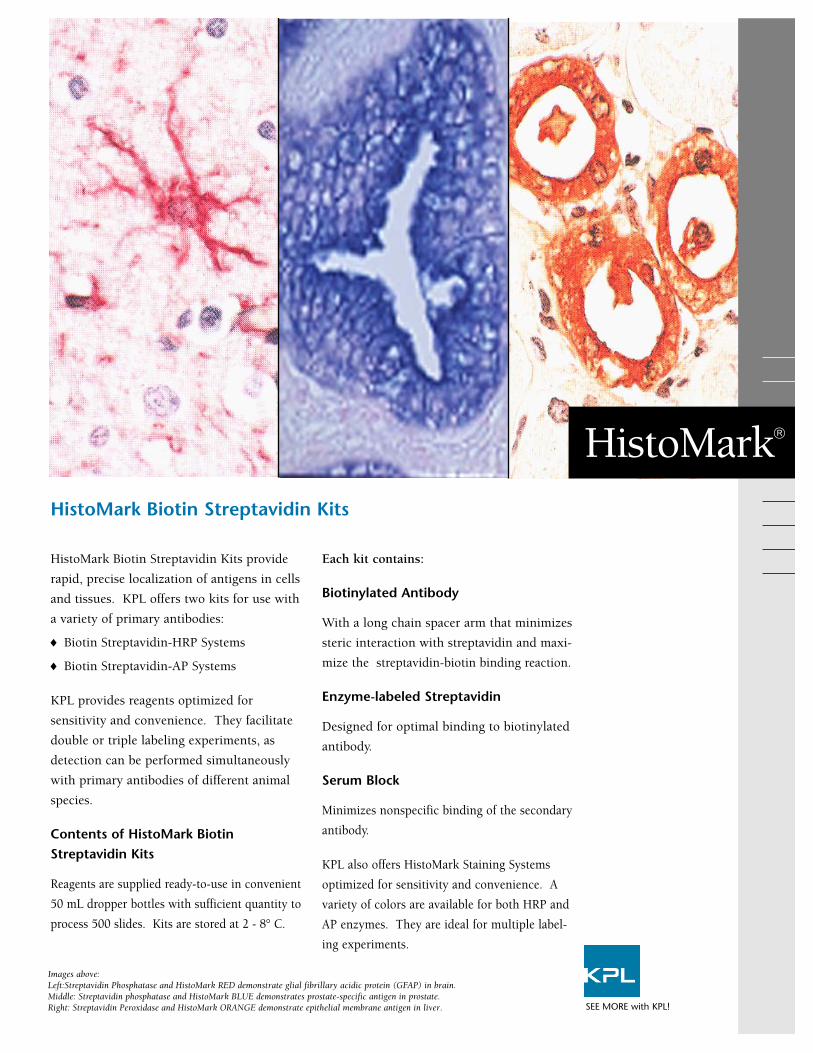
HistoMark Biotin Streptavidin Kits provide
rapid, precise localization of antigens in cells
and tissues. KPL offers two kits for use with
a variety of primary antibodies:
♦ Biotin Streptavidin-HRP Systems
♦ Biotin Streptavidin-AP Systems
KPL provides reagents optimized for
sensitivity and convenience. They facilitate
double or triple labeling experiments, as
detection can be performed simultaneously
with primary antibodies of different animal
species.
Contents of HistoMark BiotinStreptavidin Kits
Reagents are supplied ready-to-use in convenient
50 mL dropper bottles with sufficient quantity to
process 500 slides. Kits are stored at 2 - 8° C.
Each kit contains:
Biotinylated Antibody
With a long chain spacer arm that minimizes
steric interaction with streptavidin and maxi-
mize the streptavidin-biotin binding reaction.
Enzyme-labeled Streptavidin
Designed for optimal binding to biotinylated
antibody.
Serum Block
Minimizes nonspecific binding of the secondary
antibody.
KPL also offers HistoMark Staining Systems
optimized for sensitivity and convenience. A
variety of colors are available for both HRP and
AP enzymes. They are ideal for multiple label-
ing experiments.
HistoMark Biotin Streptavidin Kits
HistoMark®
Images above:Left:Streptavidin Phosphatase and HistoMark RED demonstrate glial fibrillary acidic protein (GFAP) in brain. Middle: Streptavidin phosphatase and HistoMark BLUE demonstrates prostate-specific antigen in prostate.Right: Streptavidin Peroxidase and HistoMark ORANGE demonstrate epithelial membrane antigen in liver. SEE MORE with KPL!
Ordering Information
To order or for more information on KPL’s full line of protein and nucleic acid
detection products, contact us at 800.638.3167 / 301.948.7755, FAX
301.948.0169 or visit us at www.kpl.com.
Catalog No. Description Size
Biotin-Streptavidin - Phosphatase Systems71-00-39 Mouse Primary Antibody 500 tests71-00-40 Rabbit Primary Antibody 500 tests71-00-41 Rat Primary Antibody 500 tests
Biotin-Streptavidin - Peroxidase Systems71-00-18 Mouse Primary Antibody 500 tests71-00-19 Rabbit Primary Antibody 500 tests71-00-20 Rat Primary Antibody 500 tests71-00-26 Goat Primary Antibody 500 tests
Components Available Separately71-00-29 Goat Anti-Mouse IgG (H+L) 50 mL71-00-30 Goat Anti-Rabbit IgG (H+L), HSA 50 mL71-00-31 Goat Anti-Rat IgG (H+L), MSA 50 mL71-00-37 Rabbit Anti-Goat IgG (H+L) 50 mL71-0038 HRP-Streptavidin 50 mL71-00-45 AP-Streptavidin 50 mL71-00-27 Normal Goat Serum 50 mL71-00-28 Normal Rabbit Serum 50 mL
HSA = Human Serum AdsorbedMSA = Mouse Serum Adsorbed
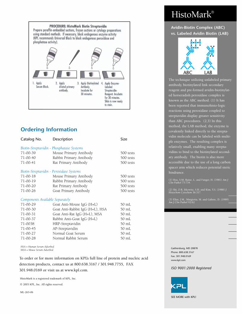
The technique utilizing unlabeled primary
antibody, biotinylated link secondary
reagent and pre-formed avidin-biotinylat-
ed horseradish peroxidase complex is
known as the ABC method. (1) It has
been reported that immunohisto-logic
reactions using peroxidase coupled to
streptavidin display greater sensitivity
than ABC procedures. (2,3) In this
method, the LAB method, the enzyme is
covalently linked directly to the strepta-
vidin molecule can be labeled with multi-
ple enzymes. The resulting complex is
relatively small, enabling many strepta-
vidins to bind to the biotinylated second-
ary antibody. The biotin is also more
accessible due to the use of a long carbon
spacer arm which reduces potential steric
hindrance.
(1) Hsu, S-M, Raine, L. and Fanger, H. (1981) Am JClin Pathol 75:734.
(2) Shi, Z-R, Itkowitz, S.H. and Kim, Y.S. (1988) JHistochem Cytochem 36:317.
(3) Elias, J.M., Margiotta, M. and Gaborc, D. (1989)Am J Clin Pathol 92:62
Avidin-Biotin Complex (ABC)vs. Labeled Avidin Biotin (LAB)
HistoMark®
Gaithersburg, MD 20878
Phone: 800.638.3167
Fax: 301.948.0169
www.kpl.com
ML-263-06
© 2003 KPL, Inc. All rights reserved.
HistoMark is a registered trademark of KPL, Inc.
ABC LAB
SEE MORE with KPL!
ISO 9001:2000 Registered
HistoMark® Staining Systems provide rapid, pre-
cise localization of antigens in cells and tissues.
Systems are available for the detection of peroxi-
dase, phosphatase and β-galactosidase-labeled
conjugates. Each kit contains the following
reagents:
HistoMark Substrate
Stable liquid concentrates provide a variety
of colors for multiple staining applications.
Substrate Buffer Solution
Maximizes color development
Counterstain
Provides sharp contrast and enhances
nuclear detail.
When used with KPL’s HistoMark Biotin
Streptavidin Kits, they offer brilliant, intense
colors and superior performance.
♦ Stable, liquid systems eliminate waste
♦ Convenient, easy-to-follow protocols
♦ Α variety of contrasting colors for
multiple staining
HistoMark® Substrate and Staining Systems
HistoMark®
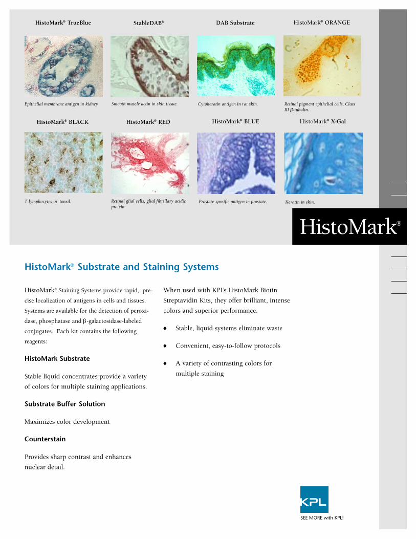
Epithelial membrane antigen in kidney. Cytokeratin antigen in rat skin.
Prostate-specific antigen in prostate.
Retinal pigment epithelial cells, ClassIII β-tubulin.
Keratin in skin.T lymphocytes in tonsil. Retinal glial cells, glial fibrillary acidicprotein.
Smooth muscle actin in skin tissue.
HistoMark® TrueBlue StableDAB® DAB Substrate HistoMark® ORANGE
HistoMark® BLACK HistoMark® RED HistoMark® BLUE HistoMark® X-Gal
SEE MORE with KPL!
KPL’s TrueBlue Substrate is highly recommended for in situ hybridization; a brilliant
blue stain with superior resolution.
Ordering Information
To order or for more information on KPL’s full line of protein and nucleic acid detection
products, contact us at 800.638.3167 / 301.948.7755, FAX 301.948.0169 or visit us at
www.kpl.com.
Catalog No. Description Size
For use with phosphatase55-69-00 HistoMark RED 1000 slides55-70-00 HistoMark BLUE 1000 slides
For use with peroxidase54-74-00 HistoMark ORANGE 1000 slides54-75-00 HistoMark BLACK 1000 slides54-78-00 HistoMark TrueBlue 500 slides54-10-00 DAB Reagent Set 500 slides54-11-00 StableDAB Peroxidase Substrate 500 slides
For use with β-galactosidase54-13-00 HistoMark X-Gal Substrate Set 200 slides
Counterstains71-01-01 Orcein 50 mL71-02-01 Eosin 10 mL71-00-11 Contrast GREEN 50 mL71-00-06 Contrast BLUE 50 mL71-00-05 Contrast RED 50 mL
KPL’s HistoMark Biotin
Streptavidin Kits provide
high signal-to-noise ratios
when used with HistoMark
Staining Systems.
KPL’s HistoMark Substrates
and staining systems can be
utilized to obtain brilliant
double-staining results. To
read more about immuno-
histochemistry double stain-
ing and for a guide to
“HistoMark Double Staining
Procedures” visit the
“Technical Info” section at
www.kpl.com.
HistoMark®
Gaithersburg, MD 20878
Phone: 800.638.3167
Fax: 301.948.0169
www.kpl.com
ML-264-05
Cytomegalovirus-infected cells detectedwith a biotinylated CMV probe and theDNADetector Chromogenic in situHybridization Kit using TrueBlue.
HistoMark Substrate and Staining Systems
StableDAB demonstrates cytokeratin19 in breast tissue. TrueBlue demon-strates smooth muscle actin in breasttissue.
HistoMark BLUE demonstrates retinalpigment epithelial cells, Class III β-tubulin. HistoMark RED demonstrateskeratin.
KPL’s staining systems offer a variety of contrasting colors for highresolution multiple labeling applications.
StableDAB and HistoMark are registered trademarks of KPL, Inc.
© 2003 KPL, Inc. All rights reserved. SEE MORE with KPL!
ISO 9001:2000 Registered
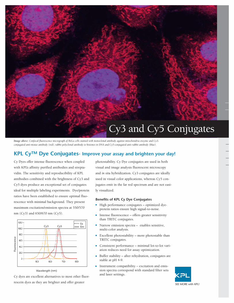
Cy Dyes offer intense fluorescence when coupled
with KPL’s affinity purified antibodies and strepta-
vidin. The sensitivity and reproducibility of KPL
antibodies combined with the brightness of Cy3 and
Cy5 dyes produce an exceptional set of conjugates
ideal for multiple labeling experiments. Dye/protein
ratios have been established to ensure optimal fluo-
rescence with minimal background. They present
maximum excitation/emission spectra at 550/570
nm (Cy3) and 650/670 nm (Cy5).
Cy dyes are excellent alternatives to most other fluor-
rescein dyes as they are brighter and offer greater
photostability. Cy Dye conjugates are used in both
visual and image analysis fluorescent microscopy
and in situ hybridization. Cy3 conjugates are ideally
used in visual color applications, whereas Cy5 con-
jugates emit in the far red spectrum and are not easi-
ly visualized.
Benefits of KPL Cy Dye Conjugates
High performance conjugates – optimized dye-protein ratios ensure high signal-to-noise.
Intense fluorescence – offers greater sensitivity than TRITC conjugates.
Narrow emission spectra – enables sensitive, multi-color analysis.
Excellent photostability – more photostable than TRITC conjugates.
Consistent performance – minimal lot-to-lot vari-ation reduces need for assay optimization.
Buffer stability – after rehydration, conjugates are stable at pH 4-9.
Instrument compatibility – excitation and emis-sion spectra correspond with standard filter sets and laser settings.
KPL CyTM Dye Conjugates- Improve your assay and brighten your day!
SEE MORE with KPL!
Cy3 and Cy5 Conjugates
Fluo
resc
ence
Wavelength (nm)
Cy3 Cy5ExEm
Image above: Confocal fluorescence micrograph of HeLa cells stained with monoclonal antibody against mitochondria enzyme and Cy3-
conjugated anti-mouse antibody (red); rabbit polyclonal antibody to histones in DNA and Cy5-conjugated anti-rabbit antibody (blue).
Ordering Information
To order or for more information on KPL’s line of unlabeled and conjugated
affinity purified antibodies, contact us at 800.638.3167 / 301.948.7755, fax
301.948.0169 or visit us at www.kpl.com.
Cy3 and Cy5 Conjugates
ML368-01For research use only.Cy Dye is a trademark of GE Healthcare.©2009 KPL, Inc. All rights reserved.
Cytomegalovirus-infected cellsdetected with a biotinylated CMVprobe and the DNADetectorTM
Fluorescent in situ Hybridization Kitusing Cy3-Strept-avidin and DAPI.
Signal DetectionCy3 is excited maximally at 550 nmand fluoresces maximally at 570 nm.It is excited to about 50% of maxi-mum with an argon laser (514 nm or528 nm lines), or to about 75% ofmaximum with a helium/neon laser(543 nm line) or mercury lamp (546nm line).
Cy5 is excited maximally at 650 nmand fluoresces maximally at 670 nm.It is excited to about 98% of maxi-mum with a krypton/argon laser (647nm line) or to about 63% of maxi-mum with a helium/neon laser (633nm line). Cy5 produces minimal auto-fluorescence of biological specimensin this region of the spectrum.
A confocal microscope equipped withthe appropriate laser for excitationand a far-red detector enable doublelabeling with Cy3 and Cy5.
Gaithersburg, MD 20878
Phone: 800.638.3167
Fax: 301.948.0169
www.kpl.com
ISO 9001:2000 Registered
SEE MORE with KPL!
Anti-Mouse IgG (γ), HSA 072-01-18-02 072-02-18-02
F(ab’)2 Anti-Mouse IgG (γ), HSA 202-01-18-02 202-02-18-02
Anti-Mouse IgG (H+L), HSA 072-01-18-06 072-02-18-06
F(ab’)2 Anti-Mouse IgG (H+L), HSA 202-01-18-06 202-02-18-06
Anti-Mouse IgG (H+L), RbSA, HSA 072-01-18-18 072-02-18-18
Anti-Mouse IgM (µ), HSA 072-01-18-03 072-02-18-03
Anti-Mouse IgG+IgM (H+L), HSA 072-01-18-09 072-02-18-09
Anti-Rabbit IgG (H+L) 072-01-15-06 072-02-15-06
F(ab’)2 Anti-Rabbit IgG (H+L), HSA 202-01-15-16 202-02-15-16
Anti-Rabbit IgG (H+L), HSA 072-01-15-16 072-02-15-16
Anti-Rat IgG (H+L) 072-01-16-06 072-02-16-06
F(ab’)2 Anti-Human IgG (H+L) 202-01-10-06 202-02-10-06
Anti-Human IgG (γ) 072-01-10-02 072-02-10-02
F(ab’)2 Anti-Human IgG (γ) 202-01-10-02 202-02-10-02
Anti-Human IgM (µ) 072-01-10-03 072-02-10-03
F(ab’)2 Anti-Human IgM (µ) 202-01-10-03 202-02-10-03
Anti-Guinea Pig IgG (H+L) 072-01-17-06 072-02-17-06
Anti-Chicken IgG (H+L) 072-01-24-06 072-02-24-06
Anti-Horse IgG (H+L) 072-01-21-06 072-02-21-06
Anti-Swine IgG (H+L) 072-01-14-06 072-02-14-06
Anti-Dog IgG (H+L) 072-01-19-06 072-02-19-06
Anti-Sheep IgG (H+L) 072-01-23-06 072-02-23-06
Anti-Goat IgG (H+L) 072-01-13-06 072-02-13-06
Streptavidin 072-01-30-00 072-02-30-00
Description Cy3 Cy5
HSA=human serum adsorbed RbSA=rabbit serum adsorbedCy Dye antibody conjugates are made in goat except anti-goat and anti-sheep antibodiesmade in rabbit. Supplied in 1 mg lyophilized form.
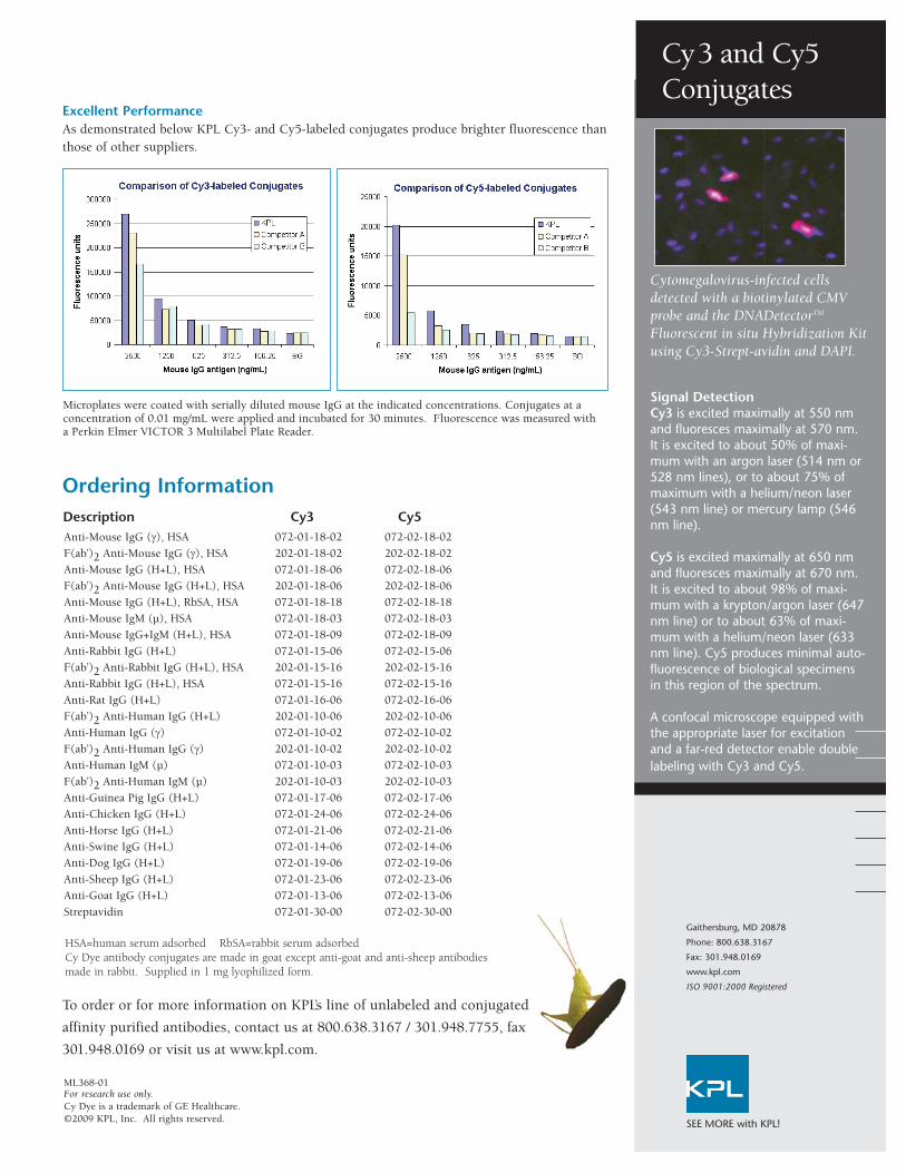
Microplates were coated with serially diluted mouse IgG at the indicated concentrations. Conjugates at aconcentration of 0.01 mg/mL were applied and incubated for 30 minutes. Fluorescence was measured witha Perkin Elmer VICTOR 3 Multilabel Plate Reader.
Excellent PerformanceAs demonstrated below KPL Cy3- and Cy5-labeled conjugates produce brighter fluorescence thanthose of other suppliers.
KPL’s DyLightTM conjugates offer a brilliant
choice in a variety of multicolor detection ap-
plications, including fluorescence microscopy,
flow cytometry, Western blotting, ELISA and
array platforms. Our affinity purified antibod-
ies combined with a series of outstanding
DyLight dyes provide superior performance
over conventional CyDyeTM fluors, fluorescein
and rhodamine, with performance comparable
to that of Alexa Fluor® dyes (Figure 1). Enjoy
these advantages when you switch to KPL’s
DyLight Conjugates:
KPL offers eight DyLight dyes, including 405,
488, 549, 594, 633, 649, 680 and 800 with
well-differentiated excitation and emission
spectra. Our extensive line of over 170 DyLight
conjugates is available across a range of animal
species immunoglobulin, including human,
mouse, rabbit, rat, other species and strepta-
vidin. See back cover to find out what sets KPL’s
antibodies apart.
SEE MORE with KPL!
KPL’s DyLightTM Conjugates - A Brilliant Choice!
DyLightTM Fluorescent Conjugates
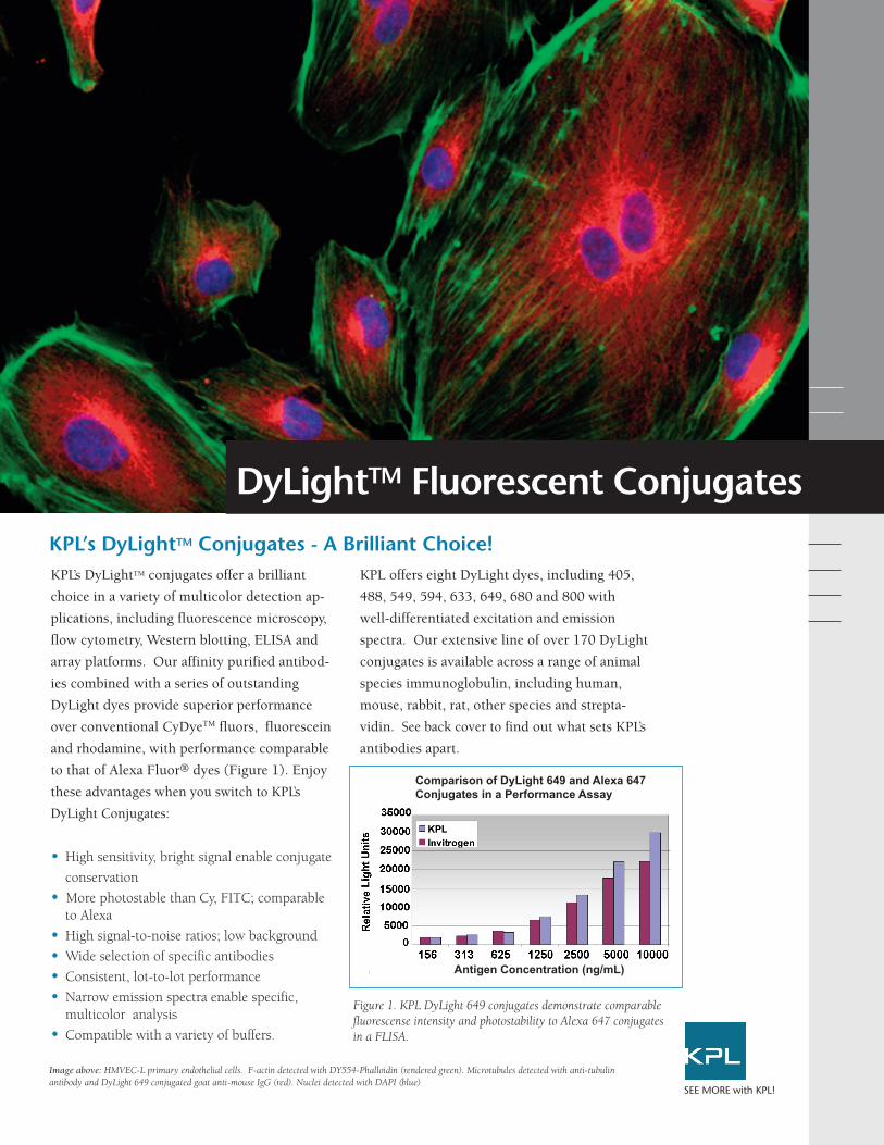
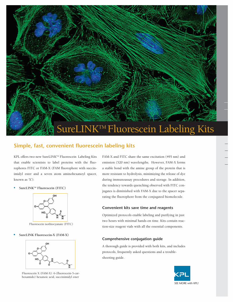
Image above: HMVEC-L primary endothelial cells. F-actin detected with DY554-Phalloidin (rendered green). Microtubules detected with anti-tubulinantibody and DyLight 649 conjugated goat anti-mouse IgG (red). Nuclei detected with DAPI (blue)
Figure 1. KPL DyLight 649 conjugates demonstrate comparablefluorescense intensity and photostability to Alexa 647 conjugatesin a FLISA.
•• High sensitivity, bright signal enable conjugate
conservation
•• More photostable than Cy, FITC; comparable to Alexa
•• High signal-to-noise ratios; low background
•• Wide selection of specific antibodies
•• Consistent, lot-to-lot performance
•• Narrow emission spectra enable specific, multicolor analysis
•• Compatible with a variety of buffers.
Antigen Concentration (ng/mL)
Comparison of DyLight 649 and Alexa 647Conjugates in a Performance Assay
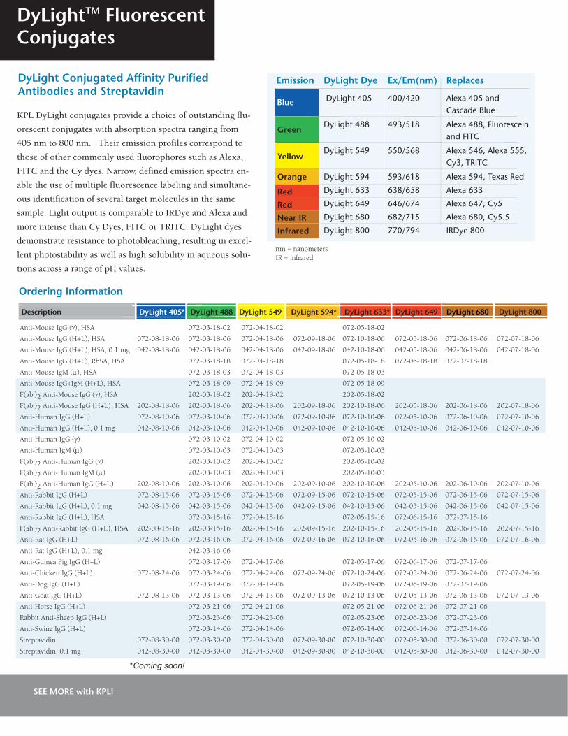
Emission DyLight Dye Ex/Em(nm) Replaces
DyLight 405 400/420 Alexa 405 and Cascade Blue
DyLight 488 493/518 Alexa 488, Fluorescein and FITC
DyLight 549 550/568 Alexa 546, Alexa 555, Cy3, TRITC
DyLight 594 593/618 Alexa 594, Texas Red
DyLight 633 638/658 Alexa 633
DyLight 649 646/674 Alexa 647, Cy5
DyLight 680 682/715 Alexa 680, Cy5.5
DyLight 800 770/794 IRDye 800
KPL DyLight conjugates provide a choice of outstanding flu-
orescent conjugates with absorption spectra ranging from
405 nm to 800 nm. Their emission profiles correspond to
those of other commonly used fluorophores such as Alexa,
FITC and the Cy dyes. Narrow, defined emission spectra en-
able the use of multiple fluorescence labeling and simultane-
ous identification of several target molecules in the same
sample. Light output is comparable to IRDye and Alexa and
more intense than Cy Dyes, FITC or TRITC. DyLight dyes
demonstrate resistance to photobleaching, resulting in excel-
lent photostability as well as high solubility in aqueous solu-
tions across a range of pH values.
Anti-Mouse IgG (γ), HSA 072-03-18-02 072-04-18-02 072-05-18-02
Anti-Mouse IgG (H+L), HSA 072-08-18-06 072-03-18-06 072-04-18-06 072-09-18-06 072-10-18-06 072-05-18-06 072-06-18-06 072-07-18-06
Anti-Mouse IgG (H+L), HSA, 0.1 mg 042-08-18-06 042-03-18-06 042-04-18-06 042-09-18-06 042-10-18-06 042-05-18-06 042-06-18-06 042-07-18-06
Anti-Mouse IgG (H+L), RbSA, HSA 072-03-18-18 072-04-18-18 072-05-18-18 072-06-18-18 072-07-18-18
Anti-Mouse IgM (µ), HSA 072-03-18-03 072-04-18-03 072-05-18-03
Anti-Mouse IgG+IgM (H+L), HSA 072-03-18-09 072-04-18-09 072-05-18-09
F(ab’)2 Anti-Mouse IgG (γ), HSA 202-03-18-02 202-04-18-02 202-05-18-02
F(ab’)2 Anti-Mouse IgG (H+L), HSA 202-08-18-06 202-03-18-06 202-04-18-06 202-09-18-06 202-10-18-06 202-05-18-06 202-06-18-06 202-07-18-06
Anti-Human IgG (H+L) 072-08-10-06 072-03-10-06 072-04-10-06 072-09-10-06 072-10-10-06 072-05-10-06 072-06-10-06 072-07-10-06
Anti-Human IgG (H+L), 0.1 mg 042-08-10-06 042-03-10-06 042-04-10-06 042-09-10-06 042-10-10-06 042-05-10-06 042-06-10-06 042-07-10-06
Anti-Human IgG (γ) 072-03-10-02 072-04-10-02 072-05-10-02
Anti-Human IgM (µ) 072-03-10-03 072-04-10-03 072-05-10-03
F(ab’)2 Anti-Human IgG (γ) 202-03-10-02 202-04-10-02 202-05-10-02
F(ab’)2 Anti-Human IgM (µ) 202-03-10-03 202-04-10-03 202-05-10-03
F(ab’)2 Anti-Human IgG (H+L) 202-08-10-06 202-03-10-06 202-04-10-06 202-09-10-06 202-10-10-06 202-05-10-06 202-06-10-06 202-07-10-06
Anti-Rabbit IgG (H+L) 072-08-15-06 072-03-15-06 072-04-15-06 072-09-15-06 072-10-15-06 072-05-15-06 072-06-15-06 072-07-15-06
Anti-Rabbit IgG (H+L), 0.1 mg 042-08-15-06 042-03-15-06 042-04-15-06 042-09-15-06 042-10-15-06 042-05-15-06 042-06-15-06 042-07-15-06
Anti-Rabbit IgG (H+L), HSA 072-03-15-16 072-04-15-16 072-05-15-16 072-06-15-16 072-07-15-16
F(ab’)2 Anti-Rabbit IgG (H+L), HSA 202-08-15-16 202-03-15-16 202-04-15-16 202-09-15-16 202-10-15-16 202-05-15-16 202-06-15-16 202-07-15-16
Anti-Rat IgG (H+L) 072-08-16-06 072-03-16-06 072-04-16-06 072-09-16-06 072-10-16-06 072-05-16-06 072-06-16-06 072-07-16-06
Anti-Rat IgG (H+L), 0.1 mg 042-03-16-06
Anti-Guinea Pig IgG (H+L) 072-03-17-06 072-04-17-06 072-05-17-06 072-06-17-06 072-07-17-06
Anti-Chicken IgG (H+L) 072-08-24-06 072-03-24-06 072-04-24-06 072-09-24-06 072-10-24-06 072-05-24-06 072-06-24-06 072-07-24-06
Anti-Dog IgG (H+L) 072-03-19-06 072-04-19-06 072-05-19-06 072-06-19-06 072-07-19-06
Anti-Goat IgG (H+L) 072-08-13-06 072-03-13-06 072-04-13-06 072-09-13-06 072-10-13-06 072-05-13-06 072-06-13-06 072-07-13-06
Anti-Horse IgG (H+L) 072-03-21-06 072-04-21-06 072-05-21-06 072-06-21-06 072-07-21-06
Rabbit Anti-Sheep IgG (H+L) 072-03-23-06 072-04-23-06 072-05-23-06 072-06-23-06 072-07-23-06
Anti-Swine IgG (H+L) 072-03-14-06 072-04-14-06 072-05-14-06 072-06-14-06 072-07-14-06
Streptavidin 072-08-30-00 072-03-30-00 072-04-30-00 072-09-30-00 072-10-30-00 072-05-30-00 072-06-30-00 072-07-30-00
Streptavidin, 0.1 mg 042-08-30-00 042-03-30-00 042-04-30-00 042-09-30-00 042-10-30-00 042-05-30-00 042-06-30-00 042-07-30-00
DyLightTM FluorescentConjugates
DyLight Conjugated Affinity PurifiedAntibodies and Streptavidin
nm = nanometersIR = infrared
Description DyLight 405* DyLight 488 DyLight 549 DyLight 594* DyLight 633* DyLight 649 DyLight 680 DyLight 800
SEE MORE with KPL!
*Coming soon!
Blue
Green
Yellow
Orange
Red
Red
Near IR
Infrared
Ordering Information
HSA = human serum adsorbedRbSA = rabbit serum adsorbedDyLight antibody conjugates are made in goat exceptanti-goat and anti-sheep antibodies are made in rabbit.Supplied in 1.0 mg lyophilized form except select 0.1mg sizes.
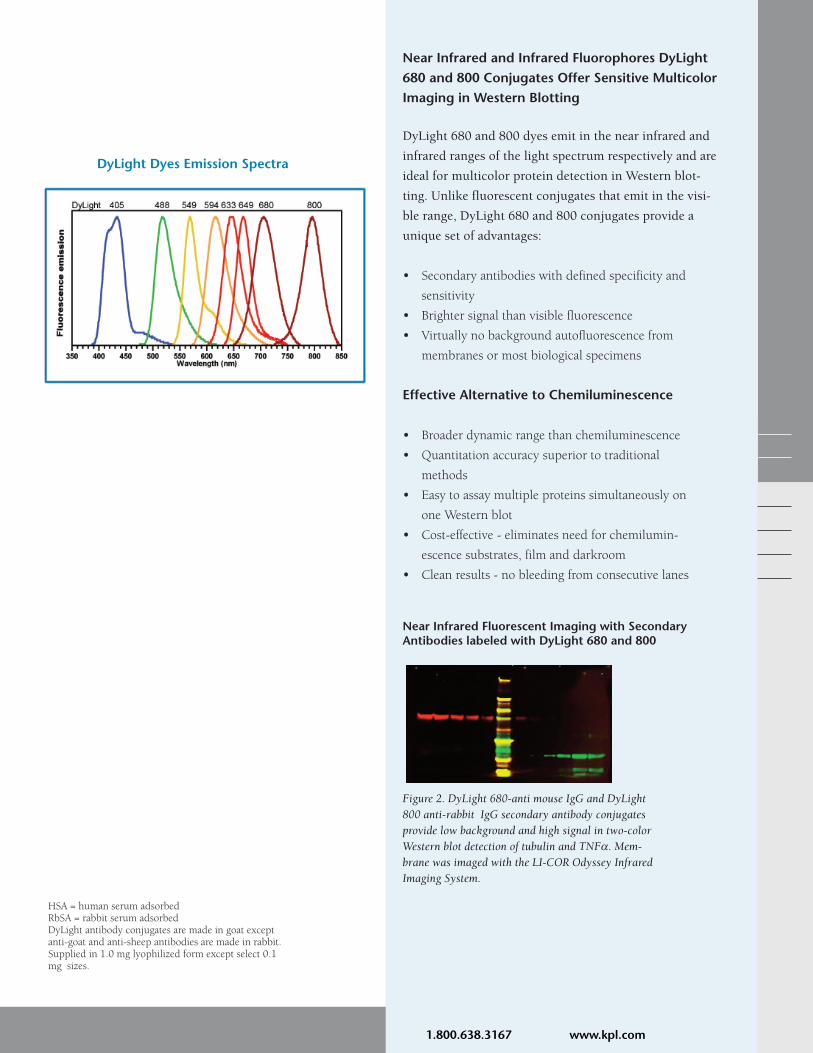
DyLight Dyes Emission Spectra
Near Infrared and Infrared Fluorophores DyLight680 and 800 Conjugates Offer Sensitive MulticolorImaging in Western Blotting
DyLight 680 and 800 dyes emit in the near infrared and
infrared ranges of the light spectrum respectively and are
ideal for multicolor protein detection in Western blot-
ting. Unlike fluorescent conjugates that emit in the visi-
ble range, DyLight 680 and 800 conjugates provide a
unique set of advantages:
•• Secondary antibodies with defined specificity and
sensitivity
•• Brighter signal than visible fluorescence
•• Virtually no background autofluorescence from
membranes or most biological specimens
Effective Alternative to Chemiluminescence
•• Broader dynamic range than chemiluminescence
•• Quantitation accuracy superior to traditional
methods
•• Easy to assay multiple proteins simultaneously on
one Western blot
•• Cost-effective - eliminates need for chemilumin-
escence substrates, film and darkroom
•• Clean results - no bleeding from consecutive lanes
Figure 2. DyLight 680-anti mouse IgG and DyLight800 anti-rabbit IgG secondary antibody conjugatesprovide low background and high signal in two-colorWestern blot detection of tubulin and TNFα. Mem-brane was imaged with the LI-COR Odyssey InfraredImaging System.
Near Infrared Fluorescent Imaging with SecondaryAntibodies labeled with DyLight 680 and 800
SEE MORE with KPL! 1.800.638.3167 www.kpl.com
Gaithersburg, MD
Phone: 800.638.3167/301.948.7755
Fax: 301.948.0169
www.kpl.com
SEE MORE with KPL!
ISO 9001:2008 Registered
DyLightTM FluorescentConjugates
KPL Antibodies-What Sets Them Apart
ML356-02For research use only.DyLight is a trademark of Thermo Fisher Scientific Inc. and its subsidiaries. Cy and Cy Dye are trademarks of GE Healthcare.Alexa Fluor is a registered trademark of Invitrogen.2009 KPL, Inc. All rights reserved.
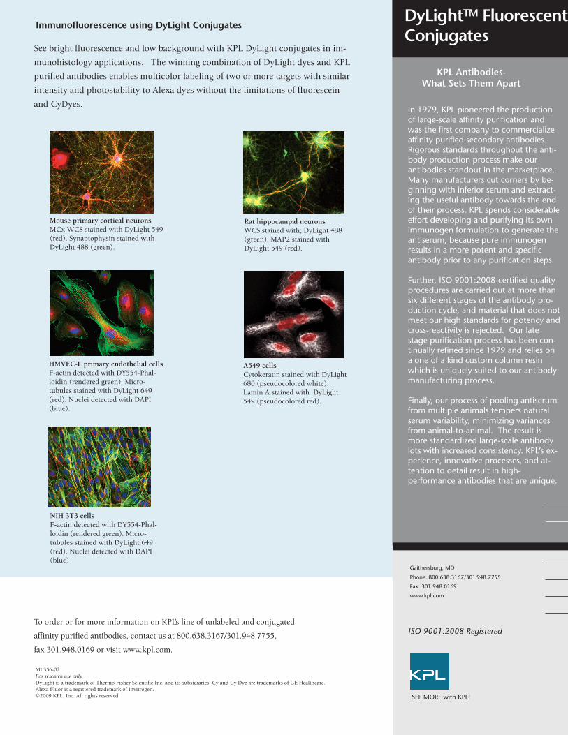
Rat hippocampal neuronsWCS stained with; DyLight 488(green). MAP2 stained withDyLight 549 (red).
Mouse primary cortical neuronsMCx WCS stained with DyLight 549(red). Synaptophysin stained withDyLight 488 (green).
HMVEC-L primary endothelial cellsF-actin detected with DY554-Phal-loidin (rendered green). Micro-tubules stained with DyLight 649(red). Nuclei detected with DAPI(blue).
NIH 3T3 cells F-actin detected with DY554-Phal-loidin (rendered green). Micro-tubules stained with DyLight 649(red). Nuclei detected with DAPI(blue)
A549 cells Cytokeratin stained with DyLight680 (pseudocolored white).Lamin A stained with DyLight549 (pseudocolored red).
Immunofluorescence using DyLight Conjugates
See bright fluorescence and low background with KPL DyLight conjugates in im-
munohistology applications. The winning combination of DyLight dyes and KPL
purified antibodies enables multicolor labeling of two or more targets with similar
intensity and photostability to Alexa dyes without the limitations of fluorescein
and CyDyes.
To order or for more information on KPL’s line of unlabeled and conjugated
affinity purified antibodies, contact us at 800.638.3167/301.948.7755,
fax 301.948.0169 or visit www.kpl.com.
In 1979, KPL pioneered the productionof large-scale affinity purification andwas the first company to commercializeaffinity purified secondary antibodies.Rigorous standards throughout the anti-body production process make ourantibodies standout in the marketplace.Many manufacturers cut corners by be-ginning with inferior serum and extract-ing the useful antibody towards the endof their process. KPL spends considerableeffort developing and purifying its ownimmunogen formulation to generate theantiserum, because pure immunogenresults in a more potent and specificantibody prior to any purification steps.
Further, ISO 9001:2008-certified qualityprocedures are carried out at more thansix different stages of the antibody pro-duction cycle, and material that does notmeet our high standards for potency andcross-reactivity is rejected. Our latestage purification process has been con-tinually refined since 1979 and relies ona one of a kind custom column resinwhich is uniquely suited to our antibodymanufacturing process.
Finally, our process of pooling antiserumfrom multiple animals tempers naturalserum variability, minimizing variancesfrom animal-to-animal. The result ismore standardized large-scale antibodylots with increased consistency. KPL’s ex-perience, innovative processes, and at-tention to detail result in high-performance antibodies that are unique.
Protein Detector ELISpot kits are designed to
provide complete, flexible systems for conduct-
ing ELISpot assays with any matched pair of
antibodies. For the beginner or the more expe-
rienced researcher, these kits are ideal due to
their comprehensive reagents and optimized
protocol.
Convenience
Each kit contains all reagents for ELISpot
detection except for matched antibody pairs
(unlabeled coating and biotinylated antibody).
Stable, ready-to-use, one-component substrates
and uncoated Multiscreen® PVDF Plates are
included. Other kit components are supplied as
concentrates; simply dilute and use.
Stability
All kit reagents are stable for a minimum of
one year, providing reliable signal development
and consistently low background.
Visualization
Visualizing immunospots has never been easier
with the exceptional localization and resolu-
tion provided by kit substrates. For sharp
localization and fine resolution of bright blue
immunospots, choose the HRP kit with
TrueBlueTM substrate. For excellent visibility
from a dark purple precipitate, choose the AP
kit with BCIP/NBT substrate.
Flexibility
Universal nature of reagents allows kits to be
used with any matched pair of antibodies.
Since labeled streptavidin is compatible with
any biotinylated antibody, the researcher sim-
ply provides the antibody pair for detection of
different cytokines or other secreted proteins.
Additional flexibility is offered through two kit
formats. Choose between the HRP kit with
TrueBlue or the AP kit with BCIP/NBT.
Protein DetectorTM ELISpot Kits
ELISpot
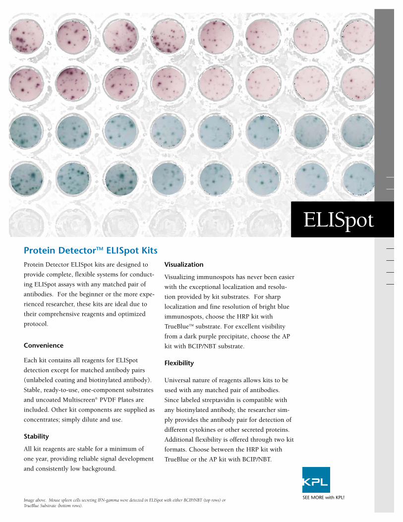
SEE MORE with KPL!Image above. Mouse spleen cells secreting IFN-gamma were detected in ELISpot with either BCIP/NBT (top rows) orTrueBlue Substrate (bottom rows).
ELISpot
Ordering Information
To order or for more information on KPL’s full line of protein and nucleic acid
detection products, contact us at 800.638.3167 / 301.948.7755, FAX 301.948.0169
or visit us at www.kpl.com.
Catalog# Description Size
40-00-05 Protein Detector HRP ELISpot Kit 5 plates
50-00-05 Protein Detector AP ELISpot Kit 5 plates
Each kit includes substrate (TrueBlue or BCIP/NBT), HRP or AP-labeled Streptavidin, 5 Multiscreen PVDF Plates, 96-well, CoatingSolution Concentrate, BSA Blocking/Diluent Solution Concentrate, and Washing Solution Concentrate.
ELISpot
Gaithersburg, MD 20878
Phone: 800.638.3167
Fax: 301.948.0169
www.kpl.com
SEE MORE with KPL!ML290-05
TrueBlue is a registered trademark and Protein Detector and Detector are trademarks of KPL, Inc.Multiscreen is a registered trademark of Millipore Corporation.
© 2003 KPL, Inc. All rights reserved.
Protein DetectorTM ELISpot Kits offer pretested reagents and a convenient, optimizedprotocol for ELISpot protein detection via either horseradish peroxidase (HRP) or alka-line phosphatase (AP) detection systems. With all necessary reagents except the pairedantibody set (unlabeled capture antibody and biotinylated detection antibody), this kitallows for minimal time and effort in developing a sensitive and reproducible ELISpotassay. Provided conjugates can detect any biotinylated antibody making these reagentseasily interchangeable with any matched antibody pair.
Substrate
TrueBlue
BCIP/NBT
AEC
Format
1-C
1-C
2-C
Enzyme
HRP
AP
HRP
Color
Blue
Dark Purple
Red
Sensitivity
√√√√
√√
√√
Comparison of Substrates for ELISpot
Related Products
*TrueBlue Peroxidase SubstrateCatalog No. 71-00-64 Size: 50 mL
*BCIP/NBT Substrate (1-Comp)Catalog No. 50-81-18 Size: 100 mL
HRP-labeled StreptavidinCatalog No. 474-3000Size: 1.0 mL
AP-labeled Streptavidin Catalog No. 475-3000 Size: 1.0 mL
*BSA Diluent/BlockingSolution Concentrate Catalog No. 50-61-00 Size: 200 mL
Coating Solution Concentrate Catalog No. 50-84-00 Size: 50 mL
Wash Solution Concentrate Catalog No. 50-63-00 Size: 800 mL
* Other sizes available
C= Component √=Least sensitive √√√√=Most Sensitive
Protein Detector HRP and AP ELISpot Kits
Development
Chromogenic Substrate
HRP Kit: TrueBlueAP Kit: BCIP/NBT
Labeled Streptavidin
HRP Kit: Peroxidase labeled StreptavidinAP Kit: Phosphatase labeled Streptavidin
5 Multiscreen PVDF Plates, 96-well
Coating Solution Concentrate
BSA Blocking/Diluent Solution Concentrate
Washing Solution Concentrate
•
•
••
•
•
Kit contents:
ISO 9001:2000 Registered
Diaminobenzidine (DAB) has long been a
standard in peroxidase localization IHC
applications. KPL’s StableDAB® offers
excellent signal-to-noise in a liquid format
that remains active and does not precipitate.
It eliminates much of the handling required
by traditional DAB.
Revolutionary Stability
Make StableDAB just once every two weeks!
Mix 2 liquid components and use it for up to
a full week when stored at room tempera-
ture, or up to 2 weeks if stored at 4° C.
Superior Performance
StableDAB forms a sharply localized brown
deposit at the reaction site and is optimized
for maximum signal and minimal background.
Unbeatable Convenience
Mix liquid chromogen with the liquid buffer
and use for the next two weeks.
Drastic Reduction in Exposure
StableDAB minimizes handling. No thawing,
weighing, dissolving, nor daily disposal. Use
it longer, make it and dispose of it less often,
reduce the risk to you and to the environ-
ment.
StableDAB®
Liquid Diaminobenzidine for Immunohistochemistry and Blotting
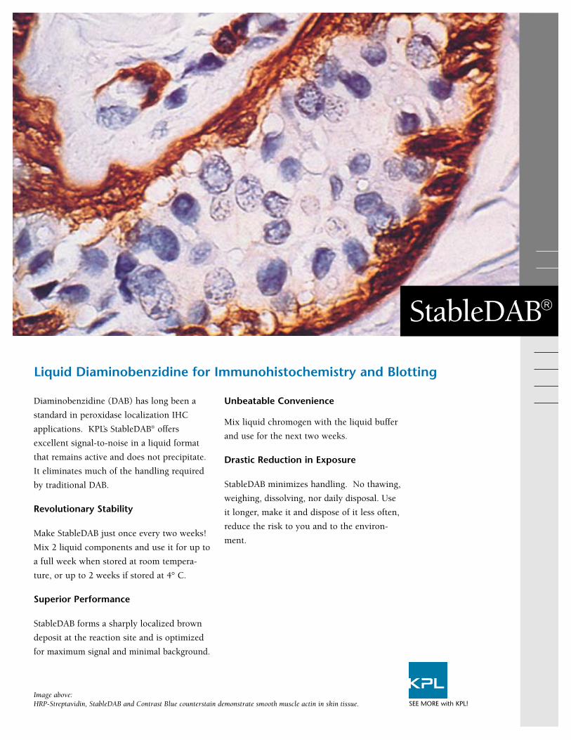
Image above: HRP-Streptavidin, StableDAB and Contrast Blue counterstain demonstrate smooth muscle actin in skin tissue. SEE MORE with KPL!
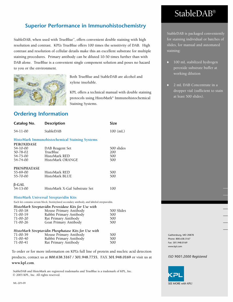
StableDAB, when used with TrueBlue™, offers convenient double staining with high
resolution and contrast. KPL’s TrueBlue offers 100 times the sensitivity of DAB. High
contrast and resolution of cellular details make this an excellent substrate for multiple
staining procedures. Primary antibody can be diluted 10-50 times further than with
DAB alone. TrueBlue is a convenient single component solution and poses no hazard
to you or the environment.
Both TrueBlue and StableDAB are alcohol and
xylene insoluble.
KPL offers a technical manual with double staining
protocols using HistoMark® Immunohistochemical
Staining Systems.
Superior Performance in Immunohistochemistry
Ordering Information
To order or for more information on KPL’s full line of protein and nucleic acid detection
products, contact us at 800.638.3167 / 301.948.7755, FAX 301.948.0169 or visit us at
www.kpl.com.
Catalog No. Description Size
54-11-00 StableDAB 100 (mL)
HistoMark Immunohistochemical Staining SystemsPEROXIDASE54-10-00 DAB Reagent Set 500 slides50-78-02 TrueBlue 20054-75-00 HistoMark RED 50054-74-00 HistoMark ORANGE 500
PHOSPHATASE55-69-00 HistoMark RED 50055-70-00 HistoMark BLUE 500
β-GAL54-13-00 HistoMark X-Gal Substrate Set 100
HistoMark Universal Streptavidin KitsEach kit contains serum block, biotinylated secondary antibody, and labeled streptavidin.
HistoMark Streptavidin Peroxidase Kits for Use with71-00-18 Mouse Primary Antibody 500 Slides71-00-19 Rabbit Primary Antibody 50071-00-20 Rat Primary Antibody 50071-00-26 Goat Primary Antibody 500
HistoMark Streptavidin Phosphatase Kits for Use with71-00-39 Mouse Primary Antibody 50071-00-40 Rabbit Primary Antibody 50071-00-41 Rat Primary Antibody 500
StableDAB®
Gaithersburg, MD 20878
Phone: 800.638.3167
Fax: 301.948.0169
www.kpl.com
ML-205-09
StableDAB is packaged conveniently
for staining individual or batches of
slides, for manual and automated
staining:
♦ 100 mL stabilized hydrogen
peroxide substrate buffer at
working dilution
♦ 2 mL DAB Concentrate in a
dropper vial (sufficient to stain
at least 500 slides).
StableDAB and HistoMark are registered trademarks and TrueBlue is a trademark of KPL, Inc.© 2003 KPL, Inc. All rights reserved.
SEE MORE with KPL!
ISO 9001:2000 Registered
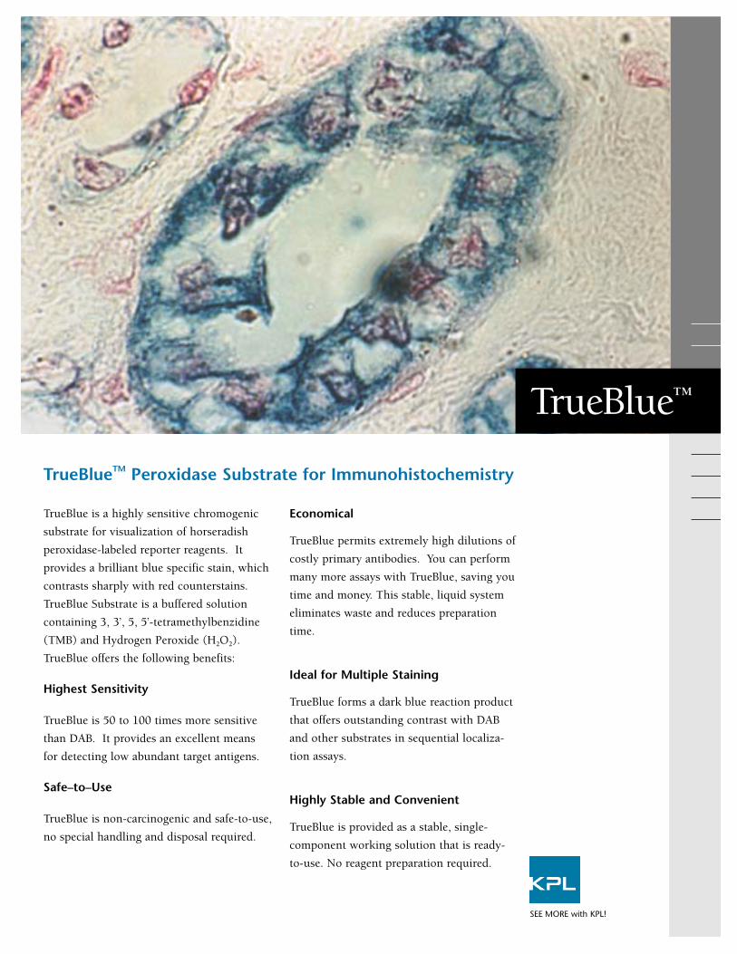
TrueBlue is a highly sensitive chromogenic
substrate for visualization of horseradish
peroxidase-labeled reporter reagents. It
provides a brilliant blue specific stain, which
contrasts sharply with red counterstains.
TrueBlue Substrate is a buffered solution
containing 3, 3’, 5, 5’-tetramethylbenzidine
(TMB) and Hydrogen Peroxide (H2O2).
TrueBlue offers the following benefits:
Highest Sensitivity
TrueBlue is 50 to 100 times more sensitive
than DAB. It provides an excellent means
for detecting low abundant target antigens.
Safe–to–Use
TrueBlue is non-carcinogenic and safe-to-use,
no special handling and disposal required.
Economical
TrueBlue permits extremely high dilutions of
costly primary antibodies. You can perform
many more assays with TrueBlue, saving you
time and money. This stable, liquid system
eliminates waste and reduces preparation
time.
Ideal for Multiple Staining
TrueBlue forms a dark blue reaction product
that offers outstanding contrast with DAB
and other substrates in sequential localiza-
tion assays.
Highly Stable and Convenient
TrueBlue is provided as a stable, single-
component working solution that is ready-
to-use. No reagent preparation required.
TrueBlueTM Peroxidase Substrate for Immunohistochemistry
TrueBlue™
SEE MORE with KPL!
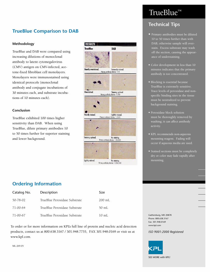
Methodology
TrueBlue and DAB were compared using
increasing dilutions of monoclonal
antibody to latent cytomegalovirus
(CMV) antigen on CMV-infected, ace-
tone-fixed fibroblast cell monolayers.
Monolayers were immunostained using
identical protocols (monoclonal
antibody and conjugate incubations of
30 minutes each, and substrate incuba-
tions of 10 minutes each).
Conclusion
TrueBlue exhibited 100 times higher
sensitivity than DAB. When using
TrueBlue, dilute primary antibodies 10
to 50 times further for superior staining
and lower background.
TrueBlue Comparison to DAB
Ordering Information
To order or for more information on KPL’s full line of protein and nucleic acid detection
products, contact us at 800.638.3167 / 301.948.7755, FAX 301.948.0169 or visit us at
www.kpl.com.
Catalog No. Description Size
50-78-02 TrueBlue Peroxidase Substrate 200 mL
71-00-64 TrueBlue Peroxidase Substrate 50 mL
71-00-67 TrueBlue Peroxidase Substrate 10 mL
• Primary antibodies must be diluted
10 to 50 times further than with
DAB, otherwise sample will over-
stain. Excess substrate may wash
off the section, causing the appear-
ance of understaining.
• Color development in less than 10
minutes indicates that the primary
antibody is too concentrated.
• Blocking is essential because
TrueBlue is extremely sensitive.
Trace levels of peroxidase and non-
specific binding sites in the tissue
must be neutralized to prevent
background staining.
• Peroxidase block solution
must be thoroughly removed by
washing; it can affect antibody
activity.
• KPL recommends non-aqueous
mounting reagent. Fading will
occur if aqueous media are used.
• Stained sections must be completely
dry or color may fade rapidly after
mounting.
Technical Tips
TrueBlue™
Gaithersburg, MD 20878
Phone: 800.638.3167
Fax: 301.948.0169
www.kpl.com
ML-269-05
SEE MORE with KPL!
ISO 9001:2000 Registered
Lighten your work load with KPL’s new line of quality
buffers. Offered as convenient liquid concentrates, they
eliminate the need for time-consuming buffer preparation.
KPL buffers are manufactured and carefully controlled for
quality and consistent performance with our ISO 9001:2008-
registered quality management system. All buffers are con-
ductivity controlled. KPL offers a variety of buffers designed
for use in general immunoassay applications such as Western
blotting, gel electrophoresis, ELISA and sample preparation.
Save preparation time with KPL’s new line of quality buffers!
Immunoassay Buffers
SEE MORE with KPL!
Description 1X Composition Applications
10X Tris-Glycine 25 mM Tris, 192 mM glycine. For preparing a standard Western blot transfer buffer Transfer Buffer pH 8.3 (Towbin) and as a gel electrophoresis buffer for native
Tris-glycine gels without SDS. .
10X Tris-Glycine-SDS 25 mM Tris, 192 mM glycine, Running buffer for sodium dodecyl sulfate – polyacrylamide0.1% SDS. pH 8.3 gel electrophoresis (SDS-PAGE) of proteins.
10X Phosphate-Buffered 10 mM Phosphate,150 mM NaCl. Wash buffer for general immunoassay applicationsSaline (PBS) pH 7.4 and sample preparation. Where applicable, Tween 20
is a nonionic detergent that reduces nonspecificPhosphate-Buffered 10 mM Phosphate, 150 mM NaCl, binding and protein-protein interaction during wash steps.Tween 20, (PBST) 0.05% Tween 20. pH 7.4
10X Tris-Buffered Saline 50 mM Tris, 150 mM NaCl. (TBS) pH 7.6
10% Tween 20 10% Tween 20 Nonionic detergent additive for PBS and Western blotting wash solutions. Reduces nonspecific binding and protein- protein interactions.
See reverse side.
Catalog# Description Size
51-10-01 10X Tris-Glycine Transfer Buffer 1 L
51-10-02 5 L
51-11-01 10X Tris-Glycine SDS Buffer 1 L
51-11-02 5 L
51-13-01 10X Phosphate-Buffered Saline (PBS) 1 L
51-13-02 5 L
51-14-01 10X Phosphate-Buffered Saline with 200 mL
51-14-02 Tween 20 (PBST) 1 L
51-17-01 10X Tris-Buffered Saline (TBS) 1 L
51-17-02 5 L
51-12-01 10% Tween 20 200 mL
51-12-02 1 L
51-20-01 5% SDS 200 mL
50-86-05 20X SSC 1 L
51-15-01 10X Dulbecco's PBS (D-PBS) 1 L
51-15-02 5 L
51-16-01 10X Dulbecco's PBS with Tween 20 (D-PBST) 200 mL
51-16-02 1 L
51-18-01 10X Tris Buffered Saline with 0.5% Tween 20 200 mL
51-18-02 (TBST) 1 L
51-19-01 10X Tris Buffered Saline with 10% Tween 20 200 mL
51-19-02 (TBST) 1 L
Ordering Information
To order or for more information on KPL’s full line of protein detection products, contact us at800.638.3167 / 301.948.7755, fax 301.948.0169 or visit us at www.kpl.com.
Other Immunoassay Support
Reagents Available from KPL
ELISA and Western Blotting
Applications:
• AP and HRP Conjugate Stabilizers
• 10% BSA Diluent/Blocking Solution
Concentrate
• Milk/Diluent Blocking Solution
Concentrate
• 20X Wash Solution Concentrate
Western Blotting Applications:
• Detector Block
• Biotin Wash Kit
ELISA Applications:
• Coating Solution Concentrate
• Stop Solutions
Immunoassay Buffers
Gaithersburg, MD 20878
Phone: 800.638.3167
Fax: 301.948.0169
www.kpl.com
ML363-03
©2009 KPL, Inc. All rights reserved.SEE MORE with KPL!
ISO 9001:2008 Registered
KPL Quality Buffers (cont’d)
Take the time and effort out of your buffer preparation. Choose from our line of popular buffers andenjoy the benefits - convenience, reliability and confidence - that KPL’s pretested, quality buffersbring to your research.
Description 1X Composition Applications
5% SDS 5% Sodium Detergent surfactant used in preparing proteins for Dodecyl Sulfate sodium dodecyl sulfate – polyacrylamide gel electro-
phoresis (SDS-PAGE) and as an additive to transfer buffers in Western blotting. SDS increases the elution rate of proteins from the gel.
20X SSC 15 mM sodium citrate, Used in a variety of molecular biology applications. 150 mM NaCl Facilitates transfer of nucleic acids to membranes. in DEPC water. pH 7.0
10X Dulbecco's 8.5 mM sodium phosphate, For sample preparation and as a wash buffer in general PBS (D-PBS) 1.5 mM potassium phosphate, immunoassay, tissue, and cell culture applications.
137 mM NaCl. pH 7.4 Not formulated with magnesium or calcium salts. Where applicable, Tween 20 is a non-ionic detergent that reducesnonspecific binding.
10X Dulbecco's 8.5 mM sodium phosphate,with Tween 20. 1.5 mM potassium phosphate,(D-PBST) 137 mM NaCl, 2.7 mM KCl.
0.05% Tween 20. pH 7.4
10X Tris-Buffered 50 mM Tris, 150 mM NaCl, As a general wash buffer in immunoassay applications. Saline with 0.5% 0.05% Tween 20. pH 7.6 Tween 20 is a non-ionic detergent that reduces nonspecific Tween 20 (TBST) binding. The 10% Tween 20 formulation provides a more
stringent wash than standard TBST formulations. 10X Tris-Buffered 50 mM Tris, 150 mM NaCl,Saline with 10% 1.0% Tween 20. pH 7.6 Tween 20 (TBST)

Versatile and Convenient Probes
KPL's line of colloidal gold-labeled reagents
include anti-human, anti-mouse, anti-rabbit,
anti-biotin antibodies and streptavidin. These
antibodies are affinity purified to minimize
cross-reactivity. Anti-biotin and streptavidin
conjugates provide a sensitive indication of
biotin with low background.
Advantages of gold conjugates
• Different sized gold particles (5 nm and 40nm) allow for a variety of uses for these con-jugates.
• Greater stability in liquid form than enzymeconjugates.
• Eliminates the need for an enzyme blockingstep.
• 5 nm gold particles permit better penetrationof tissue due to their small size.
• Highly sensitive technique detects 60picograms of protein in dot blotting.
Silver Enhancer Kits
For maximum signal, KPL's Silver Enhancer
Kits amplify the gold conjugates signal 10 to
100 times. The resulting brown/black silver
stain is permanent and offers sharp resolution
and contrast with conventional counterstains.
Applications for Gold Conjugates
40 nm gold conjugates are ideal for use inthese applications.
• Lateral flow assays
• Light Microscopy
• Blotting
• Immunohistochemistry and in situ hybridiza-tion applications
5 nm gold conjugates are ideal for use in theseapplications.
• Light Microscopy
• Electron Microscopy
Easy to use, highly sensitive systems for visualizing proteins
Gold Conjugates
SEE MORE with KPL!
Ordering Information
To order or for more information on KPL’s full line
of protein detection products, contact us at
800.638.3167 / 301.948.7755, fax 301.948.0169 or
visit us at www.kpl.com.
Catalog# Description Size
Gold LabeledEach system contains Gold Conjugate Dilution Buffer
57-10-06 Anti-Human IgG (H+L) System, 40 nm 3.0 mL
58-10-06 Anti-Human IgG (H+L) System, 5 nm 1.0 mL
57-18-06 Anti-Mouse IgG (H+L) System, 40 nm 3.0 mL
58-18-06 Anti-Mouse IgG (H+L) System, 5 nm 1.0 mL
57-15-06 Anti-Rabbit IgG (H+L) System, 40 nm 3.0 mL
58-15-06 Anti-Rabbit IgG (H+L) System, 5 nm 1.0 mL
57-40-06 Anti-Biotin System, 40 nm 3.0 mL
58-40-06 Anti-Biotin System, 5 nm 1.0 mL
57-30-06 Streptavidin System, 40 nm 3.0 mL
58-30-06 Streptavidin System, 5 nm 1.0 mL
Silver Enhancer Kits
55-22-01 Silver Enhancer Kit for Microscopy Kit includes: 1 mL of Gold Conjugate100 mL of Gold Conjugate Dilution Buffer
Solution A 25 mL
Solution B 25 mL
55-22-02 Silver Enhancer Kit for Membrane ApplicationsKit includes: 3 mL of Gold Conjugate250 mL of Gold Conjugate Dilution Buffer
Solution A 250 mL
Solution B 250 mL
Gold Conjugates
Gaithersburg, MD 20878
Phone: 800.638.3167
Fax: 301.948.0169
www.kpl.com
ML248-06
© 2005 KPL, Inc. All rights reserved.
CMV infected cells, human anti-CMVserum with gold-labeled goat anti-humanIgG (H+L), 5 nm with Microscopy SilverEnhancer Kit and Eosin.
What are the advantages ofusing gold conjugates versusenzyme-labeled conjugates?
One advantage is the possibility ofcross-reactivity with naturally-occurring enzymes is non-existent,as compared to enzyme-labeledconjugates. Another advantage is asingle-step detection versusenzyme conjugate and substrate.
FREQUENTLY ASKED
QUESTIONS
Human normal lung tissue, anti-epithelialmembrane antigen with gold-labeled goatanti-mouse IgG (H+L), 5 nm withMicroscopy Silver Enhancer Kit andContrast BLUE.
Pictured on front and above:adult grasshopper.
SEE MORE with KPL!
ISO 9001:2000 Registered
Since 1979 KPL has provided quality affinity
purified antibodies to researchers worldwide.
Over the years we have refined our production
process to provide antibodies with high potency
and consistent performance in immunoassays.
From the start KPL gives careful consideration
to immunogen preparation, using a highly
purifed formulation to generate antiserum. KPL
pools antiserum from multiple animals to
reduce natural animal to animal serum variabili-
ty. During the purification process our ISO
9001:2008-certified quality procedures impose
rigorous standards for potency and cross-reac-
tivity. The result is standardized antibody lots
with excellent reproducibility.
Our extensive line of peroxidase (HRP) conju-
gates is available across a range of animal
species, including human, mouse, rabbit and rat
antibodies, as well as other animal species and
streptavidin. They are affinity purified and in
some cases further adsorbed to minimize cross-
reactivity between animal species or shared
reactivity with other immunoglobulin classes.
HRP-labeled F(ab’)2 fragment antibodies are
offered for assays requiring extremely low back-
ground and absence of F(c)-mediated binding.
KPL reacts HRP of the highest quality with
affinity purified antibodies and streptavidin
using the periodate method of Nakane and
Kawaoi. Special features of HRP include:
• faster catalytic rate than alkaline phosphatase
• generates more product in shorter incubation
times
• provides maximum sensitivity, low nonspe-
cific binding
• ideal for ELISA, Western blotting and
immunohistology applications.
KPL Peroxidase Conjugates: Time-tested, Sensitive and Reliable
Peroxidase Conjugates
SEE MORE with KPL!
Peroxidase Conjugates Ordering Information(Partial listing)
Peroxidase Conjugates
Gaithersburg, MD 20878
Phone: 800.638.3167
Fax: 301.948.0169
www.kpl.com
ISO 9001:2008 Registered
For research use only.©2009 KPL, Inc. All rights reserved.ABTS is a registered trademark of Roche Biochemicals.ML371-01
SEE MORE with KPL!
Visit our website at www.kpl.com for a complete listing of HRP-labeled antibodies.
To order or for more information on KPL’s protein research products, contact us at
800.638.3167 / 301.948.7755, FAX 301.948.0169 or visit us at www.kpl.com.
KPL offers a range of sensitive
substrates for use with HRP conju-
gates. They provide a choice of
intense colors for ELISA, blotting
and cell staining applications.
ELISA
• ABTS® 1- and 2-Component
Microwell Peroxidase Substrates
• SureBlueTM TMB Peroxidase
Substrate
• SureBlue ReserveTM TMB
Peroxidase Substrate
• TMB Peroxidase Substrate
Blotting
• 4 CN Peroxidase Substrate
• TMB Membrane Peroxidase
Substrate
• LumiGLO®Chemiluminescent
Substrate
• LumiGLO ReserveTM
Chemiluminescent Substrate
Whichever substrate you choose,
enjoy the benefits of excellent signal-
to-noise and reproducibility.
Catalog# Description Size
04-10-06 HRP-labeled Goat Anti-Human IgG (H+L) 0.1 mg
04-10-17 HRP-labeled Goat Anti-Human IgA+IgG+IgM (H+L), MSA 0.1 mg
04-10-20 HRP-labeled Goat Anti-Human IgG (Fc) 0.1 mg
074-1002 HRP-labeled Goat Anti-Human IgG (γ) 1.0 mg
074-1003 HRP-labeled Goat Anti-Human IgM (μ) 1.0 mg
074-1004 HRP-labeled Goat Anti-Human IgE (ε) 1.0 mg
074-1006 HRP-labeled Goat Anti-Human IgG (H+L) 1.0 mg
074-1007 HRP-labeled Goat Anti-Human IgA+IgG+IgM (H+L) 1.0 mg
14-10-01 HRP-labeled Goat Anti-Human IgA (α) 0.5 mg
214-1002 HRP-labeled F(ab’)2 Goat Anti-Human IgG (γ) 0.5 mg
214-1003 HRP-labeled F(ab’)2 Goat Anti-Human IgM (μ) 0.5 mg
214-1006 HRP-labeled F(ab’)2 Goat Anti-Human IgG (H+L) 0.5 mg
474-1002 HRP-labeled Goat Anti-Human IgG (γ), Liquid 1.0 mL
474-1003 HRP-labeled Goat Anti-Human IgM (μ), Liquid 1.0 mL
474-1006 HRP-labeled Goat Anti-Human IgG (H+L), Liquid 1.0 mL
04-18-06 HRP-labeled Goat Anti-Mouse IgG (H+L), HSA 0.1 mg
04-18-15 HRP-labeled Goat Anti-Mouse IgG (H+L), RtSA, HSA 0.1 mg
04-18-18 HRP-labeled Goat Anti-Mouse IgG (H+L), RbSA, HSA 0.1 mg
074-1802 HRP-labeled Goat Anti-Mouse IgG (γ), HSA 1.0 mg
074-1803 HRP-labeled Goat Anti-Mouse IgM (μ), HSA 1.0 mg
074-1806 HRP-labeled Goat Anti-Mouse IgG (H+L), HSA 1.0 mg
074-18-061 HRP-labeled Goat Anti-Mouse IgG (H+L), XSA 1.0 mg
074-1807 HRP-labeled Goat Anti-Mouse IgA+IgG+IgM (H+L), HSA 1.0 mg
074-1809 HRP-labeled Goat Anti-Mouse IgG+IgM (H+L), HSA 1.0 mg
14-18-01 HRP-labeled Goat Anti-Mouse IgA (α), HSA 0.5 mg
214-1802 HRP-labeled F(ab’)2 Goat Anti-Mouse IgG (γ), HSA 0.5 mg
214-1806 HRP-labeled F(ab’)2 Goat Anti-Mouse IgG (H+L), HSA 0.5 mg
474-1802 HRP-labeled Goat Anti-Mouse IgG (γ), HSA, Liquid 1.0 mL
474-1806 HRP-labeled Goat Anti-Mouse IgG (H+L), HSA, Liquid 1.0 mL
074-1506 HRP-labeled Goat Anti-Rabbit IgG (H+L) 1.0 mg
074-15-061 HRP-labeled Goat Anti-Rabbit IgG (H+L), XSA 1.0 mg
074-1516 HRP-labeled Goat Anti-Rabbit IgG (H+L), HSA 1.0 mg
214-1516 HRP-labeled F(ab’)2 Goat Anti-Rabbit IgG (H+L), HSA 0.5 mg
474-1506 HRP-labeled Goat Anti-Rabbit IgG (H+L), Liquid 1.0 mL
474-1516 HRP-labeled Goat Anti-Rabbit IgG (H+L), HSA, Liquid 1.0 mL
14-30-00 HRP-labeled Streptavidin 0.5 mg
474-3000 HRP-labeled Streptavidin, Liquid, Molecular Grade 1.0 mL
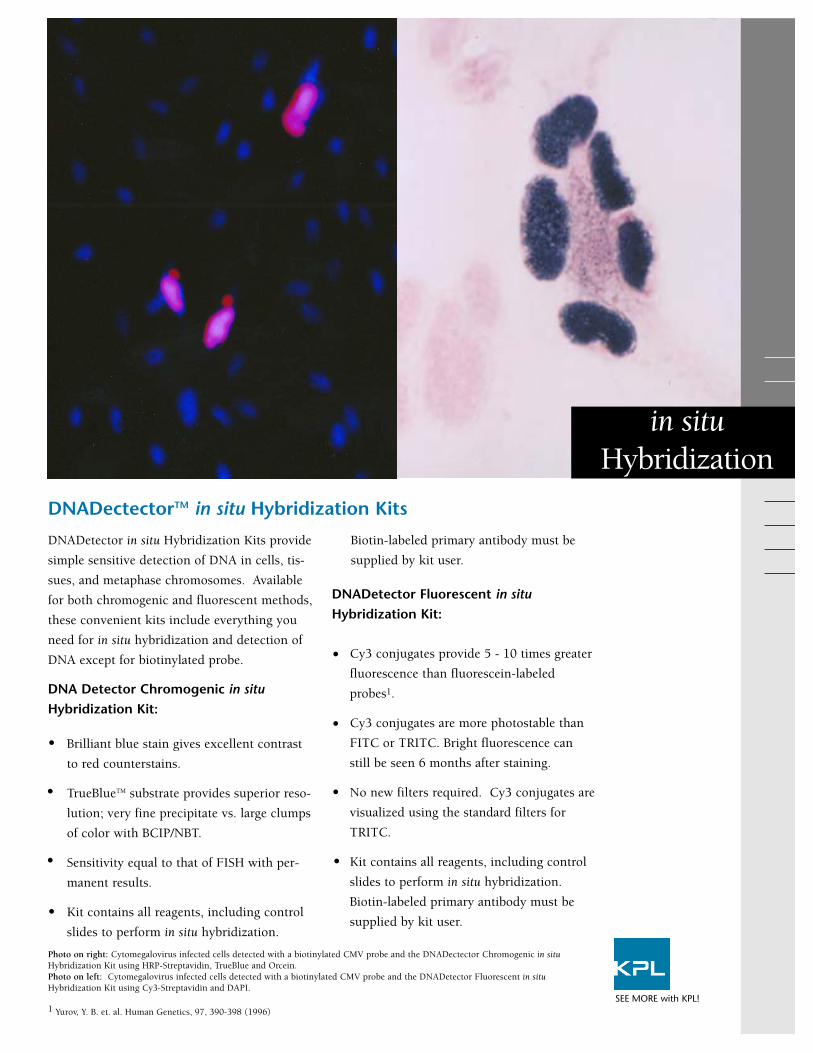
DNADetector in situ Hybridization Kits provide
simple sensitive detection of DNA in cells, tis-
sues, and metaphase chromosomes. Available
for both chromogenic and fluorescent methods,
these convenient kits include everything you
need for in situ hybridization and detection of
DNA except for biotinylated probe.
DNA Detector Chromogenic in situHybridization Kit:
Brilliant blue stain gives excellent contrast
to red counterstains.
TrueBlueTM substrate provides superior reso-
lution; very fine precipitate vs. large clumps
of color with BCIP/NBT.
Sensitivity equal to that of FISH with per-
manent results.
Kit contains all reagents, including control
slides to perform in situ hybridization.
Biotin-labeled primary antibody must be
supplied by kit user.
DNADetector Fluorescent in situHybridization Kit:
Cy3 conjugates provide 5 - 10 times greater
fluorescence than fluorescein-labeled
probes1.
Cy3 conjugates are more photostable than
FITC or TRITC. Bright fluorescence can
still be seen 6 months after staining.
No new filters required. Cy3 conjugates are
visualized using the standard filters for
TRITC.
Kit contains all reagents, including control
slides to perform in situ hybridization.
Biotin-labeled primary antibody must be
supplied by kit user.
DNADectectorTM in situ Hybridization Kits
in situHybridization
SEE MORE with KPL!
Photo on right: Cytomegalovirus infected cells detected with a biotinylated CMV probe and the DNADectector Chromogenic in situHybridization Kit using HRP-Streptavidin, TrueBlue and Orcein. Photo on left: Cytomegalovirus infected cells detected with a biotinylated CMV probe and the DNADetector Fluorescent in situHybridization Kit using Cy3-Streptavidin and DAPI.
1 Yurov, Y. B. et. al. Human Genetics, 97, 390-398 (1996)
•
•
•
•
•
•
•
•
DNADetector Chromogenic in situHybridization Kit
Contents:Hybridization Buffers20X SSCHRP Streptavidin ConjugateWash SolutionConjugate Diluent bufferTrueBlue Peroxidase SubstrateOrcein and Eosin CounterstainsControl Hybridization CocktailControl SlideSufficient reagents are provided for the hybridization
and detection of 50 samples.
DNADetector Fluorescent in situHybridization Kit
Contents:Hybridization Buffers20X SSCCy3 Streptavidin ConjugateWash SolutionConjugate Diluent bufferDAPI CounterstainFluorescent Mounting MediaControl Hybridization CocktailControl SlideSufficient reagents are provided for the hybridization
and detection of 50 samples.
DNADectectorTM in situ Hybridization Kits
Ordering Information
To order or for more information on KPL’s full line of protein and nucleic acid
detection products, contact us at 800.638.3167 / 301.948.7755, FAX 301.948.0169
or visit us at www.kpl.com.
Catalog# Description Size
60-03-00 Chromogenic in situ Hybridization Kit 50 samples
60-05-00 Fluorescent in situ Hybridization Kit, Cy3/DAPI 50 samples
Detector Probe Labeling Kits
60-01-00 Random Primer Biotinylation Kit 30 reactions
60-01-01 PCR Biotinylation Kit 30 reactions
60-01-02 RNA in vitro Transcription Kit 20 reactionsGaithersburg, MD 20878
Phone: 800.638.3167
Fax: 301.948.0169
www.kpl.com
ML224-111203
DNADetector is a trademark of KPL, Inc.Cy is a trademark of Amersham International PLC.
© 2003 KPL, Inc. All rights reserved.SEE MORE with KPL!
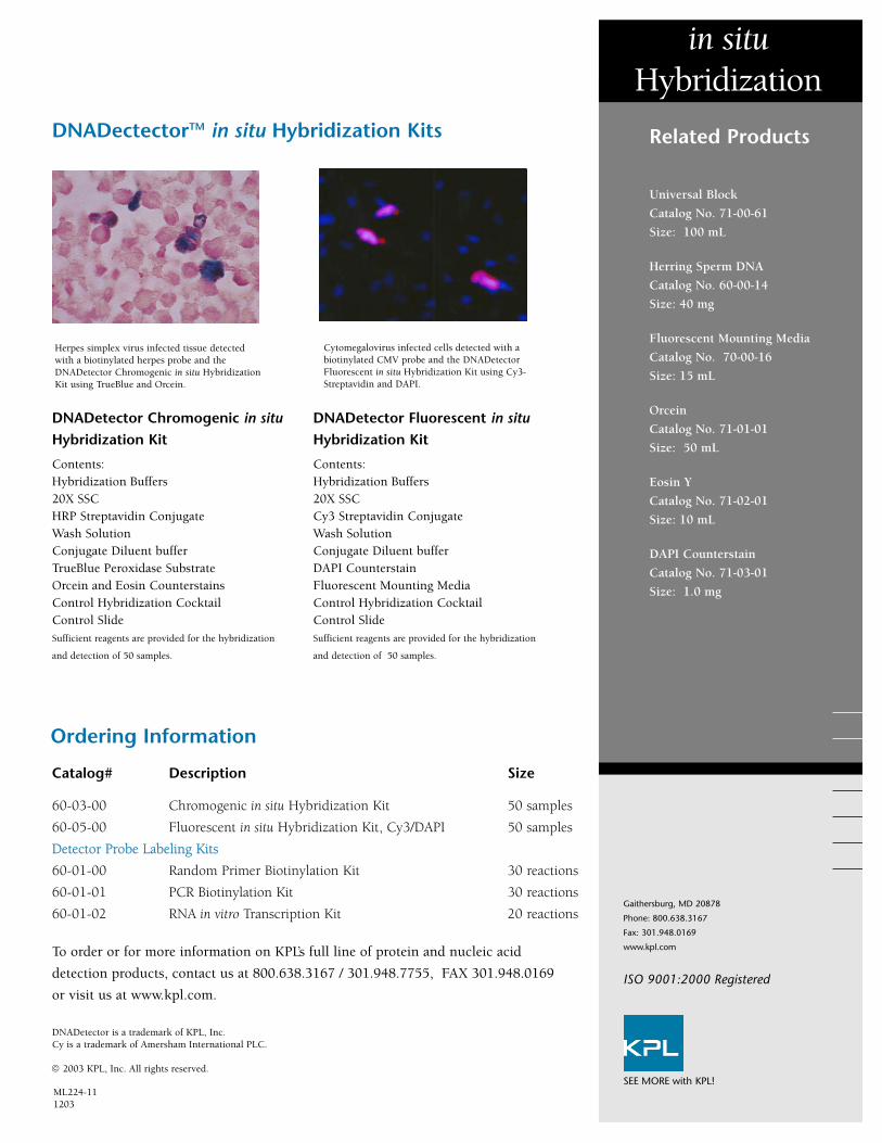
Herpes simplex virus infected tissue detectedwith a biotinylated herpes probe and theDNADetector Chromogenic in situ HybridizationKit using TrueBlue and Orcein.
in situHybridization
Cytomegalovirus infected cells detected with abiotinylated CMV probe and the DNADetectorFluorescent in situ Hybridization Kit using Cy3-Streptavidin and DAPI.
Related Products
Universal Block
Catalog No. 71-00-61
Size: 100 mL
Herring Sperm DNA
Catalog No. 60-00-14
Size: 40 mg
Fluorescent Mounting Media
Catalog No. 70-00-16
Size: 15 mL
Orcein
Catalog No. 71-01-01
Size: 50 mL
Eosin Y
Catalog No. 71-02-01
Size: 10 mL
DAPI Counterstain
Catalog No. 71-03-01
Size: 1.0 mg
ISO 9001:2000 Registered
KPL offers two new SureLINKTM Fluorescein Labeling Kits
that enable scientists to label proteins with the fluo-
rophores FITC or FAM-X (FAM fluorophore with succin-
imidyl ester and a seven atom aminohexanoyl spacer,
known as ‘X’):
SureLINKTM Fluorescein (FITC)
SureLINK Fluorescein-X (FAM-X)
FAM-X and FITC share the same excitation (495 nm) and
emission (520 nm) wavelengths. However, FAM-X forms
a stable bond with the amine group of the protein that is
more resistant to hydrolysis, minimizing the release of dye
during immunoassay procedures and storage. In addition,
the tendency towards quenching observed with FITC con-
jugates is diminished with FAM-X due to the spacer sepa-
rating the fluorophore from the conjugated biomolecule.
Convenient kits save time and reagents
Optimized protocols enable labeling and purifying in just
two hours with minimal hands-on time. Kits contain reac-
tion-size reagent vials with all the essential components.
Comprehensive conjugation guide
A thorough guide is provided with both kits, and includes
protocols, frequently asked questions and a trouble-
shooting guide.
Simple, fast, convenient fluorescein labeling kits
SEE MORE with KPL!
SureLINKTM Fluorescein Labeling Kits
Fluorescein isothiocyanate (FITC)
Fluorescein X (FAM-X): 6-(fluorescein-5-car-boxamido) hexanoic acid, succinimidyl ester
Ordering Information
To order or for more information, contact us at 800.638.3167 / 301.948.7755,
fax 301.948.0169 or visit us at www.kpl.com.
Catalog# Description Size
82-00-01 SureLINKTM Fluorescein (FITC) Labeling Kit 5 reactions
82-00-02 SureLINKTM Fluorescein-X (FAM-X) Labeling Kit 5 reactions
SureLINK Kit Components
SureLINK Fluorescein (FITC) Labeling Kit: SureLINK FITC, 5 x 0.3 mg 5 Carbonate Bicarbonate Buffer Capsules
SureLINK Fluorescein-X (FAM-X) Labeling Kit: SureLINK Fluorescein-X (FAM-X), 5 x 0.3 mg Borate Buffer
Both kits: Anhydrous DMF, 5 Spin-Pure Filters, 10 Amber Reaction Tubes
SureLINKTM
Labeling Kits
Gaithersburg, MD 20878
Phone: 800.638.3167
Fax: 301.948.0169
www.kpl.com
ISO 9001:2000 Registered
ML350-01
For research use only.©2007 KPL, Inc. All rights reserved.
SEE MORE with KPL!
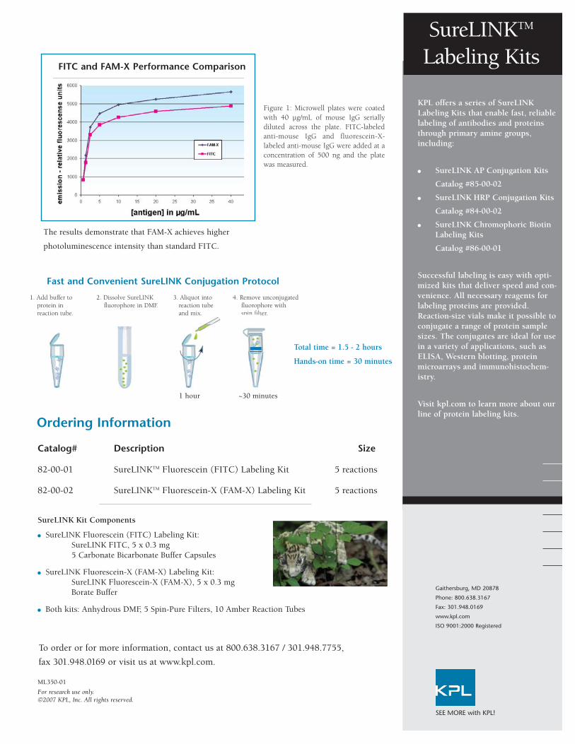
FITC and FAM-X Performance Comparison
Figure 1: Microwell plates were coatedwith 40 µg/mL of mouse IgG seriallydiluted across the plate. FITC-labeledanti-mouse IgG and fluorescein-X-labeled anti-mouse IgG were added at aconcentration of 500 ng and the platewas measured.
Fast and Convenient SureLINK Conjugation Protocol
KPL offers a series of SureLINKLabeling Kits that enable fast, reliablelabeling of antibodies and proteinsthrough primary amine groups,including:
SureLINK AP Conjugation Kits
Catalog #85-00-02
SureLINK HRP Conjugation Kits
Catalog #84-00-02
SureLINK Chromophoric Biotin Labeling Kits
Catalog #86-00-01
Successful labeling is easy with opti-mized kits that deliver speed and con-venience. All necessary reagents forlabeling proteins are provided.Reaction-size vials make it possible toconjugate a range of protein samplesizes. The conjugates are ideal for usein a variety of applications, such asELISA, Western blotting, proteinmicroarrays and immunohistochem-istry.
Visit kpl.com to learn more about ourline of protein labeling kits.
The results demonstrate that FAM-X achieves higher
photoluminescence intensity than standard FITC.
1 hour ~30 minutes
Total time = 1.5 - 2 hours
Hands-on time = 30 minutes
1. Add buffer to 2. Dissolve SureLINK 3. Aliquot into 4. Remove unconjugated protein in fluorophore in DMF. reaction tube fluorophore withreaction tube. and mix. spin filter.

KPL’s new ReserveAPTM Alkaline Phosphatase
(AP)-labeled antibody conjugates exhibit high
potency and consistent performance in
immunoassays. These conjugates are the result
of advances in our conjugation technology and
offer higher signal than our current line of AP
conjugates while meeting the same standards
for low background, stability and reproducibili-
ty. They are intended for demanding
immunoassays that require high detection sen-
sitivity, including ELISA, Western blotting and
immunohistology.
Higher Potency
ReserveAP Conjugates are affinity purified and
conjugated to the highest grade of alkaline
phosphatase. In our studies, they generate two-
to-three fold higher values than our current
line of AP conjugates in ELISA and outperform
AP conjugates offered by other manufacturers.
Higher conjugate dilutability is also observed
without loss of linearity, enabling precious
antigen or primary antibody conservation.
Consistent Performance
Reproducible antibody conjugation and consis-
tent performance are verified according to our
ISO 9001:2000-registered quality assurance
system. Lot consistency studies in which three
lots were studied by ELISA indicated minimal
variability.
Excellent Value
ReserveAP Conjugates provide high perform-
ance at an economical price.
Spice up your assay with our red hot high potency ReserveAPTM Conjugates!
ReserveAPTM Conjugates
SEE MORE with KPL!
ReserveAPTM Conjugates Ordering Information
Catalog# Description Size
0751-1001 Goat Anti-Human IgA (α) 1.0 mg
0751-1002 Goat Anti-Human IgG (γ) 1.0 mg
0751-1003 Goat Anti-Human IgM (µ) 1.0 mg
0751-1004 Goat Anti-Human IgE (ε) 1.0 mg
0751-1006 Goat Anti-Human IgG (H+L) 1.0 mg
0751-1007 Goat Anti-Human IgA+IgG+IgM (H+L) 1.0 mg
2151-1002 F(ab’)2 Anti-Human IgG (γ) 0.5 mg
4751-1002 Goat Anti-Human IgG (γ) liquid 1.0 mg
4751-1003 Goat Anti-Human IgM (µ) liquid 1.0 mg
4751-1006 Goat Anti-Human IgG (H+L) liquid 1.0 mg
0751-1802 Goat Anti-Mouse IgG (γ) HSA 1.0 mg
0751-1803 Goat Anti-Mouse IgM (µ) HSA 1.0 mg
0751-1806 Goat Anti-Mouse IgG (H+L) HSA 1.0 mg
0751-1809 Goat Anti-Mouse IgG +IgM (H+L) HSA 1.0 mg
4751-1802 Goat Anti-Mouse IgG (γ) HSA, liquid 1.0 mg
4751-1806 Goat Anti-Mouse IgG (H+L) HSA, liquid 1.0 mg
151-18-01 Goat Anti-Mouse IgA (α) HSA 0.5 mg
0751-1807 Goat Anti-Mouse IgA+IgG+IgM (H+L) HSA 1.0 mg
0751-1506 Goat Anti-Rabbit IgG (H+L) 1.0 mg
0751-1516 Goat Anti-Rabbit IgG (H+L) HSA 1.0 mg
4751-1506 Goat Anti-Rabbit IgG (H+L), liquid 1.0 mg
4751-1516 Goat Anti-Rabbit IgG (H+L) HSA, liquid 1.0 mg
HSA=Human Serum Adsorbed
AP Substrates
Gaithersburg, MD 20878
Phone: 800.638.3167
Fax: 301.948.0169
www.kpl.com
ISO 9001:2000 Registered
ML349-04
For research use only.©2007 KPL, Inc. All rights reserved.
SEE MORE with KPL!
Visit our website at www.kpl.com for a complete listing of ReserveAP conjugates. To
order or for more information on KPL’s protein research products, contact us at
800.638.3167 / 301.948.7755, FAX 301.948.0169 or visit us at www.kpl.com.
KPL offers a range of sensitive sub-
strates for the detection and quantifi-
cation of phosphatase (AP) activity.
They provide a choice of intense col-
ors for ELISA and blotting applica-
tions.
ELISA
• FirePhosTM AP Microwell
Substrate
• BluePhos® AP Microwell
Substrate
• pNPP Phosphatase Substrate
Blotting
• FirePhos AP Membrane
Substrate
• BCIP/NBT Phosphatase Substrate
• PhosphaGLO AP Substrate
• PhosphaGLOTM Reserve AP
Substrate
Whichever substrate you choose,
enjoy the benefits of excellent signal-
to-noise with higher sensitivity and
lower background than that of other
AP substrates. Visit www.kpl.com
for more information.
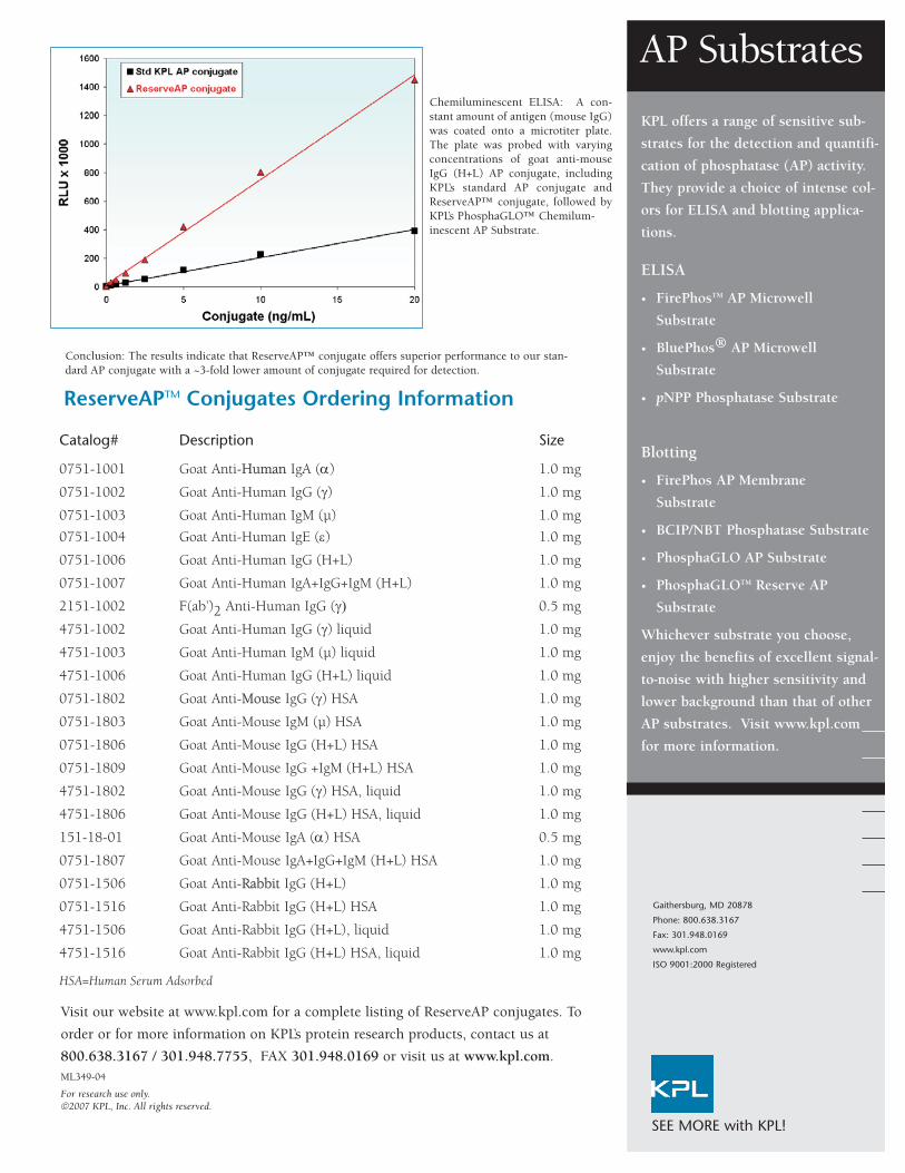
Chemiluminescent ELISA: A con-stant amount of antigen (mouse IgG)was coated onto a microtiter plate.The plate was probed with varyingconcentrations of goat anti-mouseIgG (H+L) AP conjugate, includingKPL’s standard AP conjugate andReserveAP™ conjugate, followed byKPL’s PhosphaGLO™ Chemilum-inescent AP Substrate.
Conclusion: The results indicate that ReserveAP™ conjugate offers superior performance to our stan-dard AP conjugate with a ~3-fold lower amount of conjugate required for detection.

KPL, Inc.Gaithersburg, MD
Phone: 800.638.3167 Fax: 301.948.0169www.kpl.com ML329-01
SEE MORE with KPL!Get specific signal without
background and SEE MORE!
Where Better Science Begins