Embed Size (px)
Citation preview
417
Analele Universităţii din Oradea, Fascicula Protectia Mediului Vol. XXV 2015
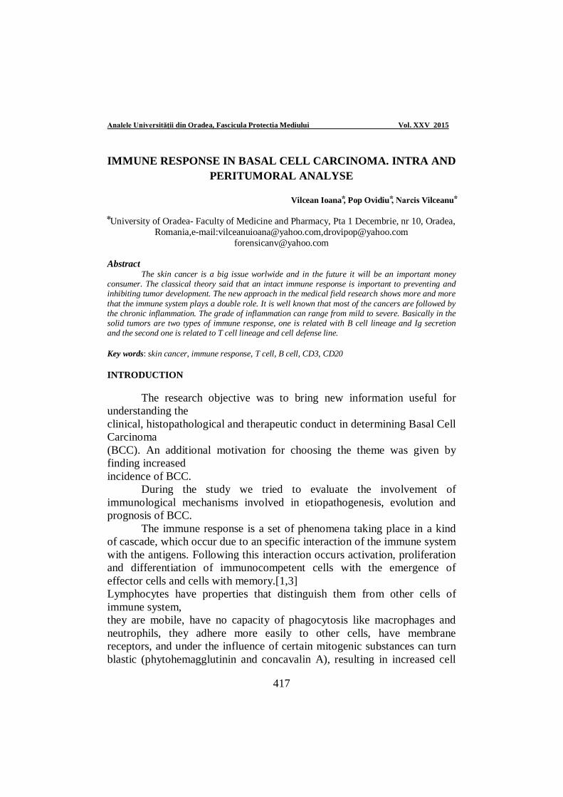
IMMUNE RESPONSE IN BASAL CELL CARCINOMA. INTRA AND PERITUMORAL ANALYSE
Vilcean Ioana , Pop Ovidiu , Narcis Vilceanu
University of Oradea- Faculty of Medicine and Pharmacy, Pta 1 Decembrie, nr 10, Oradea,
Romania,e-mail:[email protected],[email protected] [email protected]
Abstract The skin cancer is a big issue worlwide and in the future it will be an important money consumer. The classical theory said that an intact immune response is important to preventing and inhibiting tumor development. The new approach in the medical field research shows more and more that the immune system plays a double role. It is well known that most of the cancers are followed by the chronic inflammation. The grade of inflammation can range from mild to severe. Basically in the solid tumors are two types of immune response, one is related with B cell lineage and Ig secretion and the second one is related to T cell lineage and cell defense line. Key words: skin cancer, immune response, T cell, B cell, CD3, CD20 INTRODUCTION The research objective was to bring new information useful for understanding the clinical, histopathological and therapeutic conduct in determining Basal Cell Carcinoma (BCC). An additional motivation for choosing the theme was given by finding increased incidence of BCC. During the study we tried to evaluate the involvement of immunological mechanisms involved in etiopathogenesis, evolution and prognosis of BCC. The immune response is a set of phenomena taking place in a kind of cascade, which occur due to an specific interaction of the immune system with the antigens. Following this interaction occurs activation, proliferation and differentiation of immunocompetent cells with the emergence of effector cells and cells with memory.[1,3] Lymphocytes have properties that distinguish them from other cells of immune system, they are mobile, have no capacity of phagocytosis like macrophages and neutrophils, they adhere more easily to other cells, have membrane receptors, and under the influence of certain mitogenic substances can turn blastic (phytohemagglutinin and concavalin A), resulting in increased cell
418
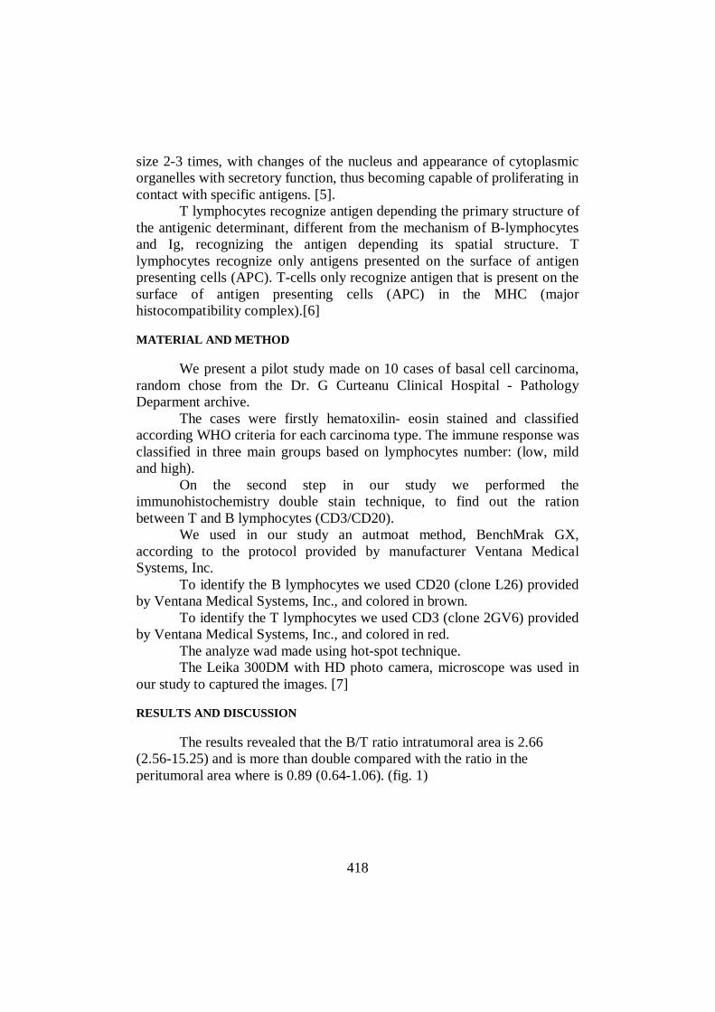
size 2-3 times, with changes of the nucleus and appearance of cytoplasmic organelles with secretory function, thus becoming capable of proliferating in contact with specific antigens. [5]. T lymphocytes recognize antigen depending the primary structure of the antigenic determinant, different from the mechanism of B-lymphocytes and Ig, recognizing the antigen depending its spatial structure. T lymphocytes recognize only antigens presented on the surface of antigen presenting cells (APC). T-cells only recognize antigen that is present on the surface of antigen presenting cells (APC) in the MHC (major histocompatibility complex).[6]
MATERIAL AND METHOD We present a pilot study made on 10 cases of basal cell carcinoma, random chose from the Dr. G Curteanu Clinical Hospital - Pathology Deparment archive. The cases were firstly hematoxilin- eosin stained and classified according WHO criteria for each carcinoma type. The immune response was classified in three main groups based on lymphocytes number: (low, mild and high). On the second step in our study we performed the immunohistochemistry double stain technique, to find out the ration between T and B lymphocytes (CD3/CD20). We used in our study an autmoat method, BenchMrak GX, according to the protocol provided by manufacturer Ventana Medical Systems, Inc. To identify the B lymphocytes we used CD20 (clone L26) provided by Ventana Medical Systems, Inc., and colored in brown. To identify the T lymphocytes we used CD3 (clone 2GV6) provided by Ventana Medical Systems, Inc., and colored in red. The analyze wad made using hot-spot technique. The Leika 300DM with HD photo camera, microscope was used in our study to captured the images. [7]
RESULTS AND DISCUSSION
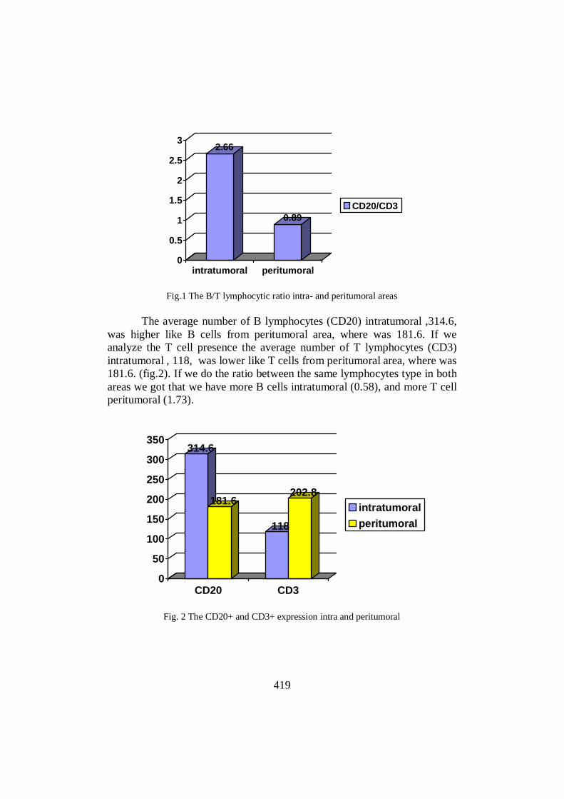
The results revealed that the B/T ratio intratumoral area is 2.66 (2.56-15.25) and is more than double compared with the ratio in the peritumoral area where is 0.89 (0.64-1.06). (fig. 1)
419
2.66
0.89
0
0.5
1
1.5
2
2.5
3
intratumoral peritumoral
CD20/CD3
Fig.1 The B/T lymphocytic ratio intra- and peritumoral areas
The average number of B lymphocytes (CD20) intratumoral ,314.6,
was higher like B cells from peritumoral area, where was 181.6. If we analyze the T cell presence the average number of T lymphocytes (CD3) intratumoral , 118, was lower like T cells from peritumoral area, where was 181.6. (fig.2). If we do the ratio between the same lymphocytes type in both areas we got that we have more B cells intratumoral (0.58), and more T cell peritumoral (1.73).
314.6
181.6
118
202.8
0
50
100
150
200
250
300
350
CD20 CD3
intratumoralperitumoral
Fig. 2 The CD20+ and CD3+ expression intra and peritumoral
420
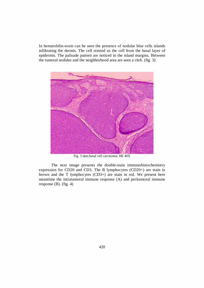
In hematohilin-eosin can be seen the presence of nodular blue cells islands infiltrating the dermis. The cell remind us the cell from the basal layer of epidermis. The palisade pattern are noticed in the island margins. Between the tumoral nodules and the neighborhood area are seen a cleft. (fig. 3)
Fig. 3 skin;basal cell carcinoma; HE 40X
The next image presents the double-stain immunohistochemistry expression for CD20 and CD3. The B lymphocytes (CD20+) are stain in brown and the T lymphocytes (CD3+) are stain in red. We present here meantime the intratumoral immune response (A) and peritumoral immune response (B). (fig. 4)

421
Fig. 4. Double-stain (brown/red) expression for CD20 and CD3 intra- and peritumoral
CONCLUSIONS
In the immune response, B cells mediate humoral immunity, whose
specific effectors are antibodies (Ab), they present specific receptors on the membrane antigens (Ag) - IgM and IgA monomer D, receptors for the Fc fragment of IgG, receptors for complement fractions.
T cells mediate cellular immunity can have direct toxic effect on the target cell, modulates the humoral immune response, helping transform of B lymphocytes into plasma cells with increased production of Ab or modulate the humoral immune response, inhibits differentiation into plasma cells, inhibits lymphocyte Th fall in production of Ab.
In our study we proof the B cell mediate humoral immunity involvement in skin cancer despite the that some study deny this theory.
Further study should be performed to set a right conclusions because our study had a limit number of cases. as we mentioned in the method chapter the study is a pilot study and becomes the starting point for next study. REFERENCES
1. Bara Constantin, 2014, Esentialul în imunologie, Editura All. 2. Curiel TJ, Coukos G, Zou L, Alvares X, Cheng P, Mottram P, Evdemon-Hogan
M., Conejo-Garcia JR, Zhang L, Burow M, Zhu Y, Wei S, Kryczek I, Daniel B, Gordon A., Mayers L, Lakner A, Disis ML, Knutson KL, Chen L, Zou W., 2004, - Specific recruitment of regulatory T cells in ovarian carcinoma fosters immune

422
privilege and predicts reduced survival. Nat Med. 2004;10(9):942-9[ PMC free article][PubMed].
3. Grivennikow SI, Greten FR, Karin M. - Immunity, Inflammation and Cancer. Cell.2010; 140(6):883-99[ PMC free article][PubMed].
4. Radu Carmen Corina, Mutiu Gabriela, Pop Ovidiu, 2014, Accessory spleen. Rom J Morphol Embryol 2014, 55(3 Suppl):1243–1246.
5. Stefănescu Ioana - Curs Imunologie, UMF Carol Davila Bucuresti.Ș. 6. Wei S, Kryzek I, Zou W, 2006, Regulatory T-cell compartmentalization and
trafficking. Blood. 2006, 108(2):426-31[ PMC free article][PubMed]. 7. www.ventana.com.