Embed Size (px)
Citation preview
Immediate/Early Loading of Zygomatic Implants:Clinical Experiences after 2 to 5 Yearsof Follow-upCarlos Aparicio, MD, DDS, MSc;* Wafaa Ouazzani, DDS;† Arnau Aparicio, DDS candidate;‡
Vanessa Fortes, DDS, MSc;† Rosa Muela, DDS, MSc;† Andres Pascual, DDS, MSc;† Maria Codesal, DDS, MSc;†
Natalia Barluenga, DDS, MSc;† Monica Franch, MD, DDS, MSc†
ABSTRACT
Background: Conventional prosthetic treatment of the edentulous and resorbed maxilla with zygomatic implants is alengthy procedure. Today, immediate/early loading is a clinical reality and it is possible that such protocols could be usedalso for zygomatic implants.
Purpose: The aim of the present study is to report on the clinical outcomes of immediate/early loading of zygomaticimplants for prosthetic rehabilitation of edentulous and severely resorbed maxillary cases.
Materials and Methods: A total of 47 zygomatic and 129 regular implants were placed in 25 consecutive patients with total(N = 23) or partial (N = 2) edentulism in the maxilla. The patients had less than 4 mm of available bone height and widthdistal to the canine pillars. Straight and angulated abutments and impression copings were attached to the implants duringsurgery. Impressions and bite registrations were made and 19 patients received a bridge within 24 hours and six patientswere rehabilitated within 5 days. Screw-retained full arch restorations were used in 23 patients and cemented in 2 patients.The patients were instructed for a soft diet during 4 months. Follow-up controls were performed at 1, 4, and 12 months andthereafter annually. All patients were followed for at least 2 years and up to 5 years in function.
Results: All zygomatic implants were stable during the follow-up (cumulative survival rate 100%). One regular implantplaced in the pterygoid plate failed after 52 months of loading (cumulative survival rate 99.2%). Apart from fracture of oneabutment screw and of anterior teeth in five patients, no other complications were noted.
Conclusions: Within the limitations of the present study, it is concluded that immediate/early loading is a viable treatmentmodality for prosthetic rehabilitation of the severely resorbed maxilla using zygomatic and conventional implants.
KEY WORDS: clinical study, edentulous maxilla, immediate loading, resorption, zygomatic implants
INTRODUCTION
Severe resorption of the maxilla may preclude routine
treatment with dental implants for support of a fixed
bridge because of the lack of bone for implant integra-
tion. There are several treatment options for these cases,
including bone grafting and the use of zygomatic
implants. The Brånemark zygomatic fixture was origi-
nally developed for prosthetic rehabilitation of cases
with extensive defects of the maxilla caused by tumor
resections, trauma, and congenital defects.1 The bone of
the zygomatic arch was used for anchorage of a long
fixture that together with ordinary fixtures could be
used as anchorage for epistheses, prostheses, and obtu-
rators.2,3 Since then the technique has been widely used
in cases with severe atrophy of the maxilla.4–10 A typical
totally edentulous case is treated with one zygomatic
implant on each side, going from the palatal aspect of
the first to the second premolar region, through the
*Private practice, Clinica Aparicio, Barcelona, Spain, and Departmentof Biomaterials, Institute for Clinical Sciences, Sahlgrenska Academy,Gothenburg University, Gothenburg, Sweden; †private practice,Clinica Aparicio, Barcelona, Spain; ‡Universidad Europea Madrid,Spain
Reprint requests: Dr. Carlos Aparicio, Clínica Aparicio, Mitre 72-74 b,08017 Barcelona, Spain; e-mail: [email protected]
© 2008, Copyright the AuthorsJournal Compilation © 2010, Wiley Periodicals, Inc.
DOI 10.1111/j.1708-8208.2008.00134.x
e77
maxillary sinus and into the body of the zygomatic
bone, and with additional regular implants in the pre-
maxilla. Today, the technique is well documented and a
recent review of studies including 1,143 zygomatic
implants showed a survival rate of 98.4% after a
follow-up from 6 months to 10 years.11 Implant treat-
ment of the maxilla was often a lengthy procedure with
healing periods of 6 to 8 months prior to loading, and
the trend now is to reduce or even to eliminate healing
periods. Immediate/early loading is a clinical reality and
good clinical outcomes have been reported on all indi-
cations and especially on totally edentulous arches.12
The advantageous for the patient are obvious since only
one surgical procedure is needed and the patient will get
an instant esthetic appearance as well as immediate
function is possible. The reasons for the good results
reported may be because of careful patient selection and
concern about primary stability. It can also be because
that the implants can be placed in an arch form that
counteracts bending forces. According to the present
authors’ experiences,10 high primary stability can also be
achieved with zygomatic implants and it is possible that
an early loading protocol could be used also for this
implant.
The aim of this study is to report on the 2 to 5 years
experiences of using an immediate/early loading proto-
col for regular and zygomatic implants to rehabilitate
the severely atrophic maxilla.
MATERIALS AND METHODS
Patients
The study group consisted of 25 consecutive patients (12
females/13 males, mean age 48 years, range 34–78 years)
with need of prosthetic rehabilitation because of missing
teeth in the maxilla. All patients were healthy. Thirteen
patients were smokers; all smoked more than 10
cigarettes/day.Twelve patients were diagnosed as bruxers.
Inclusion criteria:
• The presence of residual alveolar crest with less than
4 mm in width and height, immediately distal to the
canine pillar that precluded the use of regular
implants.
• The possibility to place regular implants in the ante-
rior regions in order to get an arch form distribu-
tion of zygomatic and regular implants.
Exclusion criteria:
• General and local health conditions that prevented
the use of general anesthesia and/or intraoral
surgery.
Surgical and Prosthetic Procedures
The presurgical radiographic examinations included
computed tomography scans and orthopantomograms.
The patients were treated under general anesthesia
and with local injections of lidocain/epinephrine.
Patients were given antibiotics prior to surgery. Crestal
and posterior vestibular releasing incisions were made
and mucoperiosteal flaps were raised to expose the
alveolar crest, the lateral wall of the maxillary sinus, and
the inferior rim of the zygomatic arch. A retractor was
used to ensure good visibility of the zygomatic bone.
The zygomatic implant site was planned by striving for
placing the implant head at or near the top of the crest,
usually in the second premolar/first molar regions using
either a classical intra-sinus path (seven patients) or a
novel extra-sinus path (18 patients).13 For the latter
technique, the implant body should preferably engage
the lateral bone wall of the maxillary sinus while enter-
ing the zygomatic bone. The implant site was prepared
without making an opening to the maxillary sinus and
followed the standard drilling steps for zygomatic
implants as described elsewhere.1, 10 Additional conven-
tional implants were placed in the anterior regions and
in some cases also posterior to the zygomatic implants. A
total of 47 zygomatic implants (Nobel Biocare AB, Goth-
enburg, Sweden), in lengths from 35 to 52.5 mm, were
placed (Table 1). A total of 129 conventional implants,
with lengths from 7 to 18 mm and in diameters of 3.75
and 4.0 mm (Nobel Biocare AB, Gothenburg, Sweden),
were used (Table 2). The zygomatic implants had a
turned surface whereas the regular implants had an
TABLE 1 Number and Length of Zygomatic Implants
Length (mm) Number
35 3
40 4
42.5 10
45 16
47.5 6
50 4
52.5 4
Total 47
e78 Clinical Implant Dentistry and Related Research, Volume 12, Supplement 1, 2010
oxidized surface (TiUnite™, Nobel Biocare AB). Abut-
ments, straight or angulated (Multiunit Abutment®,
Nobel Biocare AB) in different lengths, were connected
to the implants together with sterile impression copings.
The wound was closed by suturing. Impressions of both
jaws and bite registration were made immediately fol-
lowing surgery in order to manufacture a provisional
fixed bridge. The patients were prescribed postoperative
antibiotics and analgesics.
Nineteen patients received a temporary bridge
within 24 hours, and six patients received the definitive
metal-resin bridge within 5 days. For the former group,
the definitive metal-resin bridge was delivered 4 to 6
months after surgery. A screw retained full arch restora-
tion was used in 23 patients and two patients received
partial cemented bridges. The patients were instructed
for a soft diet during 4 months.
Removal of sutures and check-up of occlusion were
made 10 days after surgery.
Follow-Up
Follow-up controls were performed at 1, 4, and 12
months, thereafter annually. The check-up examina-
tions also included assessments of oral hygiene, soft
tissue health, prosthesis stability, and signs of mechani-
cal complications. All bridges were removed for
individual checking of the implants after 1 year in func-
tion. Standardized intraoral radiographs of the zygo-
matic implants could not be made and consequently
these implants could not be evaluated with regard to
marginal bone resorption.
An implant removed for any reason was counted as
a failure and the implants still in function were counted
as survivals.
RESULTS
The period after implant surgery was regarded as
normal in all patients with some postoperative pain and
swelling that could be controlled with analgesics.
All patients were followed for at least 2 and up to 5
years of loading and attended the follow-up appoint-
ments. All zygomatic implants were judged to be stable
during the follow-up, giving a cumulative survival rate
(CSR) of 100% (Table 3). One regular implant placed in
the pterygoid plate failed after 52 months of loading,
resulting in a CSR of 99.2% for the regular implants
(Table 4).
There were no signs of soft-tissue infections or fis-
tulae to the maxillary sinus. Some mechanical compli-
cations were experiences since anterior teeth fractured
in five patients with metal-resin (N = 4) and metal-
porcelain (N = 1) bridges. Moreover, one abutment
screw of a zygomatic implant supporting a metal-resin
bridge fractured in another patient after 3 years of
loading.
DISCUSSION
The present clinical follow-up study demonstrated good
results with immediate/early loading of 47 zygomatic
and 129 conventional implants in 25 patients as no
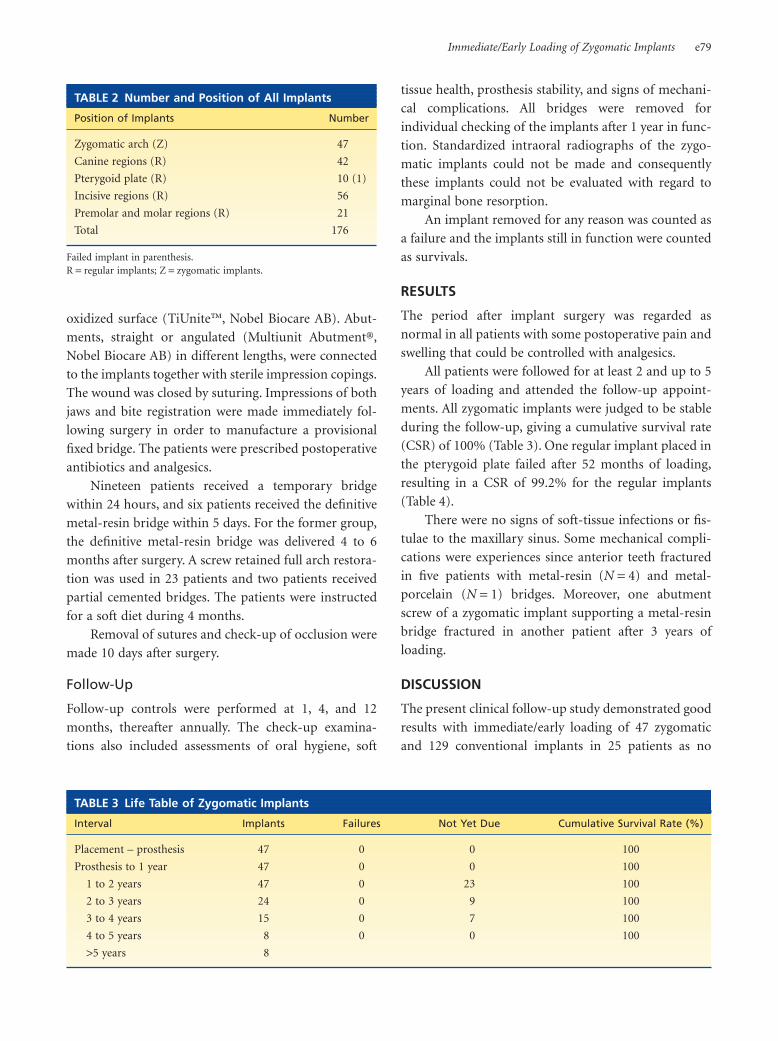
TABLE 2 Number and Position of All Implants
Position of Implants Number
Zygomatic arch (Z) 47
Canine regions (R) 42
Pterygoid plate (R) 10 (1)
Incisive regions (R) 56
Premolar and molar regions (R) 21
Total 176
Failed implant in parenthesis.R = regular implants; Z = zygomatic implants.
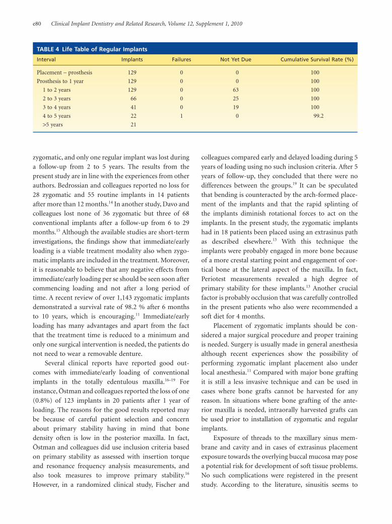
TABLE 3 Life Table of Zygomatic Implants
Interval Implants Failures Not Yet Due Cumulative Survival Rate (%)
Placement – prosthesis 47 0 0 100
Prosthesis to 1 year 47 0 0 100
1 to 2 years 47 0 23 100
2 to 3 years 24 0 9 100
3 to 4 years 15 0 7 100
4 to 5 years 8 0 0 100
>5 years 8
Immediate/Early Loading of Zygomatic Implants e79
zygomatic, and only one regular implant was lost during
a follow-up from 2 to 5 years. The results from the
present study are in line with the experiences from other
authors. Bedrossian and colleagues reported no loss for
28 zygomatic and 55 routine implants in 14 patients
after more than 12 months.14 In another study, Davo and
colleagues lost none of 36 zygomatic but three of 68
conventional implants after a follow-up from 6 to 29
months.15 Although the available studies are short-term
investigations, the findings show that immediate/early
loading is a viable treatment modality also when zygo-
matic implants are included in the treatment. Moreover,
it is reasonable to believe that any negative effects from
immediate/early loading per se should be seen soon after
commencing loading and not after a long period of
time. A recent review of over 1,143 zygomatic implants
demonstrated a survival rate of 98.2 % after 6 months
to 10 years, which is encouraging.11 Immediate/early
loading has many advantages and apart from the fact
that the treatment time is reduced to a minimum and
only one surgical intervention is needed, the patients do
not need to wear a removable denture.
Several clinical reports have reported good out-
comes with immediate/early loading of conventional
implants in the totally edentulous maxilla.16–19 For
instance, Östman and colleagues reported the loss of one
(0.8%) of 123 implants in 20 patients after 1 year of
loading. The reasons for the good results reported may
be because of careful patient selection and concern
about primary stability having in mind that bone
density often is low in the posterior maxilla. In fact,
Östman and colleagues did use inclusion criteria based
on primary stability as assessed with insertion torque
and resonance frequency analysis measurements, and
also took measures to improve primary stability.16
However, in a randomized clinical study, Fischer and
colleagues compared early and delayed loading during 5
years of loading using no such inclusion criteria. After 5
years of follow-up, they concluded that there were no
differences between the groups.19 It can be speculated
that bending is counteracted by the arch-formed place-
ment of the implants and that the rapid splinting of
the implants diminish rotational forces to act on the
implants. In the present study, the zygomatic implants
had in 18 patients been placed using an extrasinus path
as described elsewhere.13 With this technique the
implants were probably engaged in more bone because
of a more crestal starting point and engagement of cor-
tical bone at the lateral aspect of the maxilla. In fact,
Periotest measurements revealed a high degree of
primary stability for these implants.13 Another crucial
factor is probably occlusion that was carefully controlled
in the present patients who also were recommended a
soft diet for 4 months.
Placement of zygomatic implants should be con-
sidered a major surgical procedure and proper training
is needed. Surgery is usually made in general anesthesia
although recent experiences show the possibility of
performing zygomatic implant placement also under
local anesthesia.11 Compared with major bone grafting
it is still a less invasive technique and can be used in
cases where bone grafts cannot be harvested for any
reason. In situations where bone grafting of the ante-
rior maxilla is needed, intraorally harvested grafts can
be used prior to installation of zygomatic and regular
implants.
Exposure of threads to the maxillary sinus mem-
brane and cavity and in cases of extrasinus placement
exposure towards the overlying buccal mucosa may pose
a potential risk for development of soft tissue problems.
No such complications were registered in the present
study. According to the literature, sinusitis seems to
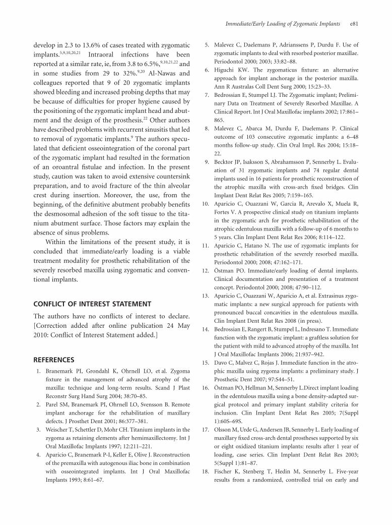
TABLE 4 Life Table of Regular Implants
Interval Implants Failures Not Yet Due Cumulative Survival Rate (%)
Placement – prosthesis 129 0 0 100
Prosthesis to 1 year 129 0 0 100
1 to 2 years 129 0 63 100
2 to 3 years 66 0 25 100
3 to 4 years 41 0 19 100
4 to 5 years 22 1 0 99.2
>5 years 21
e80 Clinical Implant Dentistry and Related Research, Volume 12, Supplement 1, 2010
develop in 2.3 to 13.6% of cases treated with zygomatic
implants.5,9,10,20,21 Intraoral infections have been
reported at a similar rate, ie, from 3.8 to 6.5%,9,10,21,22 and
in some studies from 29 to 32%.9,20 Al-Nawas and
colleagues reported that 9 of 20 zygomatic implants
showed bleeding and increased probing depths that may
be because of difficulties for proper hygiene caused by
the positioning of the zygomatic implant head and abut-
ment and the design of the prosthesis.22 Other authors
have described problems with recurrent sinusitis that led
to removal of zygomatic implants.9 The authors specu-
lated that deficient osseointegration of the coronal part
of the zygomatic implant had resulted in the formation
of an oroantral fistulae and infection. In the present
study, caution was taken to avoid extensive countersink
preparation, and to avoid fracture of the thin alveolar
crest during insertion. Moreover, the use, from the
beginning, of the definitive abutment probably benefits
the desmosomal adhesion of the soft tissue to the tita-
nium abutment surface. Those factors may explain the
absence of sinus problems.
Within the limitations of the present study, it is
concluded that immediate/early loading is a viable
treatment modality for prosthetic rehabilitation of the
severely resorbed maxilla using zygomatic and conven-
tional implants.
CONFLICT OF INTEREST STATEMENT
The authors have no conflicts of interest to declare.
[Correction added after online publication 24 May
2010: Conflict of Interest Statement added.]
REFERENCES
1. Branemark PI, Grondahl K, Ohrnell LO, et al. Zygoma
fixture in the management of advanced atrophy of the
maxilla: technique and long-term results. Scand J Plast
Reconstr Surg Hand Surg 2004; 38:70–85.
2. Parel SM, Branemark PI, Ohrnell LO, Svensson B. Remote
implant anchorage for the rehabilitation of maxillary
defects. J Prosthet Dent 2001; 86:377–381.
3. Weischer T, Schettler D, Mohr CH. Titanium implants in the
zygoma as retaining elements after hemimaxillectomy. Int J
Oral Maxillofac Implants 1997; 12:211–221.
4. Aparicio C, Branemark P-I, Keller E, Olive J. Reconstruction
of the premaxilla with autogenous iliac bone in combination
with osseointegrated implants. Int J Oral Maxillofac
Implants 1993; 8:61–67.
5. Malevez C, Daelemans P, Adrianssens P, Durdu F. Use of
zygomatic implants to deal with resorbed posterior maxillae.
Periodontol 2000; 2003; 33:82–88.
6. Higuchi KW. The zygomaticus fixture: an alternative
approach for implant anchorage in the posterior maxilla.
Ann R Australas Coll Dent Surg 2000; 15:23–33.
7. Bedrossian E, Stumpel LJ. The Zygomatic implant; Prelimi-
nary Data on Treatment of Severely Resorbed Maxillae. A
Clinical Report. Int J Oral Maxillofac implants 2002; 17:861–
865.
8. Malevez C, Abarca M, Durdu F, Daelemans P. Clinical
outcome of 103 consecutive zygomatic implants: a 6–48
months follow-up study. Clin Oral Impl. Res 2004; 15:18–
22.
9. Becktor JP, Isaksson S, Abrahamsson P, Sennerby L. Evalu-
ation of 31 zygomatic implants and 74 regular dental
implants used in 16 patients for prosthetic reconstruction of
the atrophic maxilla with cross-arch fixed bridges. Clin
Implant Dent Relat Res 2005; 7:159–165.
10. Aparicio C, Ouazzani W, Garcia R, Arevalo X, Muela R,
Fortes V. A prospective clinical study on titanium implants
in the zygomatic arch for prosthetic rehabilitation of the
atrophic edentulous maxilla with a follow-up of 6 months to
5 years. Clin Implant Dent Relat Res 2006; 8:114–122.
11. Aparicio C, Hatano N. The use of zygomatic implants for
prosthetic rehabilitation of the severely resorbed maxilla.
Periodontol 2000; 2008; 47:162–171.
12. Östman PO. Immediate/early loading of dental implants.
Clinical documentation and presentation of a treatment
concept. Periodontol 2000; 2008; 47:90–112.
13. Aparicio C, Ouazzani W, Aparicio A, et al. Extrasinus zygo-
matic implants: a new surgical approach for patients with
pronounced buccal concavities in the edentulous maxilla.
Clin Implant Dent Relat Res 2008 (in press).
14. Bedrossian E, Rangert B, Stumpel L, Indresano T. Immediate
function with the zygomatic implant: a graftless solution for
the patient with mild to advanced atrophy of the maxilla. Int
J Oral Maxillofac Implants 2006; 21:937–942.
15. Davo C, Malvez C, Rojas J. Immediate function in the atro-
phic maxilla using zygoma implants: a preliminary study. J
Prosthetic Dent 2007; 97:S44–51.
16. Östman PO, Hellman M, Sennerby L.Direct implant loading
in the edentulous maxilla using a bone density-adapted sur-
gical protocol and primary implant stability criteria for
inclusion. Clin Implant Dent Relat Res 2005; 7(Suppl
1):60S–69S.
17. Olsson M, Urde G, Andersen JB, Sennerby L. Early loading of
maxillary fixed cross-arch dental prostheses supported by six
or eight oxidized titanium implants: results after 1 year of
loading, case series. Clin Implant Dent Relat Res 2003;
5(Suppl 1):81–87.
18. Fischer K, Stenberg T, Hedin M, Sennerby L. Five-year
results from a randomized, controlled trial on early and
Immediate/Early Loading of Zygomatic Implants e81

delayed loading of implants supporting full-arch prosthesis
in the edentulous maxilla. Clin Oral Implants Res 2008;
19:433–441.
19. van Steenberghe D, Glauser R, Blombäck U, et al. A com-
puted tomographic scan-derived customized surgical tem-
plate and fixed prosthesis for flapless surgery and immediate
loading of implants in fully edentulous maxillae: a prospec-
tive multicenter study. Clin Implant Dent Relat Res 2005;
7(Suppl 1):111S–120S.
20. Farzad P, Andersson L, Gunnarsson S, Johansson B.
Rehabilitation of severely resorbed maxillae with zygomatic
implants: an evaluation of implant stability, tissue condi-
tions, and patients’ opinion before and after treatment. Int
J Oral Maxillofac Implants 2006; 21:399–404.
21. Hirsch JM, Ohrnell LO, Henry PJ, et al. A clinical evaluation
of the Zygoma fixture: one year of follow-up at 16 clinics. J
Oral Maxillofac Surg 2004; 62(9 Suppl 2):22–29.
22. Al-Nawas B, Wegener J, Bender C, Wagner W. Critical soft
tissue parameters of the zygomatic implant. J Clin Period-
ontol 2004; 31:497–500.
e82 Clinical Implant Dentistry and Related Research, Volume 12, Supplement 1, 2010