Embed Size (px)
Citation preview

Case ReportImmediate Implant Placement by Interradicular BoneDrilling before Molar Extraction: Clinical Case Report withOne-Year Follow-Up
Stuardo Valenzuela,1 Jose M. Olivares ,1 Nicolas Weiss,1 and Dafna Benadof 2
1Postgraduate Implant Dentistry Department, School of Dentistry, Universidad Andres Bello, Santiago, Chile2School of Dentistry, Universidad Andres Bello, Santiago, Chile
Correspondence should be addressed to Jose M. Olivares; [email protected]
Received 17 January 2018; Accepted 5 March 2018; Published 1 April 2018
Academic Editor: Gavriel Chaushu
Copyright © 2018 Stuardo Valenzuela et al. ,is is an open access article distributed under the Creative Commons AttributionLicense, which permits unrestricted use, distribution, and reproduction in any medium, provided the original work isproperly cited.
,e placement of immediate implants in the posterior sector is a widespread procedure where the success and survival rates aresimilar to those of traditional protocols. It has several anatomical challenges, such as the presence of interradicular bone septa thathinder a correct three-dimensional positioning of the implant and may compromise primary stability and/or cause damage ofneighboring structures. ,e aim of this article is to present the treatment and the one-year clinical follow-up of a patient whoreceived immediate implant placement using an interradicular bone-drilling technique before the molar extraction.
1. Introduction
Immediate implant placement has considerable advantagesover the conventional approach. It has fewer numbers ofsurgical procedures, reduces overall treatment time, andtherefore costs less. It also helps preserve the gingival ar-chitecture and increase the patient’s comfort, acceptance,and satisfaction [1–5].
Immediate implant placement studies in the esthetic andpremolar zone follow strict surgical protocols that have beenestablished to optimize the three-dimensional positioning ofthe implant and its primary stability and the condition of theneighboring tissue [6–8]. However, there is less informationon immediate implant placement in the posterior sectorwhere the esthetical impact is lower, but the surgical diffi-culty of the tooth extraction, drilling, and implant placementis greater [9–11].
Despite the abovementioned issue, the cumulative survivalrates reported for immediate implants placed inmolar sites aresimilar to thoseplaced inhealed sites,which ranges from93.9%to 99% [4–8, 10, 11]. An essential aspect to achieve this positive
outcome is theprimary stabilizationof the implant in the apicaland/or lateral bone,where anatomic conditions canhinder thisgoal. ,erefore, a thorough implant surgery planning, skills,and clinical experience are relevant factors in the success of thesurgical procedure [4, 12].
Modifications to the current surgical techniques arerecommended to facilitate immediate implant placement inthe posterior sector. Different authors propose implantdrilling prior to tooth extraction in order to stabilize theinterradicular bone septa through the remaining tooth roots[9, 13–15]. In 2017, a randomized pilot study of 22 patientscompared the conventional technique of dental extraction,subsequent interradicular bone drilling, and immediateimplant placement to the technique of interradicular bonedrilling using ultrasound devices. ,e results were statisti-cally higher for the implant positioning and primary stabilityusing the proposed new technique [9].
,e aim of this article is to present the treatment of apatient by means of immediate implant placement usingan interradicular bone-drilling technique and its clinicalfollow-up one-year later.
HindawiCase Reports in DentistryVolume 2018, Article ID 6412826, 5 pageshttps://doi.org/10.1155/2018/6412826
2. Case Presentation
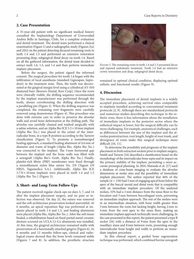
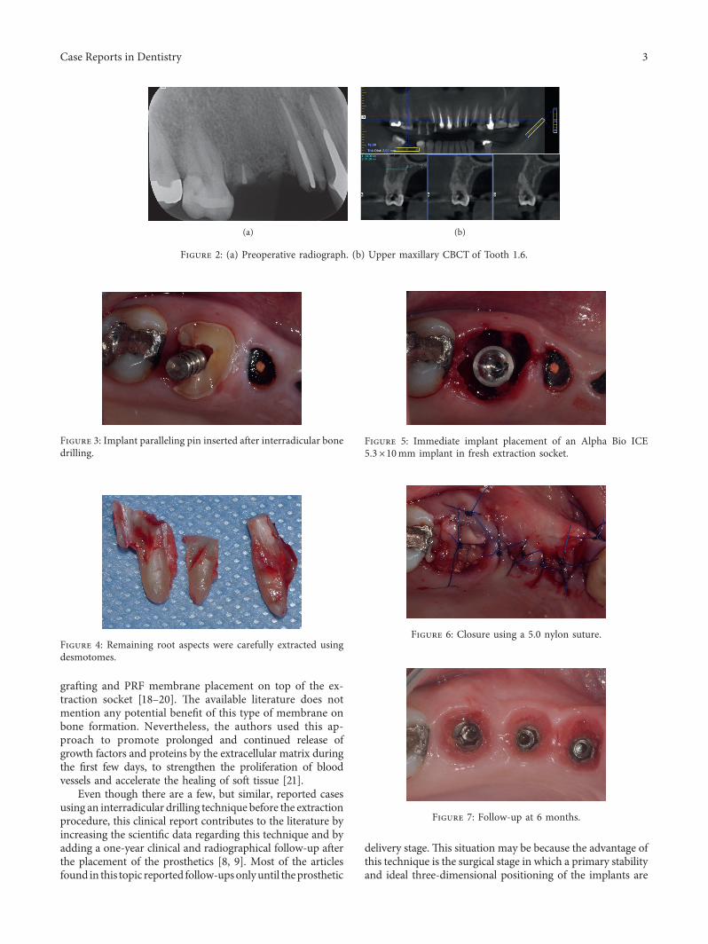
A 35-year-old patient with no significant medical historyconsulted the Implantology Department of UniversidadAndres Bello in Santiago, Chile, for a complete evaluationand dental treatment. ,e dental team performed a clinicalexamination (Figure 1) and a radiographic study (Figures 2(a)and 2(b)) on the patient detecting decayed remaining roots inteeth 1.4 and 1.5 and performed an extensive restorationpresenting deep, subgingival distal decay on tooth 1.6. Basedon all the gathered information, the dental team decided toextract teeth 1.6, 1.5, and 1.4 and then perform immediateimplant placement.
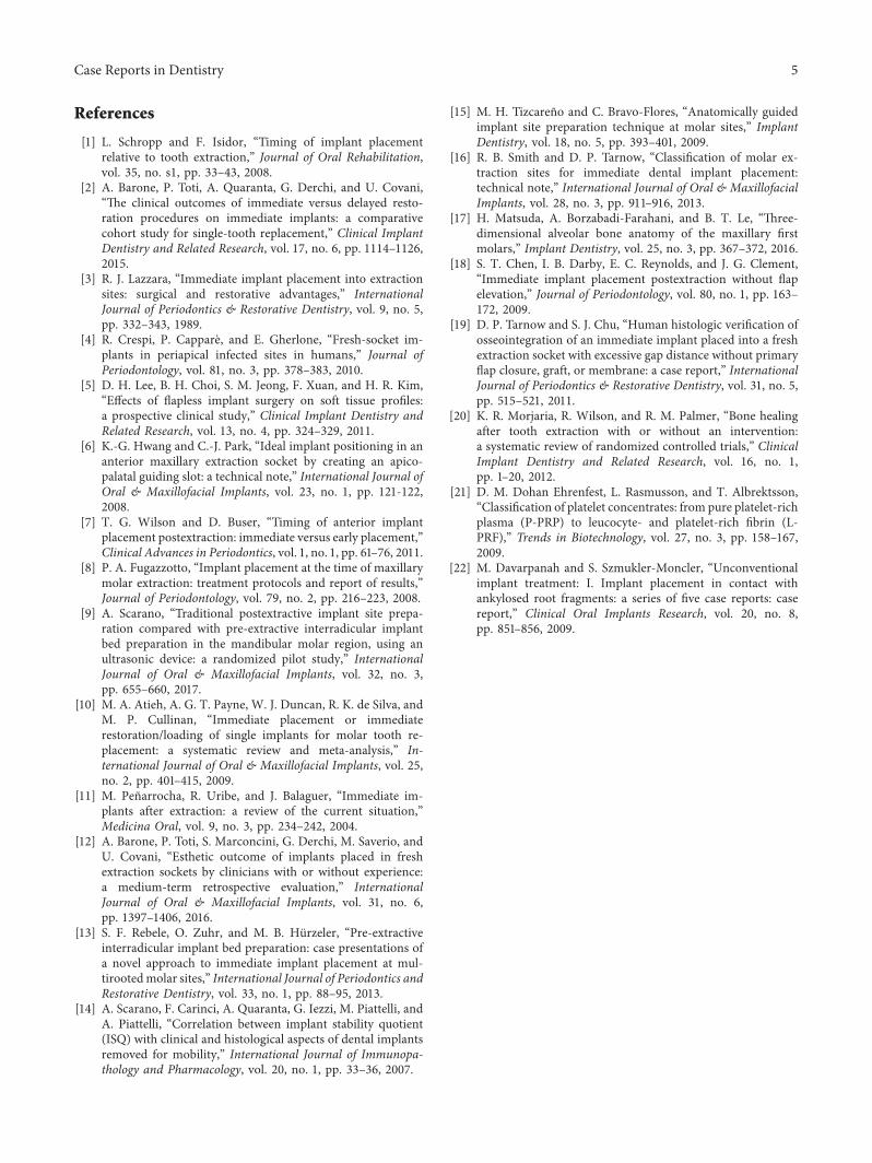
Before the surgery, the patient signed the informedconsent. ,e surgical procedure for tooth 1.6 began with theinfiltration of local anesthesia (standard Lignospan, Septo-dont) in the treatment zone. ,en, the tooth was decoro-nated at the gingival margin level using a cylindrical AV-010diamond burr (Beavers Dental, Kerr Corp). Once the rootswere clinically visible, the drilling sequence recommendedby the implant manufacturer was performed through thetooth, always corroborating the drilling direction witha paralleling pin (Figure 3). When the drilling sequence wascompleted, the remaining root fragments were carefullyremoved using desmotomes (Figure 4). ,is procedure wasdone with extreme care in order to preserve the alveolarwalls and avoid bone deformation at the drilling path. ,ealveolus was carefully cleaned and washed surgically witha saline solution, and an Alpha Bio ICE 5.3×10mm implant(Alpha Bio Tec.) was placed in the center of the inter-radicular bone, in a type B position according to the Tarnowclassification (Figure 5). To promote a nonsubmergedhealing approach, a standard healing abutment of 4.6mm ofdiameter and 4mm of length (Alpha Bio, Alpha Bio Tec.)was connected to the implant. ,e 3mm horizontal gapbetween the implant and the bone walls was filled witha xenograft (Alpha Bio’s Graft, Alpha Bio Tec.) Finally,platelet-rich fibrin (PRF) membranes were fixed througha monofilament nylon blue suture No. 5/0 (Tagum 2/0HR25, Tagumedica S.A.). Additionally, Alpha Bio ICE3.7N× 10mm implants were placed in teeth 1.4 and 1.5(Alpha Bio Tec.) (Figure 5).
3. Short- and Long-Term Follow-Ups
,e patient received regular check-ups on days 3, 7, and 14after the implant placement surgery, and no pain or in-fection was observed. On day 21, the suture was removedand the soft architecture preservation looked uneventful. At6 months, an apical reposition flap was performed at im-plants placed in teeth 1.4 and 1.5, and healing abutmentswere placed (Alpha Bio, Alpha Bio Tec.). After the soft tissuehealed, a rehabilitation based on fixed partial metal-ceramicdenture screwed on UCLA Cr-Co abutments was installed.At this time, the gingival architecture remained stable withpreservation of a functionally attached gingiva (Figure 6). At6 months and 12 months follow-ups, clinical and radio-logical exams showed that the bone levels remained stable(Figures 7 and 8). In addition, the prosthetic structure
remained in optimal clinical condition, displaying optimalesthetic and functional results (Figure 9).
4. Discussion
,e immediate placement of dental implants is a widelyaccepted procedure, achieving survival rates comparableto implants installed according to conventional treatmentprotocols [2, 9]. Although there are standardized protocolsand numerous studies describing this technique in the es-thetic zone, there is less information about the installationof immediate implants in the posterior sector where theesthetical impact is lower, but the surgical difficulty can bemore challenging. For example, anatomical challenges, suchas differences between the size of the implant and the al-veolus postextraction, root length, height of root trunk, anddivergence of roots make this surgical technique moredifficult [10, 11].
To determine the possibility and prognosis of the implantplacement in fresh extraction sockets prior to implant surgery,Smith and Tarnow [16] described a classification based on themorphology of the interradicular bone septa and its impact onthe primary stability of the implant, permitting a more ac-curate presurgical planning. In 2016, Matsuda et al. [17] useda database of cone-beam imaging to evaluate the alveolardimensions at molar sites and the possibility of immediateimplant placement. ,e author reported that 46% of thesample (n � 150) had 5mmof engaging apical bone below theapex of the buccal mesial and distal roots that is compatiblewith an immediate implant procedure. Of the analyzedmolars, 32% had a 2mm distance from the sinus floor to thefurcation and 5mm between buccolingual roots, preventingan immediate implant approach. ,e rest of the molars werein an intermediate situation, with bone width greater than5mm between the roots but lacking height, having 2mm to4mm from the root apex to the sinus floor, making animmediate implant approach technically more challenging. Inthe case presented in this report, the patient presented a type Bsocket [16] with a distance of 9mm from the apex to themaxillary sinus and 8mm between roots, presenting enoughinterradicular bone height and width to perform an imme-diate implant procedure.
In this clinical report, a guided bone regenerationtechnique was performed, which combined bovine xenograft
Figure 1: ,e remaining roots in teeth 1.4 and 1.5 presented decayand exposed endodontic treatment. Tooth 1.6 had an extensivecrown restoration and deep, subgingival distal decay.
2 Case Reports in Dentistry
grafting and PRF membrane placement on top of the ex-traction socket [18–20]. ,e available literature does notmention any potential benefit of this type of membrane onbone formation. Nevertheless, the authors used this ap-proach to promote prolonged and continued release ofgrowth factors and proteins by the extracellular matrix duringthe first few days, to strengthen the proliferation of bloodvessels and accelerate the healing of soft tissue [21].
Even though there are a few, but similar, reported casesusing an interradicular drilling technique before the extractionprocedure, this clinical report contributes to the literature byincreasing the scientific data regarding this technique and byadding a one-year clinical and radiographical follow-up afterthe placement of the prosthetics [8, 9]. Most of the articlesfound in this topic reported follow-upsonlyuntil theprosthetic
delivery stage. ,is situation may be because the advantage ofthis technique is the surgical stage in which a primary stabilityand ideal three-dimensional positioning of the implants are
(a) (b)
Figure 2: (a) Preoperative radiograph. (b) Upper maxillary CBCT of Tooth 1.6.
Figure 3: Implant paralleling pin inserted after interradicular bonedrilling.
Figure 4: Remaining root aspects were carefully extracted usingdesmotomes.
Figure 5: Immediate implant placement of an Alpha Bio ICE5.3×10mm implant in fresh extraction socket.
Figure 6: Closure using a 5.0 nylon suture.
Figure 7: Follow-up at 6 months.
Case Reports in Dentistry 3

attained. However, once osseointegration occurs, the be-havior of peri-implant tissue should not differ from tra-ditional procedures.
In Scarano’s study [9], drilling in the interradicular bonesepta before and after the extraction of molar roots wascompared. ,e author concluded that using a guide on theposition of the roots resulted in an ideal implant positioning(p< 0.05). Also, the primary stability of the implant based on
a resonance frequency analysis had significantly higherimplant stability quotient values (p< 0.05) as compared tothe traditional technique of extraction, subsequent drilling,and immediate implant placement. However, we do notknow whether other variables may affect these results. Forexample, the use of the ultrasound device is comparable todrilling with conventional rotary instruments, so these re-sults cannot be generalized. Moreover, the criteria for in-clusion were only molar sites with interradicular septa thathad crown dimensions above 2.5mm and apical dimensionsabove 3.5mm, which does not necessarily represent whatmost prevails in the population nor mean that these min-imummeasurements will suffice to attain a primary stability.
,e interradicular bone-drilling technique prior to dentalextraction could be considered a simple yet useful modifi-cation to the standard drilling procedure. Its indications areabsence of active infection, integrity of the roots, and suffi-cient remaining bone to allow an immediate implant ap-proach [15]. Contraindications are dental mobility, due tosevere loss of periodontal insertion, unfavorable root position,such as fused roots, ankyloses, and active infections [13, 15].,e authors have described that even active infections such asapical periodontitis do not lead to an increased risk ofcomplications, as long as they are asymptomatic [4].
,is procedure has an increased risk to alter the socketwall’s morphology during the extraction procedure, leadingto a deficient implant insertion. ,erefore, careful extractionusing desmotomes or ultrasonic appliances is advised toavoid any deformation of the interradicular bone that couldlead to a modification of the bone-drilling path and alter thefinal implant position.
Researchers have even stated that this technique could besuitable to nonexpert clinicians, making it simpler to obtaina correct tridimensional position of the implant and primaryinsertion torque. ,is is also supported by studies that showthe traditional approach where the level of expertise is keyfactor in the success of the procedure [12].
Some limitations of this technique are increased hard-ness of the root tissue, which may result in longer clinicaltime and greater risk of increasing intrabone temperatureand of altering the normal healing because of the remains ofdental tissue from drilling. Regarding the latter point,Davarpanah and Szmukler-Moncler [22] made a case reporton 5 patients; according to the results, dental waste did notseem to interfere with implant osseointegration, but therewas little scientific evidence on this latter point, so caution isrecommended, with an emphasis on meticulous irrigationand surgical cleaning.
Although this technique is promising and the clinicalyield has been good for the authors during intraoperationmanagement and post-op check-ups, controlled randomizedclinical testing is required, using a comparative method, toevaluate the benefits and limitations of this technique in thelong term.
Conflicts of Interest
,e authors declare that there are no conflicts of interestregarding the publication of this paper.
Figure 8: Prosthetic delivery radiograph.
Figure 9: One-year follow-up.
4 Case Reports in Dentistry
References
[1] L. Schropp and F. Isidor, “Timing of implant placementrelative to tooth extraction,” Journal of Oral Rehabilitation,vol. 35, no. s1, pp. 33–43, 2008.
[2] A. Barone, P. Toti, A. Quaranta, G. Derchi, and U. Covani,“,e clinical outcomes of immediate versus delayed resto-ration procedures on immediate implants: a comparativecohort study for single-tooth replacement,” Clinical ImplantDentistry and Related Research, vol. 17, no. 6, pp. 1114–1126,2015.
[3] R. J. Lazzara, “Immediate implant placement into extractionsites: surgical and restorative advantages,” InternationalJournal of Periodontics & Restorative Dentistry, vol. 9, no. 5,pp. 332–343, 1989.
[4] R. Crespi, P. Cappare, and E. Gherlone, “Fresh-socket im-plants in periapical infected sites in humans,” Journal ofPeriodontology, vol. 81, no. 3, pp. 378–383, 2010.
[5] D. H. Lee, B. H. Choi, S. M. Jeong, F. Xuan, and H. R. Kim,“Effects of flapless implant surgery on soft tissue profiles:a prospective clinical study,” Clinical Implant Dentistry andRelated Research, vol. 13, no. 4, pp. 324–329, 2011.
[6] K.-G. Hwang and C.-J. Park, “Ideal implant positioning in ananterior maxillary extraction socket by creating an apico-palatal guiding slot: a technical note,” International Journal ofOral & Maxillofacial Implants, vol. 23, no. 1, pp. 121-122,2008.
[7] T. G. Wilson and D. Buser, “Timing of anterior implantplacement postextraction: immediate versus early placement,”Clinical Advances in Periodontics, vol. 1, no. 1, pp. 61–76, 2011.
[8] P. A. Fugazzotto, “Implant placement at the time of maxillarymolar extraction: treatment protocols and report of results,”Journal of Periodontology, vol. 79, no. 2, pp. 216–223, 2008.
[9] A. Scarano, “Traditional postextractive implant site prepa-ration compared with pre-extractive interradicular implantbed preparation in the mandibular molar region, using anultrasonic device: a randomized pilot study,” InternationalJournal of Oral & Maxillofacial Implants, vol. 32, no. 3,pp. 655–660, 2017.
[10] M. A. Atieh, A. G. T. Payne, W. J. Duncan, R. K. de Silva, andM. P. Cullinan, “Immediate placement or immediaterestoration/loading of single implants for molar tooth re-placement: a systematic review and meta-analysis,” In-ternational Journal of Oral & Maxillofacial Implants, vol. 25,no. 2, pp. 401–415, 2009.
[11] M. Peñarrocha, R. Uribe, and J. Balaguer, “Immediate im-plants after extraction: a review of the current situation,”Medicina Oral, vol. 9, no. 3, pp. 234–242, 2004.
[12] A. Barone, P. Toti, S. Marconcini, G. Derchi, M. Saverio, andU. Covani, “Esthetic outcome of implants placed in freshextraction sockets by clinicians with or without experience:a medium-term retrospective evaluation,” InternationalJournal of Oral & Maxillofacial Implants, vol. 31, no. 6,pp. 1397–1406, 2016.
[13] S. F. Rebele, O. Zuhr, and M. B. Hurzeler, “Pre-extractiveinterradicular implant bed preparation: case presentations ofa novel approach to immediate implant placement at mul-tirootedmolar sites,” International Journal of Periodontics andRestorative Dentistry, vol. 33, no. 1, pp. 88–95, 2013.
[14] A. Scarano, F. Carinci, A. Quaranta, G. Iezzi, M. Piattelli, andA. Piattelli, “Correlation between implant stability quotient(ISQ) with clinical and histological aspects of dental implantsremoved for mobility,” International Journal of Immunopa-thology and Pharmacology, vol. 20, no. 1, pp. 33–36, 2007.
[15] M. H. Tizcareño and C. Bravo-Flores, “Anatomically guidedimplant site preparation technique at molar sites,” ImplantDentistry, vol. 18, no. 5, pp. 393–401, 2009.
[16] R. B. Smith and D. P. Tarnow, “Classification of molar ex-traction sites for immediate dental implant placement:technical note,” International Journal of Oral & MaxillofacialImplants, vol. 28, no. 3, pp. 911–916, 2013.
[17] H. Matsuda, A. Borzabadi-Farahani, and B. T. Le, “,ree-dimensional alveolar bone anatomy of the maxillary firstmolars,” Implant Dentistry, vol. 25, no. 3, pp. 367–372, 2016.
[18] S. T. Chen, I. B. Darby, E. C. Reynolds, and J. G. Clement,“Immediate implant placement postextraction without flapelevation,” Journal of Periodontology, vol. 80, no. 1, pp. 163–172, 2009.
[19] D. P. Tarnow and S. J. Chu, “Human histologic verification ofosseointegration of an immediate implant placed into a freshextraction socket with excessive gap distance without primaryflap closure, graft, or membrane: a case report,” InternationalJournal of Periodontics & Restorative Dentistry, vol. 31, no. 5,pp. 515–521, 2011.
[20] K. R. Morjaria, R. Wilson, and R. M. Palmer, “Bone healingafter tooth extraction with or without an intervention:a systematic review of randomized controlled trials,” ClinicalImplant Dentistry and Related Research, vol. 16, no. 1,pp. 1–20, 2012.
[21] D. M. Dohan Ehrenfest, L. Rasmusson, and T. Albrektsson,“Classification of platelet concentrates: from pure platelet-richplasma (P-PRP) to leucocyte- and platelet-rich fibrin (L-PRF),” Trends in Biotechnology, vol. 27, no. 3, pp. 158–167,2009.
[22] M. Davarpanah and S. Szmukler-Moncler, “Unconventionalimplant treatment: I. Implant placement in contact withankylosed root fragments: a series of five case reports: casereport,” Clinical Oral Implants Research, vol. 20, no. 8,pp. 851–856, 2009.
Case Reports in Dentistry 5

DentistryInternational Journal of
Hindawiwww.hindawi.com Volume 2018
Environmental and Public Health
Journal of
Hindawiwww.hindawi.com Volume 2018
Hindawi Publishing Corporation http://www.hindawi.com Volume 2013Hindawiwww.hindawi.com
The Scientific World Journal
Volume 2018Hindawiwww.hindawi.com Volume 2018
Public Health Advances in
Hindawiwww.hindawi.com Volume 2018
Case Reports in Medicine
Hindawiwww.hindawi.com Volume 2018
International Journal of
Biomaterials
Scienti�caHindawiwww.hindawi.com Volume 2018
PainResearch and TreatmentHindawiwww.hindawi.com Volume 2018
Preventive MedicineAdvances in
Hindawiwww.hindawi.com Volume 2018
Hindawiwww.hindawi.com Volume 2018
Case Reports in Dentistry
Hindawiwww.hindawi.com Volume 2018
Surgery Research and Practice
Hindawiwww.hindawi.com Volume 2018
BioMed Research International Medicine
Advances in
Hindawiwww.hindawi.com Volume 2018
Hindawiwww.hindawi.com Volume 2018
Anesthesiology Research and Practice
Hindawiwww.hindawi.com Volume 2018
Radiology Research and Practice
Hindawiwww.hindawi.com Volume 2018
Computational and Mathematical Methods in Medicine
EndocrinologyInternational Journal of
Hindawiwww.hindawi.com Volume 2018
Hindawiwww.hindawi.com Volume 2018
OrthopedicsAdvances in
Drug DeliveryJournal of
Hindawiwww.hindawi.com Volume 2018
Submit your manuscripts atwww.hindawi.com