Embed Size (px)
Citation preview

ORNL is managed by UT-Battelle
for the US Department of Energy
Imaging with Neutrons
Lou Santodonato Instrument Scientist
Neutron Imaging Team
19th National School on Neutron and X-ray Scattering
August 5-19, 2017

2 Santodonato - Imaging
Acknowledgement
• The Neutron Imaging Team
Hassina
Lou, Jean, Shawn, Gian, Indu
Jiao

3 Santodonato - Imaging
Goals
• See what we can do with neutron imaging
– Compare to other imaging techniques
– Know when to choose it
• Understand the basic instrument layout and principals of neutron image acquisition and analysis
• Learn by example
– Review some recent neutron imaging projects

4 Santodonato - Imaging
Imaging throughout Nobel Prize History
• 1901: Roentgen, FIRST Nobel Prize in Physics, Discovery of X-rays
• 1979: Cormack and Hounsfield, Nobel Prize in Medicine, Computed Tomography (CT)
• 1986: Ruska, Binnig, Rohrer, Nobel Prize in Physics, Electron Microscopy
• 2003: Lauterbur and Mansfield, Nobel Prize in Medicine, Magnetic Resonance Imaging (MRI)
• 2009: Boyle and Smith, Nobel Prize in Physics, Imaging semi-conductor circuit, the CCD* sensor
• (*) Charge-Coupled Device

5 Santodonato - Imaging
What about Neutron Imaging?
• The Nobel Prize for neutron imaging has yet to be won
– An opportunity for you!
• NI started in the mid 1930’s but only the past 30 years has it come to the forefront of non-destructive testing
• World conferences and workshops being held regularly

6 Santodonato - Imaging
Neutron Imaging
• Measures “shadows” based on neutron
attenuation through the object
• One shadow is a radiograph
• These “shadows” are collected at different angles
and reconstructed in 3D, called the computed
tomography or CT Watch the video
7 Santodonato - Imaging
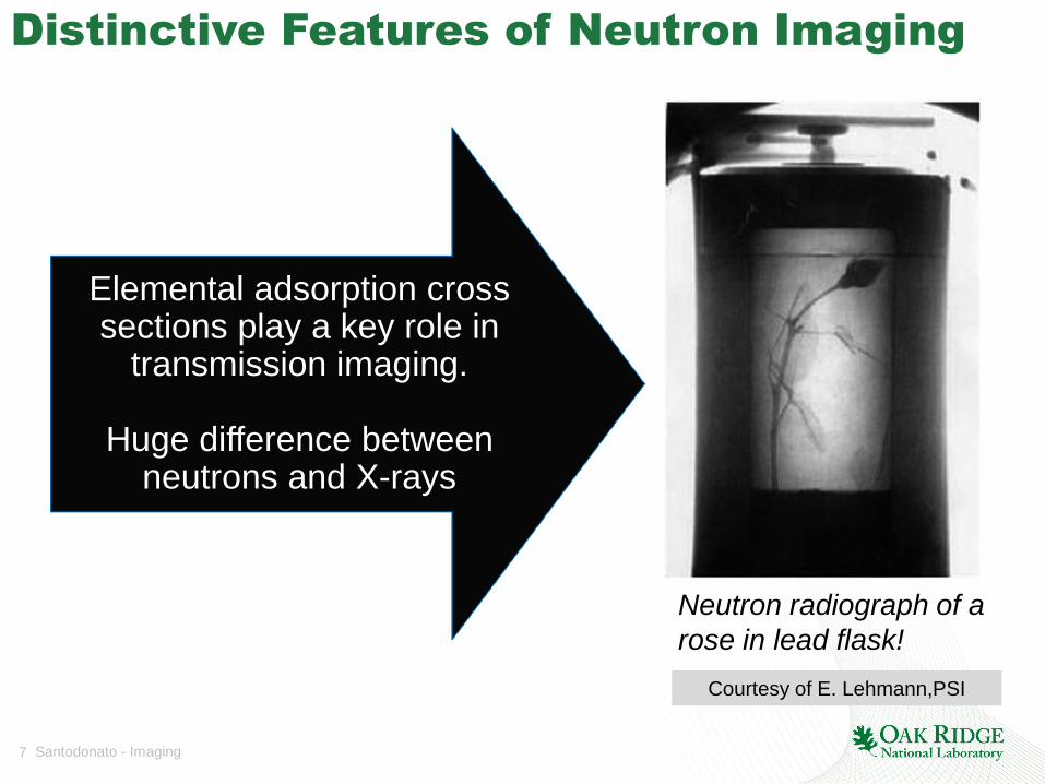
Distinctive Features of Neutron Imaging
Courtesy of E. Lehmann,PSI
Neutron radiograph of a
rose in lead flask!
Elemental adsorption cross sections play a key role in
transmission imaging.
Huge difference between neutrons and X-rays

8 Santodonato - Imaging
10-11 10-9 10-7 10-5 10-3
Dimension (meters)
0.1Å 1.0nm 1mm 0.1mm 10.0mm
Diffraction Scattering Real-space imaging
Inferred structure (indirect) Direct structure
SANS used to construct protein kinase A (PKA)
Fluid interactions in plant-groundwater systems
Characterization of biological membranes, colloids, porosity, etc.
Crystal structures Ice/water segregation in permafrost structures
You can directly see
the structure.
How easy!
Distinctive Features of Neutron Imaging

9 Managed by UT-Battelle for the U.S. Department of Energy
10-11 10-9 10-7 10-5 10-3
Dimension (meters)
0.1Å 1.0nm 1mm 0.1mm 10.0mm
Diffraction Scattering Real-space imaging
Inferred structure (indirect) Direct structure
SANS used to construct protein kinase A (PKA)
Fluid interactions in plant-groundwater systems
Characterization of biological membranes, colloids, porosity, etc.
Crystal structures Ice/water segregation in permafrost structures
Distinctive Features of Neutron Imaging
Spatial resolution is
limited!
100 mm routinely available 20 mm available with trade-off in
field-of-view and acquisition times
10 Santodonato - Imaging
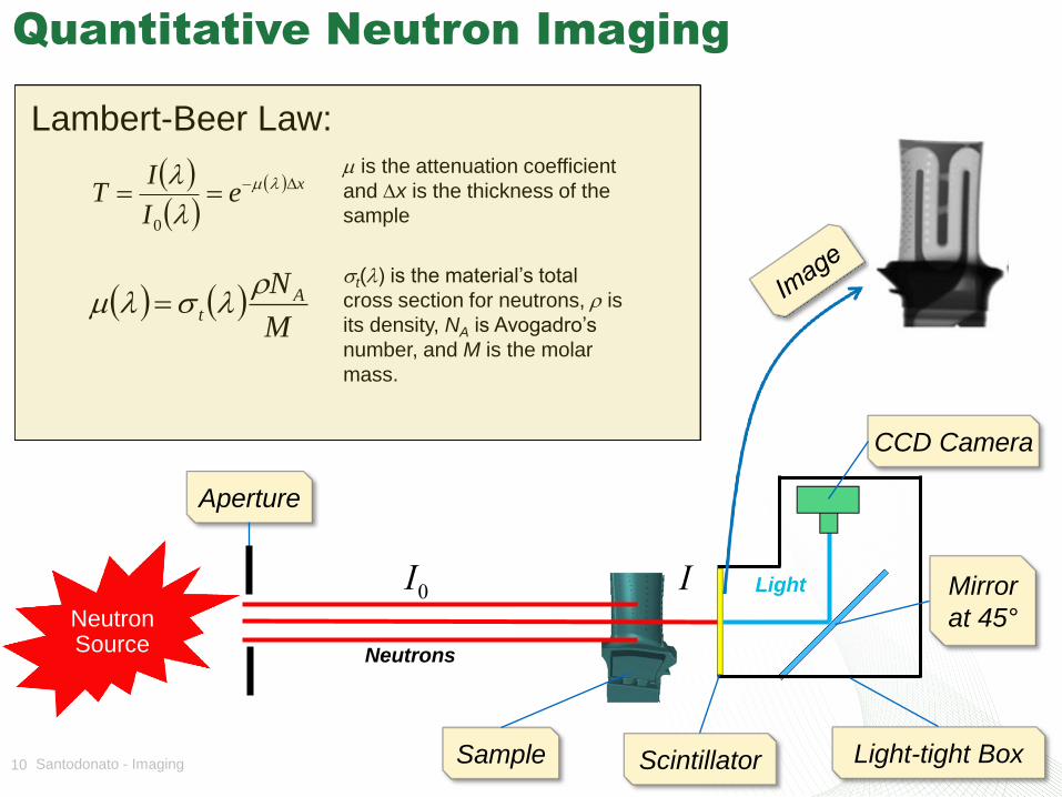
Quantitative Neutron Imaging
Sample Scintillator
Lambert-Beer Law:
CCD Camera
xeI
IT m
0
I0
I Mirror
at 45°
Neutrons
Light
Light-tight Box
Aperture
M
NAt
m
m is the attenuation coefficient
and x is the thickness of the
sample
t() is the material’s total
cross section for neutrons, is
its density, NA is Avogadro’s
number, and M is the molar
mass.
Neutron Source
11 Santodonato - Imaging
0.0
0.1
0.2
0.3
0.4
0.5
0.6
0.7
0.8
0.9
1.0
0.0 1.0 2.0 3.0 4.0 5.0 6.0 7.0 8.0 9.0 10.0
Tra
ns
mis
sio
n [
0 1
]
Wavelength (Angstroms)
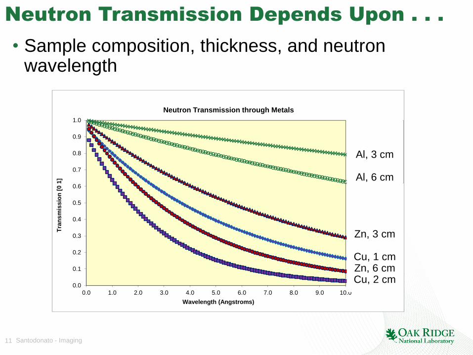
Neutron Transmission through Metals
Cu, 1 cm
Cu, 2 cm
Al, 3 cm
Al, 6 cm
Zn, 3 cm
Zn, 6 cm
Neutron Transmission Depends Upon . . .
• Sample composition, thickness, and neutron wavelength
Al, 3 cm
Al, 6 cm
Zn, 3 cm
Cu, 1 cm Zn, 6 cm Cu, 2 cm

12 Santodonato - Imaging
0.0
0.1
0.2
0.3
0.4
0.5
0.6
0.7
0.8
0.9
1.0
0.0 1.0 2.0 3.0 4.0 5.0 6.0 7.0 8.0 9.0 10.0
Tra
ns
mis
sio
n [
0 1
]
Wavelength (Angstroms)
Neutron Transmission through Metals
Cu, 1 cm
Cu, 2 cm
Al, 3 cm
Al, 6 cm
Zn, 3 cm
Zn, 6 cm
Neutron Transmission Depends Upon . . .
• Sample composition, thickness, and neutron wavelength
Al, 3 cm
Al, 6 cm
Zn, 3 cm
Cu, 1 cm Zn, 6 cm Cu, 2 cm
13 Santodonato - Imaging
Goals
• See what we can do with neutron imaging
– Compare to other imaging techniques
– Know when to choose it
• Understand the basic instrument layout and principals of neutron image acquisition and analysis
• Learn by example
– Review some recent neutron imaging projects
14 Santodonato - Imaging
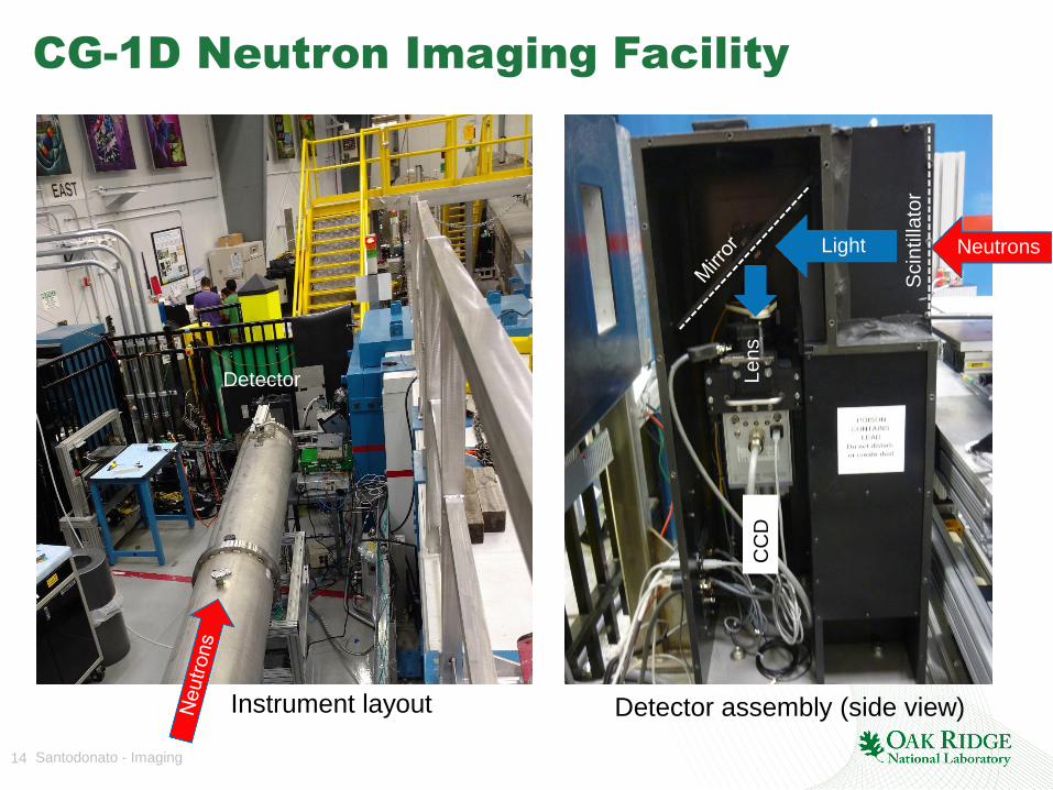
CG-1D Neutron Imaging Facility
Lens
CC
D
Scin
tilla
tor
Neutrons Light
Detector assembly (side view) Instrument layout
Detector

15 Santodonato - Imaging
Sample Area
Sample
(Automobile
part)
Rotation/
Translation
Stage
16 Santodonato - Imaging
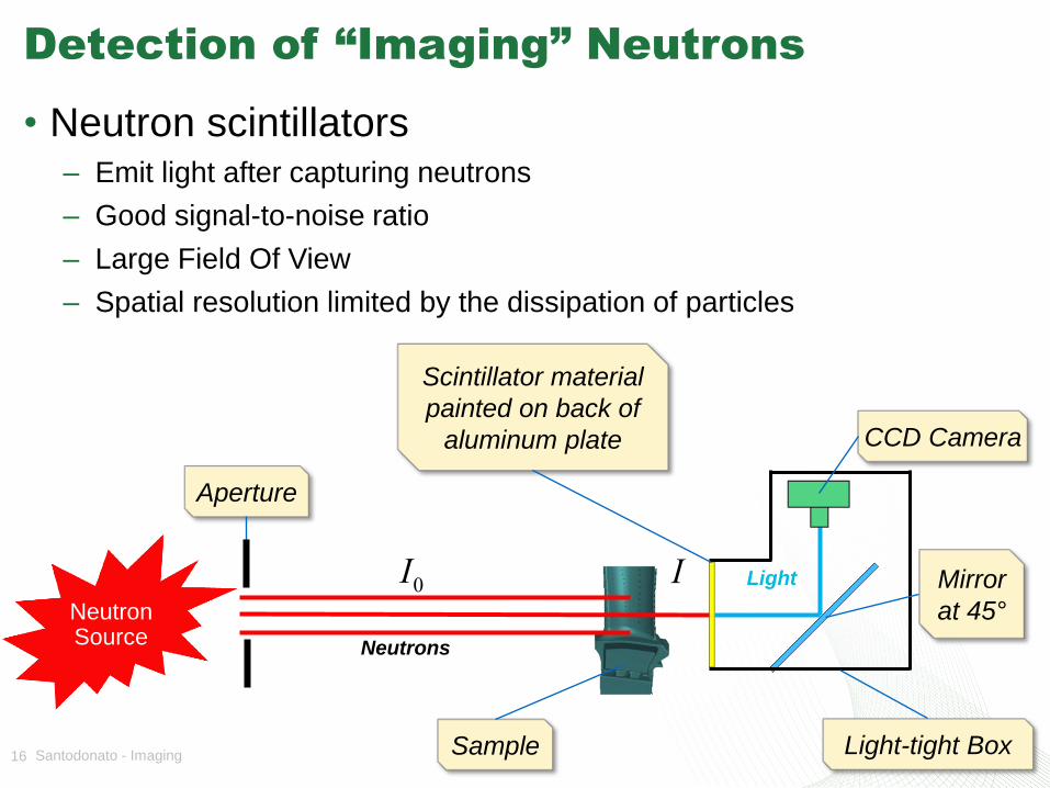
Detection of “Imaging” Neutrons
• Neutron scintillators – Emit light after capturing neutrons
– Good signal-to-noise ratio
– Large Field Of View
– Spatial resolution limited by the dissipation of particles
Sample
Scintillator material
painted on back of
aluminum plate CCD Camera
I0
I Mirror
at 45°
Neutrons
Light
Light-tight Box
Aperture
Neutron Source
17 Santodonato - Imaging
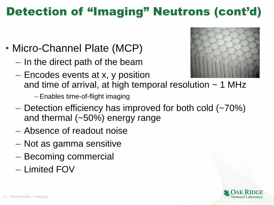
Detection of “Imaging” Neutrons (cont’d)
• Micro-Channel Plate (MCP)
– In the direct path of the beam
– Encodes events at x, y position and time of arrival, at high temporal resolution ~ 1 MHz
– Enables time-of-flight imaging
– Detection efficiency has improved for both cold (~70%) and thermal (~50%) energy range
– Absence of readout noise
– Not as gamma sensitive
– Becoming commercial
– Limited FOV

18 Santodonato - Imaging
CG-1D polychromatic beam
0.0E+00
5.0E+05
1.0E+06
1.5E+06
2.0E+06
2.5E+06
0 1 2 3 4 5 6 7 8 9 10
Neu
tro
n C
ou
nts
Wavelength (Å)
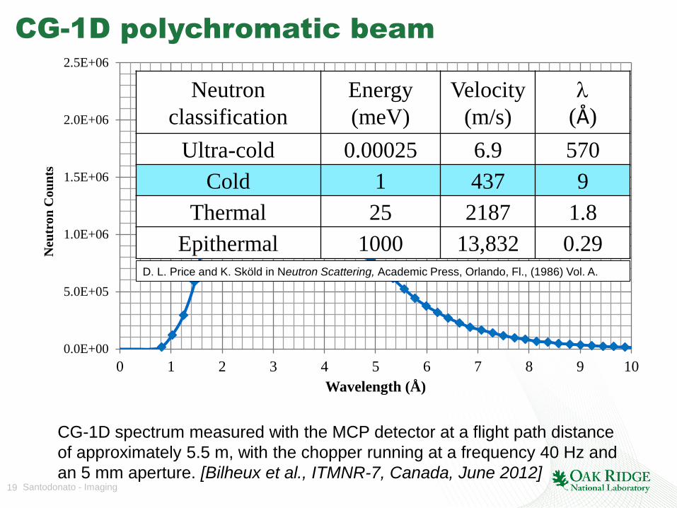
CG-1D spectrum measured with the MCP detector at a flight path distance
of approximately 5.5 m, with the chopper running at a frequency 40 Hz and
an 5 mm aperture. [Bilheux et al., ITMNR-7, Canada, June 2012]
19 Santodonato - Imaging
CG-1D polychromatic beam
0.0E+00
5.0E+05
1.0E+06
1.5E+06
2.0E+06
2.5E+06
0 1 2 3 4 5 6 7 8 9 10
Neu
tro
n C
ou
nts
Wavelength (Å)
CG-1D spectrum measured with the MCP detector at a flight path distance
of approximately 5.5 m, with the chopper running at a frequency 40 Hz and
an 5 mm aperture. [Bilheux et al., ITMNR-7, Canada, June 2012]
Neutron
classification
Energy
(meV)
Velocity
(m/s)
(Å)
Ultra-cold 0.00025 6.9 570
Cold 1 437 9
Thermal 25 2187 1.8
Epithermal 1000 13,832 0.29 D. L. Price and K. Sköld in Neutron Scattering, Academic Press, Orlando, Fl., (1986) Vol. A.
20 Santodonato - Imaging
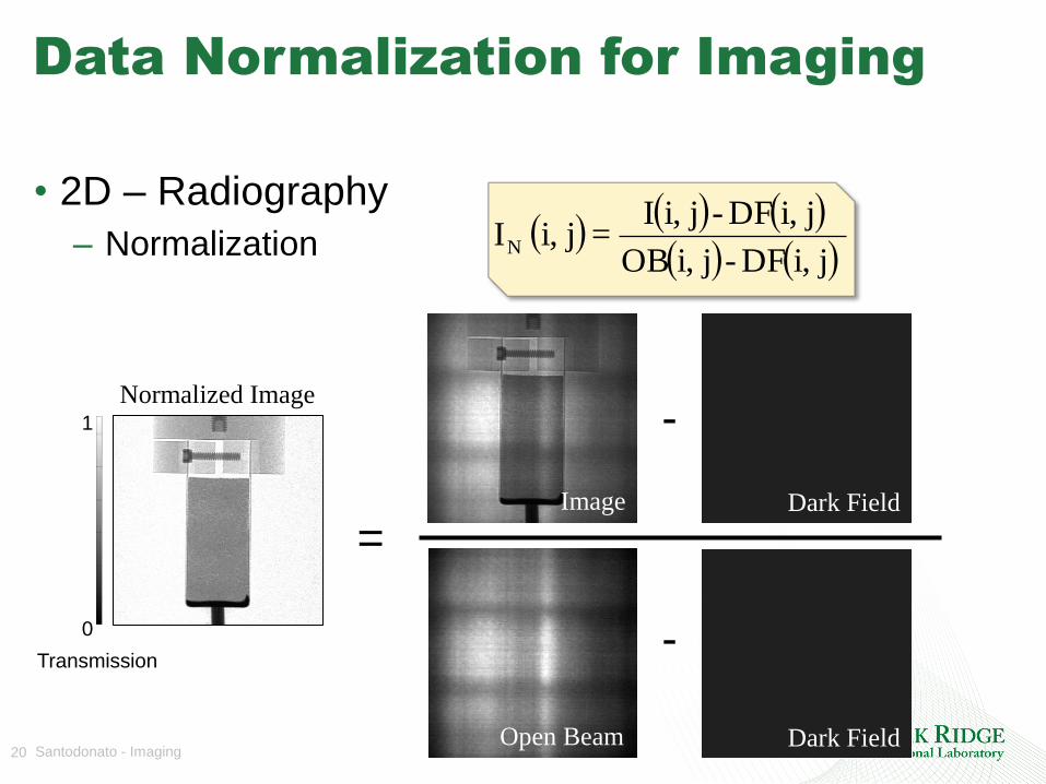
Data Normalization for Imaging
• 2D – Radiography
– Normalization
=
-
-
Image Dark Field
Dark Field Open Beam
Normalized Image 1
0
Transmission
ji,DF-ji,OB
ji,DF-ji,I=ji, IN
21 Santodonato - Imaging
Computed/Computerized Tomography
(CT)
• Several techniques:
– Filtered Back Projection
• Radon transform
• Works well with high signal to noise ration measurements
• Easy-to-use commercial, semi-automated software available
• Quick
– Iterative Reconstruction
• Direct approach
• Less artifacts
• Can reconstruct incomplete data
• High computation time
22 Santodonato - Imaging
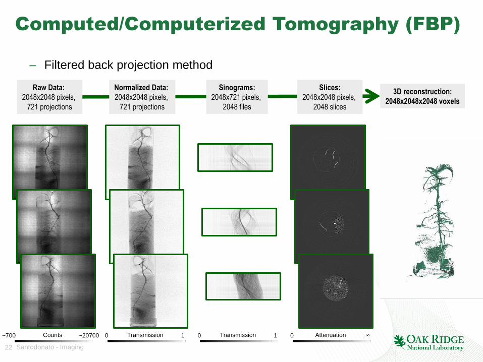
Raw Data:
2048x2048 pixels,
721 projections
Normalized Data:
2048x2048 pixels,
721 projections
Sinograms:
2048x721 pixels,
2048 files
Slices:
2048x2048 pixels,
2048 slices
3D reconstruction:
2048x2048x2048 voxels
Computed/Computerized Tomography (FBP)
– Filtered back projection method
~20700 ~700 Counts 1 0 Transmission 1 0 Transmission ∞ 0 Attenuation

23 Santodonato - Imaging
Conventional Neutron Imaging
Techniques at Steady-State Sources
• Radiography
• Tomography
• Stroboscopic Imaging
• Imaging of processes that happen fast
• Polarized Neutron Imaging
• Energy selective techniques possible with double-monochromator configuration
• Phase Contrast Imaging
– Under development
Routinely available at CG-1D
Available at CG-1D using the MCP detector
Newly implemented at CG-1D

24 Santodonato - Imaging
Goals
• See what we can do with neutron imaging
– Compare to other imaging techniques
– Know when to choose it
• Understand the basic instrument layout and principals of neutron image acquisition and analysis
• Learn by example
– Review some recent neutron imaging projects

25 Santodonato - Imaging
A Wide Range of Applications
26 Santodonato - Imaging
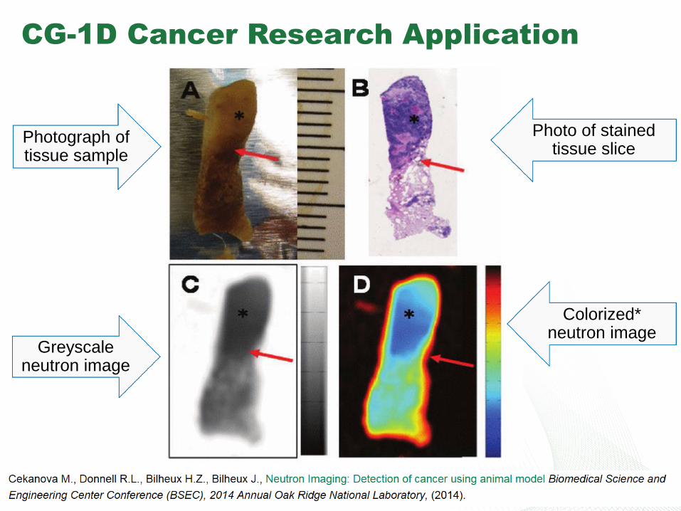
CG-1D Cancer Research Application
Photograph of tissue sample
Greyscale neutron image
Photo of stained tissue slice
Colorized* neutron image
27 Santodonato - Imaging
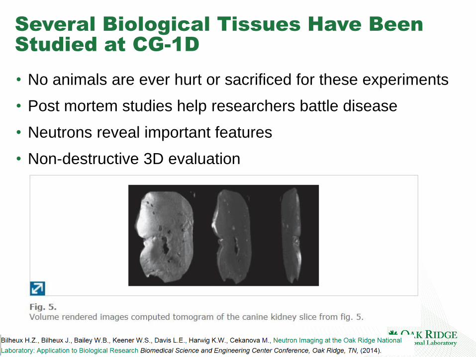
Several Biological Tissues Have Been
Studied at CG-1D
• No animals are ever hurt or sacrificed for these experiments
• Post mortem studies help researchers battle disease
• Neutrons reveal important features
• Non-destructive 3D evaluation
28 Santodonato - Imaging
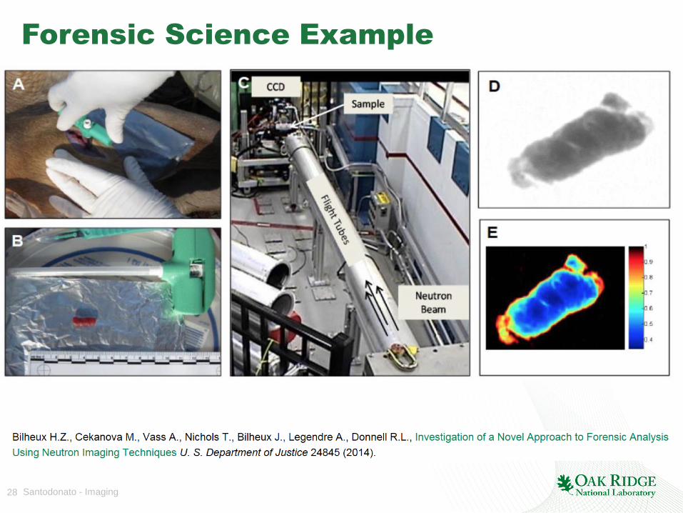
Forensic Science Example
29 Santodonato - Imaging
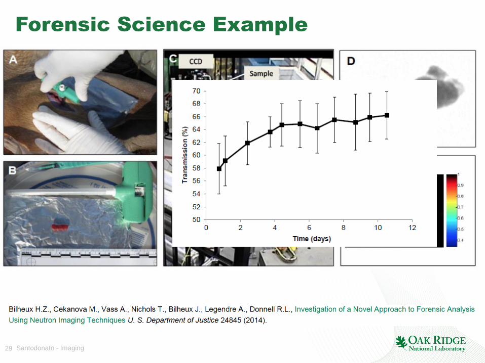
Forensic Science Example
30 Santodonato - Imaging
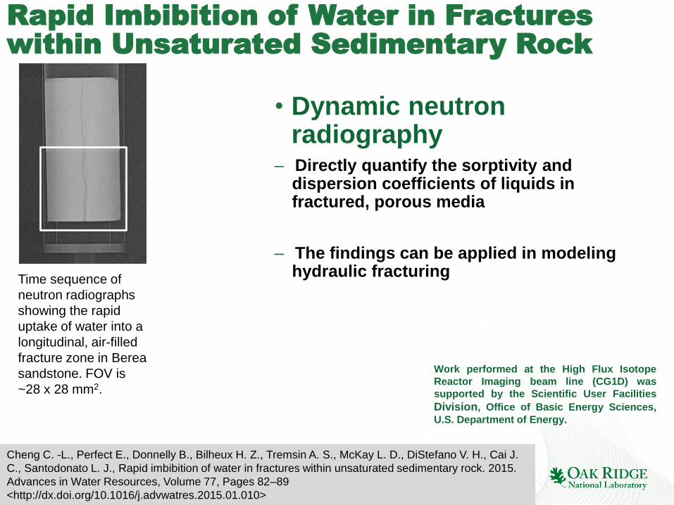
Rapid Imbibition of Water in Fractures
within Unsaturated Sedimentary Rock
Work performed at the High Flux Isotope
Reactor Imaging beam line (CG1D) was
supported by the Scientific User Facilities
Division, Office of Basic Energy Sciences,
U.S. Department of Energy.
Cheng C. -L., Perfect E., Donnelly B., Bilheux H. Z., Tremsin A. S., McKay L. D., DiStefano V. H., Cai J.
C., Santodonato L. J., Rapid imbibition of water in fractures within unsaturated sedimentary rock. 2015.
Advances in Water Resources, Volume 77, Pages 82–89
<http://dx.doi.org/10.1016/j.advwatres.2015.01.010>
Time sequence of
neutron radiographs
showing the rapid
uptake of water into a
longitudinal, air-filled
fracture zone in Berea
sandstone. FOV is
~28 x 28 mm2.
• Dynamic neutron radiography
– Directly quantify the sorptivity and dispersion coefficients of liquids in fractured, porous media
– The findings can be applied in modeling hydraulic fracturing

31 Santodonato - Imaging
Fabrication tolerance studies comparing CAD
drawing to neutron computed tomography
+
Engineering drawing Neutron CT
=
In orange/yellow: AUTOCAD
outline
In gray: neutron data
32 Santodonato - Imaging
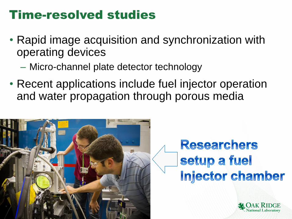
Time-resolved studies
• Rapid image acquisition and synchronization with operating devices
– Micro-channel plate detector technology
• Recent applications include fuel injector operation and water propagation through porous media

33 Santodonato - Imaging
More Examples
• More examples may be presented at the live talk

35 Santodonato - Imaging
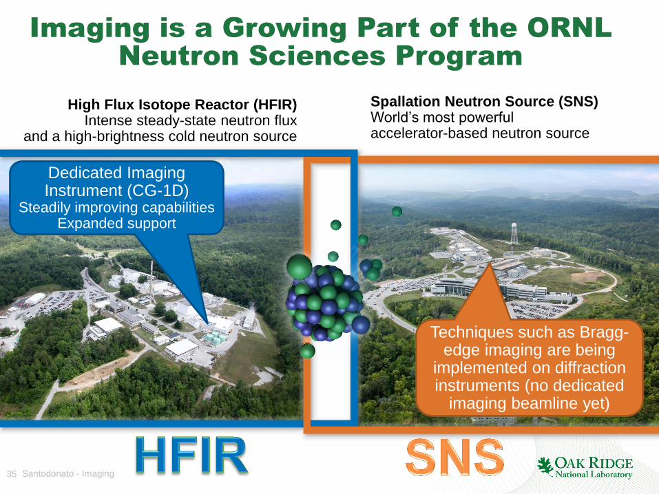
High Flux Isotope Reactor (HFIR) Intense steady-state neutron flux
and a high-brightness cold neutron source
Spallation Neutron Source (SNS) World’s most powerful accelerator-based neutron source
Imaging is a Growing Part of the ORNL
Neutron Sciences Program
Dedicated Imaging Instrument (CG-1D)
Steadily improving capabilities Expanded support
Techniques such as Bragg-edge imaging are being
implemented on diffraction instruments (no dedicated
imaging beamline yet)

36 Santodonato - Imaging
Diverse Science and Engineering
Applications
• Trends at CG-1D are similar other facilities
• Are we missing any opportunities? Your science!
Based upon recent publications
37 Santodonato - Imaging
Summary
• Neutrons are ideal for certain imaging applications, especially those requiring
– Sensitivity to hydrogen and other light elements
– Isotope sensitivity
– Penetration into large samples and/or sample environments
• Spatial resolution is a key consideration
– CG-1D routine capability of ~ 80 mm
– Radiography at ~ 20 mm (with the trade-off of long counting time) is now available
• Imaging capabilities are steadily improving

38 Santodonato - Imaging
Thank you
• Lou Santodonato [email protected]
• Hassina Bilheux [email protected]