Embed Size (px)
Citation preview
Imaging of TriathlonInjuries
Michael J. Tuite, MDKEYWORDS
� Triathlon � Swimming � Running � Cycling� Magnetic resonance imaging
The modern triathlon is a race in which athletesswim, cycle, and run in succession, and their over-all time includes the transition time between eachsport. Triathlons have become a popular athleticevent both in the United States and worldwide. Inthe United States there are about 300,000 athletesperforming triathlons every year, with more than1000 races, including approximately 15 Ironman-length events per year.1,2
Triathlon races can be variable in length, rangingfrom a “sprint triathlon,” to the longer Olympiclength, to Ironman, and to even ultradistance.These races became popular after the originalHawaii Ironman Triathlon was established tocombine into a single race the Waikiki 2.4-mile“Roughwater swim,” a 112-mile bike race aroundOahu, and a 26.2-mile marathon. However, themost popular triathlons are the shorter sprinttriathlons, which usually involve a 750-m swim,a 20-km cycling course, and a 5-km run. Althoughtermed a “sprint,” this race is really an enduranceevent, which requires training and stamina.
s.com
INJURY OVERVIEW
The modern triathlon was devised by runners whowere cross-training in swimming and cycling toprovide a variety to their workouts and to reduceoveruse injuries due to running.2 When the firsttriathlons began, many believed that training inthe 3 disciplines would mean fewer injuries thanin athletes who train in a single sport. In fact, over-use injuries among triathletes are more commonthan injuries in single-sport athletes.1,3e5 Thishigher incidence is thought to be for 2 reasons.First, triathletes train for more hours on averagethan single-sport athletes, averaging 10 to 14
Musculoskeletal Division, Department of Radiology, UE3/311, 600 Highland Avenue, Madison, WI 53792, USAE-mail address: [email protected]
Radiol Clin N Am 48 (2010) 1125–1135doi:10.1016/j.rcl.2010.07.0080033-8389/10/$ e see front matter � 2010 Elsevier Inc. Al
hours of training per week.6,7 As discussed inthis article, the 3 disciplines can contribute tosome of the same overuse injuries. Second, triath-letes sometimes have poorer technique or equip-ment than dedicated single-sport athletes, whichhas been shown to predispose them to more over-use injuries.1,2 An example is the distance runnerwho decides to train for an Ironman triathlon buthas a poorly fitted bike and develops patellartendinosis.
There are several medical issues that can occurin people either training for or racing in a triathlon(Box 1). In this article, the main focus is on thosemusculoskeletal injuries that may require medicalimaging.
About 90% of actively training triathletes willhave an acute or overuse injury over the courseof the year.3,8,9 An injury is typically defined asa musculoskeletal symptom that causes anunplanned stop in training or failure to completea race, or leads the individual to seek medicalcare.1,2 Acute injuries in triathletes include, forinstance, an acromioclavicular (AC) joint separa-tion from falling off a bike. Overuse injuries havean insidious onset and are much more commonin triathletes, representing 75% to 80% ofinjuries.1,2,6 Overuse injuries usually occur duringtraining, but 15% to 25% of injuries either presentduring a race or are exacerbated during the race toa point that it forces the athlete to discontinue.1,2,6
The most common site of injury in a triathlete isthe knee, accounting for over a quarter of allinjuries.1,2,6,10 Foot, ankle, lumbar spine, andshoulder injuries are also common (Table 1).Although overlap exists, the 3 different sportstend to be associated with somewhat distinctinjuries. Running has the highest triathlete injury
niversity of Wisconsin Medical School, UW Health,
l rights reserved. radiologic.th
eclinic
Box 1Examples of injuries and illnesses in triathletes
Nonmusculoskeletal
Dehydration
Hyperthermia
Blisters
Abrasions
Muscle cramps
Female athletic triad (anorexia, amenorrhea,osteoporosis)
Musculoskeletal, nonimaged
Short-duration low back pain
Muscle strains
Ulnar neuropathy (cyclist’s palsy)
Musculoskeletal, imaged
Rotator cuff impingement
Stress fractures
Tendinosis
Plantar fasciitis
Radiculopathy
Osteitis pubis
Meniscal tear
Iliotibial band syndrome
Tuite1126
rate and swimming the lowest (Fig. 1), althoughmost of the training hours are typically spentcycling.1,2,11
In this article, the imaging appearance of injuriesin triathletes commonly seen with each of the 3sports is discussed. The authors highlight thoseinjuries that are associated with more than one of
Table 1Site and percentage of injuries in triathletes
Site Percentage of All Injuries (%)
Foot 10e15
Ankle 10e15
Lower leg 5e10
Knee 25e30
Thigh 5e10
Hip/groin 5e10
Shoulder 10e15
Lumbar spine 10e15
Cervical spine 5
Other 5
the disciplines, which often lead to the triathleteseeking medical care because they significantlyaffect their training regimen. This article alsodiscusses injuries that tend to occur with one sportbut are then exacerbated by training in the otherdisciplines.
SWIMMING
Acute injuries are rare during swim training or duringthe swim leg of a triathlon.1,2,6,8 One of the mostserious concerns is drowning, because triathletesoften train and compete in open water where theunderwater visibility is poor.12 Acute musculoskel-etal injuries from shark attacks have also beendescribed in triathletes training in the ocean (http://www.msnbc.msn.com/id/24313314/).Overuse injuries from swimming are less
common than injuries from cycling and running,accounting for only 5% to 10% of injuries in triath-letes.1,2,13 Most clinical complaints are of shouldertendinitis and impingement pain.1,2,14 The swim-ming motion involves repetitive extremes ofabduction, flexion, and extension, and “swimmer’sshoulder” is common in competitive swimmers.Swimmer’s shoulder refers to a combination ofrotator cuff tendinosis/impingement and laxityfrom stretched anterior glenohumeral ligaments.15
Shoulder impingement pain in triathletes can beexacerbated during cycling when using aerobars,in which the cyclist leans forward and rests theelbows on a pad to maintain an aerodynamic posi-tion. This position causes the humeral head tocompress against the supraspinatus tendon andsubacromial bursa, and may aggravate impinge-ment symptoms.In one of the few imaging articles on triathletes,
shoulder magnetic resonance (MR) imaging find-ings of triathletes at the Hawaii Ironman Racewere assessed.14 These investigators performedMR imaging scans on athletes with shouldersymptoms and found rotator cuff tendinopathy in50%, AC joint marrow edema in 62%, and partial
Fig. 1. Percentage of injuries in triathletes that occurfrom the 3 sports.
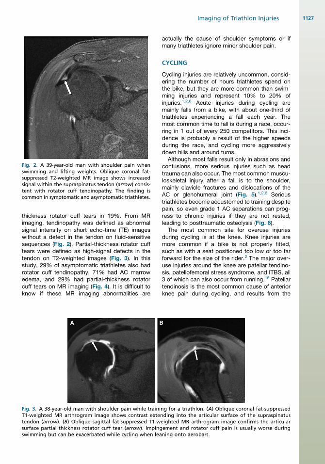
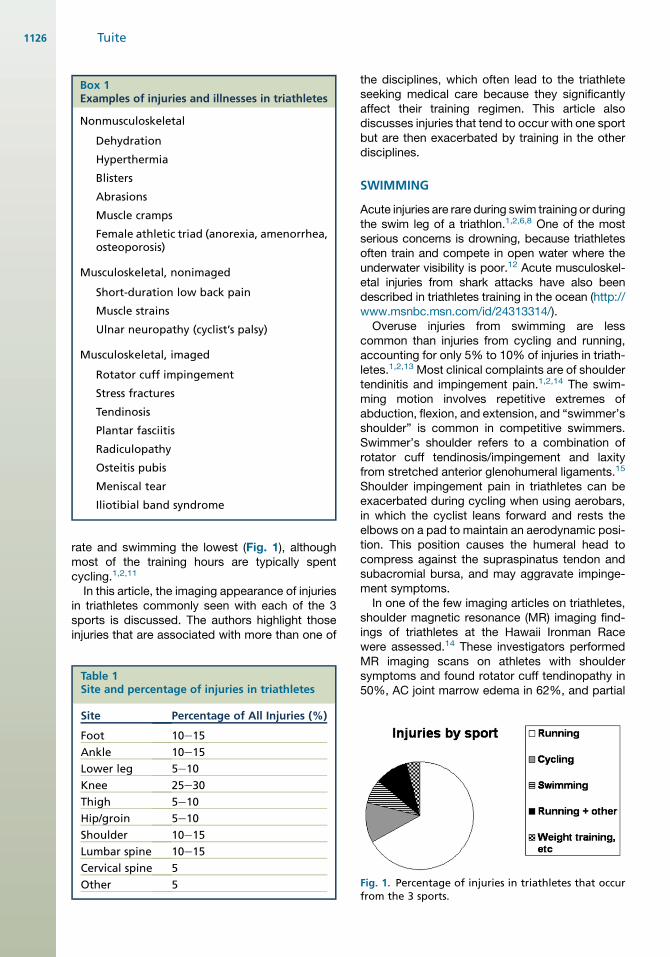
Fig. 2. A 39-year-old man with shoulder pain whenswimming and lifting weights. Oblique coronal fat-suppressed T2-weighted MR image shows increasedsignal within the supraspinatus tendon (arrow) consis-tent with rotator cuff tendinopathy. The finding iscommon in symptomatic and asymptomatic triathletes.
Imaging of Triathlon Injuries 1127
thickness rotator cuff tears in 19%. From MRimaging, tendinopathy was defined as abnormalsignal intensity on short echo-time (TE) imageswithout a defect in the tendon on fluid-sensitivesequences (Fig. 2). Partial-thickness rotator cufftears were defined as high-signal defects in thetendon on T2-weighted images (Fig. 3). In thisstudy, 29% of asymptomatic triathletes also hadrotator cuff tendinopathy, 71% had AC marrowedema, and 29% had partial-thickness rotatorcuff tears on MR imaging (Fig. 4). It is difficult toknow if these MR imaging abnormalities are
Fig. 3. A 38-year-old man with shoulder pain while trainiT1-weighted MR arthrogram image shows contrast extetendon (arrow). (B) Oblique sagittal fat-suppressed T1-wsurface partial thickness rotator cuff tear (arrow). Impingswimming but can be exacerbated while cycling when lea
actually the cause of shoulder symptoms or ifmany triathletes ignore minor shoulder pain.
CYCLING
Cycling injuries are relatively uncommon, consid-ering the number of hours triathletes spend onthe bike, but they are more common than swim-ming injuries and represent 10% to 20% ofinjuries.1,2,6 Acute injuries during cycling aremainly falls from a bike, with about one-third oftriathletes experiencing a fall each year. Themost common time to fall is during a race, occur-ring in 1 out of every 250 competitors. This inci-dence is probably a result of the higher speedsduring the race, and cycling more aggressivelydown hills and around turns.
Although most falls result only in abrasions andcontusions, more serious injuries such as headtrauma can also occur. The most common muscu-loskeletal injury after a fall is to the shoulder,mainly clavicle fractures and dislocations of theAC or glenohumeral joint (Fig. 5).1,2,6 Serioustriathletes become accustomed to training despitepain, so even grade 1 AC separations can prog-ress to chronic injuries if they are not rested,leading to posttraumatic osteolysis (Fig. 6).
The most common site for overuse injuriesduring cycling is at the knee. Knee injuries aremore common if a bike is not properly fitted,such as with a seat positioned too low or too farforward for the size of the rider.2 The major over-use injuries around the knee are patellar tendino-sis, patellofemoral stress syndrome, and ITBS, all3 of which can also occur from running.16 Patellartendinosis is the most common cause of anteriorknee pain during cycling, and results from the
ng for a triathlon. (A) Oblique coronal fat-suppressednding into the articular surface of the supraspinatuseighted MR arthrogram image confirms the articularement and rotator cuff pain is usually worse duringning onto aerobars.
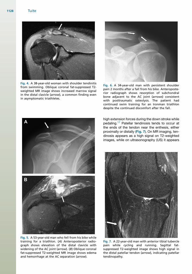
Fig. 4. A 38-year-old woman with shoulder tendinitisfrom swimming. Oblique coronal fat-suppressed T2-weighted MR image shows increased marrow signalin the distal clavicle (arrow), a common finding evenin asymptomatic triathletes.
Fig. 5. A 53-year-old man who fell from his bike whiletraining for a triathlon. (A) Anteroposterior radio-graph shows elevation of the distal clavicle withwidening of the AC joint (arrow). (B) Oblique coronalfat-suppressed T2-weighted MR image shows edemaand hemorrhage at the AC separation (arrow).
Fig. 6. A 34-year-old man with persistent shoulderpain 2 months after a fall from his bike. Anteroposte-rior radiograph shows resorption of subchondralbone adjacent to the AC joint (arrows) consistentwith posttraumatic osteolysis. The patient hadcontinued swim training for an Ironman triathlondespite the continued discomfort after the fall.
Tuite1128
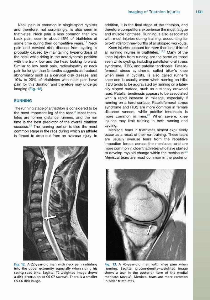
high extension forces during the down stroke whilepedaling.17 Patellar tendinosis tends to occur atthe ends of the tendon near the enthesis, eitherproximally or distally (Fig. 7). On MR imaging, ten-dinosis appears as a high signal on T2-weightedimages, while on ultrasonography (US) it appears
Fig. 7. A 22-year-old man with anterior tibial tuberclepain while cycling and running. Sagittal fat-suppressed T2-weighted image shows high signal inthe distal patellar tendon (arrow), indicating patellartendinopathy.
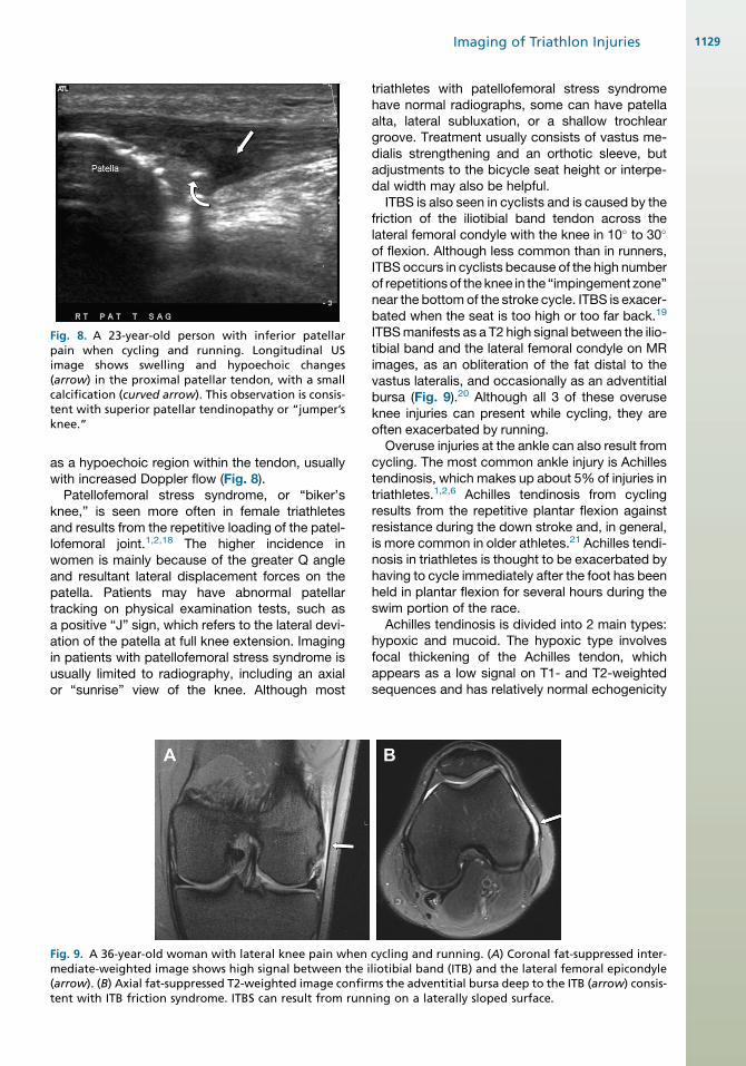
Fig. 8. A 23-year-old person with inferior patellarpain when cycling and running. Longitudinal USimage shows swelling and hypoechoic changes(arrow) in the proximal patellar tendon, with a smallcalcification (curved arrow). This observation is consis-tent with superior patellar tendinopathy or “jumper’sknee.”
Imaging of Triathlon Injuries 1129
as a hypoechoic region within the tendon, usuallywith increased Doppler flow (Fig. 8).
Patellofemoral stress syndrome, or “biker’sknee,” is seen more often in female triathletesand results from the repetitive loading of the patel-lofemoral joint.1,2,18 The higher incidence inwomen is mainly because of the greater Q angleand resultant lateral displacement forces on thepatella. Patients may have abnormal patellartracking on physical examination tests, such asa positive “J” sign, which refers to the lateral devi-ation of the patella at full knee extension. Imagingin patients with patellofemoral stress syndrome isusually limited to radiography, including an axialor “sunrise” view of the knee. Although most
Fig. 9. A 36-year-old woman with lateral knee pain whenmediate-weighted image shows high signal between the i(arrow). (B) Axial fat-suppressed T2-weighted image confirtent with ITB friction syndrome. ITBS can result from runn
triathletes with patellofemoral stress syndromehave normal radiographs, some can have patellaalta, lateral subluxation, or a shallow trochleargroove. Treatment usually consists of vastus me-dialis strengthening and an orthotic sleeve, butadjustments to the bicycle seat height or interpe-dal width may also be helpful.
ITBS is also seen in cyclists and is caused by thefriction of the iliotibial band tendon across thelateral femoral condyle with the knee in 10� to 30�
of flexion. Although less common than in runners,ITBS occurs in cyclists because of the high numberof repetitionsof the knee in the “impingement zone”near the bottom of the stroke cycle. ITBS is exacer-bated when the seat is too high or too far back.19
ITBSmanifests as a T2 high signal between the ilio-tibial band and the lateral femoral condyle on MRimages, as an obliteration of the fat distal to thevastus lateralis, and occasionally as an adventitialbursa (Fig. 9).20 Although all 3 of these overuseknee injuries can present while cycling, they areoften exacerbated by running.
Overuse injuries at the ankle can also result fromcycling. The most common ankle injury is Achillestendinosis, which makes up about 5% of injuries intriathletes.1,2,6 Achilles tendinosis from cyclingresults from the repetitive plantar flexion againstresistance during the down stroke and, in general,is more common in older athletes.21 Achilles tendi-nosis in triathletes is thought to be exacerbated byhaving to cycle immediately after the foot has beenheld in plantar flexion for several hours during theswim portion of the race.
Achilles tendinosis is divided into 2 main types:hypoxic and mucoid. The hypoxic type involvesfocal thickening of the Achilles tendon, whichappears as a low signal on T1- and T2-weightedsequences and has relatively normal echogenicity
cycling and running. (A) Coronal fat-suppressed inter-liotibial band (ITB) and the lateral femoral epicondylems the adventitial bursa deep to the ITB (arrow) consis-ing on a laterally sloped surface.
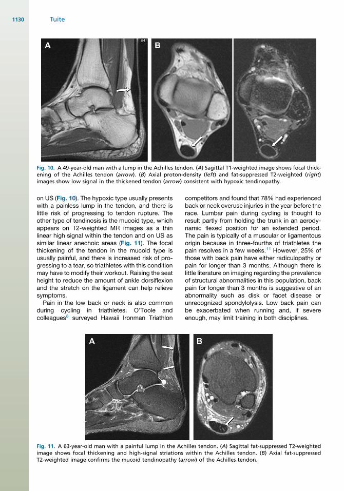
Fig. 10. A 49-year-old man with a lump in the Achilles tendon. (A) Sagittal T1-weighted image shows focal thick-ening of the Achilles tendon (arrow). (B) Axial proton-density (left) and fat-suppressed T2-weighted (right)images show low signal in the thickened tendon (arrow) consistent with hypoxic tendinopathy.
Tuite1130
on US (Fig. 10). The hypoxic type usually presentswith a painless lump in the tendon, and there islittle risk of progressing to tendon rupture. Theother type of tendinosis is the mucoid type, whichappears on T2-weighted MR images as a thinlinear high signal within the tendon and on US assimilar linear anechoic areas (Fig. 11). The focalthickening of the tendon in the mucoid type isusually painful, and there is increased risk of pro-gressing to a tear, so triathletes with this conditionmay have to modify their workout. Raising the seatheight to reduce the amount of ankle dorsiflexionand the stretch on the ligament can help relievesymptoms.Pain in the low back or neck is also common
during cycling in triathletes. O’Toole andcolleagues8 surveyed Hawaii Ironman Triathlon
Fig. 11. A 63-year-old man with a painful lump in the Acimage shows focal thickening and high-signal striationsT2-weighted image confirms the mucoid tendinopathy (a
competitors and found that 78% had experiencedback or neck overuse injuries in the year before therace. Lumbar pain during cycling is thought toresult partly from holding the trunk in an aerody-namic flexed position for an extended period.The pain is typically of a muscular or ligamentousorigin because in three-fourths of triathletes thepain resolves in a few weeks.11 However, 25% ofthose with back pain have either radiculopathy orpain for longer than 3 months. Although there islittle literature on imaging regarding the prevalenceof structural abnormalities in this population, backpain for longer than 3 months is suggestive of anabnormality such as disk or facet disease orunrecognized spondylolysis. Low back pain canbe exacerbated when running and, if severeenough, may limit training in both disciplines.
hilles tendon. (A) Sagittal fat-suppressed T2-weightedwithin the Achilles tendon. (B) Axial fat-suppressedrrow) of the Achilles tendon.
Imaging of Triathlon Injuries 1131
Neck pain is common in single-sport cyclistsand therefore, not surprisingly, is also seen intriathletes. Neck pain is less common than lowback pain, seen in about 45% of triathletes atsome time during their competitive years.11 Neckpain and cervical disk disease from cycling isprobably caused by maintaining hyperlordosis ofthe neck while riding in the aerodynamic positionwith the trunk low and the head looking forward.Similar to low back pain, radiculopathy or neckpain for longer than 3months suggests a structuralabnormality such as a cervical disk disease, and10% to 20% of triathletes with neck pain havepain for this duration and therefore may undergoimaging (Fig. 12).
RUNNING
The running stage of a triathlon is considered to bethe most important leg of the race.2 Most triath-letes are former distance runners, and the runtime is the best predictor of the overall triathlonsuccess.22 The running portion is also the mostcommon stage in the race during which an athleteis forced to drop out from an overuse injury. In
Fig. 12. A 22-year-old man with neck pain radiatinginto the upper extremity, especially when riding hisracing road bike. Sagittal T2-weighted image showsa disk protrusion at C6-C7 (arrow). There is a smallerC5-C6 disk bulge.
addition, it is the final stage of the triathlon, andtherefore competitors experience the most fatigueand muscle tightness. Running is also associatedwith most injuries during training, accounting fortwo-thirds to three-fourths of all skipped workouts.
Knee injuries account for more than one-third ofall running injuries in triathletes.1,2,6 Many of theknee injuries from running are the same as thoseseen while cycling, including patellofemoral stresssyndrome, ITBS, and patellar tendinosis. Patello-femoral stress syndrome, called biker’s kneewhen seen in cyclists, is also called runner’sknee and is usually worse when running on hills.ITBS tends to be aggravated by running on a later-ally sloped surface, such as a steeply crownedroad. Patellar tendinosis appears to be associatedwith a rapid increase in mileage, especially ifrunning on a hard surface. Patellofemoral stresssyndrome and ITBS are more common in femaledistance runners, while patellar tendinosis ismore common in men.23 When severe, kneeinjuries may limit training in both running andcycling.
Meniscal tears in triathletes almost exclusivelyoccur as a result of their run training. These tearsare usually overuse tears from the repetitiveimpaction forces across the meniscus, and aremore common in older triathletes who have startedto develop myxoid change within the meniscus.24
Meniscal tears are most common in the posterior
Fig. 13. A 45-year-old man with knee pain whenrunning. Sagittal proton-densityeweighted imageshows a tear in the posterior horn of the medialmeniscus (arrow). Meniscal tears are more commonin older triathletes.
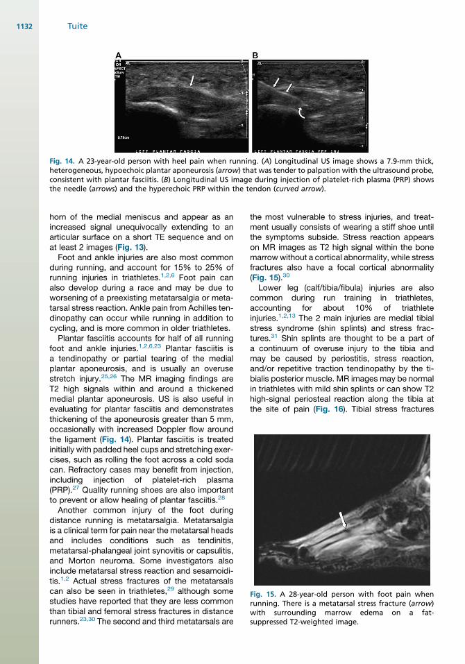
Fig. 14. A 23-year-old person with heel pain when running. (A) Longitudinal US image shows a 7.9-mm thick,heterogeneous, hypoechoic plantar aponeurosis (arrow) that was tender to palpation with the ultrasound probe,consistent with plantar fasciitis. (B) Longitudinal US image during injection of platelet-rich plasma (PRP) showsthe needle (arrows) and the hyperechoic PRP within the tendon (curved arrow).
Fig. 15. A 28-year-old person with foot pain whenrunning. There is a metatarsal stress fracture (arrow)with surrounding marrow edema on a fat-suppressed T2-weighted image.
Tuite1132
horn of the medial meniscus and appear as anincreased signal unequivocally extending to anarticular surface on a short TE sequence and onat least 2 images (Fig. 13).Foot and ankle injuries are also most common
during running, and account for 15% to 25% ofrunning injuries in triathletes.1,2,6 Foot pain canalso develop during a race and may be due toworsening of a preexisting metatarsalgia or meta-tarsal stress reaction. Ankle pain from Achilles ten-dinopathy can occur while running in addition tocycling, and is more common in older triathletes.Plantar fasciitis accounts for half of all running
foot and ankle injuries.1,2,6,23 Plantar fasciitis isa tendinopathy or partial tearing of the medialplantar aponeurosis, and is usually an overusestretch injury.25,26 The MR imaging findings areT2 high signals within and around a thickenedmedial plantar aponeurosis. US is also useful inevaluating for plantar fasciitis and demonstratesthickening of the aponeurosis greater than 5 mm,occasionally with increased Doppler flow aroundthe ligament (Fig. 14). Plantar fasciitis is treatedinitially with padded heel cups and stretching exer-cises, such as rolling the foot across a cold sodacan. Refractory cases may benefit from injection,including injection of platelet-rich plasma(PRP).27 Quality running shoes are also importantto prevent or allow healing of plantar fasciitis.28
Another common injury of the foot duringdistance running is metatarsalgia. Metatarsalgiais a clinical term for pain near the metatarsal headsand includes conditions such as tendinitis,metatarsal-phalangeal joint synovitis or capsulitis,and Morton neuroma. Some investigators alsoinclude metatarsal stress reaction and sesamoidi-tis.1,2 Actual stress fractures of the metatarsalscan also be seen in triathletes,29 although somestudies have reported that they are less commonthan tibial and femoral stress fractures in distancerunners.23,30 The second and third metatarsals are
the most vulnerable to stress injuries, and treat-ment usually consists of wearing a stiff shoe untilthe symptoms subside. Stress reaction appearson MR images as T2 high signal within the bonemarrowwithout a cortical abnormality, while stressfractures also have a focal cortical abnormality(Fig. 15).30
Lower leg (calf/tibia/fibula) injuries are alsocommon during run training in triathletes,accounting for about 10% of triathleteinjuries.1,2,13 The 2 main injuries are medial tibialstress syndrome (shin splints) and stress frac-tures.31 Shin splints are thought to be a part ofa continuum of overuse injury to the tibia andmay be caused by periostitis, stress reaction,and/or repetitive traction tendinopathy by the ti-bialis posterior muscle. MR images may be normalin triathletes with mild shin splints or can show T2high-signal periosteal reaction along the tibia atthe site of pain (Fig. 16). Tibial stress fractures
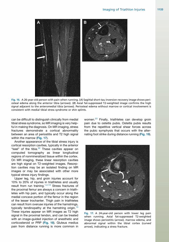
Fig. 16. A 26-year-old person with pain when running. (A) Sagittal short-tau inversion recovery image shows peri-osteal edema along the anterior tibia (arrows). (B) Axial fat-suppressed T2-weighted image confirms the highsignal adjacent to the anteromedial tibia (arrows). Periosteal edema without marrow or cortical involvement isconsistent with medial tibial stress syndrome or shin splints.
Fig. 17. A 24-year-old person with lower leg painwhen running. Axial fat-suppressed T2-weightedimage shows periostitis (arrow), marrow edema, andabnormal signal within the tibial cortex (curvedarrow), indicating a stress fracture.
Imaging of Triathlon Injuries 1133
can be difficult to distinguish clinically from medialtibial stress syndrome, soMR imaging is very help-ful in making the diagnosis. On MR imaging, stressfractures demonstrate a cortical abnormalitybetween an area of periostitis and T2 high signalwithin the marrow (Fig. 17).
Another appearance of the tibial stress injury iscortical resorption cavities, typically in the anterior“keel” of the tibia.32 These cavities appear oncomputed tomography as linear longitudinalregions of nonmineralized tissue within the cortex.On MR imaging, these linear resorption cavitiesare high signal on T2-weighted images. Resorp-tion cavities may be an isolated finding on MRimages or may be associated with other moretypical stress injury findings.
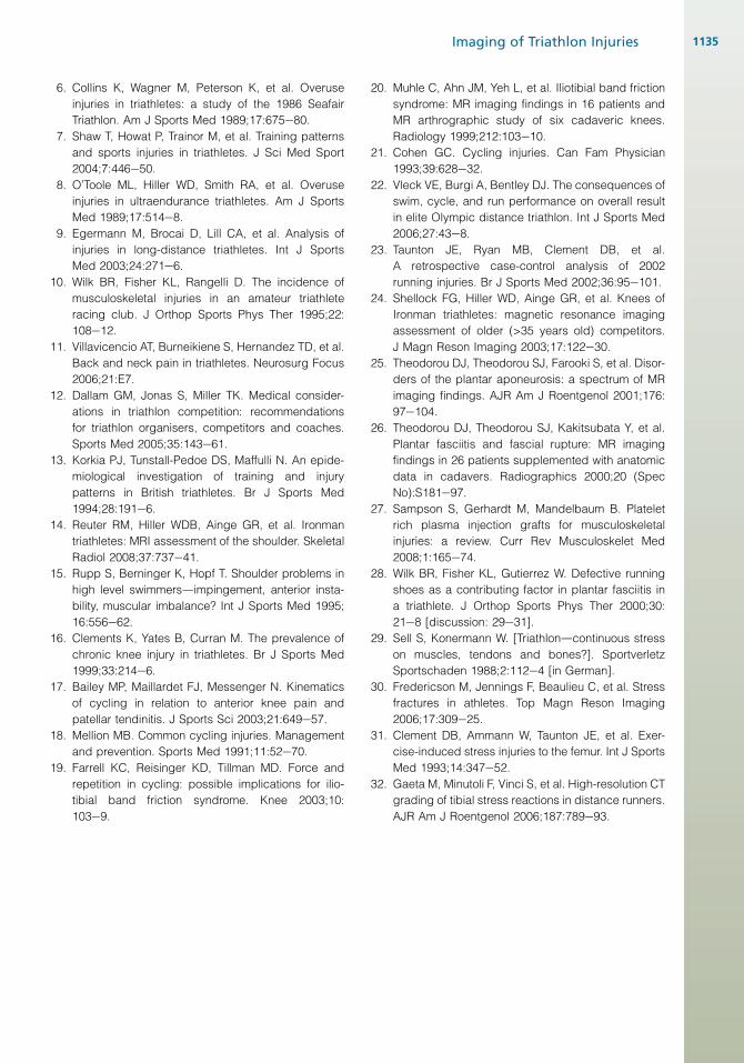
Upper leg, hip, and groin injuries account for10% to 20% of injuries in triathletes and usuallyresult from run training.1,2,13 Stress fractures ofthe proximal femur are always a concern in triath-letes with hip pain, and typically occur along themedial concave portion of the femur in the regionof the lesser trochanter. Thigh pain in triathletescan result from overuse injuries of the hamstrings,typically tendinopathy at the hamstring origin.13
These injuries appear on MR images as T2 highsignal in the proximal tendon, and can be treatedwith an image-guided injection of anesthetic andcorticosteroid or PRP (Fig. 18). Gluteus mediuspain from distance running is more common in
women.23 Finally, triathletes can develop groinpain due to osteitis pubis. Osteitis pubis resultsfrom the repetitive vertical shear forces acrossthe pubic symphysis that occurs with the alter-nating foot strike during distance running (Fig. 19).
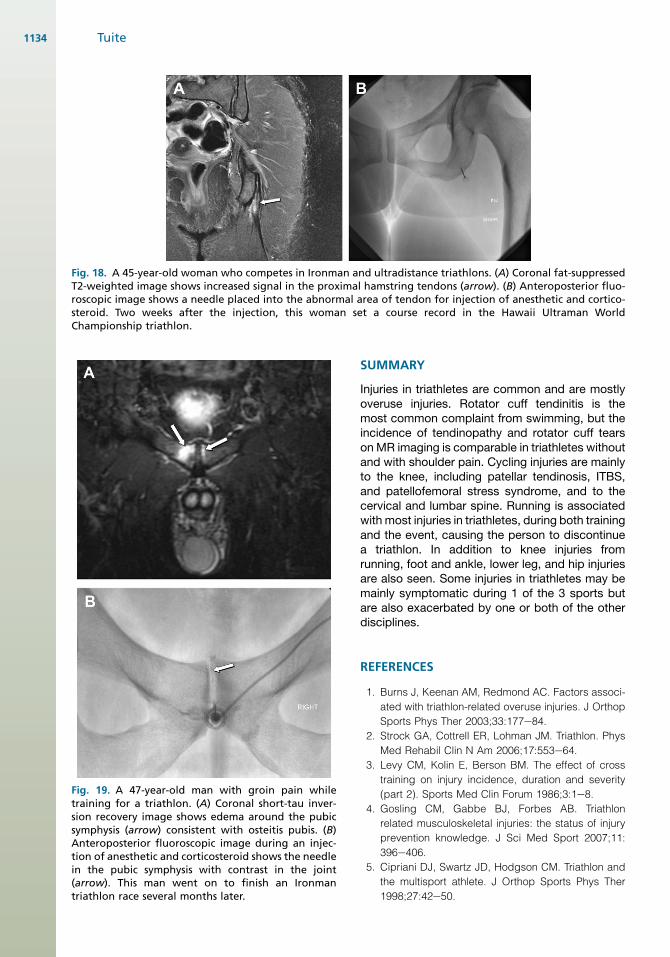
Fig. 18. A 45-year-old woman who competes in Ironman and ultradistance triathlons. (A) Coronal fat-suppressedT2-weighted image shows increased signal in the proximal hamstring tendons (arrow). (B) Anteroposterior fluo-roscopic image shows a needle placed into the abnormal area of tendon for injection of anesthetic and cortico-steroid. Two weeks after the injection, this woman set a course record in the Hawaii Ultraman WorldChampionship triathlon.
Fig. 19. A 47-year-old man with groin pain whiletraining for a triathlon. (A) Coronal short-tau inver-sion recovery image shows edema around the pubicsymphysis (arrow) consistent with osteitis pubis. (B)Anteroposterior fluoroscopic image during an injec-tion of anesthetic and corticosteroid shows the needlein the pubic symphysis with contrast in the joint(arrow). This man went on to finish an Ironmantriathlon race several months later.
Tuite1134
SUMMARY
Injuries in triathletes are common and are mostlyoveruse injuries. Rotator cuff tendinitis is themost common complaint from swimming, but theincidence of tendinopathy and rotator cuff tearson MR imaging is comparable in triathletes withoutand with shoulder pain. Cycling injuries are mainlyto the knee, including patellar tendinosis, ITBS,and patellofemoral stress syndrome, and to thecervical and lumbar spine. Running is associatedwith most injuries in triathletes, during both trainingand the event, causing the person to discontinuea triathlon. In addition to knee injuries fromrunning, foot and ankle, lower leg, and hip injuriesare also seen. Some injuries in triathletes may bemainly symptomatic during 1 of the 3 sports butare also exacerbated by one or both of the otherdisciplines.
REFERENCES
1. Burns J, Keenan AM, Redmond AC. Factors associ-
ated with triathlon-related overuse injuries. J Orthop
Sports Phys Ther 2003;33:177e84.
2. Strock GA, Cottrell ER, Lohman JM. Triathlon. Phys
Med Rehabil Clin N Am 2006;17:553e64.
3. Levy CM, Kolin E, Berson BM. The effect of cross
training on injury incidence, duration and severity
(part 2). Sports Med Clin Forum 1986;3:1e8.
4. Gosling CM, Gabbe BJ, Forbes AB. Triathlon
related musculoskeletal injuries: the status of injury
prevention knowledge. J Sci Med Sport 2007;11:
396e406.
5. Cipriani DJ, Swartz JD, Hodgson CM. Triathlon and
the multisport athlete. J Orthop Sports Phys Ther
1998;27:42e50.
Imaging of Triathlon Injuries 1135
6. Collins K, Wagner M, Peterson K, et al. Overuse
injuries in triathletes: a study of the 1986 Seafair
Triathlon. Am J Sports Med 1989;17:675e80.
7. Shaw T, Howat P, Trainor M, et al. Training patterns
and sports injuries in triathletes. J Sci Med Sport
2004;7:446e50.
8. O’Toole ML, Hiller WD, Smith RA, et al. Overuse
injuries in ultraendurance triathletes. Am J Sports
Med 1989;17:514e8.
9. Egermann M, Brocai D, Lill CA, et al. Analysis of
injuries in long-distance triathletes. Int J Sports
Med 2003;24:271e6.
10. Wilk BR, Fisher KL, Rangelli D. The incidence of
musculoskeletal injuries in an amateur triathlete
racing club. J Orthop Sports Phys Ther 1995;22:
108e12.
11. Villavicencio AT, Burneikiene S, Hernandez TD, et al.
Back and neck pain in triathletes. Neurosurg Focus
2006;21:E7.
12. Dallam GM, Jonas S, Miller TK. Medical consider-
ations in triathlon competition: recommendations
for triathlon organisers, competitors and coaches.
Sports Med 2005;35:143e61.
13. Korkia PJ, Tunstall-Pedoe DS, Maffulli N. An epide-
miological investigation of training and injury
patterns in British triathletes. Br J Sports Med
1994;28:191e6.
14. Reuter RM, Hiller WDB, Ainge GR, et al. Ironman
triathletes: MRI assessment of the shoulder. Skeletal
Radiol 2008;37:737e41.
15. Rupp S, Berninger K, Hopf T. Shoulder problems in
high level swimmersdimpingement, anterior insta-
bility, muscular imbalance? Int J Sports Med 1995;
16:556e62.
16. Clements K, Yates B, Curran M. The prevalence of
chronic knee injury in triathletes. Br J Sports Med
1999;33:214e6.
17. Bailey MP, Maillardet FJ, Messenger N. Kinematics
of cycling in relation to anterior knee pain and
patellar tendinitis. J Sports Sci 2003;21:649e57.
18. Mellion MB. Common cycling injuries. Management
and prevention. Sports Med 1991;11:52e70.
19. Farrell KC, Reisinger KD, Tillman MD. Force and
repetition in cycling: possible implications for ilio-
tibial band friction syndrome. Knee 2003;10:
103e9.
20. Muhle C, Ahn JM, Yeh L, et al. Iliotibial band friction
syndrome: MR imaging findings in 16 patients and
MR arthrographic study of six cadaveric knees.
Radiology 1999;212:103e10.
21. Cohen GC. Cycling injuries. Can Fam Physician
1993;39:628e32.
22. Vleck VE, Burgi A, Bentley DJ. The consequences of
swim, cycle, and run performance on overall result
in elite Olympic distance triathlon. Int J Sports Med
2006;27:43e8.
23. Taunton JE, Ryan MB, Clement DB, et al.
A retrospective case-control analysis of 2002
running injuries. Br J Sports Med 2002;36:95e101.
24. Shellock FG, Hiller WD, Ainge GR, et al. Knees of
Ironman triathletes: magnetic resonance imaging
assessment of older (>35 years old) competitors.
J Magn Reson Imaging 2003;17:122e30.
25. Theodorou DJ, Theodorou SJ, Farooki S, et al. Disor-
ders of the plantar aponeurosis: a spectrum of MR
imaging findings. AJR Am J Roentgenol 2001;176:
97e104.
26. Theodorou DJ, Theodorou SJ, Kakitsubata Y, et al.
Plantar fasciitis and fascial rupture: MR imaging
findings in 26 patients supplemented with anatomic
data in cadavers. Radiographics 2000;20 (Spec
No):S181e97.
27. Sampson S, Gerhardt M, Mandelbaum B. Platelet
rich plasma injection grafts for musculoskeletal
injuries: a review. Curr Rev Musculoskelet Med
2008;1:165e74.
28. Wilk BR, Fisher KL, Gutierrez W. Defective running
shoes as a contributing factor in plantar fasciitis in
a triathlete. J Orthop Sports Phys Ther 2000;30:
21e8 [discussion: 29e31].
29. Sell S, Konermann W. [Triathlondcontinuous stress
on muscles, tendons and bones?]. Sportverletz
Sportschaden 1988;2:112e4 [in German].
30. Fredericson M, Jennings F, Beaulieu C, et al. Stress
fractures in athletes. Top Magn Reson Imaging
2006;17:309e25.
31. Clement DB, Ammann W, Taunton JE, et al. Exer-
cise-induced stress injuries to the femur. Int J Sports
Med 1993;14:347e52.
32. Gaeta M, Minutoli F, Vinci S, et al. High-resolution CT
grading of tibial stress reactions in distance runners.
AJR Am J Roentgenol 2006;187:789e93.