Embed Size (px)
Citation preview
ORIGINAL ARTICLE
Imaging of chemokine receptor CXCR4 expression in culpritand nonculprit coronary atherosclerotic plaque using motion-corrected[68Ga]pentixafor PET/CT
Thorsten Derlin1& Daniel G. Sedding2
& Jochen Dutzmann2& Arash Haghikia2 & Tobias König2
& L. Christian Napp2&
Christian Schütze1& Nicole Owsianski-Hille1 & Hans-Jürgen Wester4 & Saskia Kropf5 & James T. Thackeray1
&
Jens P. Bankstahl1 & Lilli Geworski3 & Tobias L. Ross1 & Johann Bauersachs2 & Frank M. Bengel1
Received: 27 February 2018 /Accepted: 19 June 2018 /Published online: 3 July 2018# The Author(s) 2018
AbstractPurpose The chemokine receptor CXCR4 is a promising target for molecular imaging of CXCR4+ cell types, e.g. inflammatorycells, in cardiovascular diseases. We speculated that a specific CXCR4 ligand, [68Ga]pentixafor, along with novel techniques formotion correction, would facilitate the in vivo characterization of CXCR4 expression in small culprit and nonculprit coronaryatherosclerotic lesions after acute myocardial infarction by motion-corrected targeted PET/CT.Methods CXCR4 expression was analysed ex vivo in separately obtained arterial wall specimens. [68Ga]Pentixafor PET/CTwasperformed in 37 patients after stent-based reperfusion for a first acute ST-segment elevation myocardial infarction. List-modePET data were reconstructed to five different datasets using cardiac and/or respiratory gating. Guided by CT for localization, thePET signals of culprit and various groups of nonculprit coronary lesions were analysed and compared.Results Ex vivo, CXCR4 was upregulated in atherosclerotic lesions, and mainly colocalized with CD68+ inflammatory cells.In vivo, elevated CXCR4 expressionwas detected in culprit and nonculprit lesions, and the strongest CXCR4 PETsignal (medianSUVmax 1.96; interquartile range, IQR, 1.55–2.31) was observed in culprit coronary artery lesions. Stented nonculprit lesions(median SUVmax 1.45, IQR 1.23–1.88; P = 0.048) and hot spots in naive remote coronary segments (median SUVmax 1.34, IQR1.23–1.74; P = 0.0005) showed significantly lower levels of CXCR4 expression. Dual cardiac/respiratory gating provided thestrongest CXCR4 PET signal and the highest lesion detectability.Conclusion We demonstrated the basic feasibility of motion-corrected targeted PET/CT imaging of CXCR4 expression incoronary artery lesions, which was triggered by vessel wall inflammation but also by stent-induced injury. This novelmethodology may serve as a platform for future diagnostic and therapeutic clinical studies targeting the biology of coronaryatherosclerotic plaque.
Keywords Positron emission tomography . Atherosclerosis . Plaque . CXCR4 .Myocardial infarction
Electronic supplementary material The online version of this article(https://doi.org/10.1007/s00259-018-4076-2) contains supplementarymaterial, which is available to authorized users.
* Thorsten [email protected]
1 Department of Nuclear Medicine, Hannover Medical School,Carl-Neuberg-Str. 1, 30625 Hannover, Germany
2 Department of Cardiology and Angiology, Hannover MedicalSchool, Carl-Neuberg-Str. 1, 30625 Hannover, Germany
3 Department of Radiation Protection and Medical Physics, HannoverMedical School, Carl-Neuberg-Str. 1, 30625 Hannover, Germany
4 Radiopharmaceutical Chemistry, Technical University of Munich,Munich, Germany
5 Scintomics GmbH, Fürstenfeldbruck, Germany
European Journal of Nuclear Medicine and Molecular Imaging (2018) 45:1934–1944https://doi.org/10.1007/s00259-018-4076-2
Introduction
The C-X-C chemokine receptor 4 (CXCR4) is a transmem-brane G-protein-coupled receptor that plays a pivotal role inrecruitment of immune and progenitor cells to injured andinflamed tissue via interaction with its ligand CXCL12 [1,2]. In atherosclerosis, the CXCL12/CXCR4 axis exerts ath-erogenic, prothrombotic, and plaque-destabilizing effects [3,4]. CXCR4 is highly expressed by monocytes, differentiatedmacrophages and lymphocytes migrating into arterial lesions[4–6], and also by platelets [7]. In porcine models of coronaryinjury, CXCR4+ leucocytes have been shown to enter the in-jured tissue [6]. Furthermore, CXCR4 is also expressed bydifferent cell types including smooth muscle cell progenitorsand endothelial progenitor cells which contribute to plaqueevolution [4]. Accordingly, CXCR4 may be a useful targetfor noninvasive molecular imaging, e.g. to determine the de-gree of inflammation, the likelihood of lesion progression orrepair, and the effect of potential targeted therapies in injuredatherosclerotic plaques.
To this end, combined PET/CT has emerged as a well-characterized imaging technique to assess plaque biologyvia mechanisms such as increased metabolism andmicrocalcification [8–10]. Recently, a promising CXCR4-specific ligand, [68Ga]pentixafor [11], has been introducedfor clinical molecular imaging of CXCR4 expression.[68Ga]Pentixafor identifies myocardial inflammation earlyafter acute myocardial infarction [12, 13], and first both ex-perimental and clinical studies support its use to determinevessel wall CXCR4 expression [14–16]. However, the use ofPET/CT for imaging coronary vessels is complicated by thesmall target size and by blurring from respiratory and cardiacmotion. Techniques for motion correction have been pro-posed to overcome these challenges [17], but have only re-cently become available for routine clinical application.
Supported by histological verification of the molecular tar-get in plaque specimens, we speculated that CXCR4-targetedPET/CT combined with novel motion-correction techniquesmay enable in vivo detection of CXCR4-expressing cells incoronary atherosclerotic plaque in the clinical setting. We test-ed this specific hypothesis in a retrospective analysis of pa-tients who had undergone PETearly after reperfusion for acutemyocardial infarction, where a spectrum of coronary plaquesranging from culprit injured to nonculprit lesions are readilyavailable as imaging targets.
Materials and methods
Ex vivo tissue analysis
For ex vivo validation, cadaveric coronary artery specimenswith or without atherosclerotic lesions and carotid plaque
specimens from patients with symptomatic or asymptomaticsevere carotid stenosis who had undergone carotid endarter-ectomy were analysed by immunofluorescence microscopy,real-time PCR, immunoblotting and autoradiography forCXCR4 expression and its cellular substrate. A detailed de-scription of the ex vivo techniques is available in theSupplementary material.
Patients
[68Ga]Pentixafor PET/CT scans were performed in 37 patients(median 62.4 years; interquartile range, IQR, 51.8–70.8 years)within 1 week of stent-based reperfusion therapy for a firstacute ST-segment elevation (STEMI) myocardial infarction,for clinical assessment of postinfarction myocardial inflamma-tion. Datasets were retrospectively analysed for the presence ofuptake within the coronary arteries. Relevant clinical charac-teristics of the study population are shown in Table 1. Nopatients with a history of large vessel vasculitis were includedin this study. At the time of infarction, ten patients were receiv-ing treatment with β-blockers, nine with angiotensin-converting-enzyme (ACE) inhibitors, nine with angiotensin IIreceptor antagonists, seven with statins, and six with low-doseacetylsalicylic acid. One patient was receiving treatment with
Table 1 Patient characteristics
Parameter Value
No. of patients 37
Age (years), median (IQR) 62.4 (51.8–70.8)
Gender (male/female), n 30/7
Cardiovascular risk profile, n (%)
Arterial hypertension 20 (54)
Hyperlipidaemia 14 (38)
Diabetes mellitus 9 (24)
Smoking 20 (54)
Obesitya 8 (22)
Renal Insufficiencyb 2 (5)
Culprit vessel, n (%)c
LAD 24 (63)
LCX 3 (8)
RCA 11 (29)
Time intervals (h), median (IQR)
Symptoms to intervention 3 (2–12)
Intervention to PET 96 (73–128)
Symptoms to PET 105 (75–133)
IQR interquartile range, LAD left anterior descending coronary artery,LCX left circumflex coronary artery, RCA right coronary arterya Body mass index >30 kg/m2
b Estimated glomerular filtration rate <60 ml/min/1.73 m2
c 38 culprit lesions
Eur J Nucl Med Mol Imaging (2018) 45:1934–1944 1935
ibuprofen. No patient was treated with other antiinflammatorydrugs. Noninvasive imaging had been performed for clinicalpurposes, i.e. to determine inflammatory burden in the infarctregion [12, 13], since suppressed or unrestricted postinfarctioninflammation increases the risk of adverse remodelling, mayresult in impaired cardiac function and heart failure [13], andidentifies subjects who require more careful follow up. Patientsprovided written informed consent before imaging.[68Ga]Pentixafor was used clinically according to Section13.2b of the German Medicinal Products Act. The local insti-tutional review board approved the data analysis, and the needof a formal review was waived. The study complied with theprinciples of the Declaration of Helsinki.
PET/CT imaging
[68Ga]Pentixafor was synthesized as previously described [18,19] using a 68Ge/68Ga generator (Eckert & Ziegler,Braunschweig, Germany) connected to an automated module(Scintomics, Fürstenfeldbruck, Germany). All studies wereconducted using a dedicated PET/CT system (BiographmCT 128 Flow; Siemens, Knoxville, TN). Patients receivedan intravenous injection of [68Ga]pentixafor (median dose129 MBq, IQR 107–150 MBq). Imaging began with a low-dose CT scan (120 kV, mA modulated, pitch 1.2, reconstruct-ed axial slice thickness 5.0 mm) for attenuation correction ofPET images. List-mode PET was acquired starting 60 minafter injection over 30 min, with electrocardiographic and re-spiratory gating (Anzai AZ733 V system; Anzai Medical Co,Tokyo, Japan). In addition to ungated PET images, list-modedata were resampled to various gated datasets, to correct formotion. Specifically, datasets were created using cardiac [20],amplitude-based respiratory [21, 22], list-mode data-drivenrespiratory [23, 24], and dual cardiac and respiratory gating[25]. For cardiac gating, eight time bins were created and theend-diastolic bin was used for analysis. For amplitude-basedrespiratory gating, a duty cycle of 35% was employed thatprovided balance between image quality and motion rejection[21, 22]. List-mode data-driven gating (MFL, Bmotion fromlist-mode^; Siemens, Knoxville, TN) was also performedwitha duty cycle of 35%, combined with an optimal respiratorygating algorithm to determine the best amplitude range. Fordual respiratory and cardiac gating, a combination ofamplitude-based respiratory duty cycles of 35% and cardiacend-diastolic-phase was used [21, 25]. All studies were recon-structed using time-of-flight and point-spread function infor-mation combined with an ordered subsets expectation maxi-mization algorithm (TrueX®; Siemens Healthcare).
PET/CT analysis
Transaxial [68Ga]pentixafor PET, CT and fused PET/CT im-ages were analysed using commercial software (syngo.via;
Siemens Healthcare). Images were analysed by an experi-enced reader (T.D., with >10 years experience of PET plaqueimaging). PET images were read in conjunction with CT im-ages to identify the stented lesion and with coronary angio-grams to identify the site of the culprit lesion. First, ungatedimages were evaluated for the presence of focally increasedtracer uptake (higher than background) in the stented culpritlesion. Then, the different gated datasets were likewise evalu-ated for the presence of focal tracer uptake in the stentedculprit lesion. Using the dataset with the highest detection rate(dual-gated images), four groups of atherosclerotic lesionswere then evaluated accordingly:
– Group 1 consisted of the culprit lesions, which led tocoronary occlusion on angiography and were identifiedon PET/CT images by CT-based localization of stentsplaced for reperfusion: 38 lesions were identified in 37patients, 24 (63%) in the left anterior descending coro-nary artery (LAD), 11 (29%) in the right coronary artery(RCA), and 3 (8%) in the left circumflex coronary artery(LCX).
– Group 2 consisted of nonculprit lesions which did notlead to coronary occlusion but were stented to treat sig-nificant stenosis (at least 50% diameter narrowing of amajor coronary artery) in the same session: 12 lesionswere identified.
– Group 3 consisted of nonculprit nonstented coronary le-sions (<50% diameter narrowing of a major coronaryartery) which were identified on PET/CT images as afocal hot spot of CXCR4 upregulation fusing to a coro-nary artery: 36 lesions were identified in 22 patients.
– Group 4 consisted of nonculprit nonstented coronary le-sions (<50% diameter narrowing of a major coronaryartery), which were identified on PET/CT images ascalcified lesions in a noninfarct vessel: 37 lesions wereidentified (one intra-individual control lesion per patient).
All PET images were visually evaluated for the presence offocal radiotracer uptake (higher than background).Additionally, maximum standardized uptake values(SUVmax) as a measure of signal intensity in target regionswere obtained by manually placing an individual circular vol-ume of interest (VOI) around the lesion. Tracer uptake inmyocardial tissue was determined using an additional VOIplaced in the infarcted area. Mean SUVs were also obtainedfor thoracic vertebra bone marrow and spleen using VOIs ofdiameter 2 cm.
Statistical analysis
Continuous variables are expressed as medians with interquar-tile ranges (IQR). Categorical variables are presented withabsolute and relative frequencies. The D’Agostino-Pearson
1936 Eur J Nucl Med Mol Imaging (2018) 45:1934–1944
omnibus normality test was used to confirm that values werenormally distributed. Lesion detectability using different gat-ed and ungated reconstructions was compared using Fisher’sexact test. A repeated-measures one-way ANOVA with theGreenhouse-Geisser correction with Dunett’s multiple com-parison test was also applied. As retrospective reconstructionof PET data using dual cardiac and respiratory gating couldnot be performed in three patients (8%) because of missingvalues, these data were compared with ungated data using apaired t test. A one-way ANOVAwith Dunett’s multiple com-parison test was used to compare tracer uptake betweenstented culprit lesions, stented nonculprit lesions, nonculpritCXCR4+ lesions, and nonculprit calcified control lesions.Multiplicity-adjusted P values are reported. Arterial tracer up-take was correlated with time intervals and tracer uptake inorgans using Pearson correlation. Ex vivo data among studygroups were analysed using the unpaired Student’s t test.Statistical significance was considered established for Pvalues of less than 0.05. Data were stored and analysed onpersonal computers using Microsoft Excel 2010. Statisticalanalysis was performed using GraphPad Prism (version 6.01for Microsoft Windows; GraphPad Software).
Results
Ex vivo CXCR4 is upregulated in atheroscleroticplaque specimens and localizes mainly to CD68+ cells
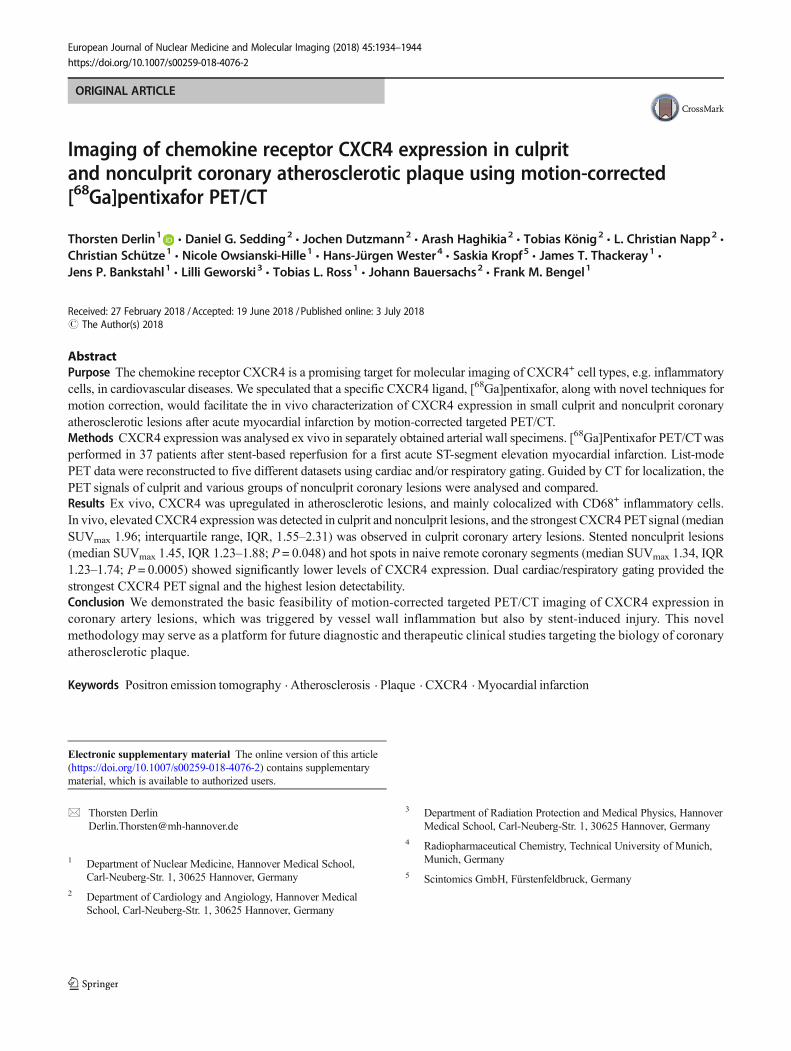
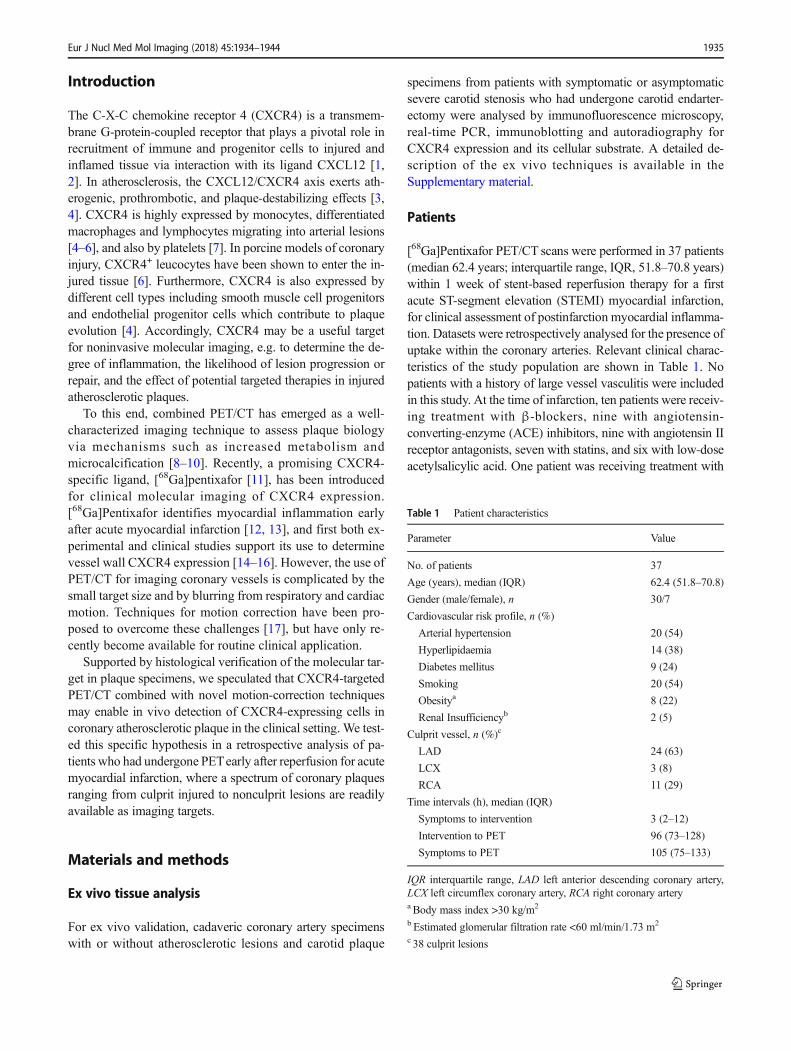
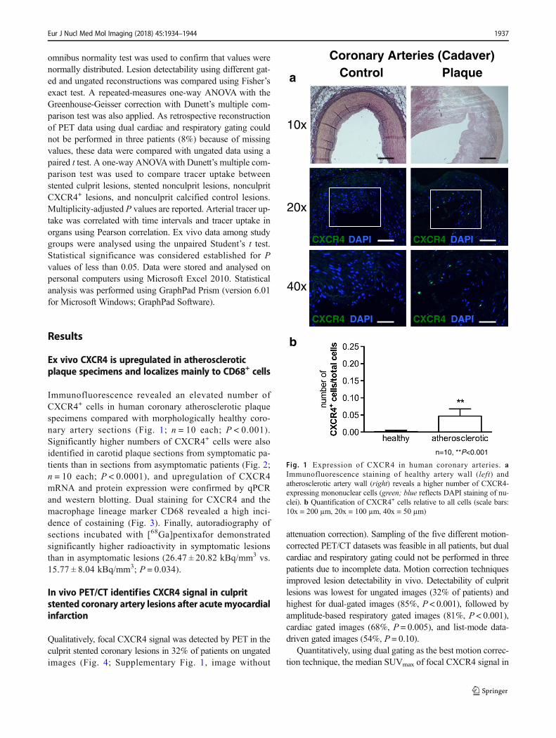
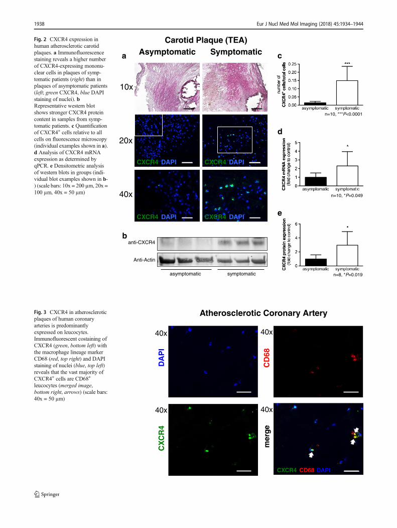
Immunofluorescence revealed an elevated number ofCXCR4+ cells in human coronary atherosclerotic plaquespecimens compared with morphologically healthy coro-nary artery sections (Fig. 1; n = 10 each; P < 0.001).Significantly higher numbers of CXCR4+ cells were alsoidentified in carotid plaque sections from symptomatic pa-tients than in sections from asymptomatic patients (Fig. 2;n = 10 each; P < 0.0001), and upregulation of CXCR4mRNA and protein expression were confirmed by qPCRand western blotting. Dual staining for CXCR4 and themacrophage lineage marker CD68 revealed a high inci-dence of costaining (Fig. 3). Finally, autoradiography ofsections incubated with [68Ga]pentixafor demonstratedsignificantly higher radioactivity in symptomatic lesionsthan in asymptomatic lesions (26.47 ± 20.82 kBq/mm3 vs.15.77 ± 8.04 kBq/mm3; P = 0.034).
In vivo PET/CT identifies CXCR4 signal in culpritstented coronary artery lesions after acute myocardialinfarction
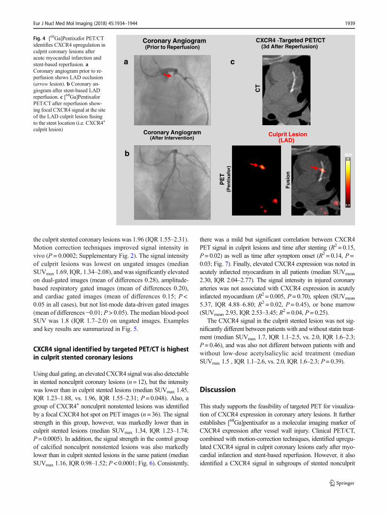
Qualitatively, focal CXCR4 signal was detected by PET in theculprit stented coronary lesions in 32% of patients on ungatedimages (Fig. 4; Supplementary Fig. 1, image without
attenuation correction). Sampling of the five different motion-corrected PET/CT datasets was feasible in all patients, but dualcardiac and respiratory gating could not be performed in threepatients due to incomplete data. Motion correction techniquesimproved lesion detectability in vivo. Detectability of culpritlesions was lowest for ungated images (32% of patients) andhighest for dual-gated images (85%, P < 0.001), followed byamplitude-based respiratory gated images (81%, P < 0.001),cardiac gated images (68%, P = 0.005), and list-mode data-driven gated images (54%, P = 0.10).
Quantitatively, using dual gating as the best motion correc-tion technique, the median SUVmax of focal CXCR4 signal in
CXCR4 DAPICXCR4 DAPI
10x
40x
CXCR4 DAPICXCR4 DAPI
20x
a
b
Coronary Arteries (Cadaver)Control Plaque
n=10, **P<0.001
Fig. 1 Expression of CXCR4 in human coronary arteries. aImmunofluorescence staining of healthy artery wall (left) andatherosclerotic artery wall (right) reveals a higher number of CXCR4-expressing mononuclear cells (green; blue reflects DAPI staining of nu-clei). b Quantification of CXCR4+ cells relative to all cells (scale bars:10x = 200 µm, 20x = 100 µm, 40x = 50 µm)
Eur J Nucl Med Mol Imaging (2018) 45:1934–1944 1937
n=10, ***P<0.0001
CXCR4 DAPICXCR4 DAPI
40x
10x
anti-CXCR4
Anti-Actin
asymptomatic symptomatic
b
a c
n=10, *P=0.049
d
n=8, *P=0.019
e
CXCR4 DAPICXCR4 DAPI
20x
Carotid Plaque (TEA)Asymptomatic Symptomatic
Fig. 2 CXCR4 expression inhuman atherosclerotic carotidplaques. a Immunofluorescencestaining reveals a higher numberof CXCR4-expressing mononu-clear cells in plaques of symp-tomatic patients (right) than inplaques of asymptomatic patients(left; green CXCR4, blue DAPIstaining of nuclei). bRepresentative western blotshows stronger CXCR4 proteincontent in samples from symp-tomatic patients. c Quantificationof CXCR4+ cells relative to allcells on fluorescence microscopy(individual examples shown in a).d Analysis of CXCR4 mRNAexpression as determined byqPCR. e Densitometric analysisof western blots in groups (indi-vidual blot examples shown in b-) (scale bars: 10x = 200 µm, 20x =100 µm, 40x = 50 µm)
CXCR4 CD68 DAPI
DA
PI
CX
CR
4
CD
68m
erg
e
Atherosclerotic Coronary Artery
40x
40x
40x
40x
Fig. 3 CXCR4 in atheroscleroticplaques of human coronaryarteries is predominantlyexpressed on leucocytes.Immunofluorescent costaining ofCXCR4 (green, bottom left) withthe macrophage lineage markerCD68 (red, top right) and DAPIstaining of nuclei (blue, top left)reveals that the vast majority ofCXCR4+ cells are CD68+
leucocytes (merged image,bottom right, arrows) (scale bars:40x = 50 µm)
1938 Eur J Nucl Med Mol Imaging (2018) 45:1934–1944
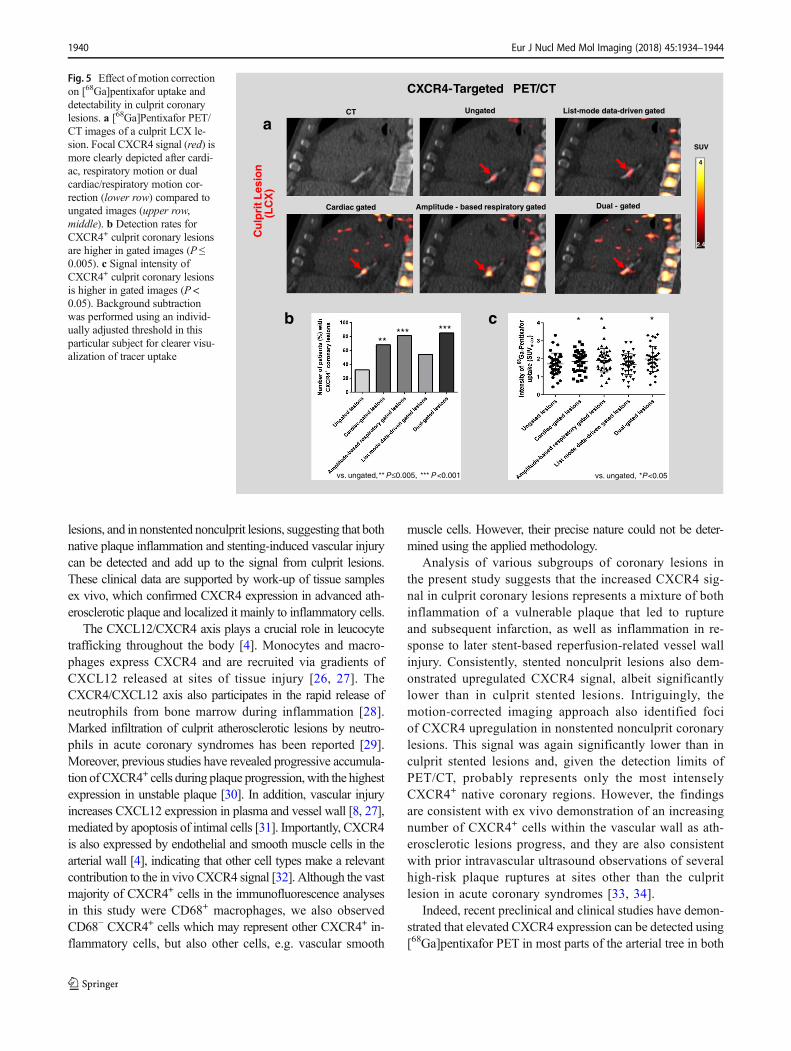
the culprit stented coronary lesions was 1.96 (IQR 1.55–2.31).Motion correction techniques improved signal intensity invivo (P = 0.0002; Supplementary Fig. 2). The signal intensityof culprit lesions was lowest on ungated images (medianSUVmax 1.69, IQR, 1.34–2.08), and was significantly elevatedon dual-gated images (mean of differences 0.28), amplitude-based respiratory gated images (mean of differences 0.20),and cardiac gated images (mean of differences 0.15; P <0.05 in all cases), but not list-mode data-driven gated images(mean of differences −0.01; P > 0.05). The median blood-poolSUV was 1.8 (IQR 1.7–2.0) on ungated images. Examplesand key results are summarized in Fig. 5.
CXCR4 signal identified by targeted PET/CT is highestin culprit stented coronary lesions
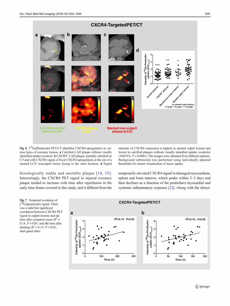
Using dual gating, an elevatedCXCR4 signalwas also detectablein stented nonculprit coronary lesions (n= 12), but the intensitywas lower than in culprit stented lesions (median SUVmax 1.45,IQR 1.23–1.88, vs. 1.96, IQR 1.55–2.31; P = 0.048). Also, agroup of CXCR4+ nonculprit nonstented lesions was identifiedby a focal CXCR4 hot spot on PET images (n = 36). The signalstrength in this group, however, was markedly lower than inculprit stented lesions (median SUVmax 1.34, IQR 1.23–1.74;P = 0.0005). In addition, the signal strength in the control groupof calcified nonculprit nonstented lesions was also markedlylower than in culprit stented lesions in the same patient (medianSUVmax 1.16, IQR 0.98–1.52; P < 0.0001; Fig. 6). Consistently,
there was a mild but significant correlation between CXCR4PET signal in culprit lesions and time after stenting (R2 = 0.15,P = 0.02) as well as time after symptom onset (R2 = 0.14, P =0.03; Fig. 7). Finally, elevated CXCR4 expression was noted inacutely infarcted myocardium in all patients (median SUVmean
2.30, IQR 2.04–2.77). The signal intensity in injured coronaryarteries was not associated with CXCR4 expression in acutelyinfarcted myocardium (R2 = 0.005, P = 0.70), spleen (SUVmean
5.37, IQR 4.88–6.80; R2 = 0.02, P = 0.45), or bone marrow(SUVmean 2.93, IQR 2.53–3.45; R2 = 0.04, P = 0.25).
The CXCR4 signal in the culprit stented lesion was not sig-nificantly different between patients with andwithout statin treat-ment (median SUVmax 1.7, IQR 1.1–2.5, vs. 2.0, IQR 1.6–2.3;P = 0.46), and was also not different between patients with andwithout low-dose acetylsalicylic acid treatment (medianSUVmax 1.5 , IQR 1.1–2.6, vs. 2.0, IQR 1.6–2.3; P = 0.39).
Discussion
This study supports the feasibility of targeted PET for visualiza-tion of CXCR4 expression in coronary artery lesions. It furtherestablishes [68Ga]pentixafor as a molecular imaging marker ofCXCR4 expression after vessel wall injury. Clinical PET/CT,combined with motion-correction techniques, identified upregu-lated CXCR4 signal in culprit coronary lesions early after myo-cardial infarction and stent-based reperfusion. However, it alsoidentified a CXCR4 signal in subgroups of stented nonculprit
Coronary Angiogram(Prior to Reperfusion)
CXCR4 -Targeted PET/CT (3d After Reperfusion)
CT
Fu
sio
n
PE
T(P
enti
xafo
r)
Culprit Lesion(LAD)
3.5
2.3
SUV
ca
Coronary Angiogram(After Intervention)
b
Fig. 4 [68Ga]Pentixafor PET/CTidentifies CXCR4 upregulation inculprit coronary lesions afteracute myocardial infarction andstent-based reperfusion. aCoronary angiogram prior to re-perfusion shows LAD occlusion(arrow lesion). b Coronary an-giogram after stent-based LADreperfusion. c [68Ga]PentixaforPET/CT after reperfusion show-ing focal CXCR4 signal at the siteof the LAD culprit lesion fusingto the stent location (i.e. CXCR4+
culprit lesion)
Eur J Nucl Med Mol Imaging (2018) 45:1934–1944 1939
lesions, and in nonstented nonculprit lesions, suggesting that bothnative plaque inflammation and stenting-induced vascular injurycan be detected and add up to the signal from culprit lesions.These clinical data are supported by work-up of tissue samplesex vivo, which confirmed CXCR4 expression in advanced ath-erosclerotic plaque and localized it mainly to inflammatory cells.
The CXCL12/CXCR4 axis plays a crucial role in leucocytetrafficking throughout the body [4]. Monocytes and macro-phages express CXCR4 and are recruited via gradients ofCXCL12 released at sites of tissue injury [26, 27]. TheCXCR4/CXCL12 axis also participates in the rapid release ofneutrophils from bone marrow during inflammation [28].Marked infiltration of culprit atherosclerotic lesions by neutro-phils in acute coronary syndromes has been reported [29].Moreover, previous studies have revealed progressive accumula-tion of CXCR4+ cells during plaque progression,with the highestexpression in unstable plaque [30]. In addition, vascular injuryincreases CXCL12 expression in plasma and vessel wall [8, 27],mediated by apoptosis of intimal cells [31]. Importantly, CXCR4is also expressed by endothelial and smooth muscle cells in thearterial wall [4], indicating that other cell types make a relevantcontribution to the in vivo CXCR4 signal [32]. Although the vastmajority of CXCR4+ cells in the immunofluorescence analysesin this study were CD68+ macrophages, we also observedCD68− CXCR4+ cells which may represent other CXCR4+ in-flammatory cells, but also other cells, e.g. vascular smooth
muscle cells. However, their precise nature could not be deter-mined using the applied methodology.
Analysis of various subgroups of coronary lesions inthe present study suggests that the increased CXCR4 sig-nal in culprit coronary lesions represents a mixture of bothinflammation of a vulnerable plaque that led to ruptureand subsequent infarction, as well as inflammation in re-sponse to later stent-based reperfusion-related vessel wallinjury. Consistently, stented nonculprit lesions also dem-onstrated upregulated CXCR4 signal, albeit significantlylower than in culprit stented lesions. Intriguingly, themotion-corrected imaging approach also identified fociof CXCR4 upregulation in nonstented nonculprit coronarylesions. This signal was again significantly lower than inculprit stented lesions and, given the detection limits ofPET/CT, probably represents only the most intenselyCXCR4+ native coronary regions. However, the findingsare consistent with ex vivo demonstration of an increasingnumber of CXCR4+ cells within the vascular wall as ath-erosclerotic lesions progress, and they are also consistentwith prior intravascular ultrasound observations of severalhigh-risk plaque ruptures at sites other than the culpritlesion in acute coronary syndromes [33, 34].
Indeed, recent preclinical and clinical studies have demon-strated that elevated CXCR4 expression can be detected using[68Ga]pentixafor PET in most parts of the arterial tree in both
cb
vs. ungated, **P≤0.005, ***P<0.001
********
Cu
lpri
tLes
ion
(LC
X)
aSUV
4
2.4
CXCR4-Targeted PET/CT
Ungated List-mode data-driven gated
Dual - gated
CT
Cardiac gated Amplitude - based respiratory gated
vs. ungated, *P<0.05
** *
Fig. 5 Effect of motion correctionon [68Ga]pentixafor uptake anddetectability in culprit coronarylesions. a [68Ga]Pentixafor PET/CT images of a culprit LCX le-sion. Focal CXCR4 signal (red) ismore clearly depicted after cardi-ac, respiratory motion or dualcardiac/respiratory motion cor-rection (lower row) compared toungated images (upper row,middle). b Detection rates forCXCR4+ culprit coronary lesionsare higher in gated images (P ≤0.005). c Signal intensity ofCXCR4+ culprit coronary lesionsis higher in gated images (P <0.05). Background subtractionwas performed using an individ-ually adjusted threshold in thisparticular subject for clearer visu-alization of tracer uptake
1940 Eur J Nucl Med Mol Imaging (2018) 45:1934–1944
histologically stable and unstable plaque [14, 15].Interestingly, the CXCR4 PET signal in injured coronaryplaque tended to increase with time after reperfusion in theearly time-frame covered in this study, and it differed from the
temporarily elevated CXCR4 signal in damaged myocardium,spleen and bone marrow, which peaks within 3–5 days andthen declines as a function of the postinfarct myocardial andsystemic inflammatory response [12]. Along with the detect-
CXCR4+ plaque(LAD)
Stented non-culpritplaque (LCX)
a c
Calcified controlplaque (LAD)
3
1.5
SUV
SUV
b
SUVSUV34
1.51
d
vs. stented culprit lesions* P<0.05; **P<0.001; ***P<0.0001
******
CXCR4-TargetedPET/CT
Fig. 6 [68Ga]Pentixafor PET/CT identifies CXCR4 upregulation in var-ious types of coronary lesions. a Calcified LAD plaque without visuallyidentified uptake (control). bCXCR4+ LAD plaque, partially calcified onCT and with CXCR4 signal. c Focal CXCR4 upregulation at the site of astented LCX nonculprit lesion fusing to the stent location. d Signal
intensity of CXCR4 expression is highest in stented culprit lesions andlowest in calcified plaques without visually identified uptake (controls)(ANOVA,P < 0.0001). The images were obtained from different patients.Background subtraction was performed using individually adjustedthresholds for clearer visualization of tracer uptake
CXCR4-TargetedPET/CT
baR2=0.14, P=0.03 R2=0.15, P=0.02
Fig. 7 Temporal evolution of[68Ga]pentixafor signal. Therewas a mild but significantcorrelation between CXCR4 PETsignal in culprit lesions and (a)time after symptom onset (R2 =0.14, P = 0.03, and (b) time afterstenting (R2 = 0.15, P = 0.02;dual-gated data)
Eur J Nucl Med Mol Imaging (2018) 45:1934–1944 1941
able CXCR4 signal in stented nonculprit lesions, this obser-vation might be a result of differences in the evolution ofinflammatory processes in the myocardium and plaque overtime, but also suggests a relevant contribution of stent-inducedinjury to the signal. Future studies including patients beforestent-based reperfusion are needed to determine the relativecontribution of native plaque inflammation versus stent-induced injury to the measured composite CXCR4 signal.The current study provides proof-of-principle that CXCR4-targeted imaging of CXCR4+ coronary plaque is feasible,and it thereby lays the foundation for such future projects.Speculatively, [68Ga]pentixafor coronary PET/CT may evenbe useful for guiding antiinflammatory treatment or even treat-ment targeting CXCR4. This has been suggested, for example,to reduce neointimal lesion size, smooth muscle progenitorcell mobilization and neointimal proliferation after experi-mental arterial injury [35], and noninvasive detection of thepresence of the target may be helpful for clinical translation ofsuch interventions.
Besides the novel molecular target, our study also in-cluded motion correction as a technical innovation in PETthat is highly relevant for noninvasive coronary artery im-aging. The detection of small lesions on PET is generallycomplicated by three-dimensional image blurring intro-duced by the finite spatial resolution of the imaging sys-tem and by image sampling on a voxel grid that does notmatch the actual contours of the tracer distribution [36].Because PET acquisition is not fast enough to enablebreath-hold imaging, respiratory motion along with intrin-sic cardiac motion causes further blurring. Together withthe limited spatial resolution, this leads to deviation of theapparent signal intensity from true values as a result ofpartial volume effects. Motion correction can significantlyreduce blurring by accounting for both respiration-inducedand cardiac movement [20]. Of note, the detectability ofculprit coronary lesions, which provided the strongest sig-nal, was still low when using ungated images, but in-creased significantly when using motion correction. Dualcorrection of cardiac and respiratory motion yielded thebest results. Relatively small differences in SUVmax be-tween different gating approaches led to relatively highdifferences in detection rates. This may be explained bythe fact that blood-pool signal is usually relatively highusing [68Ga]pentixafor PET (SUVmean 1.8, IQR 1.7–2.0)and the lesion to blood-pool signal ratio may be relativelylow. Therefore, relatively small increases in signal inten-sity may lead to markedly higher detection rates (e.g. themean of differences for dual-gated images compared toungated images was 0.28). It is also important to keep inmind that localization of culprit lesions was guided by CT(i.e. stents), and not by the PET signal itself. Therefore,knowing precisely which lesion to assess may allow a
lesion with even a small increase in tracer intensity to bejudged as visually [68Ga]pentixafor-positive when the sig-nal exceeds that of the blood pool.
In addition to gating, we also applied point-spread func-tion modelling and time-of-flight reconstruction techniques(Ultra High-Definition, UHD) which have been shown toconsiderably improve signal intensity for PET analysis[37]. This study established and evaluated a sophisticateddual-gated imaging approach for coronary artery imagingon UHD PET. By contrast, list-mode data-driven gating didnot significantly improve lesion detectability. This ap-proach is based directly on frame-by-frame analysis ofPET emission data, without the use of external trigger sig-nals [23, 24]. It requires clear contours of the organ to betracked, such as the myocardium in case of 18F-FDG imag-ing, but such myocardial contours are not visible in[68Ga]pentixafor PET images owing to lack of uptake innormal myocardium, which on the other hand is an advan-tage for delineation of coronary uptake without spillover.
Some limitations of the present study should be acknowl-edged. Obviously, not all lesions observed on PET in vivocan be verified by histology (which applies to all clinicalatherosclerosis studies). However, there are recent clinicalstudies evaluating the usefulness of pentixafor for CXCR4imaging in plaque [14, 38], and also studies which havedemonstrated that the arterial wall uptake can be blocked,is specific, reproducible, and correlates with the presence ofCXCR4+ cells [16, 38]. In addition, the ex vivo data pre-sented in this work confirm the presence of CXCR4+ cells inthe coronary wall, and demonstrate an increasing number ofthese cells with advancing pathology of lesions, in line withthe observed in vivo PET data. Second, the signal mighthave been enhanced by attenuation correction related tothe proximity of a metallic stent. However, the signal seenin stented culprit lesions was also seen on images without -attenuation correction, and was significantly higher than instented nonculprit lesions, supporting the basic hypothesisof this work, and confirming that the stent signal was notcaused by attenuation correction. The precise cell popula-tion contributing to the in vivo [68Ga]pentixafor signalcould not be identified. However, ex vivo data showed up-regulated CXCR4 expression in atherosclerotic coronaryarteries and/or in carotid arteries from symptomatic sub-jects. This finding was confirmed by mRNA expression,protein expression and immunofluorescence. Moreover, up-take of [68Ga]pentixafor was validated by ex vivo autoradi-ography in carotid arteries, and immunofluorescence dual-staining of CD68 confirmed CXCR4 expression byleucocytes. In addition, two recent experimental studies in-vestigating carotid arteries [15, 16] have shown thatCXCR4 expression in injured plaque is predominantlyfound on leucocytes, supporting our conclusions.
1942 Eur J Nucl Med Mol Imaging (2018) 45:1934–1944
Conclusion
In this proof-of-principle study, we demonstrated the feasibil-ity of motion-corrected targeted PET imaging with[68Ga]pentixafor for identifying increased CXCR4 expressionin injured culprit and nonculprit coronary artery plaques trig-gered by vessel wall inflammation, but also by stent-inducedinjury. This novel noninvasive approach may refine the clini-cal characterization of atherosclerotic lesions, serving as aplatform for novel diagnostic and therapeutic approachestargeting coronary atherosclerotic plaque biology.
Acknowledgements The publication of this article was supported byfunds of the European Association of Nuclear Medicine (EANM).
Funding This study was supported by the Excellence Cluster REBIRTH-2, and by KFO311 of the German Research Foundation.
Compliance with ethical standards
Conflicts of interest Dr. Wester is a shareholder of Scintomics GmbH,Fürstenfeldbruck, Germany. The other authors declare that they have norelationships relevant to the contents of this paper to disclose.
Ethical approval All procedures performed in studies involving humanparticipants were in accordance with the ethical standards of the institu-tional research committee and with the principles of the 1964 Declarationof Helsinki and its later amendments or comparable ethical standards.
Open Access This article is distributed under the terms of the CreativeCommons At t r ibut ion 4 .0 In te rna t ional License (h t tp : / /creativecommons.org/licenses/by/4.0/), which permits unrestricted use,distribution, and reproduction in any medium, provided you giveappropriate credit to the original author(s) and the source, provide a linkto the Creative Commons license, and indicate if changes were made.
References
1. Wang JF, Liu ZY, Groopman JE. The alpha-chemokine receptorCXCR4 is expressed on the megakaryocytic lineage from progen-itor to platelets and modulates migration and adhesion. Blood.1998;92:756–64.
2. Teicher BA, Fricker SP. CXCL12(SDF-1)/CXCR4 pathway in can-cer. Clin Cancer Res. 2010;16:2927–31.
3. Swirski FK, Nahrendorf M. Leukocyte behavior in atherosclerosis,myocardial infarction, and heart failure. Science. 2013;339:161–6.
4. Döring Y, Pawig L, Weber C, Noels H. The CXCL12/CXCR4chemokine ligand/receptor axis in cardiovascular disease. FrontPhysiol. 2014;5:212.
5. Gupta SK, Pillarisetti K, Lysko PG. Modulation of CXCR4expression and SDF-1 alpha functional activity during differ-entiation of human monocytes and macrophages. J LeukocBiol. 1999;66:135–43.
6. Jabs A, Okamoto E, Vinten-Johansen J, Bauriedel G, Wilcox JN.Sequential patterns of chemokine- and chemokine receptor-synthesis following vessel wall injury in porcine coronary arteries.Atherosclerosis. 2007;192:75–84.
7. Abi-Younes S, Sauty A,Mach F, Sukhova GK, Libby P, Luster AD.The stromal cell-derived factor-1 chemokine is a potent plateletagonist highly expressed in atherosclerotic plaques. Circ Res.2000;86:131–8.
8. Derlin T, Tóth Z, Papp L, Wisotzki C, Apostolova I, HabermannCR, et al. Correlation of inflammation assessed by 18F-FDG PET,active mineral deposition assessed by 18F-fluoride PET, and vas-cular calcification in atherosclerotic plaque: a dual-tracer PET/CTstudy. J Nucl Med. 2011;52:1020–7.
9. Derlin T, Richter U, Bannas P, Begemann P, Buchert R, Mester J, etal. Feasibility of 18F-sodium fluoride PET/CT for imaging of ath-erosclerotic plaque. J Nucl Med. 2010;51:862–5.
10. Joshi NV, Vesey AT, Williams MC, Shah AS, Calvert PA,Craighead FH, et al. 18F-fluoride positron emission tomographyfor identification of ruptured and high-risk coronary atheroscleroticplaques: a prospective clinical trial. Lancet. 2014;383:705–13.
11. Demmer O, Gourni E, Schumacher U, Kessler H, Wester HJ. PETimaging of CXCR4 receptors in cancer by a new optimized ligand.ChemMedChem. 2011;6:1789–91.
12. Thackeray JT, Derlin T, Haghikia A,NappLC,WangY, Ross TL, et al.Molecular imaging of the chemokine receptorCXCR4 after acutemyo-cardial infarction. JACC Cardiovasc Imaging. 2015;8:1417–26.
13. Reiter T, Kircher M, Schirbel A, Werner RA, Kropf S, Ertl G, et al.Imaging of C-X-C motif chemokine receptor CXCR4 expressionafter myocardial infarction with [68Ga]pentixafor-PET/CT in cor-relation with cardiac MRI. JACC Cardiovasc Imaging. 2018.https://doi.org/10.1016/j.jcmg.2018.01.001.
14. Weiberg D, Thackeray JT, DaumG, Sohns JM, Kropf S,Wester HJ,et al. Clinical molecular imaging of chemokine receptor CXCR4expression in atherosclerotic plaque using 68Ga-pentixafor PET:correlation with cardiovascular risk factors and calcified plaqueburden. J Nucl Med. 2018;59:266–72.
15. Merckelbach S, van der Vorst EPC, Kallmayer M, Rischpler C,Burgkart R, Döring Y, et al. Expression and cellular localizationof CXCR4 and CXCL12 in human carotid atherosclerotic plaques.Thromb Haemost. 2018;118:195–206.
16. Hyafil F, Pelisek J, Laitinen I, SchotteliusM,MohringM, Döring Y,et al. Imaging the cytokine receptor CXCR4 in atheroscleroticplaques with the radiotracer 68Ga-pentixafor for positron emissiontomography. J Nucl Med. 2017;58:499–506.
17. Rubeaux M, Joshi NV, Dweck MR, Fletcher A, Motwani M,Thomson LE, et al. Motion correction of 18F-NaF PET for imagingcoronary atherosclerotic plaques. J Nucl Med. 2016;57:54–9.
18. Martin R, Jüttler S, Müller M, Wester HJ. Cationic eluate pretreat-ment for automated synthesis of [68Ga]CPCR4.2. Nucl Med Biol.2014;41:84–9.
19. Gourni E, Demmer O, Schottelius M, D’Alessandria C, Schulz S,Dijkgraaf I, et al. PET of CXCR4 expression by a (68)Ga-labeledhighly specific targeted contrast agent. J NuclMed. 2011;52:1803–10.
20. Slomka PJ, RubeauxM, LeMeunier L, DeyD, Lazewatsky JL, PanT, et al. Dual-gated motion-frozen cardiac PET with Flurpiridaz F18. J Nucl Med. 2015;56:1876–81.
21. Grootjans W, de Geus-Oei LF, Meeuwis AP, van der Vos CS,Gotthardt M, Oyen WJ, et al. Amplitude-based optimal respiratorygating in positron emission tomography in patients with primarylung cancer. Eur Radiol. 2014;24:3242–50.
22. van Elmpt W, Hamill J, Jones J, Ruysscher D, Lambin P, Ollers M.Optimal gating compared to 3D and 4D PET reconstruction forcharacterization of lung tumours. Eur J Nucl Med Mol Imaging.2011;5:843–55.
23. Büther F, Dawood M, Stegger L, Wübbeling F, Schäfers M,Schober O, et al. List mode-driven cardiac and respiratory gatingin PET. J Nucl Med. 2009;50:674–81.
24. Jin X, Chan C, Mulnix T, Panin V, Casey ME, Liu C, et al. List-modereconstruction for the biographmCTwith physics modeling and event-by-event motion correction. Phys Med Biol. 2013;58:5567–91.
Eur J Nucl Med Mol Imaging (2018) 45:1934–1944 1943
25. Teräs M, Kokki T, Durand-Schaefer N, Noponen T, Pietilä M, KissJ, et al. Dual-gated cardiac PET – clinical feasibility study. Eur JNucl Med Mol Imaging. 2010;37:505–16.
26. van der Vorst EP, Döring Y, Weber C. Chemokines. ArteriosclerThromb Vasc Biol. 2015;35:e52–6.
27. Yin Y, Zhao X, Fang Y, Yu S, Zhao J, Song M, et al. SDF-1alphainvolved in mobilization and recruitment of endothelial progenitorcells after arterial injury in mice. Cardiovasc Pathol. 2010;19:218–27.
28. Suratt BT, Petty JM, Young SK, Malcolm KC, Lieber JG, Nick JA,et al. Role of the CXCR4/SDF-1 chemokine axis in circulatingneutrophil homeostasis. Blood. 2004;104:565–71.
29. Naruko T, Ueda M, Haze K, van der Wal AC, van der Loos CM,Itoh A, et al. Neutrophil infiltration of culprit lesions in acute cor-onary syndromes. Circulation. 2002;106:2894–900.
30. Bot I, Daissormont IT, Zernecke A, van Puijvelde GH, Kramp B, deJager SC, et al. CXCR4 blockade induces atherosclerosis by affect-ing neutrophil function. J Mol Cell Cardiol. 2014;74:44–52.
31. Zernecke A, Schober A, Bot I, von Hundelshausen P, Liehn EA,Möpps B, et al. SDF-1alpha/CXCR4 axis is instrumental in neoin-timal hyperplasia and recruitment of smooth muscle progenitorcells. Circ Res. 2005;96:784–91.
32. Shi X, Guo LW, Seedial S, Takayama T, Wang B, Zhang M, et al.Local CXCR4 upregulation in the injured arterial wall contributesto intimal hyperplasia. Stem Cells. 2016;34:2744–57.
33. Hong MK, Mintz GS, Lee CW, Lee BK, Yang TH, Kim YH, etal. The site of plaque rupture in native coronary arteries: a three-vessel intravascular ultrasound analysis. J Am Coll Cardiol.2005;46:261–5.
34. Xie Y, Mintz GS, Yang J, Doi H, Iñiguez A, Dangas GD, et al.Clinical outcome of nonculprit plaque ruptures in patients withacute coronary syndrome in the PROSPECT study. JACCCardiovasc Imaging. 2014;7:397–405.
35. Karshovska E, Zagorac D, Zernecke A, Weber C, Schober A. Asmall molecule CXCR4 antagonist inhibits neointima formationand smooth muscle progenitor cell mobilization after arterial injury.J Thromb Haemost. 2008;6:1812–5.
36. Soret M, Bacharach SL, Buvat I. Partial-volume effect in PET tu-mor imaging. J Nucl Med. 2007;48:932–45.
37. Armstrong IS, Kelly MD, Williams HA, Matthews JC. Impact ofpoint spread function modelling and time of flight on FDG uptakemeasurements in lung lesions using alternative filtering strategies.EJNMMI Phys. 2014;1:99.
38. Li X, Heber D, Leike T, Beitzke D, Lu X, Zhang X, et al.[68Ga]Pentixafor-PET/MRI for the detection of chemokine recep-tor 4 expression in atherosclerotic plaques. Eur J Nucl Med MolImaging. 2018;45:558–66.
1944 Eur J Nucl Med Mol Imaging (2018) 45:1934–1944