Embed Size (px)
Citation preview
Image Registration and Assessment for Adaptive
Therapy
Kristy K. Brock, PhD, DABRAssociate Professor
Department of Radiation OncologyUniversity of Michigan
Disclosure
• I have financial interest in deformable registration technology through a licensing agreement with RaySearch Laboratories
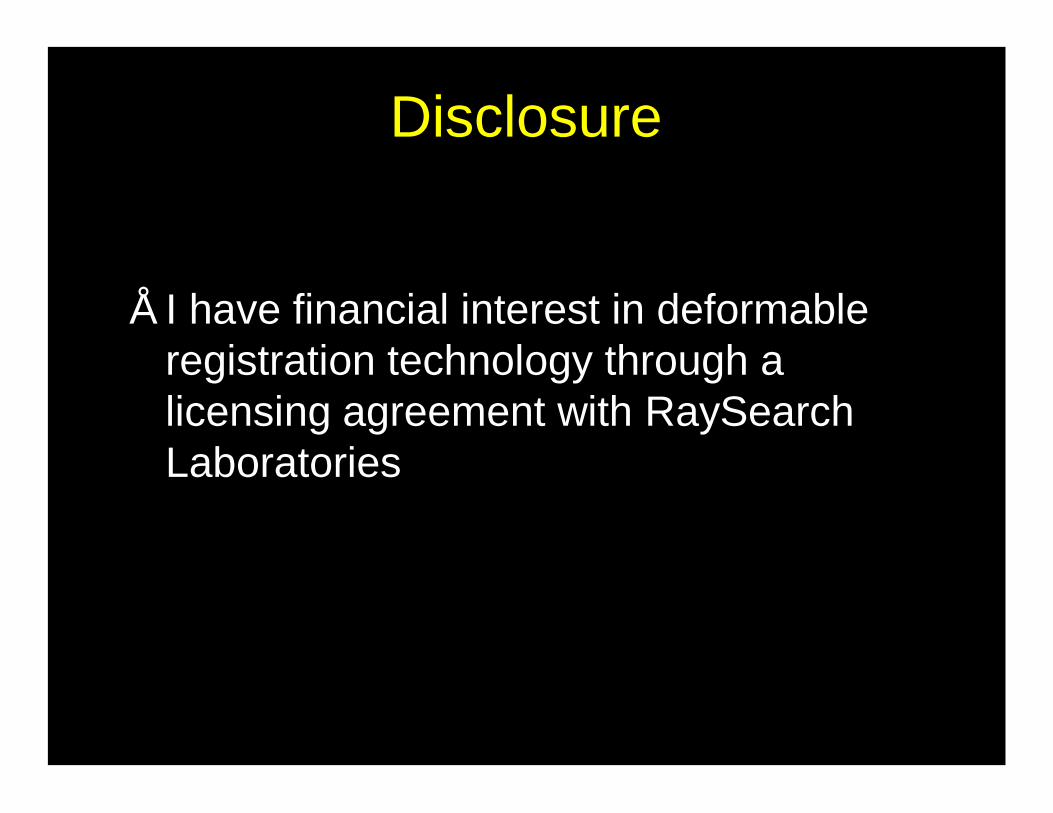
Beautiful, Streamlined System Provides clear information
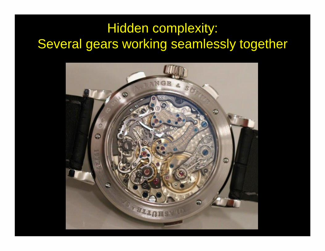
Hidden complexity:Several gears working seamlessly together
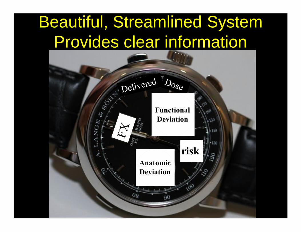
Beautiful, Streamlined System Provides clear information
Functional Deviation
Anatomic Deviation
risk
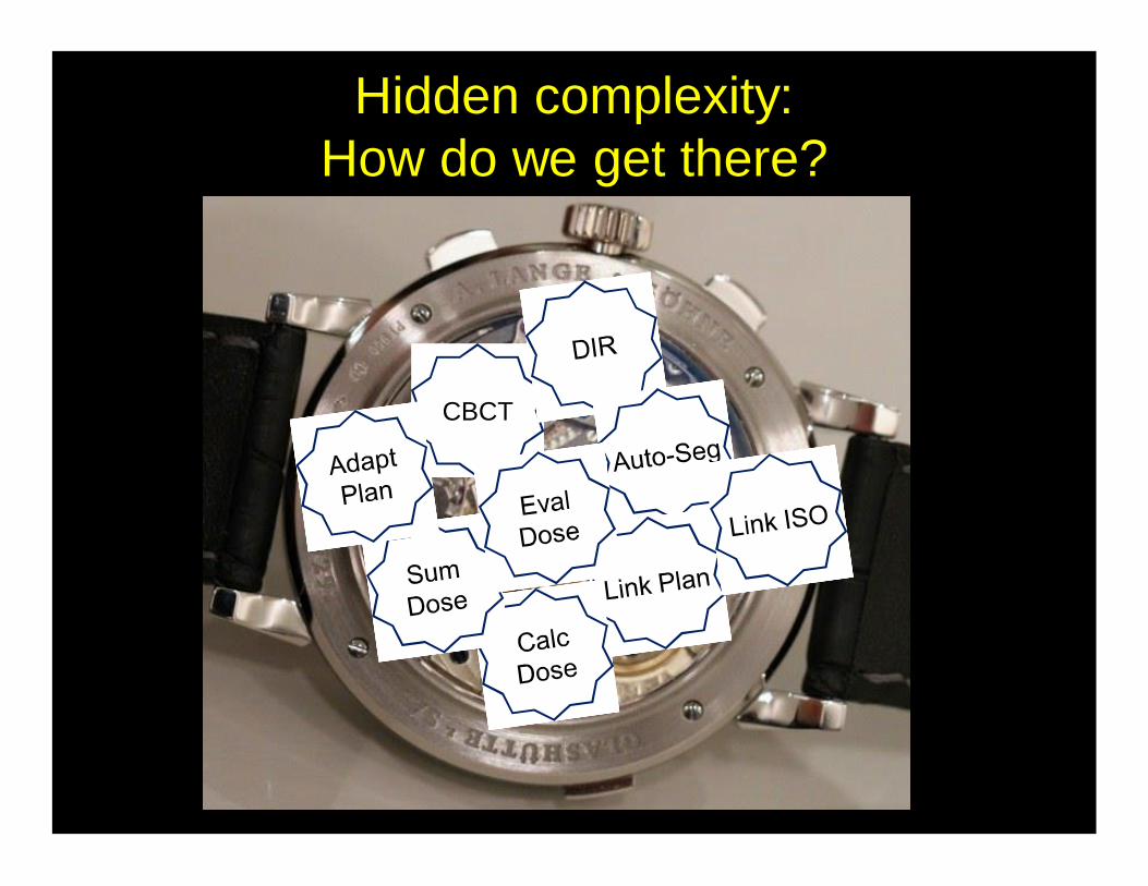
Hidden complexity:How do we get there?
CBCT
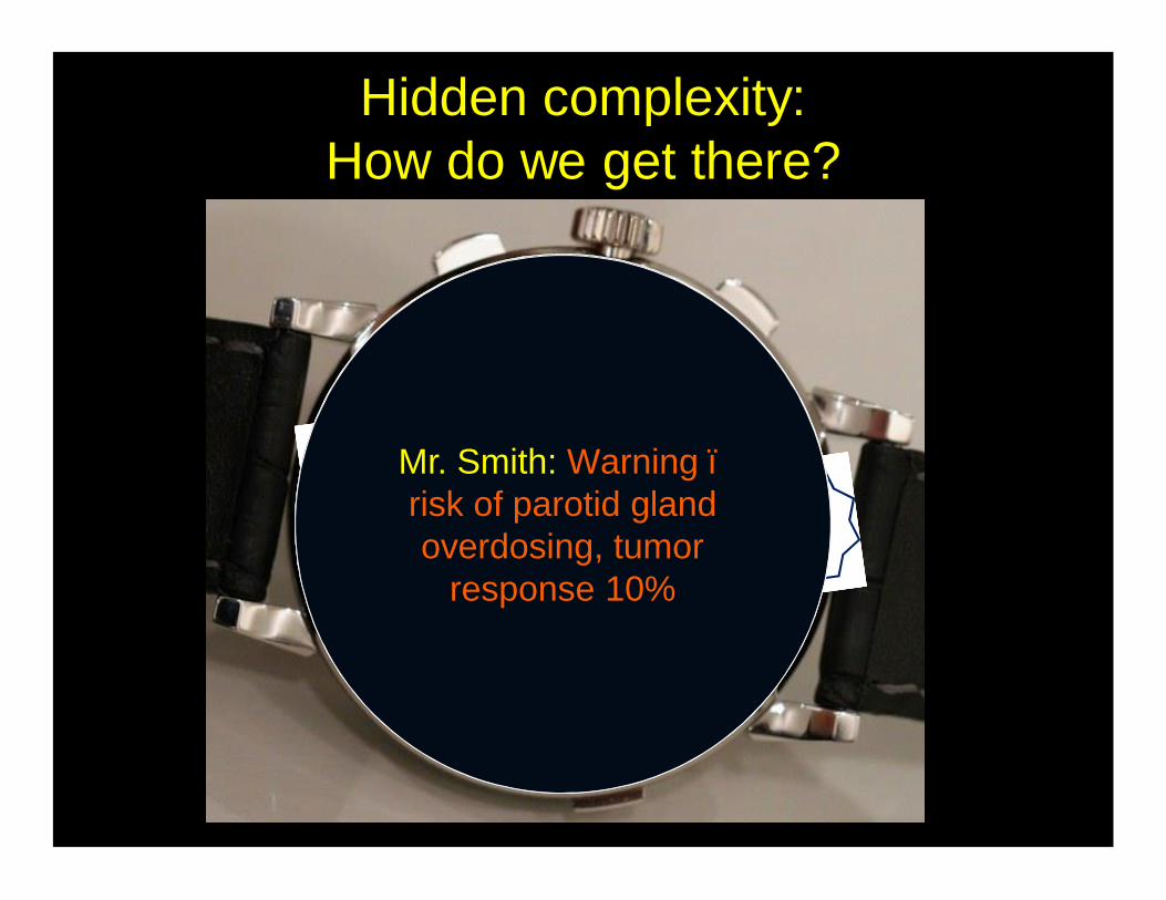
Hidden complexity:How do we get there?
CBCT
Mr. Smith: On Target
Mr. Smith: Warning –risk of parotid gland overdosing, tumor
response 10%
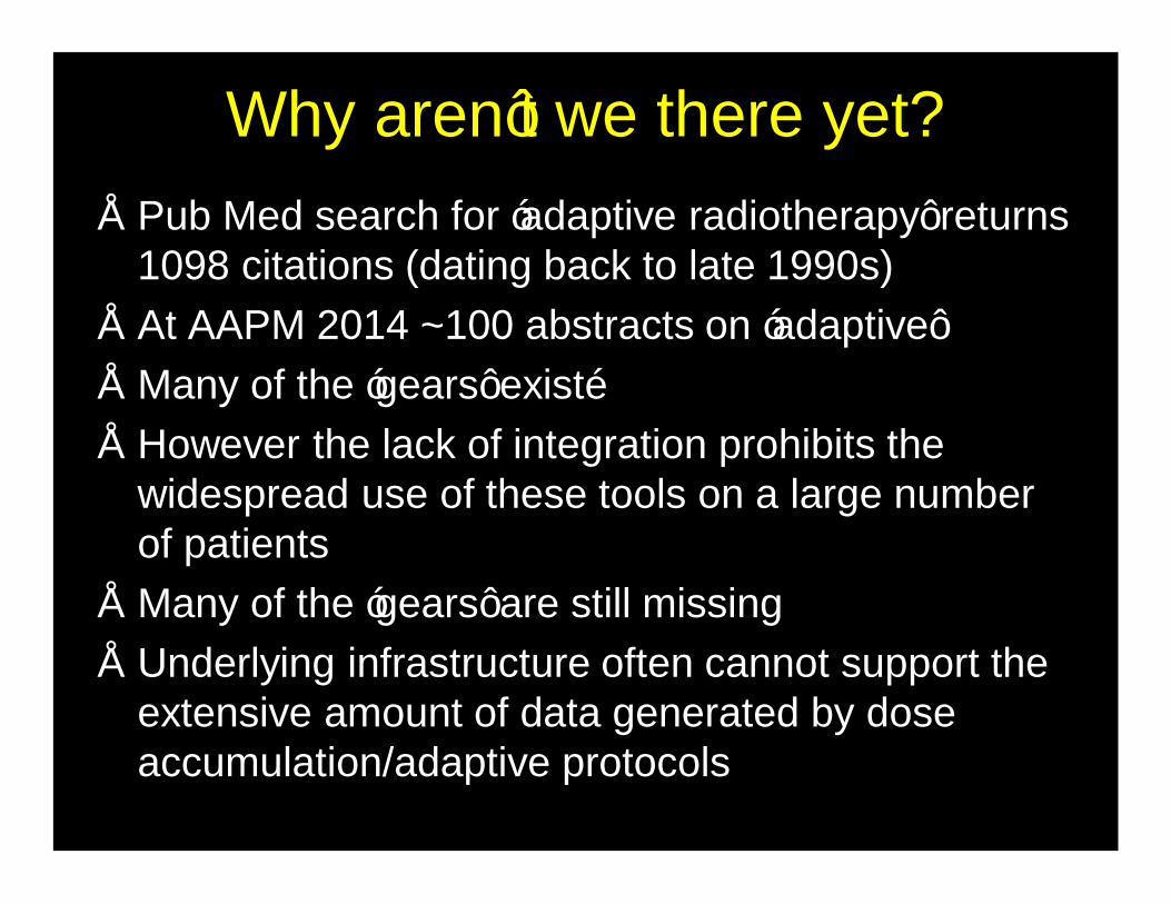
Why aren’t we there yet?• Pub Med search for ‘adaptive radiotherapy’ returns
1098 citations (dating back to late 1990s)• At AAPM 2014 ~100 abstracts on ‘adaptive’• Many of the ‘gears’ exist…• However the lack of integration prohibits the
widespread use of these tools on a large number of patients
• Many of the ‘gears’ are still missing• Underlying infrastructure often cannot support the
extensive amount of data generated by dose accumulation/adaptive protocols
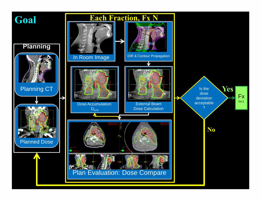
DIR & Contour Propagation
External BeamDose Calculation
In Room Image
Plan Evaluation: Dose Compare
Each Fraction, Fx N
Planning CT
Planned Dose
Planning
Is the dose
deviation acceptable
?
FxN+1
No
Yes
Dose Accumulation: DA,N
Goal
DIR & Contour Propagation
External Beam Dose Calculation
CBCT
Plan Evaluation: Dose Compare
Each Fraction, Fx N
Planning CT
Planned Dose
Planning
Is the dose deviation
acceptable?
Fx N+1
No
Yes
Dose Accumulation: DA,N
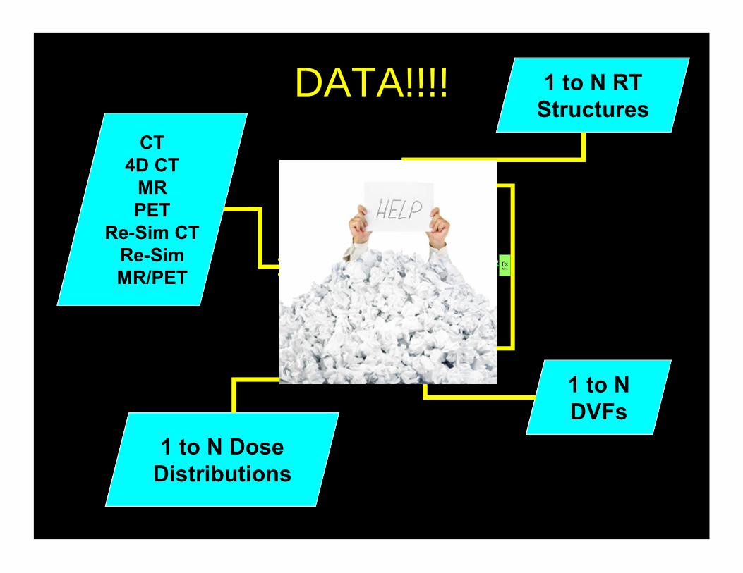
DATA!!!!CT
4D CTMRPET
Re-Sim CTRe-SimMR/PET
1 to N Dose Distributions
1 to N RT Structures
1 to N DVFs
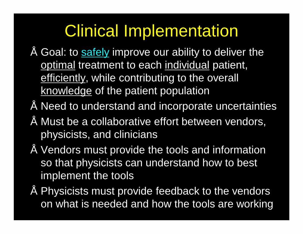
Clinical Implementation• Goal: to safely improve our ability to deliver the
optimal treatment to each individual patient, efficiently, while contributing to the overall knowledge of the patient population
• Need to understand and incorporate uncertainties• Must be a collaborative effort between vendors,
physicists, and clinicians• Vendors must provide the tools and information
so that physicists can understand how to best implement the tools
• Physicists must provide feedback to the vendors on what is needed and how the tools are working
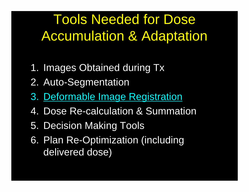
Tools Needed for Dose Accumulation & Adaptation
1. Images Obtained during Tx2. Auto-Segmentation3. Deformable Image Registration4. Dose Re-calculation & Summation5. Decision Making Tools6. Plan Re-Optimization (including
delivered dose)
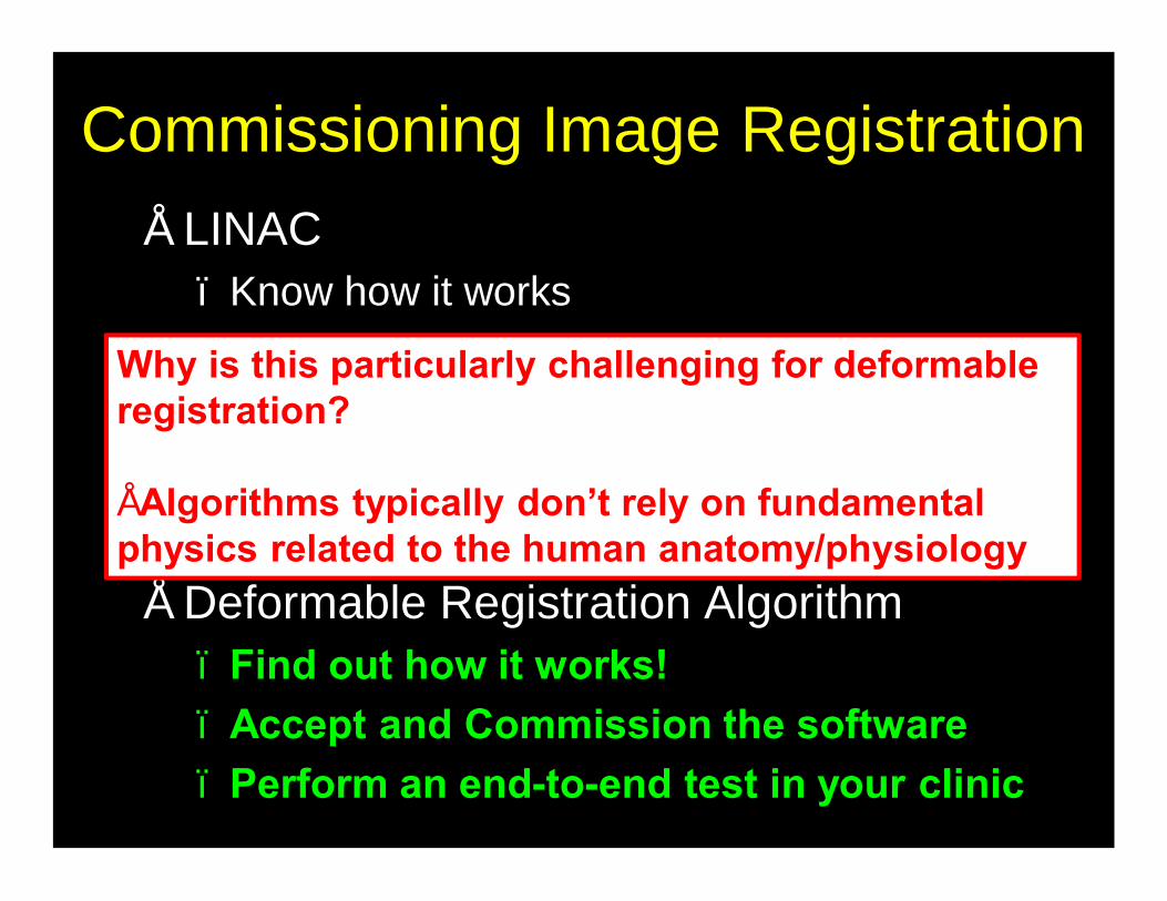
Commissioning Image Registration• LINAC
– Know how it works– Accept and Commission
• Planning System– Know the dose calculation algorithm– Accept and Commission
• Deformable Registration Algorithm– Find out how it works!– Accept and Commission the software– Perform an end-to-end test in your clinic
Why is this particularly challenging for deformable registration?
• Algorithms typically don’t rely on fundamental physics related to the human anatomy/physiology
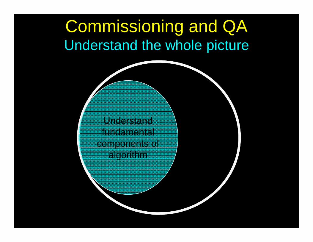
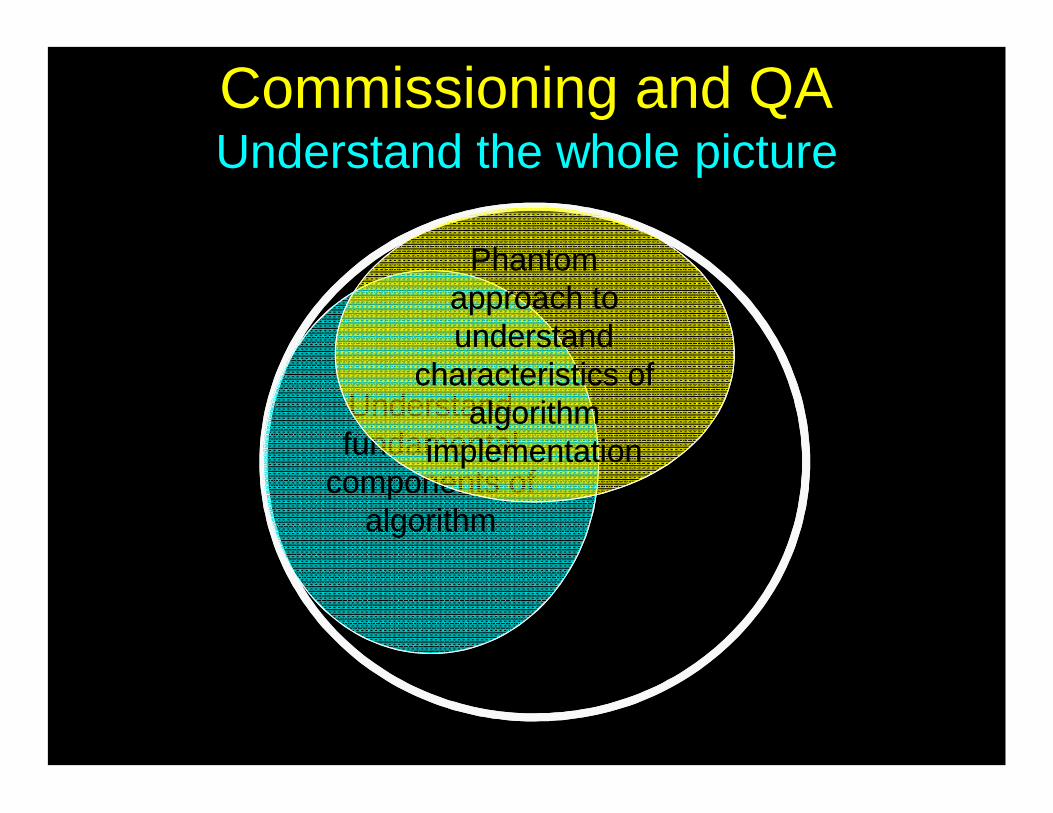
Commissioning and QAUnderstand the whole picture
Understand fundamental
components of algorithm
Understand fundamental
components of algorithm
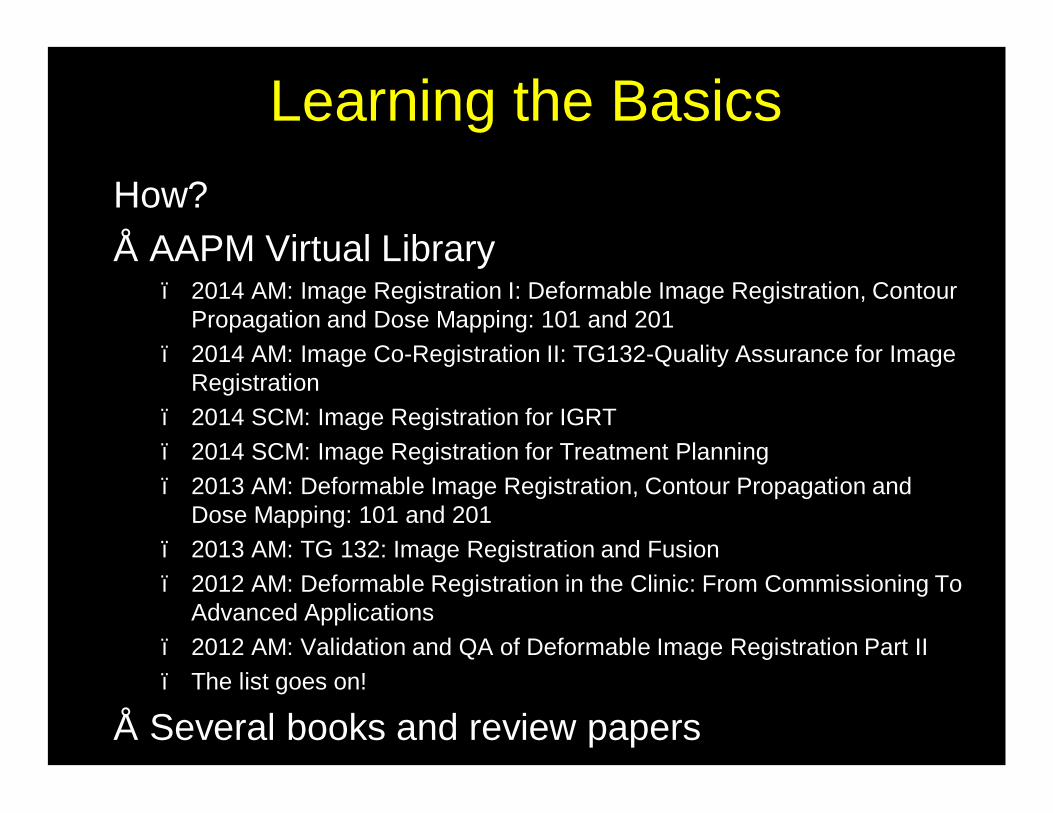
Learning the BasicsHow?• AAPM Virtual Library
– 2014 AM: Image Registration I: Deformable Image Registration, Contour Propagation and Dose Mapping: 101 and 201
– 2014 AM: Image Co-Registration II: TG132-Quality Assurance for Image Registration
– 2014 SCM: Image Registration for IGRT– 2014 SCM: Image Registration for Treatment Planning– 2013 AM: Deformable Image Registration, Contour Propagation and
Dose Mapping: 101 and 201– 2013 AM: TG 132: Image Registration and Fusion– 2012 AM: Deformable Registration in the Clinic: From Commissioning To
Advanced Applications– 2012 AM: Validation and QA of Deformable Image Registration Part II– The list goes on!
• Several books and review papers
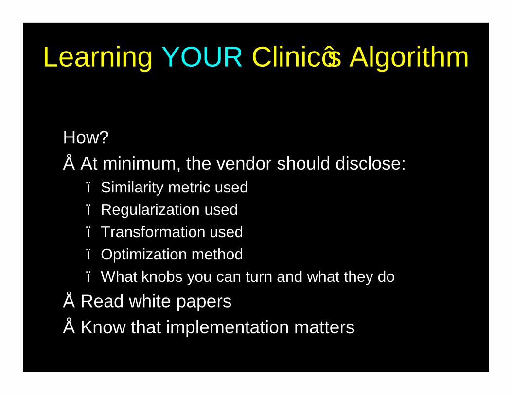
Learning YOUR Clinic’s Algorithm
How?• At minimum, the vendor should disclose:
– Similarity metric used– Regularization used– Transformation used– Optimization method– What knobs you can turn and what they do
• Read white papers• Know that implementation matters
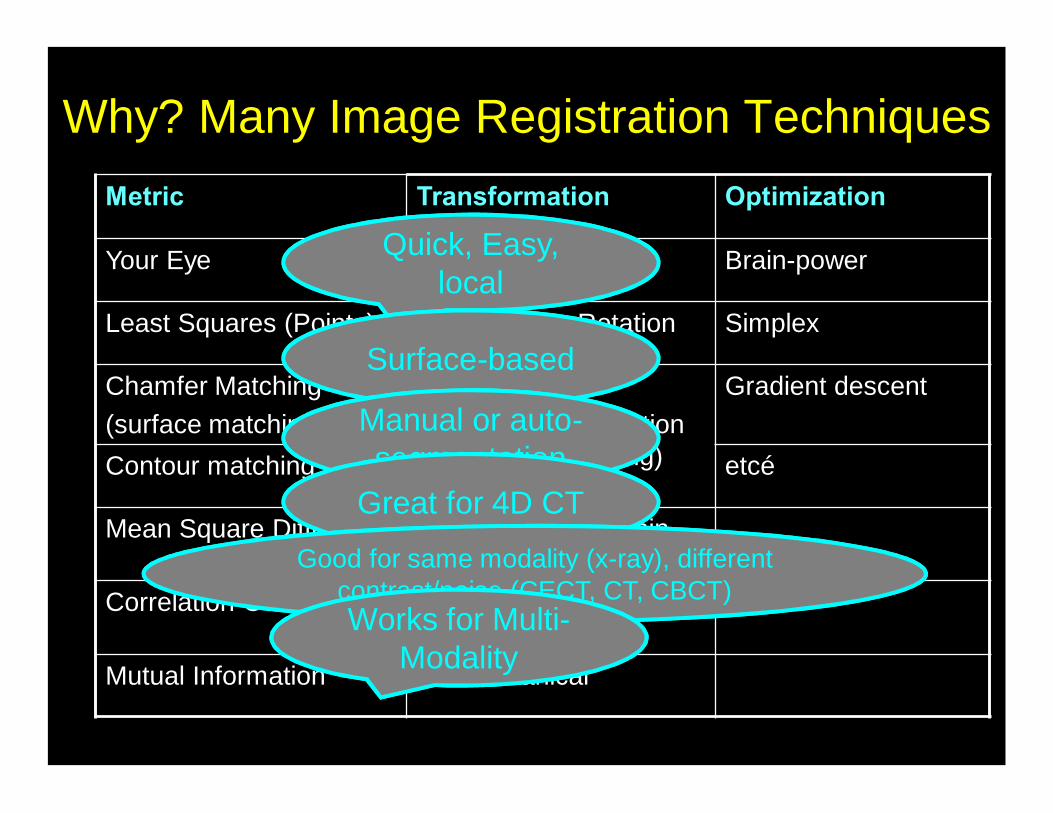
Why? Many Image Registration Techniques Metric Transformation Optimization
Your Eye Translation Brain-power
Least Squares (Points) Translation + Rotation Simplex
Chamfer Matching(surface matching)
Affine (Translation + Rotation + scaling + shearing)
Gradient descent
Contour matching etc…
Mean Square Difference Spline (B-spline, Thin plate spline)
Correlation Coefficient Physical (optical/fluid flow, elastic body)
Mutual Information Biomechanical
Quick, Easy, local
Surface-based
Manual or auto-segmentation
Great for 4D CT
Good for same modality (x-ray), different contrast/noise (CECT, CT, CBCT)Works for Multi-
Modality
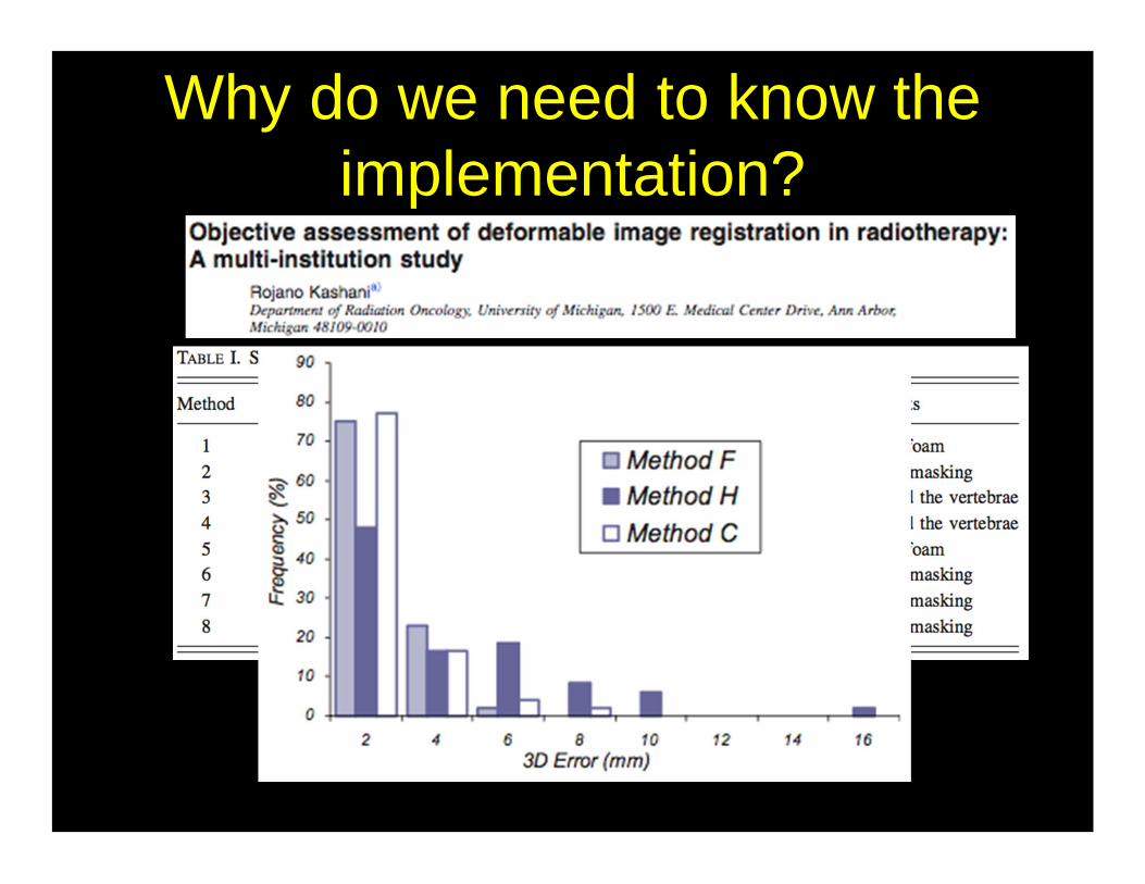
Why do we need to know the implementation?
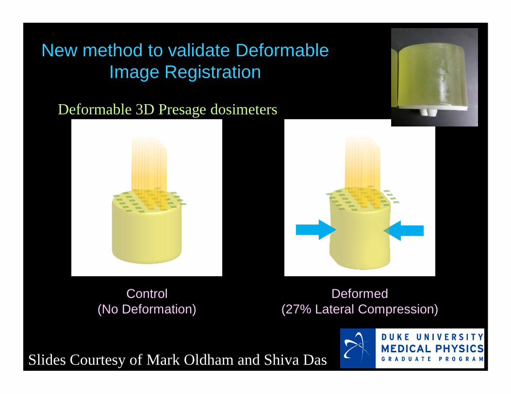
New method to validate Deformable Image Registration
Control(No Deformation)
Deformed(27% Lateral Compression)
Deformable 3D Presage dosimeters
Slides Courtesy of Mark Oldham and Shiva Das
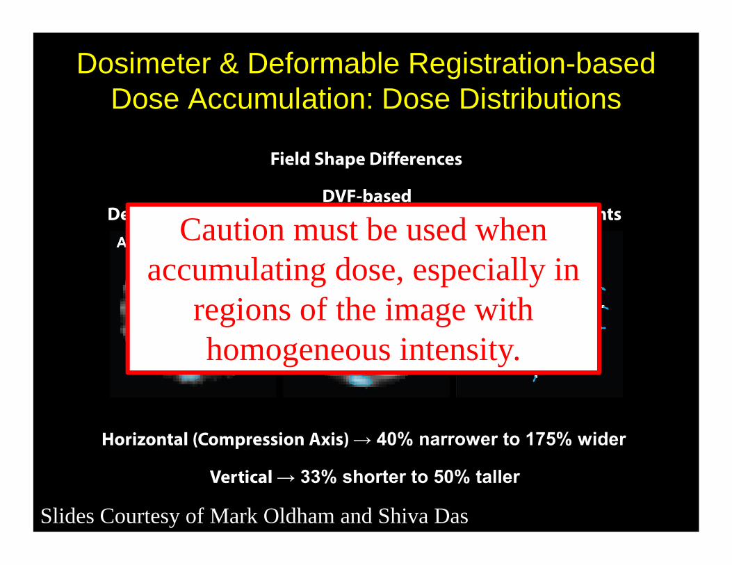
Dosimeter & Deformable Registration-based Dose Accumulation: Dose Distributions
Field DisplacementsDeformed DosimeterDVF-based
Accumulation
Field Shape Differences
Horizontal (Compression Axis) → 40% narrower to 175% wider
Vertical → 33% shorter to 50% taller
Slides Courtesy of Mark Oldham and Shiva Das
Caution must be used when accumulating dose, especially in
regions of the image with homogeneous intensity.
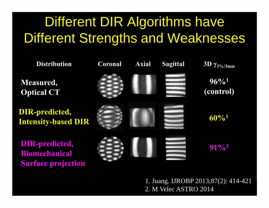
Distribution Coronal Axial Sagittal 3D γ3%/3mm
Measured,Optical CT
DIR-predicted,BiomechanicalSurface projection
96%1
(control)
1. Juang. IJROBP 2013;87(2): 414-4212. M Velec ASTRO 2014
91%2
DIR-predicted, Intensity-based DIR 60%1
Different DIR Algorithms have Different Strengths and Weaknesses
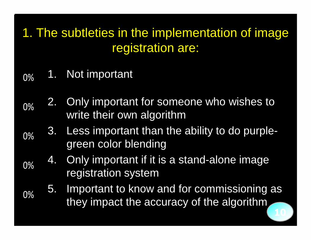
1. The subtleties in the implementation of image registration are:
0%
0%
0%
0%
0% 1. Not important
2. Only important for someone who wishes to write their own algorithm
3. Less important than the ability to do purple-green color blending
4. Only important if it is a stand-alone image registration system
5. Important to know and for commissioning as they impact the accuracy of the algorithm
10
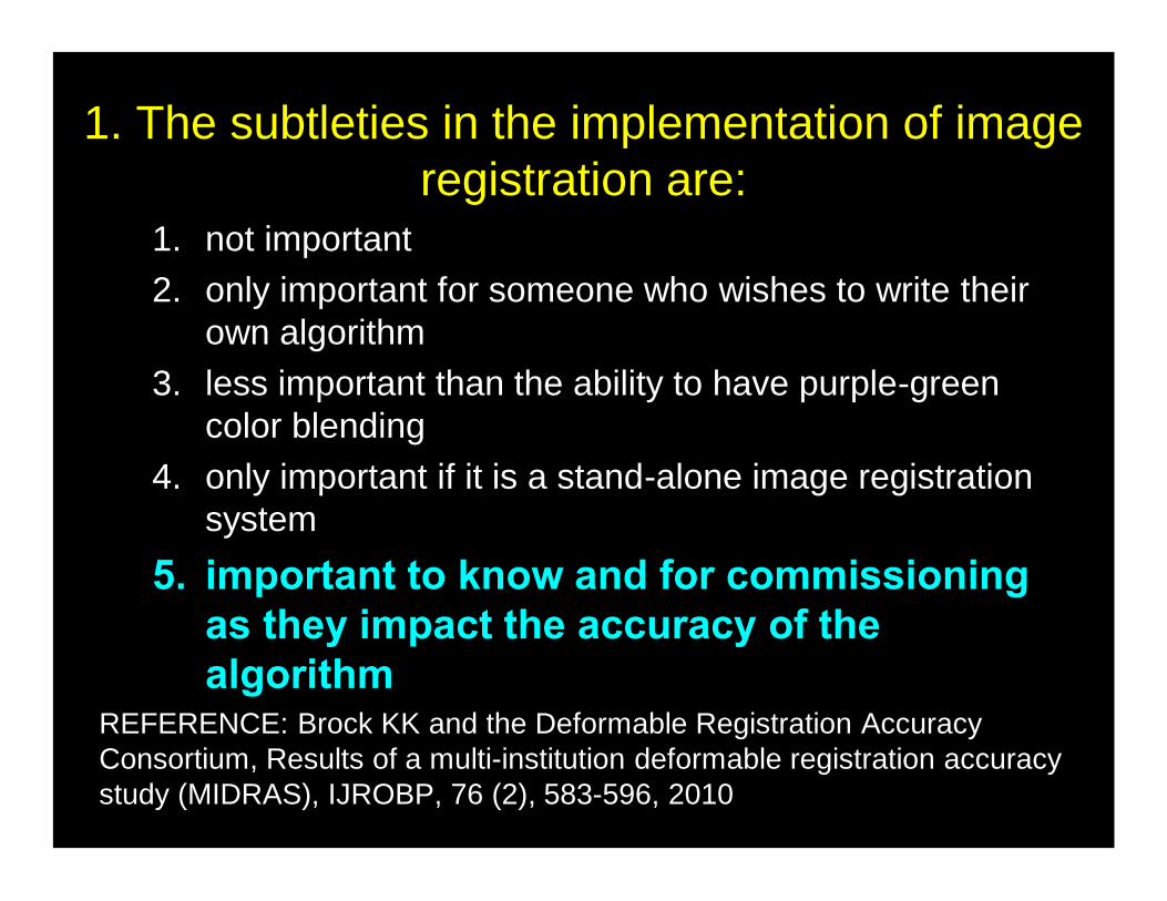
1. not important2. only important for someone who wishes to write their
own algorithm3. less important than the ability to have purple-green
color blending4. only important if it is a stand-alone image registration
system5. important to know and for commissioning
as they impact the accuracy of the algorithm
REFERENCE: Brock KK and the Deformable Registration Accuracy Consortium, Results of a multi-institution deformable registration accuracy study (MIDRAS), IJROBP, 76 (2), 583-596, 2010
1. The subtleties in the implementation of image registration are:

Commissioning Toolbox
• What tools do we have?
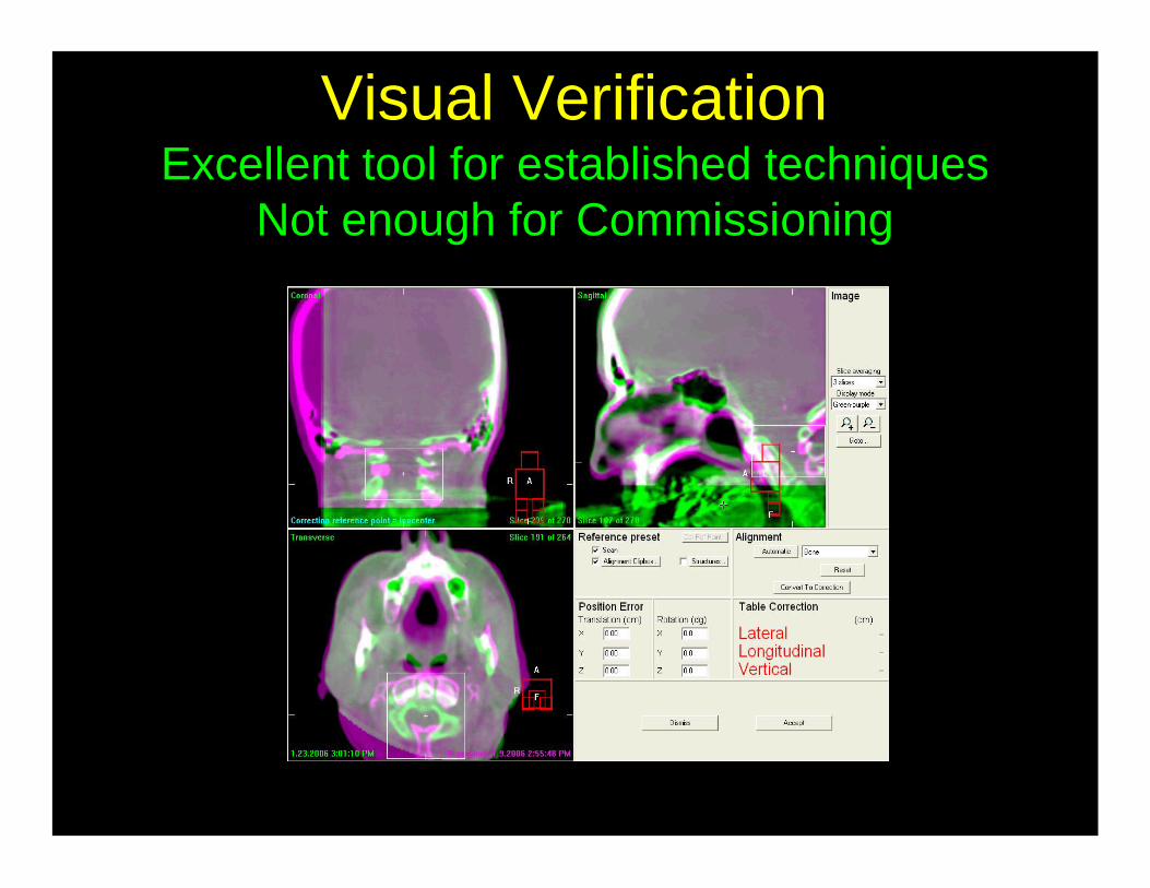
Visual VerificationExcellent tool for established techniques
Not enough for Commissioning
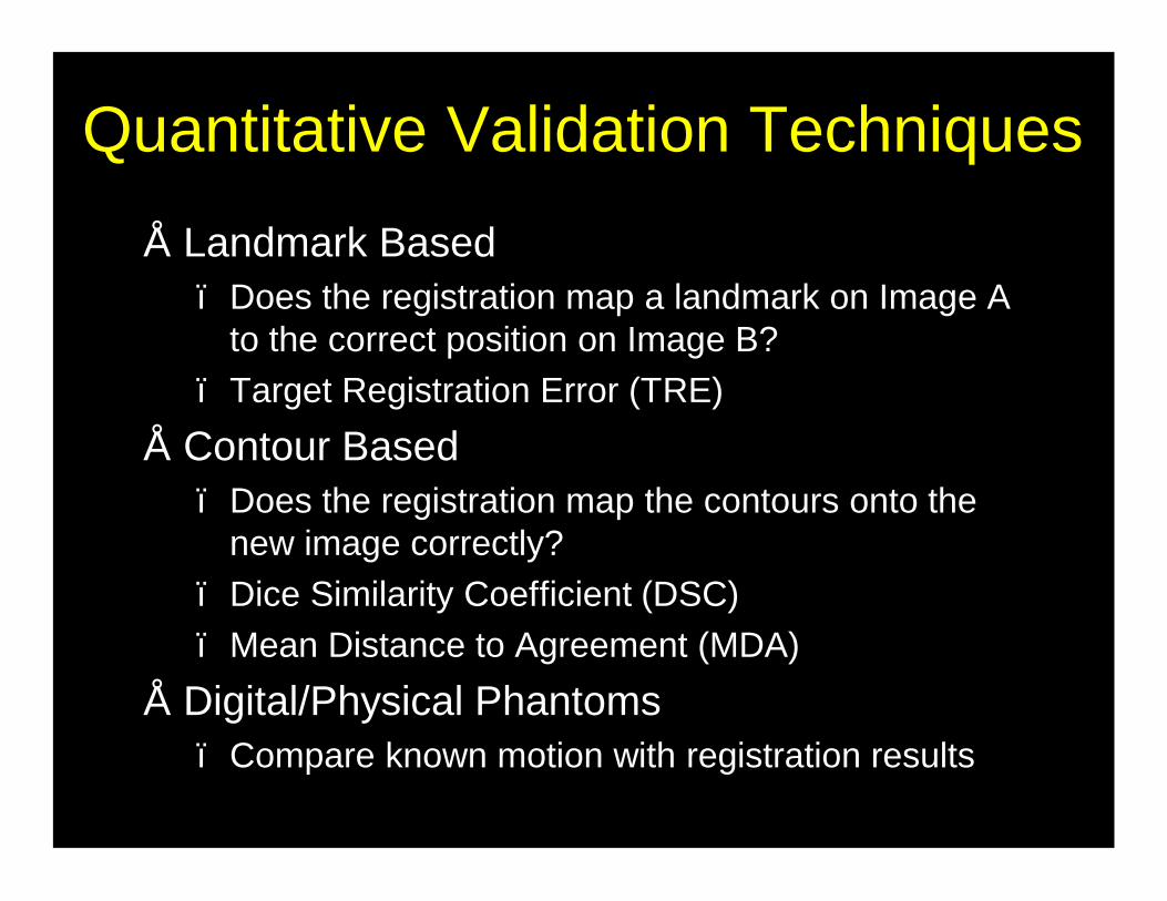
Quantitative Validation Techniques• Landmark Based
– Does the registration map a landmark on Image A to the correct position on Image B?
– Target Registration Error (TRE)• Contour Based
– Does the registration map the contours onto the new image correctly?
– Dice Similarity Coefficient (DSC)– Mean Distance to Agreement (MDA)
• Digital/Physical Phantoms– Compare known motion with registration results
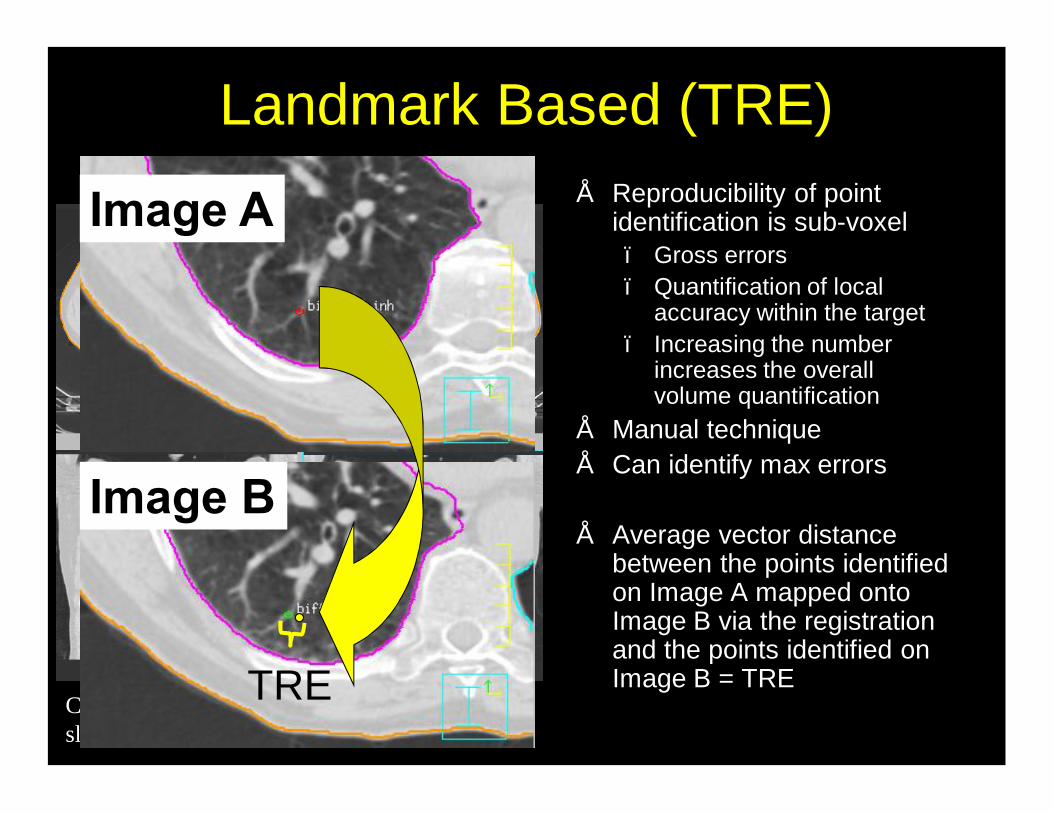
Landmark Based (TRE)• Reproducibility of point
identification is sub-voxel– Gross errors – Quantification of local
accuracy within the target– Increasing the number
increases the overall volume quantification
• Manual technique• Can identify max errors
• Average vector distance between the points identified on Image A mapped onto Image B via the registration and the points identified on Image B = TRE
CT: 512x512x152; 0.09 cm in plane, 0.25 cm slice; GE scanner; 4D CT with Varian RPM
TRE
Image A
Image B

That sounds great! Is that enough?
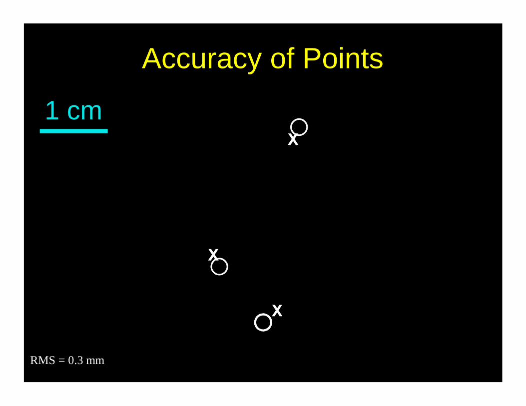
Accuracy of Points
X
X
X
1 cm
RMS = 0.3 mm
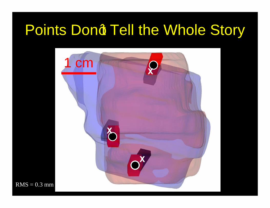
Points Don’t Tell the Whole Story
X
X
X
1 cm
RMS = 0.3 mm
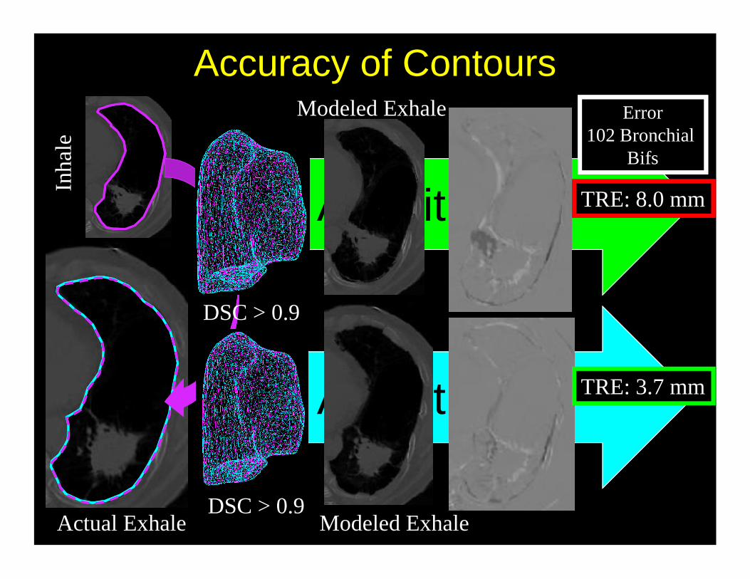
Algorithm 2
Algorithm 1
Accuracy of Contours
Actual Exhale Modeled Exhale
Modeled Exhale Error102 Bronchial
Bifs
TRE: 3.7 mm
TRE: 8.0 mm
Inha
le
DSC > 0.9
DSC > 0.9
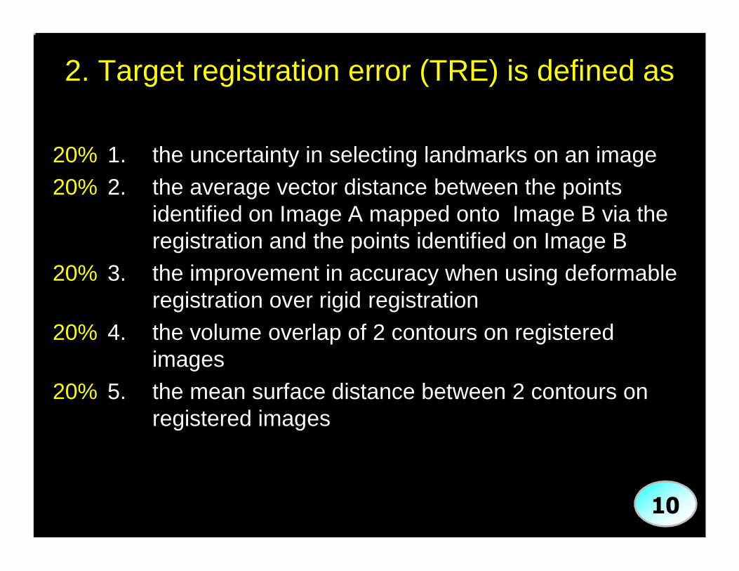
2. Target registration error (TRE) is defined as
1. the uncertainty in selecting landmarks on an image 2. the average vector distance between the points
identified on Image A mapped onto Image B via the registration and the points identified on Image B
3. the improvement in accuracy when using deformable registration over rigid registration
4. the volume overlap of 2 contours on registered images
5. the mean surface distance between 2 contours on registered images
10
20%20%
20%
20%
20%
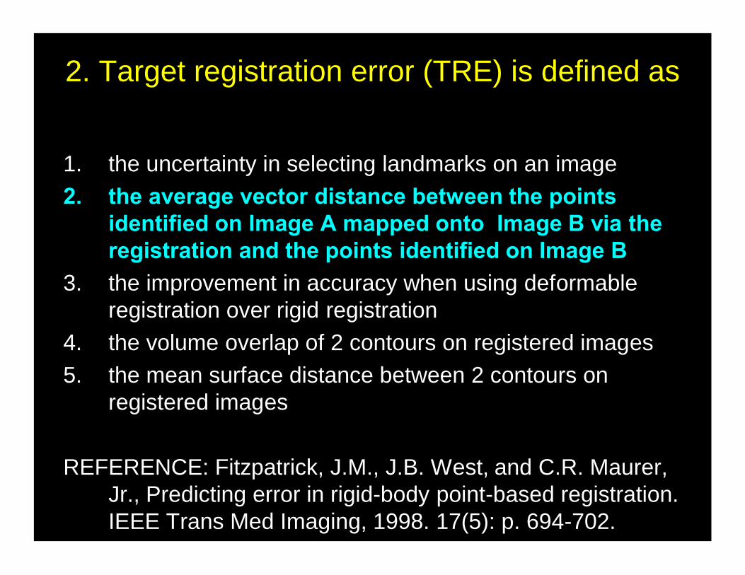
1. the uncertainty in selecting landmarks on an image 2. the average vector distance between the points
identified on Image A mapped onto Image B via the registration and the points identified on Image B
3. the improvement in accuracy when using deformable registration over rigid registration
4. the volume overlap of 2 contours on registered images5. the mean surface distance between 2 contours on
registered images
REFERENCE: Fitzpatrick, J.M., J.B. West, and C.R. Maurer, Jr., Predicting error in rigid-body point-based registration. IEEE Trans Med Imaging, 1998. 17(5): p. 694-702.
2. Target registration error (TRE) is defined as
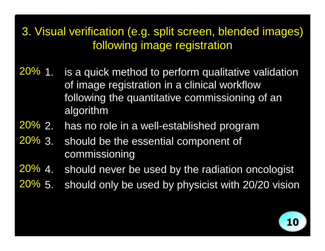
3. Visual verification (e.g. split screen, blended images) following image registration
1. is a quick method to perform qualitative validation of image registration in a clinical workflow following the quantitative commissioning of an algorithm
2. has no role in a well-established program3. should be the essential component of
commissioning4. should never be used by the radiation oncologist5. should only be used by physicist with 20/20 vision
10
20%
20%20%
20%20%

1. is a quick method to perform qualitative validation of image registration in a clinical workflow following the quantitative commissioning of an algorithm
2. has no role in a well-established program3. should be the essential component of commissioning4. should never be used by the radiation oncologist5. should only be used by physicist with 20/20 vision
REFERENCE: REFERENCE: Kessler ML, Image Registration and Data Fusion in Radiation Therapy (Review Article), BJR 79:S99-S108 2006
3. Visual verification (e.g. split screen, blended images) following image registration
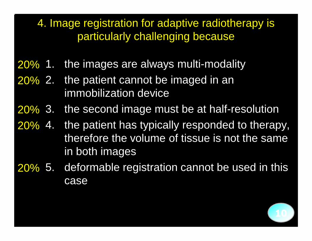
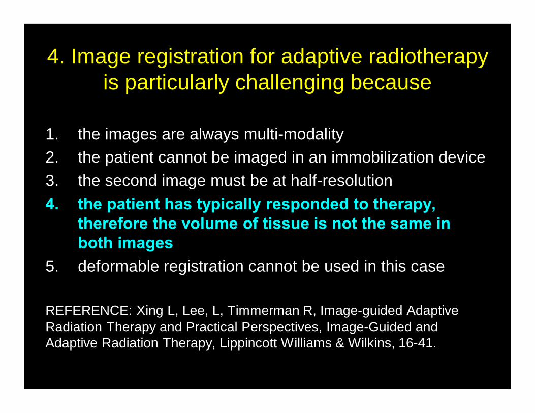
4. Image registration for adaptive radiotherapy is particularly challenging because
1. the images are always multi-modality2. the patient cannot be imaged in an
immobilization device3. the second image must be at half-resolution4. the patient has typically responded to therapy,
therefore the volume of tissue is not the same in both images
5. deformable registration cannot be used in this case
10
20%20%
20%20%
20%
1. the images are always multi-modality2. the patient cannot be imaged in an immobilization device3. the second image must be at half-resolution4. the patient has typically responded to therapy,
therefore the volume of tissue is not the same in both images
5. deformable registration cannot be used in this case
REFERENCE: Xing L, Lee, L, Timmerman R, Image-guided Adaptive Radiation Therapy and Practical Perspectives, Image-Guided and Adaptive Radiation Therapy, Lippincott Williams & Wilkins, 16-41.
4. Image registration for adaptive radiotherapy is particularly challenging because
Commissioning and QAUnderstand the whole picture
Understand fundamental
components of algorithm
Understand fundamental
components of algorithm
Phantom approach to understand
characteristics of algorithm
implementation
Phantom approach to understand
characteristics of algorithm
implementation
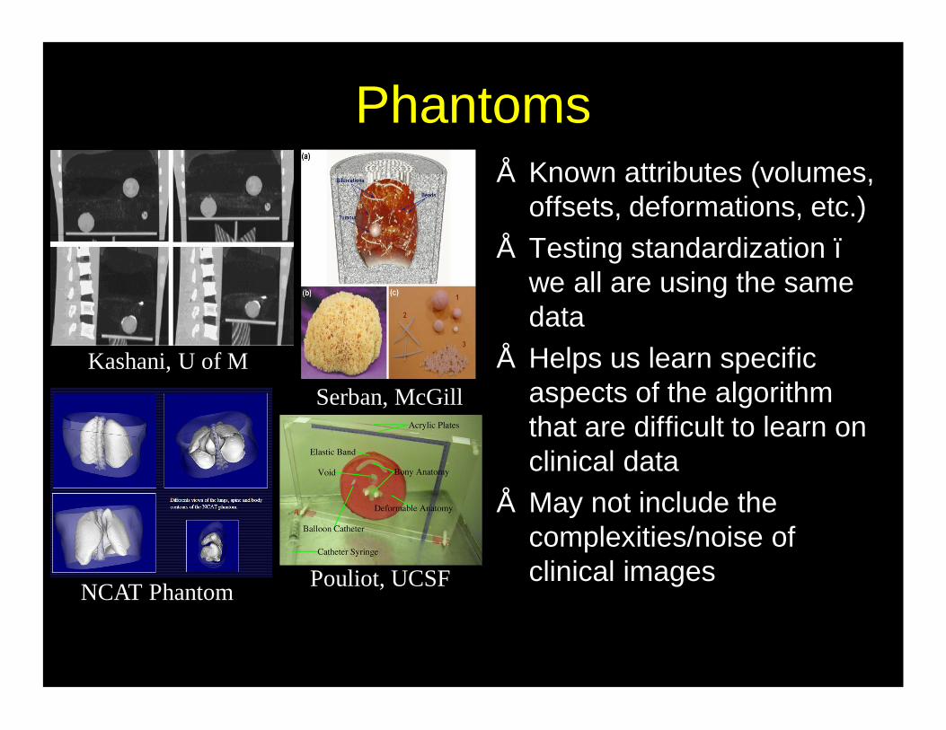
Phantoms• Known attributes (volumes,
offsets, deformations, etc.)• Testing standardization –
we all are using the same data
• Helps us learn specific aspects of the algorithm that are difficult to learn on clinical data
• May not include the complexities/noise of clinical images
NCAT Phantom
Kashani, U of MSerban, McGill
Pouliot, UCSF
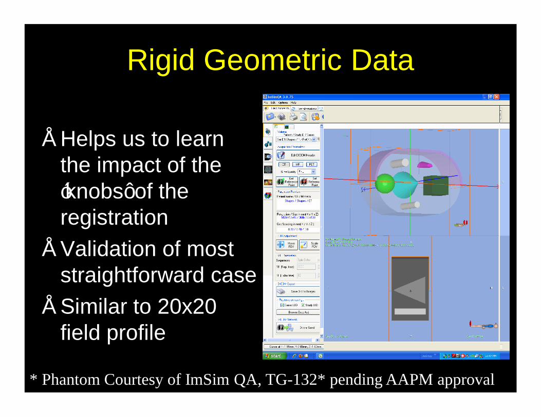
Rigid Geometric Data
• Helps us to learn the impact of the ‘knobs’ of the registration
• Validation of most straightforward case
• Similar to 20x20 field profile
* Phantom Courtesy of ImSim QA, TG-132* pending AAPM approval
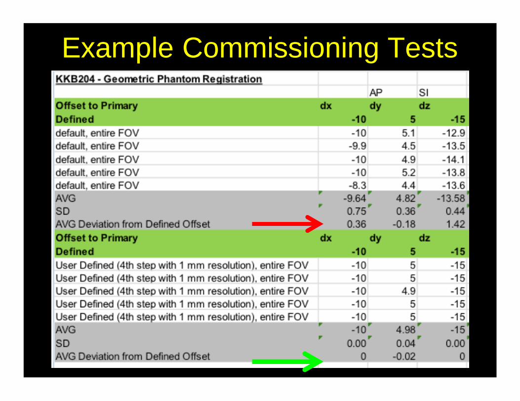
Example Commissioning Tests
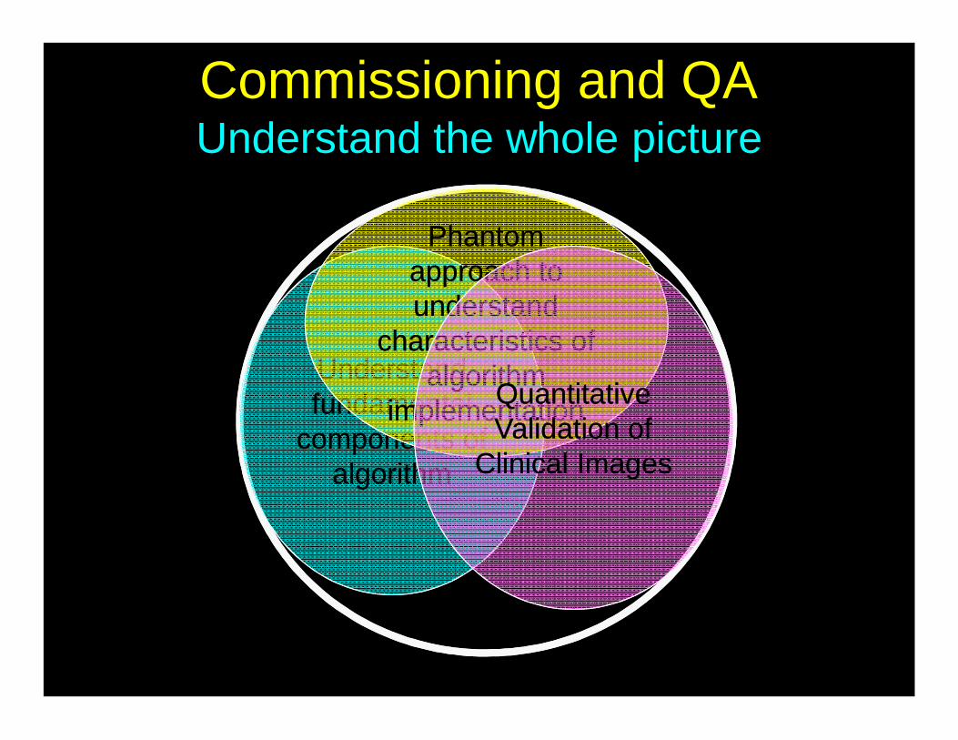
Commissioning and QAUnderstand the whole picture
Understand fundamental
components of algorithm
Understand fundamental
components of algorithm
Phantom approach to understand
characteristics of algorithm
implementation
Phantom approach to understand
characteristics of algorithm
implementationQuantitative Validation of
Clinical Images
Quantitative Validation of
Clinical Images
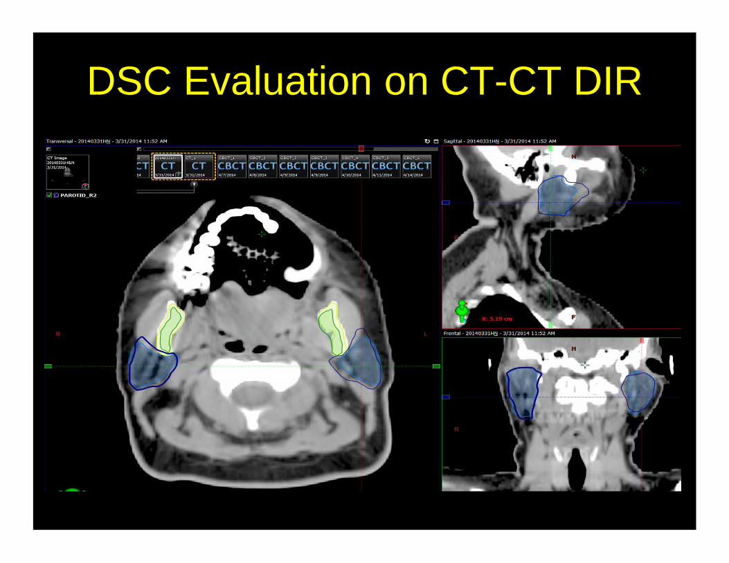
DSC Evaluation on CT-CT DIR
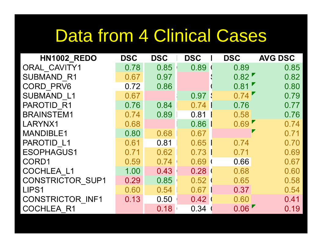
Data from 4 Clinical Cases
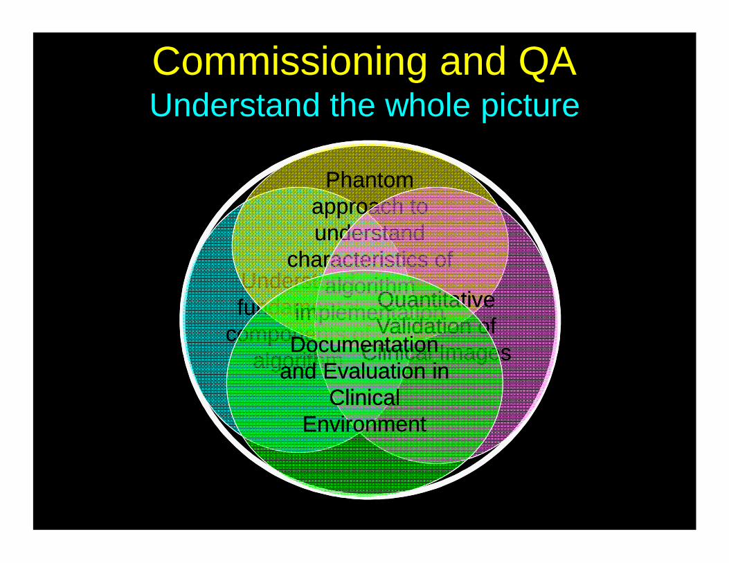
Commissioning and QAUnderstand the whole picture
Understand fundamental
components of algorithm
Understand fundamental
components of algorithm
Phantom approach to understand
characteristics of algorithm
implementation
Phantom approach to understand
characteristics of algorithm
implementationQuantitative Validation of
Clinical Images
Quantitative Validation of
Clinical ImagesDocumentation and Evaluation in
Clinical Environment
Documentation and Evaluation in
Clinical Environment
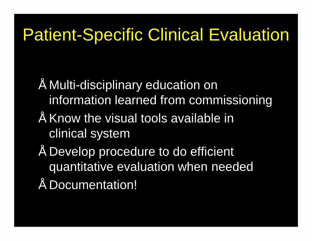
Patient-Specific Clinical Evaluation
• Multi-disciplinary education on information learned from commissioning
• Know the visual tools available in clinical system
• Develop procedure to do efficient quantitative evaluation when needed
• Documentation!
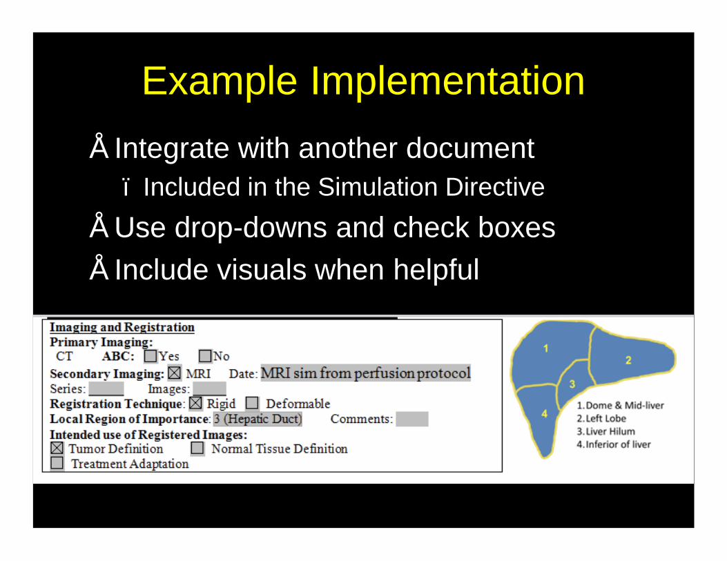
Example Implementation• Integrate with another document
– Included in the Simulation Directive• Use drop-downs and check boxes• Include visuals when helpful
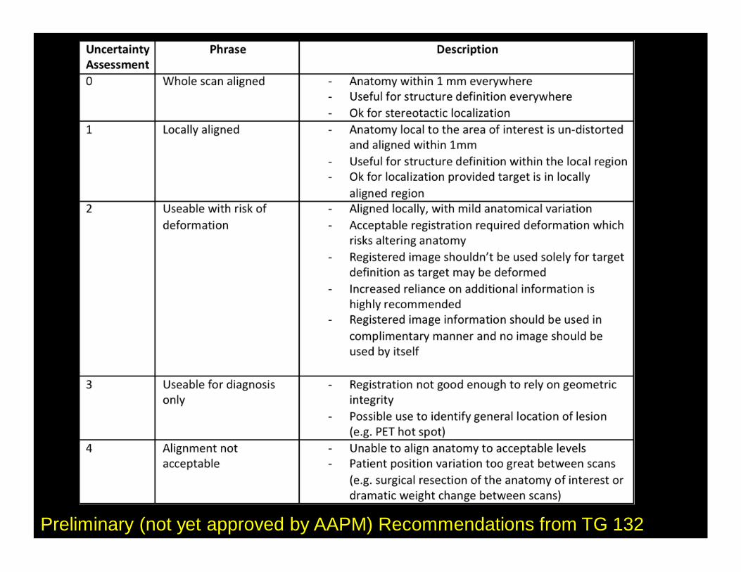
Preliminary (not yet approved by AAPM) Recommendations from TG 132
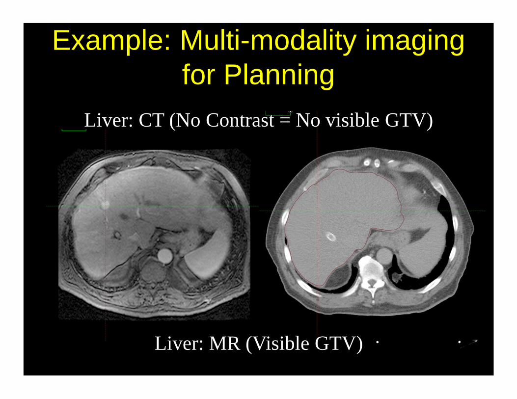
Example: Multi-modality imaging for Planning
Liver: CT (No Contrast = No visible GTV)
Liver: MR (Visible GTV)
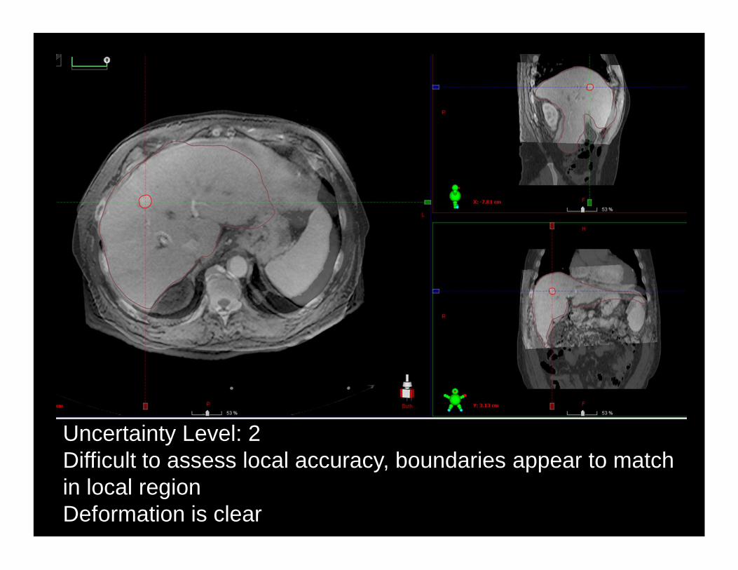
Uncertainty Level: 2Difficult to assess local accuracy, boundaries appear to match in local regionDeformation is clear
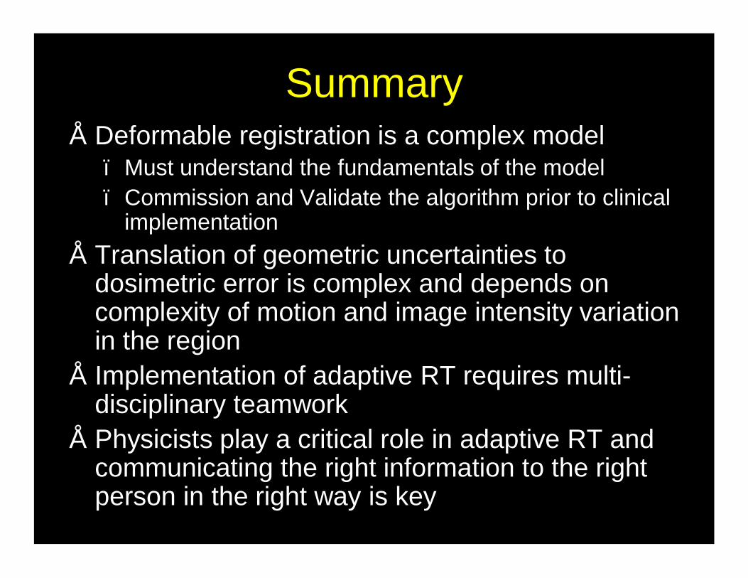
Summary• Deformable registration is a complex model
– Must understand the fundamentals of the model– Commission and Validate the algorithm prior to clinical
implementation• Translation of geometric uncertainties to
dosimetric error is complex and depends on complexity of motion and image intensity variation in the region
• Implementation of adaptive RT requires multi-disciplinary teamwork
• Physicists play a critical role in adaptive RT and communicating the right information to the right person in the right way is key


![[XLS]hsbte.org.inhsbte.org.in/pdf/docs/List of UMCs Jan 2014 website... · Web viewAnkit Kaushik S/o Rakesh Kaushik, Add. #116, Main Bazar, Najafgarh, New Delhi-110043 Shashikant](https://img.pdfslide.us/doc/110x75/5afebfb47f8b9a256b8d7f12/xlshsbteorg-of-umcs-jan-2014-websiteweb-viewankit-kaushik-so-rakesh-kaushik.jpg)