Embed Size (px)
Citation preview
A Web‐based Computer‐aided Diagnosis Tool for Bone Age Assessment:
Clinical Implementation and Lessons Learned
K Ma*; P Moin, MD*; M Fleshman*; L Vachon, MD**;A Zhang, PhD*; B Liu, PhD*; HK Huang, DSc*;
*Image Processing and Informatics LaboratoryDepartment of RadiologyKeck School of Medicine
University of Southern California 1450 San Pablo Street, Suite 2100
Los Angeles, CA 90033
**LAC+USC Medical CenterDepartment of RadiologyKeck School of Medicine
University of Southern California
Image Processing and Informatics Lab
Society of Pediatric Radiology 2009
Bone Age AssessmentIntroduction
Bone Age Assessment (BAA) Computer‐Aided Diagnosis (CAD) project goals
digital hand atlas (DHA)CAD algorithm
Clinical ValidationLAC+USC clinical workflowgraphical user interface (GUI)radiologist validation
Preliminary Results and Analysisintegration and lessons learned
Future Workreproducibility at other institutions
Image Processing and Informatics Lab
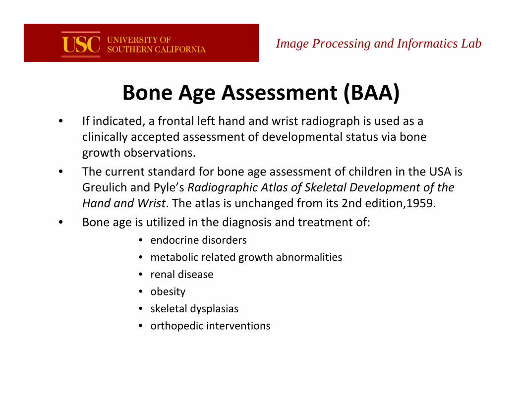
Bone Age Assessment (BAA) • If indicated, a frontal left hand and wrist radiograph is used as a
clinically accepted assessment of developmental status via bone growth observations.
• The current standard for bone age assessment of children in the USA is Greulich and Pyle’s Radiographic Atlas of Skeletal Development of the Hand and Wrist. The atlas is unchanged from its 2nd edition,1959.
• Bone age is utilized in the diagnosis and treatment of:• endocrine disorders
• metabolic related growth abnormalities
• renal disease
• obesity
• skeletal dysplasias
• orthopedic interventions
Image Processing and Informatics Lab
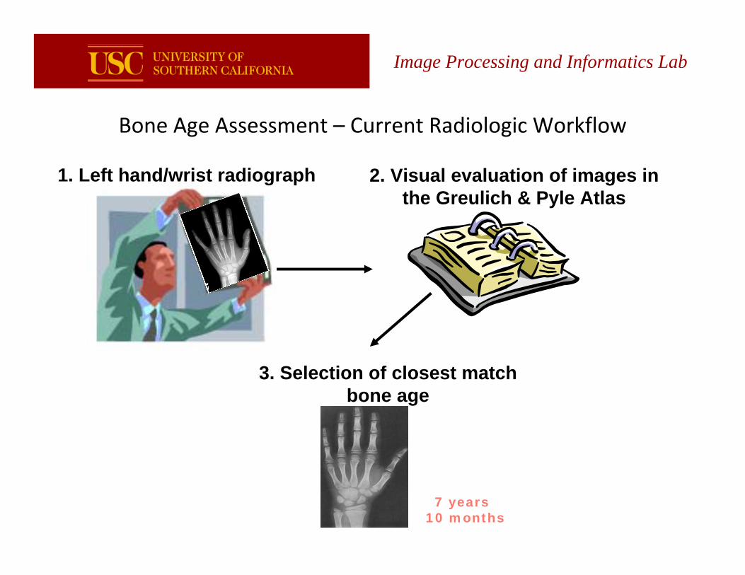
Bone Age Assessment – Current Radiologic Workflow
1. Left hand/wrist radiograph 2. Visual evaluation of images in the Greulich & Pyle Atlas
3. Selection of closest match bone age
7 years 10 months
Image Processing and Informatics Lab
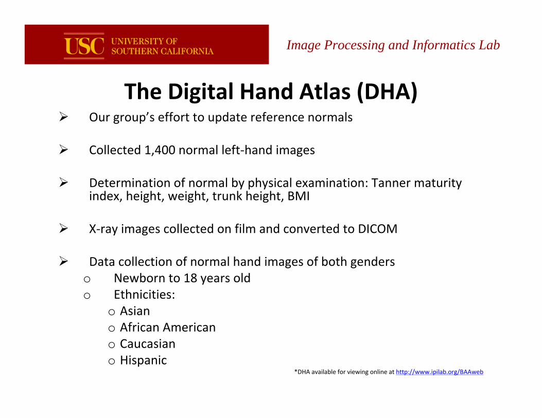
Image Processing and Informatics Lab
The Digital Hand Atlas (DHA)Our group’s effort to update reference normals
Collected 1,400 normal left‐hand images
Determination of normal by physical examination: Tanner maturityindex, height, weight, trunk height, BMI
X‐ray images collected on film and converted to DICOM
Data collection of normal hand images of both genderso Newborn to 18 years oldo Ethnicities:
o Asiano African Americano Caucasiano Hispanic
*DHA available for viewing online at http://www.ipilab.org/BAAweb
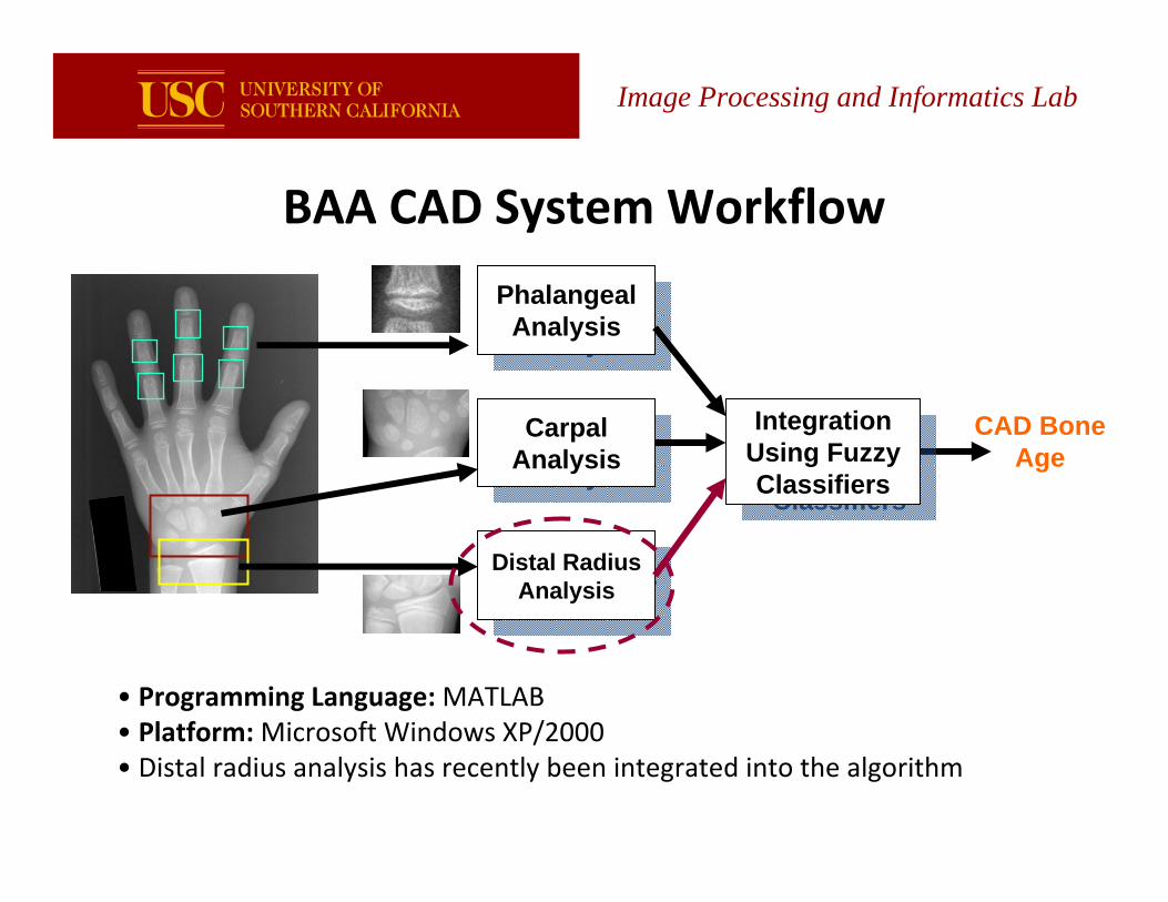
Image Processing and Informatics Lab
CAD Bone Age
Phalangeal Analysis
Phalangeal Analysis
Carpal Analysis
Carpal Analysis
Distal Radius Analysis
Distal Radius Analysis
IntegrationUsing Fuzzy Classifiers
IntegrationUsing Fuzzy Classifiers
• Programming Language: MATLAB• Platform: Microsoft Windows XP/2000• Distal radius analysis has recently been integrated into the algorithm
BAA CAD System Workflow
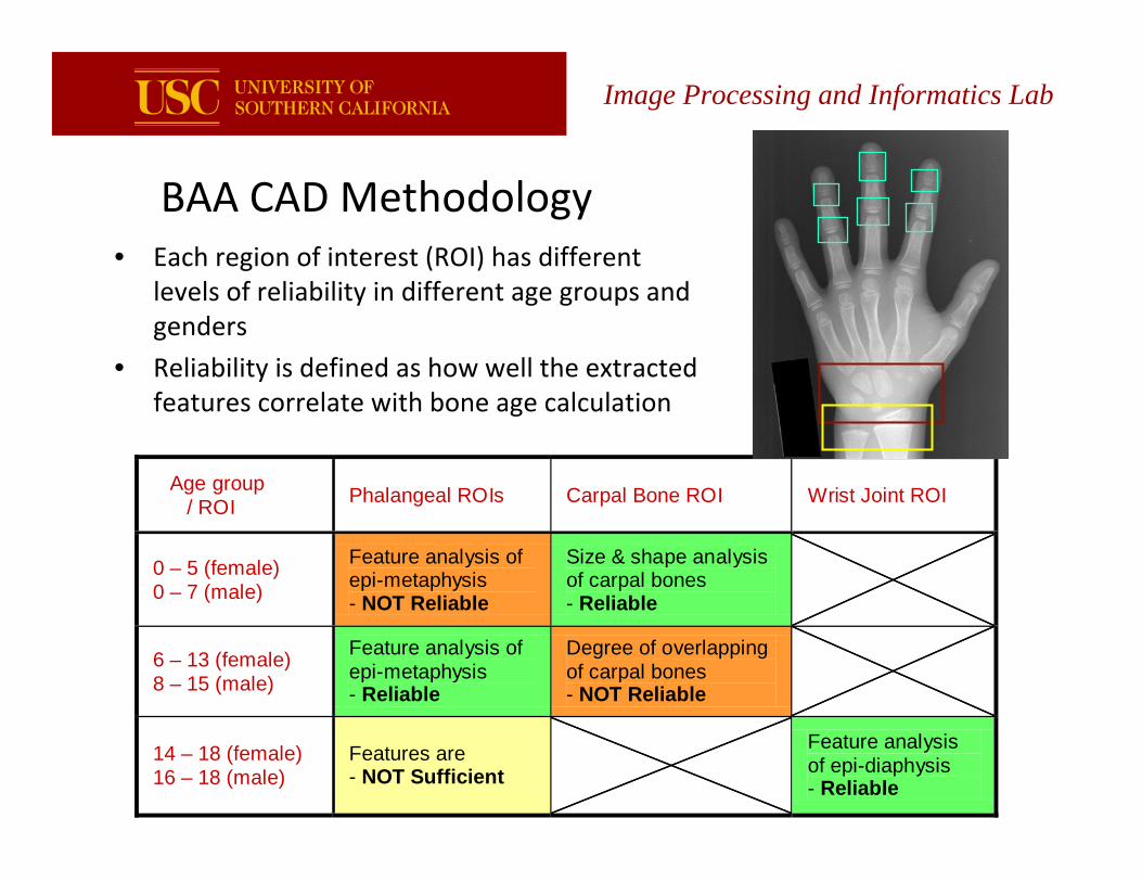
BAA CAD Methodology
Age group / ROI Phalangeal ROIs
0 – 5 (female) 0 – 7 (male)
Feature analysis of epi-metaphysis - NOT Reliable
6 – 13 (female) 8 – 15 (male)
Feature analysis of epi-metaphysis - Reliable
14 – 18 (female) 16 – 18 (male)
Features are - NOT Sufficient
Carpal Bone ROI
Size & shape analysis of carpal bones - Reliable
Degree of overlappingof carpal bones - NOT Reliable
Wrist Joint ROI
Feature analysis of epi-diaphysis - Reliable
Image Processing and Informatics Lab
• Each region of interest (ROI) has different levels of reliability in different age groups and genders
• Reliability is defined as how well the extracted features correlate with bone age calculation
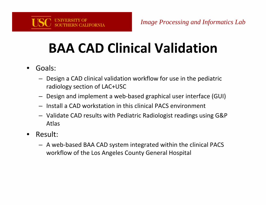
BAA CAD Clinical Validation• Goals:
– Design a CAD clinical validation workflow for use in the pediatric radiology section of LAC+USC
– Design and implement a web‐based graphical user interface (GUI)
– Install a CAD workstation in this clinical PACS environment
– Validate CAD results with Pediatric Radiologist readings using G&P Atlas
• Result: – A web‐based BAA CAD system integrated within the clinical PACS
workflow of the Los Angeles County General Hospital
Image Processing and Informatics Lab
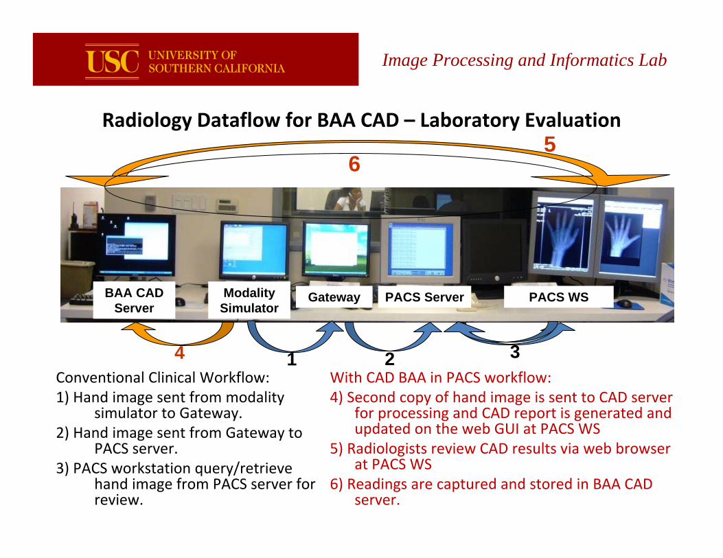
Image Processing and Informatics Lab
Radiology Dataflow for BAA CAD – Laboratory Evaluation
With CAD BAA in PACS workflow:4) Second copy of hand image is sent to CAD server
for processing and CAD report is generated and updated on the web GUI at PACS WS
5) Radiologists review CAD results via web browser at PACS WS
6) Readings are captured and stored in BAA CAD server.
BAA CAD Server
Modality Simulator
Gateway PACS Server PACS WS
1 2 34Conventional Clinical Workflow:1) Hand image sent from modality
simulator to Gateway.2) Hand image sent from Gateway to
PACS server.3) PACS workstation query/retrieve
hand image from PACS server for review.
56
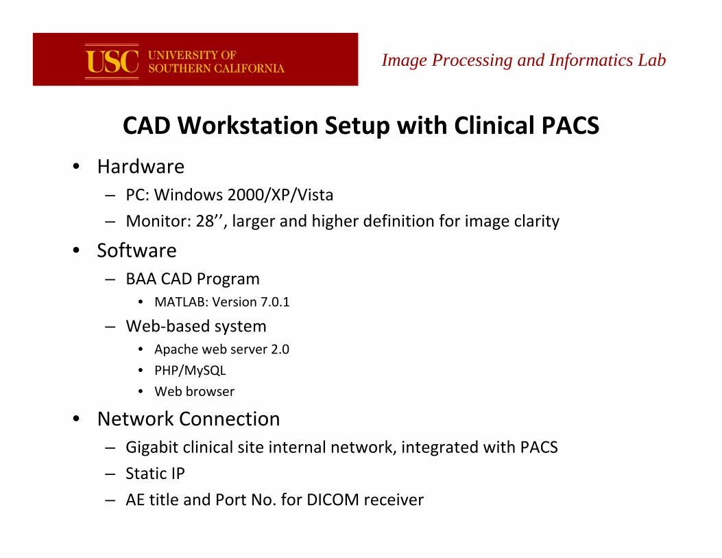
CAD Workstation Setup with Clinical PACS
• Hardware– PC: Windows 2000/XP/Vista
– Monitor: 28’’, larger and higher definition for image clarity
• Software– BAA CAD Program
• MATLAB: Version 7.0.1
– Web‐based system• Apache web server 2.0
• PHP/MySQL
• Web browser
• Network Connection– Gigabit clinical site internal network, integrated with PACS
– Static IP
– AE title and Port No. for DICOM receiver
Image Processing and Informatics Lab
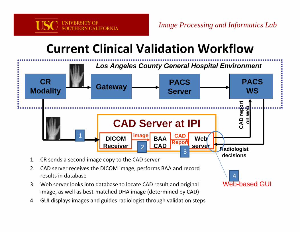
Image Processing and Informatics Lab
Current Clinical Validation Workflow
CAD Server at IPICAD Server at IPIDICOM
ReceiverBAA CAD
Web server
image CAD Report
CA
D re
port
on
web
Radiologist decisions
CRModality Gateway PACS
ServerPACS
WS
Los Angeles County General Hospital Environment
Web-based GUI
1. CR sends a second image copy to the CAD server
2. CAD server receives the DICOM image, performs BAA and record results in database
3. Web server looks into database to locate CAD result and originalimage, as well as best‐matched DHA image (determined by CAD)
4. GUI displays images and guides radiologist through validation steps
1
23
4
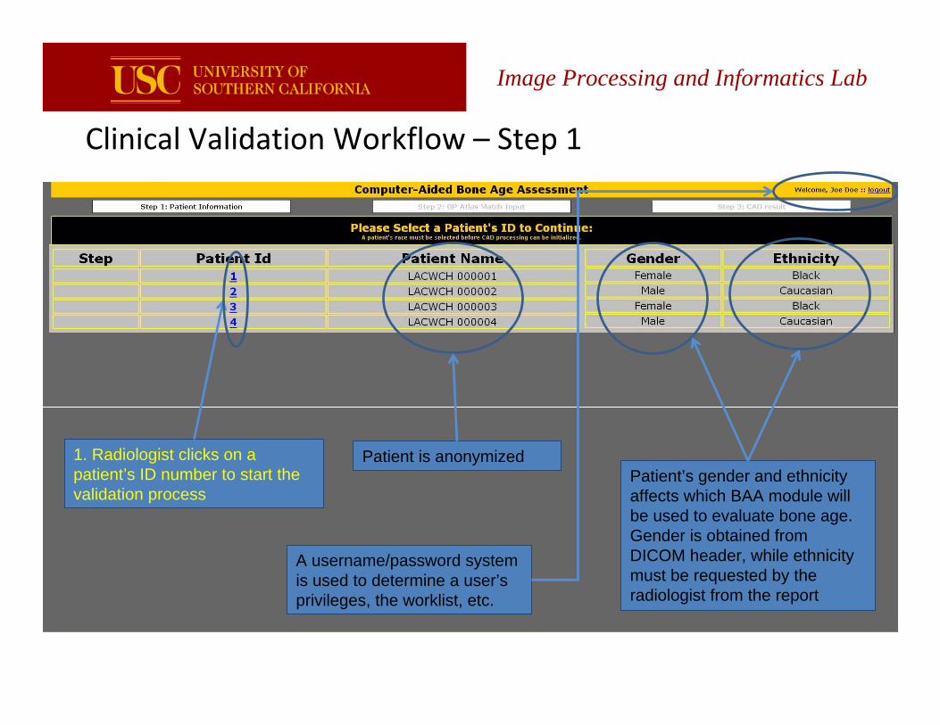
Clinical Validation Workflow – Step 1
Image Processing and Informatics Lab
1. Radiologist clicks on a patient’s ID number to start the validation process
Patient is anonymizedPatient’s gender and ethnicity affects which BAA module will be used to evaluate bone age. Gender is obtained from DICOM header, while ethnicity must be requested by the radiologist from the report
A username/password system is used to determine a user’s privileges, the worklist, etc.
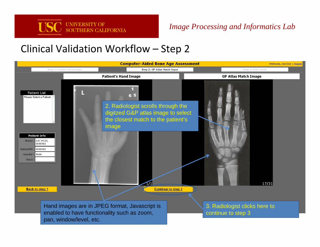
Clinical Validation Workflow – Step 2
Image Processing and Informatics Lab
2. Radiologist scrolls through the digitzed G&P atlas image to select the closest match to the patient’s image
Hand images are in JPEG format, Javascript is enabled to have functionality such as zoom, pan, window/level, etc.
3. Radiologist clicks here to continue to step 3
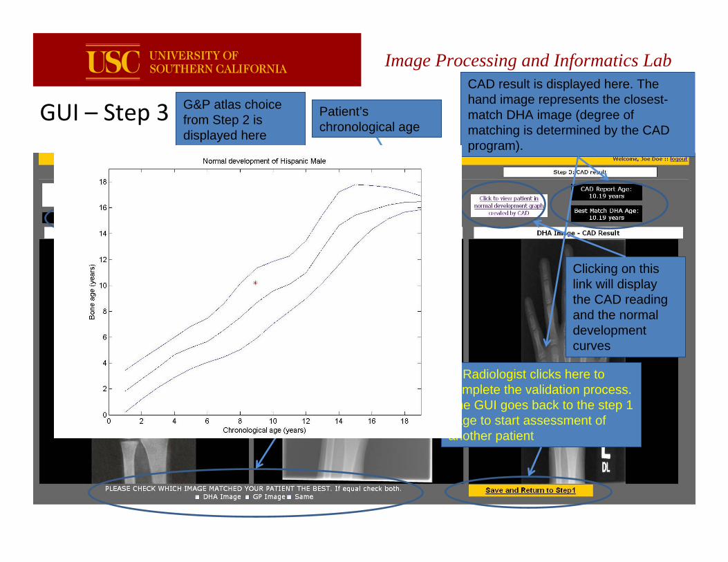
GUI – Step 3
Image Processing and Informatics Lab
G&P atlas choice from Step 2 is displayed here
CAD result is displayed here. The hand image represents the closest-match DHA image (degree of matching is determined by the CAD program).
Patient’s chronological age
4. Radiologist chooses which method (G&P or CAD) provides the better assessment by checking the 3 options: G&P, DHA, or both equally
5. Radiologist clicks here to complete the validation process. The GUI goes back to the step 1 page to start assessment of another patient
Clicking on this link will display the CAD reading and the normal development curves
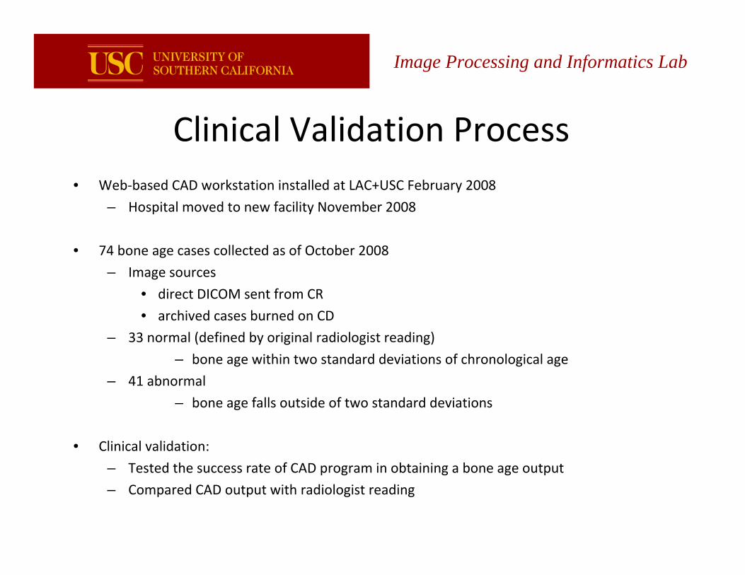
Clinical Validation Process• Web‐based CAD workstation installed at LAC+USC February 2008
– Hospital moved to new facility November 2008
• 74 bone age cases collected as of October 2008
– Image sources
• direct DICOM sent from CR
• archived cases burned on CD
– 33 normal (defined by original radiologist reading)
– bone age within two standard deviations of chronological age
– 41 abnormal
– bone age falls outside of two standard deviations
• Clinical validation:
– Tested the success rate of CAD program in obtaining a bone age output
– Compared CAD output with radiologist reading
Image Processing and Informatics Lab
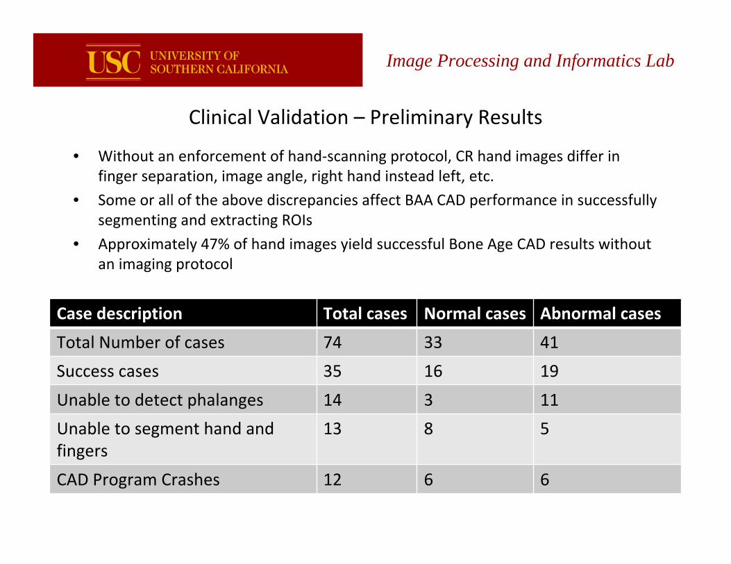
Clinical Validation – Preliminary Results
• Without an enforcement of hand‐scanning protocol, CR hand images differ in finger separation, image angle, right hand instead left, etc.
• Some or all of the above discrepancies affect BAA CAD performance in successfully segmenting and extracting ROIs
• Approximately 47% of hand images yield successful Bone Age CAD results without an imaging protocol
Case description Total cases Normal cases Abnormal cases
Total Number of cases 74 33 41
Success cases 35 16 19
Unable to detect phalanges 14 3 11
Unable to segment hand and fingers
13 8 5
CAD Program Crashes 12 6 6
Image Processing and Informatics Lab
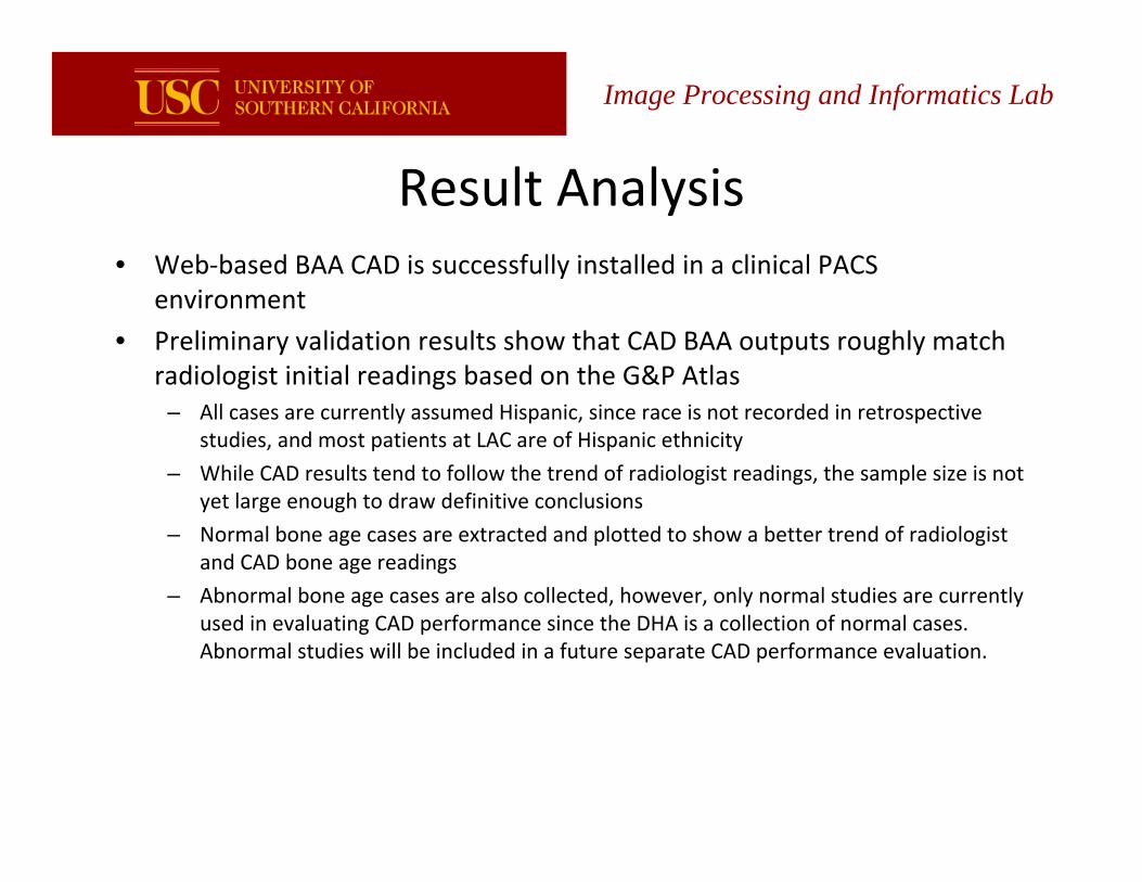
Result Analysis• Web‐based BAA CAD is successfully installed in a clinical PACS
environment
• Preliminary validation results show that CAD BAA outputs roughly match radiologist initial readings based on the G&P Atlas
– All cases are currently assumed Hispanic, since race is not recorded in retrospective studies, and most patients at LAC are of Hispanic ethnicity
– While CAD results tend to follow the trend of radiologist readings, the sample size is not yet large enough to draw definitive conclusions
– Normal bone age cases are extracted and plotted to show a better trend of radiologist and CAD bone age readings
– Abnormal bone age cases are also collected, however, only normal studies are currently used in evaluating CAD performance since the DHA is a collection of normal cases. Abnormal studies will be included in a future separate CAD performance evaluation.
Image Processing and Informatics Lab
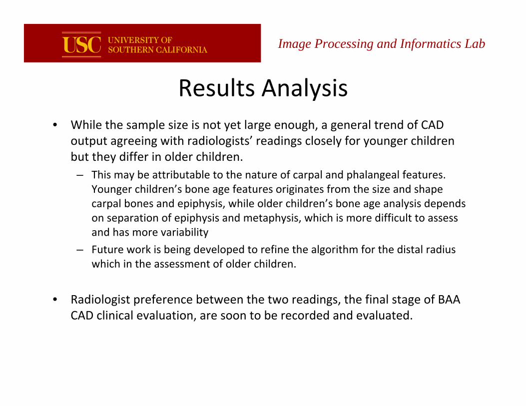
Results Analysis • While the sample size is not yet large enough, a general trend of CAD
output agreeing with radiologists’ readings closely for younger children but they differ in older children. – This may be attributable to the nature of carpal and phalangeal features.
Younger children’s bone age features originates from the size and shape carpal bones and epiphysis, while older children’s bone age analysis depends on separation of epiphysis and metaphysis, which is more difficult to assess and has more variability
– Future work is being developed to refine the algorithm for the distal radius which in the assessment of older children.
• Radiologist preference between the two readings, the final stage of BAA CAD clinical evaluation, are soon to be recorded and evaluated.
Image Processing and Informatics Lab
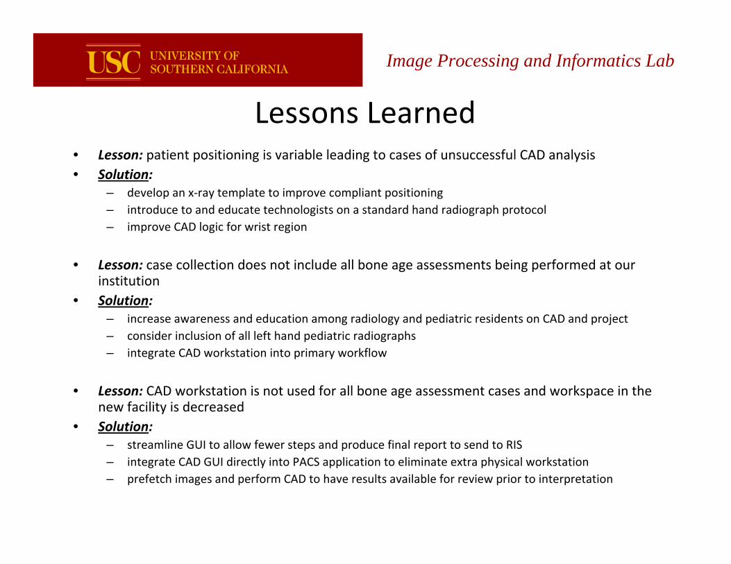
Image Processing and Informatics Lab
Lessons Learned• Lesson: patient positioning is variable leading to cases of unsuccessful CAD analysis• Solution:
– develop an x‐ray template to improve compliant positioning– introduce to and educate technologists on a standard hand radiograph protocol– improve CAD logic for wrist region
• Lesson: case collection does not include all bone age assessments being performed at our institution
• Solution:– increase awareness and education among radiology and pediatric residents on CAD and project– consider inclusion of all left hand pediatric radiographs– integrate CAD workstation into primary workflow
• Lesson: CAD workstation is not used for all bone age assessment cases and workspace in the new facility is decreased
• Solution:– streamline GUI to allow fewer steps and produce final report to send to RIS– integrate CAD GUI directly into PACS application to eliminate extra physical workstation– prefetch images and perform CAD to have results available for review prior to interpretation
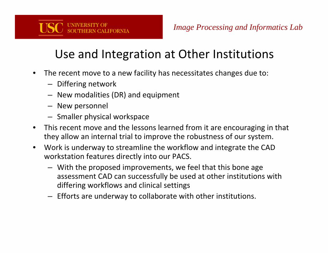
Image Processing and Informatics Lab
Use and Integration at Other Institutions• The recent move to a new facility has necessitates changes due to:
– Differing network– New modalities (DR) and equipment– New personnel– Smaller physical workspace
• This recent move and the lessons learned from it are encouraging in that they allow an internal trial to improve the robustness of our system.
• Work is underway to streamline the workflow and integrate the CAD workstation features directly into our PACS.– With the proposed improvements, we feel that this bone age
assessment CAD can successfully be used at other institutions with differing workflows and clinical settings
– Efforts are underway to collaborate with other institutions.

Summary
– An updated, ethnically diverse bone age assessment CAD has been developed by IPILab at and is now in the advanced clinical validation stage.
– A web‐based GUI has been developed to aid radiologists in comparing CAD bone age results with results from the GP atlas.
– The web‐based CAD validation system has been installed in USC+LAC and integrated into the clinical PACS environment.
– Cases collected so far have produced promising results for BAA CAD. Further refinement of the workflow and efforts to implement this tool at other institutions are underway.
Image Processing and Informatics Lab
Thank you.
Portions of this research project supported by NIH R01 EB 00298.
More information available at:
http://www.ipilab.org/BAAweb
http://www.ipilab.org/BAAgraph
http://www.ipilab.org/Research/BAA/BAAindex.php
Image Processing and Informatics Lab


![Digital Archiving and Cloud Museum E-Piloti OISHI LAB. · Institute of Industrial Science OISHI LAB. [Spatiotemporal Modeling and Visualization] Dept. of Informatics and Electronics](https://img.pdfslide.us/doc/110x75/5b87fa897f8b9aa0218e1144/digital-archiving-and-cloud-museum-e-piloti-oishi-lab-institute-of-industrial.jpg)


![09-CV-00298-Settlement Agreement [pt. 1]securities.stanford.edu/.../2016627_o01x_09CV00298.pdfCase 3:09-cv-00298-N Document 2325-1 Filed 06/27/16 Page 5 of 100 PageID 66308 “Stanford](https://img.pdfslide.us/doc/110x75/5f2e44483ef79f5e34612bf1/09-cv-00298-settlement-agreement-pt-1-case-309-cv-00298-n-document-2325-1-filed.jpg)