Embed Size (px)
Citation preview

1
Image Processing and Analysis at IPAGI. INTRODUCTION
Medical image analysis has grown and evolved tremendouslyin the last 30 years. The distinctive nature of the problems en-countered have led to the development of a significant body ofwork addressing such issues as fully three-dimensional data,nonrigid models for motion, deformation and comparison, andthe statistical variation of normal and abnormal structure. Thisarea of research derives from the clinical and scientific appli-cations which must be well understood. However, the method-ologies developed encompass an array of techniques that haveadvanced image analysis independent of the application.
This paper will describe the development of the Image Pro-cessing and Analysis Group (IPAG) at Yale and the broad rangeof work in the area of medical image analysis that has beenpursued. Throughout our work, we have endeavored to take ad-vantage of all available information both from prior knowledgeof physical properties, geometric constraints or statistical vari-ation as well as imaging data from various modalities. Medicalimaging problems typically lie in a well-defined domain. Thechallenge is to take best advantage of the given domain to solvethe often complex and subtle problems posed.
II. ORIGIN AND HISTORY
1970’s to 1983: Initial Ph.D. Faculty in Diagnostic Radiology
During the 1970’s, several Ph.D. faculty were hired into theDepartment of Diagnostic Radiology in the medical school atYale with their primary tasks being aimed at providing physicssupport for the clinical operations in the burgeoning fields ofnuclear medicine (Bob Lange), ultrasound (Fred Kremkau) andcomputed tomography (Stelios Orphanoudakis). Each of thesefaculty members were able to initiate and maintain their ownresearch programs as well, using different methods to supporttheir efforts, including industrial and some federal funding. In1982, as Magnetic Resonance was just beginning to be consid-ered for clinical use, John Gore was hired as the NMR physi-cist, now with a clear charge to develop a research program inthis area. Soon after (1982-1983), three more hires were madewithin the Department of Diagnostic Radiology. Art Gmitroand Gene Gindi were hired initially to help with image/signalprocessing and hardware issues related to the development of aDigital Subtraction Angiography (DSA) system, funded by anindustrial grant and Jim Duncan was hired jointly by the De-partment and the Section of Cardiology to work on a number ofimage analysis issues related to the TIMI (Thrombolysis in My-ocardial Infarction) project funded by the National Institutes ofHealth. While all of these faculty were hired for separate rea-sons, this group began to get together and develop a number ofinteractions to start to form a medical imaging methodology-based research community at Yale.
As the social and professional relationships among thisgroup of seven or so faculty members matured, these peopleformed, with support from clinical colleagues such as CarlJaffe, Richard Greenspan and Barry Zaret, the substrate for the
bioimaging science research now firmly established at Yale in2003.
While there were a variety of attempts to integrate bioimag-ing science into a single entity within Diagnostic Radiology,by and large these research efforts clustered into three primaryareas: nuclear medicine, magnetic resonance imaging and im-age post-processing. In this paper, we will focus on the evolu-tion of the latter area, that became the Yale Image Processingand Analysis Group (IPAG). However, at the end of the paper,we will describe how the early possibilities of an integratedbioimaging science community were re-kindled, bringing theevolution of effort full circle.
1983-1989: Biomedical Image Processing Early Years
One subset of the Ph.D. researchers in the bioimaging sci-ences areas overlapped significantly enough to form a core ef-fort in medical image processing: Drs. Duncan, Gindi, Gmitroand Orphanoudakis. Stelios Orphanoudakis obtained a joint ap-pointment with the Department of Electrical Engineering (EE)within the Faculty of Arts and Sciences at Yale in the late1970’s. Jim Duncan and Gene Gindi also received joint ap-pointments in Electrical Engineering. These faculty establishednew courses in the EE department in both Digital Image Pro-cessing and Computer Vision. While both had a biomedicalflavor in the examples given, the courses were established in or-der to interest Yale graduate and undergraduate students in thegeneral areas of image processing and image analysis. At thegraduate level, this effort soon paid off. Several new graduatestudents with focused interests in image processing and analysis(Larry Staib, Kathy Andriole, Dimitris Gerogiannis and VolkerTresp) joined Scott Holland (already working with Stelios Or-phanoudakis on ultrasound signal processing) as the core stu-dents of this new group. Early work on parallel image process-ing algorithms [1] and vessel tracking [2] appeared during thisperiod. Other work by Art Gmitro and Gene Gindi focused onoptical image processing strategies for feature extraction gar-nering a best paper award from the journal Optical Engineer-ing [3]. In addition, a body of work on image reconstructionmethods began at this time ranging from optical methods [4] tostatistical methods in the work of Mindy Lee [5], [6] in collab-oration with Gene Gindi and George Zubal, nuclear medicinephysicist. This work also included the development of theZubal Phantom [7], [8], a high-resolution three-dimensional an-thropomorphic image phantom derived from segmented humananatomy of the full body representing all of the major struc-tures. This data was designed for use as a phantom in imagingsimulations such as for Monte Carlo methods.
The emphasis, however, from the outset was to bridge areasof general image processing and computer vision with medical-imaging-specific knowledge. As the Yale group was forming,much of the efforts in the medical image analysis field seemedto be coming from one of two directions: i.) computer visionand image processing specialists with an interest in, but limiteddetailed knowledge of, medical imaging and its applications

2
Fig. 1. In 2D, the elliptic Fourier parameters define a curve and, by way ofprobability distributions, can be used to constrain the boundary finding process.
or ii.) medical imaging physicists who understood the princi-ples of image acquisition and formation, but who typically didnot have the specific applied mathematics, computer science orsignal and image processing background to contribute in thealgorithm development areas. By being physically located inthe medical school while maintaining close ties to engineeringthrough teaching and research, we strove to advance the fieldby developing new image processing and analysis methodologyfully grounded with knowledge of image acquisition physics,anatomy and physiology, and the clinical and scientific ques-tions.
From about 1985 to 1988 there was a strong influence ofwork from artificial intelligence in computer vision and med-ical image analysis. Drs. Gindi and Duncan developed a courseproject in their computer vision course in which students im-plemented a “blocks world” image understanding system tak-ing the problem from image acquisition to high level modelmatching [9]. Jim Duncan incorporated explicit knowledge-based concepts modeling the walls of the left ventricle of theheart in early work [10]. Advances in the application of neuralnetworks to computer vision and medical image analysis werealso made at this time in the group by Gene Gindi with his grad-uate student Joachim Utans [11]. They developed approachesto object recognition using hierarchical matching networks.
1989-present: IPAG Flourishes
As 1990 approached, the group began to explore more deeplysome of the key areas in medical image analysis. Larry Staib,Amir Amini and Hemant Tagare became faculty members inthe group around this time creating a critical mass of imageanalysis researchers. Workstation computational and graphi-cal power was also increasing tremendously enabling an arrayof techniques that were impossible before. The developmentof the web started in this period and we created one of thefirst web sites in medical image analysis in December 1993(http://noodle.med.yale.edu/). The web has obviously since be-come an essential aspect of dissemination and research.
Below we will discuss some of the key areas that IPAG haspursued including deformable models for segmentation, non-rigid motion analysis aimed primarily at cardiac motion, imageregistration and structural measurement.
Fig. 2. On the left, a mean curve is shown in the center along with curvesrepresenting plus and minus one standard deviation of the model parameters.On the right this model is used to segment the corpus callosum from a magneticresonance image.
Fig. 3. 2D deformable Fourier model segmenting the myocardium from mag-netic resonance images (left: initialization; right: final curves).
Deformable Models: A strong focus of the group aroseat this time aimed at developing mathematical strategies forextracting quantitative measurements from medical images.Boundary finding for anatomical structure is a central problemthat pervades many measurement problems. A chapter on themethodologies discussed below was included in a collection onmedical imaging [12]. The key themes that emerged from thiswork were the incorporation of prior information of shape asconstraints or biases and the integration of multiple informationsources [13], [14]. Such information is crucial to the solutionof medical image segmentation problems in the face of noise,ambiguity and structural complexity. We approached such seg-mentation problems from the perspective of mathematical opti-mization and (Bayesian) estimation theory, solved using a vari-ety of numerical approaches. Perhaps the earliest clear exampleof this trend was the Ph.D. thesis work of Larry Staib [15]. Thiswork was aimed at developing a concise parametrized model,using Fourier descriptors, of a contour or surface that could thenbe deformed or modified to find a specific boundary within animage [16], [17], [18]. This model easily allows the incorpo-ration of prior shape information and was the first technique touse prior shape information in a general and flexible way.
A maximum a posteriori objective function of parameters, � ,was used and allows a trade-off or compromise between priorinformation, ����� ��� , and image-derived information, ����� � ��� .For a uniform prior, this formulation reduces to the maximumlikelihood solution.����������� ����� � ���� � � ���� �!�#" � � �����$ � �!�&% (1)
Prior probability distributions on the parameters are used tointroduce a global shape model with a bias toward an expectedrange of shapes. This approach is illustrated in Figures 1, 2,3 and 4. This work was initially presented in 1988 [19], [20].

3
Fig. 4. Spatiotemporal models can be used to solve boundary finding prob-lems in temporal sequences as in this cardiac left ventricle magnetic resonanceexample showing three orthogonal views with the initialization of the left andthe final result on the right.
Fig. 5. Original MR cardiac image (top left) with mean contour (top middle)and expert tracing (bottom left). Note the improvement in performance mov-ing from an independent covariance (bottom middle) to smoothness covariance(bottom right) and finally the prior shape covariance (top right).
Interestingly from a historical perspective, this was being devel-oped in parallel with the deformable “snake” boundary findingapproaches of Kass, Witkin, and Terzopoulos.
Later, we studied active shape models [21] and showed therelationship to Fourier models and ways of exploiting the useof the covariance matrix [22], [23] (see Figure 5).
Two alternate approaches to using prior knowledge were pur-sued by Hemant Tagare. When the prior probability distribu-tions of the boundaries are unknown, an alternate approachis available which uses only a single canonical shape for theboundary. The canonical shape is encoded in a template whichfits the image boundary by deforming to minimize an energyfunction. Using conformal maps, normals to a deformable 2-d template can be extended smoothly to a large region of space[24]. The template is deformed by moving it along the extendednormals – which are called “orthogonal curves”. Interestingly,it is rather straightforward to show that the orthogonal curvesand dynamic programming gives the global minimum of the en-ergy function. Finding the global minimum is crucial to avoid-ing local minima that arise from noise and give grossly incorrectanswers. This algorithm has been successfully used for manyyears to segment carpal bones in CT scans. Figure 6 shows anexample.
Fig. 6. A wrist CT image showing carpal bones (left) and a deformable tem-plate with orthogonal curves and an example deformation (right).
Fig. 7. Functional activation map with global registration (left) and with localregistration (right) determined using the level set algorithm with shape priors tosegment and register the corpus callosum.
In the second approach, prior knowledge about the shape ofthe boundaries was incorporated into a level set formulationof active contours in a variational framework. The shape ofa boundary is all of the information that is left when location,orientation, and size information is removed. The shape prior ismapped into the image space by a translation, rotation and scal-ing and it appears as an additional term in the energy functionof the level set [25]. During level set evolution, the translation,rotation and scaling are continuously estimated and updated.The algorithm simultaneously achieves segmentation of an im-age and its registration with a standard coordinate system. Thismethod can be exploited in fMRI studies where simultaneoussegmentation and registration of local brain structures leads toa lowering of false positives in the activation map as shown inFigure 7.
The results of active contours are often evaluated by com-parison with manual segmentation. Unfortunately, experimen-tal evaluations often do not shed light on when and why activecontours become inaccurate. A theoretical analysis is requiredfor understanding this. In his Ph.D. thesis, Tianyun Ma theoreti-cally explored accuracy and consistency in active contours [26],[27]. He was able to show that some of the common forms ofexternal energy functions can lead to a significant bias in seg-mentation. Some energy functions can also make the contourunstable. By suitable modifications of the energy function, thebias and stability problems can be eliminated [26], [27].
Integration: Boundary finding based on edge features canbe augmented by considering the incorporation of region-basedinformation and developing an integrated Maximum a posteri-ori (MAP) probability method in order to determine the surface(or curve, in 2D) parameters which correspond to the structurewhich matches both the boundary strength in the image and theregion homogeneity properties [13], [28]. This work on inte-gration was the thesis work of Amit Chakraborty.
We consider two modules: one related to boundary findingand the other region growing where each contains a coupling

4
p
Nash Equilibrium
2
p1
p1
F = constant1
F = constant2
l2
l1
Fig. 8. In a simple 2D game theoretic formulation, constant level curves for')(+*-, .and
'�/0*$, .and the corresponding reaction curves ( 1 ( and 1 / ) of the two
players are shown. For a fixed 2 (43652 (, the best player 2 can do is to minimize' /
along the line 2 ( 3652 (. For each different
52 (, a different optimal response
can be found for player 2, and the collection of all these points form 1 / , thereaction curve of player 2. The Nash equilibrium lies at the intersection of theplayers reaction curves.
term that feeds information related to the other module:�87 �9;:�< �>=@?$A �B� �C7 �9 � D < �-= �E"6F DHG < �>=@?$A �I%�87 �J : G �KA#?L= �M� �C7 �J � D G �NA �4"PO D < G �NA4?�= �I% (2)= represents the output of the region-based process and A rep-resents the output of the boundary finding module. In thefirst equation, D < �>= � represents region-based classification in-formation [13]. In the second, D G �KA � represents the boundarystrength in the shape prior-driven contribution. D < G �KA4?�= � andD G < �-=@?-A � represent interaction terms between the two modules.
Initially, these equations were solved sequentially with FQ�R. The region based segmentation was determined first and then
that information was used to optimize the boundary.A more powerful approach to integration comes from a game
theoretic formulation [29], [30]. We were able to derive a moregeneral formulation pioneering the use of game theory (startedby the thesis work of Is.ıl Bozma [31]) as a means for integratingimage information [14].
When F is non-zero, the first equation includes an additionalinteraction term, D G < �-=@?-A � , which feeds back the latest avail-able output A of the boundary module and represents the agree-ment of the voxels within the current boundary with the as-sumed gray level distribution for the indicated tissue type. Inthe second equation, the interaction term D < G �KA4?�= � uses thelatest available output = of the region process. The modulesassume the roles of players in a 2-player game and are opti-mized in parallel. The game continues until the players cannot improve their positions without cooperation from the otherplayer. This natural stopping point of the parallel decision mak-ing process constitutes the Nash equilibrium solution [32] (seeFigure 8).
The rational decision provided by the Nash equilibrium so-lution is the natural counterpart of the optimum found with se-quential objective optimization. We have found it to be morerobust to noise and initialization in a variety of 2D and 3D prob-lems [13].
In order to compute the contribution of the region based in-formation, we need to compute an integral over the region. Wecan compute this efficiently if we convert the volume integral
Fig. 9. Results of surface finding for the head of the left caudate nucleus (toprow) and the right thalamus (bottom row) in an MR image. The wireframe andthree perpendicular slices through the 3D image (1.2mm S voxels) are shownwith the surface obtained using both boundary and region information.
to an area integral using Gauss’ divergence theorem [33]. Weconstruct a function whose divergence is the function we wishto integrate by integrating in each of the coordinate directions.Then we can simply compute the area integral of this functionduring the optimization process (see [28] for details), greatlyreducing the necessary computation.
In the brain, some subcortical structures often have poor con-trast between gray and white matter because they are striate andappear with intermediate intensity. These structures, however,are less variable, in terms of shape, than the cortex. Thus, priorshape information can be of great value in identifying subcor-tical boundaries. In Figure 9, we demonstrate the performanceof the 3D integrated method on subcortical structure examples:the head of the right caudate nucleus and the left thalamus.Using the integrated method with a prior shape model, alongwith region and boundary information, the proper boundariesare found.
Level Set Method Incorporating Thickness Constraint:While some segmentation problems are well suited to the con-straints that global shape information provides, some involvestructures whose shapes are highly variable or have no consis-tent shape at all and thus require more generic constraints [12].
Xiaolan Zeng, in her graduate work in the group, developed acoupled surfaces approach for automatically segmenting a vol-umetric layer from a 3D image [34], [35], [36]. This approachuses a set of coupled differential equations, with each equationdetermining the evolution or propagation of a surface within alevel set framework. In the case of the cortex, one surface at-tempts to localize the white matter/gray matter (WM/GM) innercortical boundary and the other the gray matter/cerebrospinalfluid (GM/CSF) outer boundary. Coupling between the surfacesincorporates the notion of an approximately fixed thickness sep-arating the surfaces everywhere in the cortex. This soft con-straint helps in ensuring that the GM/CSF boundary is capturedeven in the deeper cortical folds in the brain. A further assump-tion is that across each surface, there is a local difference in thegray level values, while in between the two surfaces there isa homogeneity of gray levels. By evolving two embedded sur-faces simultaneously, each driven by its own image-based infor-

5
Fig. 10. Evolution of coupled level sets: outer (magenta) and inner (yellow)surfaces propagate from initialized pairs of concentric spheres in a 3D MR brainimage to localize the gray matter.
mation while maintaining the coupling, we are able to achievean automatic and robust segmentation of the cortex, and simul-taneously obtain a representation of the inner and outer corticalsurfaces.
Starting from inside the inner bounding surface (gray/whiteboundary), with an offset in between, the interfaces propa-gate along the normal direction stopping at the desired place,while maintaining the distance between them. Embeddingeach surface as the zero level set in its own level function,we have two equations: TEUWV XZYHT\[ " : V X]� ^_U`V X4� � R
andTEUWa�bcdYHT\[ " : a�b�cE� ^_UWa�bce� � Rwhere : V X and : aLb�c are func-
tions of the surface normal direction, image-derived informa-
Fig. 11. Top: Surfaces resulting from single surface approach finding the innerand outer cortical surfaces separately; Bottom: Coupled surfaces approach runon same data overlaid on a sagittal slice masked by expert tracing of the outercortical surface showing good agreement. Coupling prevents the inner surfacefrom collapsing into CSF (1) and the outer surface from penetrating non-braintissue (2).
tion and the distance between the two surfaces. The couplingis embedded in the design of : V X and : a�b�c . Where the dis-tance between the two surfaces is within the normal range forcortical thickness, the two surfaces propagate according to theimage-based information; where the distance between the twosurfaces is out of the normal range, the distance constrains thepropagation. We define:: V X � f �KA\g#hjilkmhn�poqsr �L�Lt ��UWa�b�c �: aLb�c � f �KA\u#vsw!ixg#h@�poqsr �L��t �IU VyX �ez (3)
Function f smoothly maps larger gray level transition probabil-ity to slower speed. Function t smoothly penalizes the distanceoutside of the normal range. Thus, each surface moves withconstant speed along the normal direction, and slows down orstops when either the image-based information becomes strongor the distance to the other surface moves away from the normalrange.
The ability of the evolving level set to change topology,break, merge and form sharp corners greatly assists in the con-vergence to complex structures. In addition, the level set ap-proach facilitates structural measurement through the directcomputation of geometric parameters, such as curvature andthickness, via the level set function [36] (see, for example, Fig-ure 18). Surface renderings showing the evolution of the innerand outer cortical layers found from a normal 3D brain MR im-age using this approach are shown in Figure 10. This methodhas been extensively tested in our lab and elsewhere [37] withaccurate results under a range of conditions.

6
Fig. 12. Strain computed using a biophysical model from canine cardiac MRdata comparing baseline to infarcted left ventricle.
Fig. 13. Echo ultrasound data can also be analyzed with a physical model toestimate strain. Shown here are points derived from the model overlaid on theultrasound image to give “echo tissue tags” analogous to MR tagging.
Nonrigid Motion Leading to Deformation: Analyzing in-formation embedded in temporal sequences of images had longbeen of interest to medical image analysis researchers, withmany of the driving applications coming from the need to quan-tify cardiovascular function. The IPAG efforts focused primar-ily on developing strategies to follow left ventricular (LV) mo-tion, and ultimately deformation, in attempts to stratify differ-ences in LV performance as derived from any one of severalnon-invasive imaging modalities. A comprehensive review ofmethods for estimation of cardiac motion and deformation in-cluding the methods described below was included in a recentbook on medical image analysis [38].
In the early 1990’s, a number of groups had begun to developtechniques to find correspondences between pairs of framesfrom 2D cardiac image sequences [39], [40] as well as to de-velop useful parametrizations for spatiotemporal datasets [41],[42] in attempts to model and more fully determine nonrigidmotion. In the computer vision community, these efforts wereinitially reported at the first conference session on nonrigid mo-tion analysis at the IEEE Computer Society’s 1991 Conferenceon Computer Vision and Pattern Recognition (CVPR91). To theclinical cardiology community, however, many of these ideaswere seen as the most recent attempts to address the difficultproblem of stratifying left ventricular function.
Within IPAG, our focus was to develop a strategy for quanti-
fying LV function that could be used to derive information fromany one of a number of noninvasive image sequences. Whileother groups focused almost solely on the promising techniquesof analyzing MR tagging [43], [44] or MR phase velocity [45],we chose to try to develop approaches that were potentiallymodality-independent. The common thread we saw was thatif we could somehow accurately segment out the endocardialand epicardial boundaries from each frame of a cardiac imagesequence, and sample often enough, we could assume that localsurface shape would be preserved between any two frames andhence be used as a tracking metric.
This effort began by analyzing two-dimensional cardiac im-age sequences. Due to its spatial resolution, and in part to beable to compare to tagging and phase velocity tracking results,we chose to use two-dimensional cine-gradient echo MR imagesequences as our initial data. Endocardial and epicardial bound-ary segmentation in each frame was performed using modifiedversions of the Bayesian boundary finding strategy describedabove (see [16]). Curvatures, { , were derived at each bound-ary point and used as tracking tokens. Matching was performedby finding the best local segment of length | surrounding pointA V } < at frame i+1 within a plausible search region ~ whoseshape best matches a segment surrounding candidate point A V attime i via a squared curvature (i.e. bending energy) metric:�A Vy} < ��� �����87 �J0�y����� k �� � � < �${ � �KA Vy} < ��� { � �KA V �L� G (4)
Once the optimal A Vy} < is found in the search region, the vec-tor � that connects these two points can be defined. Similardisplacement vectors can be located at all points A V with confi-dences, � , assigned by the strength and uniqueness of the shapematch. Uncertainty, noise and out-of-plane motion were han-dled by employing a regularization strategy to estimate a finalset of smoothed displacements:�� �NA ����� �����C7 �������m����� � �� �KA �x� o� �NA �!� o�#� �KA �+� G " T o� �KA �T �M��� �
(5)Early results using this approach on 2D MR and echocardio-
graphic image sequence data were encouraging [40], but it wasappreciated that there were a number of difficulties with the ap-proach, including the lack of including temporal information inthe modeling, the difficulties deriving motion from segmentedboundaries and perhaps most importantly, the need to addressthe full 3D motion problem.
This work evolved in several directions. One effort focusedon the integration of temporal and full spatiotemporal infor-mation/modeling into the motion recovery process [46]. Thiswork, by John McEachen for his thesis, continued to work on2D image sequence data and took on a decidedly classical Elec-trical Engineering flavor as the basic strategy was based on re-cursive filtering incorporating a periodicity constraint. A sec-ond effort was centered on integrating several sources of track-ing cues into the frame-to-frame LV motion estimation process.The initial idea was aimed at using the boundary-based shapecues described above, along with Eulerian estimates of dis-placement found by integrating Magnetic Resonance phase ve-

7
locity information. The integration of shape with direct, densemeasurements of three-dimensional velocity provides a power-ful set of constraints on the interpretation of motion from imagedata. This work was primarily done by Fran cois Meyer, a post-doctoral fellow in IPAG at this time (later a faculty member inthe group), who had worked on rigid body motion problems incomputer vision during his Ph.D. studies in France. While thesedata provide direct measures of velocity, they are extremelynoisy. A Kalman filtering approach was developed here usingspatial and temporal constraints to determine an optimal esti-mate of the cardiac kinematics [47].
The third research direction that evolved from our initialshape-based 2D cardiac motion tracking work was anchored intwo key concepts. The first concept was grounded in the un-derstanding in the cardiology clinical research literature at thistime that the quantification of LV function from noninvasiveimage sequences was found to be more robust if one lookedat the relative measurement of LV myocardial wall thicken-ing as opposed to just absolute point displacement (see e.g.[48]). The second concept was that in order to fully under-stand LV function, one had to appreciate that the heart is a three-dimensional, nonrigidly deforming object and that the quantifi-cation of this deformation likely had to be derived from the full4-dimensional (3 spatial dimensions plus time) image informa-tion. Wall thickening was thus only one component of a morecomplete measure of myocardial deformation: the local straintensor.
These same basic ideas were also taking hold at the time inthe MR imaging community, driven by the development of MRgrid tagging for following tissue deformation [49], [50].
Thus, IPAG efforts on cardiac nonrigid motion in the mid-1990’s further developed by extending our 2D shape-based mo-tion tracking strategies to three dimensions using 3D curva-ture to track surface-based motion derived from cine-gradientecho MRI. The earliest efforts were performed by Amir Aminiand Jim Duncan, during Amir’s postdoctoral studies at Yale[51]. These ideas were further developed in the Ph.D. theses ofPengcheng Shi [52] and then Xenios Papademetris [53]. Thecurvature matching process represented by equation 5 abovewas now based on matching the similarity of the principal cur-vatures between an endocardial or epicardial surface patch attime � to a set of candidate patches within a search region at time� " � . A key next step, anchored in Pengcheng Shi’s efforts, wasthe development of an interpolation approach that could acceptthese image-derived, surface-based displacements and then cre-ate quantitative measures of mechanical strain across the entireLV myocardial volume. This work was published at the firstCVRMed conference in 1995 [54] and moved IPAG into thearea of using more realistic physical models to try to capturethe underlying myocardial tissue properties and serve as a ba-sis for integrating information and presenting quantitative mea-surements of strain. The advantage of this direction was thatthere was an entire cardiac biomechanics community [55] thathad spent decades trying to develop accurate forward modelsof the LV. The initial IPAG efforts in this area used simple, lin-ear elastic biomechanical models (realized via a stiffness matrix¡
), assumed infinitesimal strains between any two time framesand assumed that a sparse set of displacements, ¢ £ , could be
found from 3D shape tracking. The volume between the endo-cardial and epicardial surfaces at each time frame was meshedand the following Newtonian equation was then solved usinga finite element strategy at all points at each time frame to geta dense set of displacements ¤ , where the ¥ matrix is used toweight the confidence in the shape matches:¦ ¤ � ¥��I¤ � ¤ £ � (6)
Further extensions were then incorporated to integrate shapebased estimates of displacement with velocity estimates mea-sured using phase velocity MR, now in a fully 4D frameworkand using a continuum mechanical model of the heart [56], [57].
As the physical modeling work continued to mature andevolve in the graduate work of Xenios Papademetris [58], [53],[59], the formulation was viewed from a Bayesian standpointand the biomechanical model of the heart was augmented to in-clude muscle fiber directions. Quantified 3D measures of straincould now be reliably derived from a moving sequence of seg-mented myocardial surfaces on a desktop workstation. An ex-ample of these derived strains are shown in Figure 12. Thismethodology was implemented in a software platform that isusable by collaborators and has led to advances in the cardiol-ogy literature on regional myocardial deformation [60].
Critical to these efforts was the notion that displacement in-formation could come from a variety of data sources, includingshape cues, MR tags, and/or MR phase velocity information.The shape tracking strategy permits the approach to be usedacross imaging platforms. For example, further work extendedthese techniques directly to 3D ultrasound data enabling thepossibility of such measurements from a low cost and portablemodality [61], shown in Figure 13.
These ideas in realistic soft tissue modeling were further de-veloped and applied to the related problem of computing braindeformation during neurosurgery [62], [63]. Biomechanicalmodels are again of extreme value here in order to accuratelymodel the brain and track its deformation incorporating theeffects of gravity. This work stems from the thesis of OskarSkrinjar. First, intraoperative sparse point measurements wereused to drive the model [62], [64]. A finite element compu-tational approach was used to implement the biomechanicalmodel. Later, stereo images of the exposed brain surface wereused to provide a dense set of measurements for driving themodel. Stereo reconstruction methods were derived for this pur-pose [65] to determine the changing surface of the brain. Thisprocess is shown in Figure 14.
Registration Methods: Over the years, a number of us inIPAG have worked on different registration problems with dif-ferent approaches reflecting both the application area and theunderlying methodology, starting with rigid methods and mov-ing to nonrigid. We developed methods for rigid registration in-corporating constraints using linear programming [66] as wellas methods using genetic methods for improved optimization[67]. Nonrigid methods were developed using elastic and fluidmodels that were augmented with structural constraints thuscombining gray level matching with features [68]. This work,by Yongmei Wang during her graduate work, used these con-straints to help to bring the gray level matching into concor-dance with the underlying anatomy. During his postdoctoral

8
Fig. 14. Intraoperative stereo image pairs (top) can be used to reconstruct the exposed brain surface (bottom left) which can then be used as a constraint in thebiomechanical model for determining brain shift (bottom right).
study, Colin Studholme, worked on the problem of magneticresonance (MR) distortion correction mapping echo planar im-age data to conventional MR. He developed methods to accountfor both geometric and intensity distortions within the EPI databy constraining nonrigid mutual information based registrationusing the physical basis of the distortion [69].
Anand Rangarajan, a faculty member in the group at this pe-riod, and his student Haili Chiu, developed the technique ro-bust point matching (RPM) for nonrigid registration [70], [71].They formulated registration completely in terms of point fea-tures and a corresponding match matrix. The match matrixconverges to a specification of correspondence, and a corre-sponding nonrigid transformation, with outlier rejection usingthe “soft-assign” technique.
Another approach to non-rigid correspondence betweenclosed curves was pursued by Hemant Tagare. Generalizationsof monotonic one-to-one correspondences, called bimorphisms,were shown to be a class of “monotonic” curves on the torus,which is a product space of two closed curves. By suitablydefining a metric on the torus, the shapes of the two curves canbe compared and an optimal correspondence can be found [72],[73]. Figure 15 shows an example. There are several advan-tages of this idea. The theory clearly lays out the geometry ofcorrespondences. Also, the optimal correspondence is symmet-ric, i.e. the same answer is obtained irrespective of the order inwhich the two curves are compared. The last property is impor-
µ
C2
Correspondenceas a set
Projection (p )
Projection (p )
Product space C X C1 2
C1
1
2 An element ofthe correspondence
Fig. 15. The correspondence between two closed curves is shown to be acurve on the torus, which is the product space of the curves (left). An optimalcorrespondence from one corpus callosum to another is shown on the right.
tant in applying non-rigid correspondences to motion analysis.An information theoretic framework was developed for the
specific registration problem in radiation therapy treatmentplanning where one or more low quality 2D portal x-rays (gen-erated from therapeutic energy x-rays) need to be registered to a3D CT image [74], [75]. Ravi Bansal, in his thesis work, devel-oped an information theoretic approach for this problem wheresimultaneous segmentation and registration were carried out inan alternating iterative minimax entropy algorithm.
We have also pursued methods for the determination of sur-face point correspondence for shape comparison based on cur-vature features and geodesic interpolation [76], [77].

9
a e
b c f g
d h
Fig. 16. Registration of portal images with digitally reconstructed radiographs(DRR) from CT before (left) and after (right) registration. Manual traces inred indicate degree of correspondence of features. (a) anterior-posterior (AP)portal (b) lateral portal (c) original lateral DRR (d) original AP DRR (e) APsegmentation (f) lateral segmentation (g) registered lateral DRR (h) registeredAP DRR.
Measurement: Structure can be characterized in manyways from simple volume measurements to curvature. We havedeveloped a number of techniques for specific structural mea-surement.
The level set representation is particularly convenient forsome structural computations, such as thickness. For any pointon the outer cortical surface, the absolute value of U VyX at thepoint is simply the distance from the point to the inner corticalsurface. Using this measure, we obtain a thickness map be-tween the inner and outer cortical surfaces, which can be usedto study the normal thickness variations in different regions aswell as abnormalities, as seen in Figure 17. Cortical surfacearea is also easily captured and measured from our segmenta-tion approach.
Cortical surface determination facilitates further analysis bythe determination of sulcal surfaces [78]. First, sulcal curvesat the top and bottom of the sulcus can be automatically tracedafter the specification of start and end points using dynamicprogramming based on a surface maximum principal curvaturecost function. Sulcal ribbon surfaces (shown in Figure 19) canthen be determined between these curves deforming the surfacebased on the distance function of the surface within the sulcus.
Neuroanatomic measurement applications apply these meth-ods to clinical and scientific tasks such as for structural volumemeasurement [79], [80]. We have developed methods for shapeanalysis using factor analysis for the characterization of shapeapplied to the corpus callosum [81].
Image Databases: Digital medical imaging has made itpossible to create and maintain large collections of medicalimages for research and browsing. Medical image content is
Region thickness(Lobe) (mm) ( § SD)
Left Frontal 3.40(.43)Right Frontal 3.25(.42)
Left Posterior ¨ 3.06(.41)Right Posterior ¨ 3.00(.40)
Fig. 17. Measurement of Cortical Thickness. Top: Table reporting meanthickness values for N=30 normal control males. Note: ¨ Posterior region en-compasses all parietal, temporal and occipital cortical tissue. Bottom: Thick-ness plots of 2 normal brains (one brain: a,b; second brain: c) showing markedthinning in the postcentral gyrus and primary and secondary visual cortices inthe occipital lobe.
difficult to describe in words and a medical image database ismost effective if it exploits image content. Developing the tech-nology for such databases has been a focus of extensive workby Hemant Tagare. An overview of the problems in this areaand our overall methodology is given in [82]. Much of our re-search in this area focuses on indexing strategies for similarityretrieval. Classical indexing strategies assume that features arein a Euclidean space and a metric is available for comparingthem. We extend indexing strategies to the non-metric case us-ing interval-valued arithmetic [83] and propose non-Euclideanmetric spaces for indexing [84]. Complicated feature compar-isons, for example with dynamic programming, can also be in-dexed [85]. In high dimensional spaces, indexing suffers fromthe curse of dimension; it becomes increasingly difficult to in-dex uniformly distributed data. Efficient indexing needs to ex-ploit non-uniformity in data distribution and we have developedadaptive indexing strategies for this purpose [86], [87]. Thesestrategies modify the indexing tree so that the resulting tree isguaranteed to be more efficient than the original one.
Other aspects of medical image databases have also been de-veloped. While visiting our group, Sennay Ghebreab devel-oped a tool for creating and editing graphical image databaseschemas [88]. Some interesting medical image features havealso been developed. One example is the notion of arrange-ments of organs, which describe the manner in which differentorgans appear embedded in the image [89]. Arrangement is re-lated to the Voronoi diagram of the organs and it is possible todefine a metric comparing different arrangements.
Although our group has focused on developing imagedatabases in the context of medical images, a surprising andgratifying application of this technology has been in biology inmarine mammal research [90], [91].
10
−1 −7/8 −5/8 −3/8 −1/8 1/8 3/8 5/8 7/8 1 si
spherical trough rut saddle saddl e saddle ridge d ome spherical cup rut rid ge cap
Fig. 18. The inner and outer cortical surfaces of a brain colored according tothe corresponding curvature measure [92].
Fig. 19. Sulcal surfaces shown with cut-away view of brain (top) and on outercortical rendering (bottom).
III. RECENT DEVELOPMENTS
Two developments at Yale have led to the better integrationof education and research in medical imaging: the formationof a program in biomedical engineering within the Faculty ofEngineering and a section of bioimaging sciences within theDepartment of Diagnostic Radiology.
In 1996, with the generous support of the Whitaker Founda-tion, we embarked on the formation of a biomedical engineer-ing program at Yale with medical imaging as a core area (alongwith biotechnology and biomechanics). This program was de-signed to combine the strengths of engineering and medicine atYale and has steadily grown since then. The program providesboth undergraduate and graduate students who can participatein research in medical imaging.
In the early Spring of 2001, the Image Processing and Anal-ysis Group was formally united with groups in the areas of MR
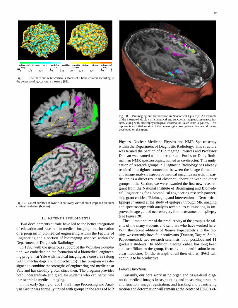
Fig. 20. Bioimaging and Intervention in Neocortical Epilepsy: An exampleof the integrated display of anatomical and functional magnetic resonance im-ages, along with electrophysiological information taken from a patient. Thisrepresents an initial version of the neurosurgical navigational framework beingdeveloped on this grant.
Physics, Nuclear Medicine Physics and NMR Spectroscopywithin the Department of Diagnostic Radiology. This structurewas termed the Section of Bioimaging Sciences and ProfessorDuncan was named as the director and Professor Doug Roth-man, an NMR spectroscopist, named as co-director. This unifi-cation of research groups in Diagnostic Radiology has alreadyresulted in a tighter connection between the image formationand image analysis aspects of medical imaging research. In par-ticular, as a direct result of closer collaboration with the othergroups in the Section, we were awarded the first new researchgrant from the National Institute of Bioimaging and Biomedi-cal Engineering for a biomedical engineering research partner-ship grant entitled “Bioimaging and Intervention in NeocorticalEpilepsy” aimed at the study of epilepsy through MR imagingand spectroscopy with analysis techniques culminating in im-proved image guided neurosurgery for the treatment of epilepsy(see Figure 20).
The ultimate source of the productivity of the group is the tal-ents of the many students and scholars who have worked here.With the recent addition of Xenios Papademetris to the fac-ulty, we currently have four professors (Duncan, Tagare, Staib,Papademetris), two research scientists, four postdocs and 11graduate students. In addition, George Zubal, has long beena close affiliate to the group, focusing on quantification in nu-clear medicine. On the strength of all their efforts, IPAG willcontinue to be productive.
Future Directions
Certainly, our core work using organ and tissue-level diag-nostic medical images in segmenting and measuring structureand function, image registration, and tracking and quantifyingmotion and deformation will remain at the center of IPAG’s ef-

11
forts in the years to come. Continuing to explore how our ap-proaches can be integrated or economized is always somethingat the forefront of our thinking: e.g. how can image intensityand image-derived feature information be combined to developmore robust segmentation and registration algorithms or howcan parameters found at a higher level of abstraction (e.g. strainin an infarcted region of the left ventricle) be used to guide theextraction of useful low level image features?
However, we also expect to be drawn in new and excitingdirections based on our exposure to emerging collaborationswith our colleagues in MR spectroscopy and physics, differentclinical areas related to image-guided intervention and struc-tural and functional imaging at the cellular and molecular level.While we will be able to dovetail some of these efforts withmethodological approaches we are already developing, we ex-pect to be drawn to entirely new directions that include: track-ing multiple nonrigid moving objects with complex evolvingrelationships (e.g. cell body motion and tubule growth), esti-mating statistical mixtures of biochemical information repre-sented at each voxel in a variety of image datasets attemptingto probe biologically meaningful information (e.g. MR spec-troscopy, molecular imaging using fluorescent and/or radiola-beled probes) and designing close-to-real-time updating strate-gies regarding tissue and tool movement in interventional pro-cedures.
IV. GUIDING PRINCIPLES
Within IPAG, we feel fortunate to have been able to cre-ate a stable scientific, financial and administrative environmentwhere medical image analysis researchers interested in apply-ing well-grounded mathematical and computational concepts toproblems in medical imaging can flourish. There have been sev-eral guiding principles that have helped us:
1) Take the time to find and maintain good clinical and tech-nical collaborators who have interesting problems that arepushing the current limits of medical image analysis andprocessing technology. The profile of a good collaboratorcan vary, but generally has these traits:© he/she realizes that you are looking for interesting
problems in your own (medical image analysis) re-search area, related to the methodology and under-lying formulation of these issues – and they want towalk side-by-side with you toward common goals.© is someone who you can genuinely resonate with andget along with.© is someone who has an appreciation and some under-standing of the field of medical image analysis.
We have had longstanding and successful collaborationswith a number of scientists at Yale including RobertSchultz (Child Study Center), Albert Sinusas (Cardiol-ogy), Dennis Spencer (Neurosurgery) and Todd Consta-ble (Diagnostic Radiology).
2) Go after problems that push the current state-of-the-art;avoid problems that apply established/routine ideas incomputer vision to problems in medical image analysisthat don’t require at least some original thinking. How-ever, keep in mind the requirements of medical imageanalysis in terms of the need to validate.
3) Maintain a presence, including publications, conferenceattendance, etc., in both the basic fields of computer vi-sion and image processing as well as the more focusedfield of medical image analysis. The primary medicalimage analysis conferences have been IPMI (Informa-tion Processing in Medical Imaging) and MICCAI (Med-ical Image Computing and Computer Assisted Interven-tion) (and its predecessors). Participation in the clini-cal/biomedical application communities is also important(e.g. cardiology, neurology, neuroscience, etc).
4) Permit junior faculty to develop in an independent man-ner, but provide guidance to try to develop a cohesive andcomplementary group of researchers.
5) Develop a stable internal source of funds from the De-partment/School that carries over year-to-year.
6) Encourage collaboration and cooperation with other med-ical image analysis groups. The intellectual life atIPAG has periodically been enriched by visiting studentsand scholars. We have had particularly good relation-ships with visitors from Guy’s Hospital through DaveHawkes (Glynn Robinson, Colin Studholme), Universityof Amsterdam through Arnold Smeulders (Marcel Wor-ring, Sennay Ghebreab), Utrecht University through MaxViergever (Wiro Niessen, Josien Pluim, Rik Stokking)and INRIA-Sophia Antipolis through Nicholas Ayache(Eric Bardinet).
7) Insure access to graduate students either through partic-ipation in a medical imaging sciences graduate programor through appointments and teaching with an appropri-ate department (e.g. EE, BME, CS, Physics, etc.)
ACKNOWLEDGEMENTS
Special thanks to Hemant Tagare for his help with this docu-ment.
James S. Duncan, Guest EditorImage Processing and Analysis GroupSection of Bioimaging SciencesDepartments of Diagnostic Radiology andElectrical EngineeringYale UniversityNew Haven, CT 06520 USA
Lawrence H. Staib, Guest EditorImage Processing and Analysis GroupSection of Bioimaging SciencesDepartments of Diagnostic Radiology andElectrical EngineeringYale UniversityNew Haven, CT 06520 USA
REFERENCES
[1] D. Gerogiannis and S. C. Orphanoudakis, “Load balancing requirementsin parallel implementations of image feature extraction tasks,” IEEETransactions on Parallel & Distributed Systems, vol. 4, no. 9, pp. 994–1013, Sept. 1993.

12
[2] K. P. Andriole, J. S. Duncan, S. C. Orphanoudakis, and T. Shibata, “De-velopment of a 3-D discontinuity detector and linking algorithm for track-ing vessels in medical images,” in Proc. Twelfth Annual Northeast Bio-engineering Conf., 1986, pp. 137–140.
[3] G. R. Gindi and A. F. Gmitro, “Optical feature extraction via the radontransform,” Optical Engineering, vol. 23, no. 5, pp. 499–506, Sep-Oct1984.
[4] A. F. Gmitro, V. Tresp, and G. R. Gindi, “Videographic tomography –I: Reconstruction with parallel-beam projection data,” IEEE Trans. Med.Imaging, vol. 9, no. 4, pp. 366–375, 1990.
[5] M. Lee, A. Rangarajan, I. G. Zubal, and G. Gindi, “A continuation methodfor emission tomography,” IEEE Trans. Nucl. Sci., vol. 40, no. 6, pp.2049–2058, 1993.
[6] G. Gindi, M. Lee, A. Rangarajan, and I. G. Zubal, “Bayesian reconstruc-tion of functional images using anatomical information as priors,” IEEETrans. Med. Imaging, vol. 12, no. 4, pp. 670–680, 1993.
[7] I. G. Zubal, C. R. Harrell, E. O. Smith, Z. Rattner, G. R. Gindi, and P. B.Hoffer, “Computerized 3-dimensional segmented human anatomy,” Med.Phys., vol. 21, no. 2, pp. 299–302, 1994.
[8] Zubal Phantom: http://ipagwww.med.yale.edu/zubal.[9] G. R. Gindi and J. S. Duncan, “A complete object recognition system as
a computer vision course project,” IEEE Transactions on Education, vol.30, no. 3, pp. 142–150, August 1987.
[10] J. S. Duncan, “Knowledge directed left ventricular boundary detection inequilibrium radionuclide angiocardiography,” IEEE Trans. Med. Imaging,vol. 6, no. 4, pp. 325–336, 1987.
[11] J. Utans and G. R. Gindi, “Improving convergence in hierarchical match-ing networks for object recognition,” in Advances in Neural InformationProcessing 5, S. Hanson, J. Cowan, and L. Giles, Eds. Morgan Kaufmann,San Mateo, CA, 1993.
[12] L. H. Staib, X. Zeng, A. Chakraborty, R. T. Schultz, and J. S. Dun-can, “Shape constraints in deformable models,” in Handbook of MedicalImaging: Processing and Analysis, I. Bankman, Ed. Academic Press, SanDiego, 2000.
[13] A. Chakraborty, L. H. Staib, and J. S. Duncan, “Deformable boundaryfinding in medical images by integrating gradient and region informa-tion,” IEEE Trans. Med. Imaging, vol. 15, no. 6, pp. 859–870, 1996.
[14] A. Chakraborty and J. S. Duncan, “Game theoretic integration for imagesegmentation,” IEEE Trans. Pattern Anal. Machine Intell., vol. 21, no. 1,pp. 12–30, 1999.
[15] L. H. Staib, Parametrically Deformable Contour Models for Image Anal-ysis, Ph.D. thesis, Yale University, New Haven, CT, 1990.
[16] L. H. Staib and J. S. Duncan, “Boundary finding with parametricallydeformable models,” IEEE Trans. Pattern Anal. Machine Intell., vol. 14,no. 11, pp. 1061–1075, 1992.
[17] M. Worring, A. W. M. Smeulders, L. H. Staib, and J. S. Duncan, “Param-eterized feasible boundaries in gradient vector fields,” Comp. Vision andImage Understanding, vol. 63, no. 1, pp. 135–144, Jan. 1996.
[18] L. H. Staib and J. S. Duncan, “Model-based deformable surface findingfor medical images,” IEEE Trans. Med. Imaging, vol. 15, no. 5, pp. 720–731, 1996.
[19] L. H. Staib and J. S. Duncan, “Left ventricular analysis using parametri-cally deformable models,” in AAAI Spring Symposium Series: ArtificialIntelligence in Medicine, Stanford University, Mar. 1988.
[20] L. H. Staib and J. S. Duncan, “Left ventricular analysis from cardiacimages using deformable models,” in Proc. Computers in Cardiology,pp. 427–430. IEEE Comp. Soc., Los Alamitos, CA, 1988.
[21] T. Cootes, A. Hill, C. Taylor, and J. Haslam, “The use of active shapemodels for locating structures in medical images,” in Information Proc.Med. Imaging, H. H. Barrett and A. F. Gmitro, Eds., pp. 33–47. LNCS687, Springer, Berlin, 1993.
[22] Y. Wang and L. H. Staib, “Boundary finding with correspondence usingstatistical shape models,” in Proc. Comp. Vision Pattern Recog., 1998,pp. 338–345.
[23] Y. Wang and L. H. Staib, “Statistical shape and smoothness models forboundary finding with correspondence,” IEEE Trans. Pattern Anal. Ma-chine Intell., vol. 22, no. 7, pp. 738–743, 2000.
[24] H. D. Tagare, “Deformable 2-d template matching using orthogonalcurves,” IEEE Trans. Med. Imaging, vol. 16, no. 1, pp. 108–117, 1997.
[25] Y. Chen, H. D. Tagare, S. Thiruvenkadam, F. Huang, D. Wilson,A. Geiser, K. Gopinath, and R. Briggs, “Using prior shapes in geometricactive contours in a variational framework,” Int. J. Computer Vision, vol.50, no. 3, pp. 315–328, 2002.
[26] T. Ma, Active Contour Models: Consistency, Stability, and ParameterEstimation, Ph.D. thesis, Yale University, 1997.
[27] T. Ma and H. D. Tagare, “Bias and stability of active contours with eu-clidean and non-euclidean arc-lengths,” IEEE Trans. Image Proc., vol. 8,no. 11, pp. 1549–1559, 1999.
[28] L. H. Staib, A. Chakraborty, and J. S. Duncan, “An integrated approachfor locating neuroanatomical structure from MRI,” Int. J. Patt. Recog.Art. Intell., vol. 11, no. 8, pp. 1247–1269, 1997, (Special Issue on MRBrain Image Analysis).
[29] G. Owen, Game Theory, Academic Press, 1982.[30] T. Basar and G. J. Olsder, Dynamic noncooperative game theory, Aca-
demic Press, 1982.[31] H. I. Bozma and J. S. Duncan, “A game-theoretic approach to integration
of modules,” IEEE Trans. Pattern Anal. Machine Intell., vol. 16, pp.1074–1086, 1994.
[32] J. Nash, “Equilibrium points in n-person games,” Proc. Natl. Acad. Sci.USA, vol. 36, pp. 48–49, July 1950.
[33] P. Baxandall and H. Liebeck, Vector Calculus, Oxford University Press,1986.
[34] X. Zeng, L. H. Staib, R. T. Schultz, and J. S. Duncan, “Volumetric layersegmentation using coupled surfaces propagation,” in Computer Visionand Pattern Recognition, 1998, pp. 708–715.
[35] X. Zeng, L. H. Staib, R. T. Schultz, and J. S. Duncan, “Segmentation andmeasurement of the cortex from 3D MR images,” in Medical Image Com-puting and Computer-Assisted Intervention (LNCS 1496), Berlin, 1998,pp. 519–530, Springer.
[36] X. Zeng, L. H. Staib, R. T. Schultz, and J. S. Duncan, “Segmentation andmeasurement of the cortex from 3D MR images using coupled surfacespropagation,” IEEE Trans. Med. Imaging, vol. 18, no. 10, 1999.
[37] D. W. Shattuck and R. M. Leahy, “Brainsuite: An automated corticalsurface identification tool,” in Medical Image Computing and Computer-Assisted Intervention (MICCAI), S. L. Delp, A. M. DiGoia, and B. Jara-maz, Eds., Berlin, 2000, vol. Lecture Notes in Computer Science, Vol.1935, pp. 51–61, Springer-Verlag.
[38] X. Papademetris and J. S. Duncan, “Cardiac image analysis: Motionand deformation,” in Handbook of Medical Imaging, Volume 2: MedicalImage Processing and Analysis, M. Sonka and J. M. Fitzpatrick, Eds.SPIE Press, 2000.
[39] G. E. Mailloux, A. Bleau, M. Bertrand, and R. Petitclerc, “Computeranalysis of heart motion from two-dimensional echocardiograms,” IEEETrans. Biomed. Eng., vol. 34, pp. 356–364, 1987.
[40] J. S. Duncan, R. L. Owen, L. H. Staib, and P. Anandan, “Measurementof non-rigid motion in images using contour shape descriptors,” in Proc.Comp. Vision Pattern Recog., June 1991, pp. 318–324.
[41] D. Metaxas and D. Terzopoulos, “Constrained deformable superquadricsand nonrigid motion tracking,” in Computer Vision and Pattern Recogni-tion, G. Medioni and B. Horn, Eds. 1991, pp. 337–343, IEEE Press.
[42] A. Pentland and S. Sclaroff, “Closed-form solutions for physically basedshape modeling and recognition,” IEEE Trans. Pattern Anal. MachineIntell., vol. 13, no. 7, pp. 715–729, July 1991.
[43] J. Park, D. Metaxas, and L. Axel, “Analysis of left ventricular wall motionbased on volumetric deformable models and MRI-SPAMM,” MedicalImage Analysis, vol. 1, no. 1, pp. 53–71, 1996.
[44] S. N. Gupta and J. L. Prince, “On variable brightness optical flow fortagged MRI,” in Information Processing in Medical Imaging, June 1995,pp. 323–334.
[45] N. Pelc, M. Drangova, L. Pelc, Y. Zhu, D. Noll, B. Bowman, and R. Her-fkens, “Tracking of cyclic motion with phase-contrast cine mr velocitydata,” Journal of Magnetic Resonance Imaging, vol. 5, pp. 339–345,1995.
[46] J. McEachen and J.S. Duncan, “Shape-based tracking of left ventricularwall motion,” IEEE Trans. Med. Imaging, vol. 16, no. 3, pp. 270–283,June 1997.
[47] F. Meyer, R. T. Constable, A. Sinusas, and J. Duncan, “Tracking my-ocardial deformation using phase contrast velocity fields: A stochasticapproach,” IEEE Transactions on Medical Imaging, vol. 15, no. 4, pp.453–465, 1996.
[48] F. H. Sheehan, E. L. Bolson, H. T. Dodge, D. G. Mathey, J. Schofer, andH. W. Woo, “Advantages and applications of the centerline method forcharacterizing regional ventricular function,” Circulation, vol. 74, pp.293–305, 1986.
[49] E. Zerhouni, D. Parish, W. Rogers, A. Yang, and E. Shapiro, “Humanheart tagging with MR imaging-a method for noninvasive assessment ofmyocardial motion,” Radiology, vol. 169, pp. 59–63, 1988.
[50] L. Axel and L. Dougherty, “MR imaging of motion with spatial modula-tion of magnetization,” Radiology, vol. 171, pp. 841–845, 1989.
[51] A. Amini, R. L. Owen, P. Anandan, and J. S. Duncan, “Non-rigid mo-tion models for tracking the left ventricular wall,” in Information Pro-cessing in Medical Imaging, Alan C. F. Colchester and David J. Hawkes,Eds. 1991, vol. 511 of Lecture Notes in Computer Science, pp. 343–357,Springer.
[52] P. Shi, Image Analysis of 3D Cardiac Motion Using Physical and Geo-metrical Models, Ph.D. thesis, Yale University, May 1996.

13
[53] X. Papademetris, Estimation of 3D Left Ventricular Deformation fromMedical Images Using Biomechanical Models., Ph.D. thesis, Yale Uni-versity, May 2000.
[54] P. Shi, G. Robinson, A. Chakraborty, L. Staib, R. Constable, A. Sinusas,and J. Duncan, “A unified framework to assess myocardial functionfrom 4D images,” in Comp. Vision, Virtual Reality and Robotics in Med.(CVRMed ’95, LNCS 905), N. Ayache, Ed., Berlin, 1995, pp. 327–340,Springer.
[55] P. J. Hunter, A. McCulloch, and P. Nielsen, Eds., Theory of Heart,Springer-Verlag, Berlin, 1991.
[56] P. Shi, A.J. Sinusas, R.T. Constable, and J.S. Duncan, “Volumetric de-formation analysis using mechanics-based data fusion: Applications incardiac motion recovery,” Int. J. Computer Vision, vol. 35, no. 1, pp.87–107, Nov. 1999.
[57] P. Shi, A. Sinusas, R. Constable, E. Ritman, and J. Duncan, “Point-tracked quantitative analysis of left ventricular motion from 3D imagesequences,” IEEE Trans. Med. Imaging, vol. 19, no. 1, pp. 36–50, 2000.
[58] X. Papademetris, P. Shi, D. P. Dione, A. J. Sinusas, and J. S. Duncan,“Recovery of soft tissue object deformation using biomechanical mod-els,” in Information Proc. Med. Imaging, M. Samal A. Kuba and A. Todd-Pokropek, Eds., Berlin, 1999, pp. 352–357, Springer-Verlag.
[59] X. Papademetris, A. J. Sinusas, D. P. Dione, R. T. Constable, and J. S.Duncan, “Estimation of 3D left ventricular deformation from medicalimages using biomechanical models,” IEEE Trans. Med. Imaging, vol.21, no. 7, pp. 786–800, July 2002.
[60] A. J. Sinusas, X. Papademetris, R. T. Constable, D. P. Dione, M. D. Slade,P. Shi, and J. S. Duncan., “Quantification of 3-d regional myocardial de-formation: Shape-based analysis of magnetic resonance images,” Amer-ican Journal of Physiology: Heart and Circulatory Physiology, vol. 281,pp. H698–H714, August 2001.
[61] X. Papademetris, A. J. Sinusas, D. P. Dione, and J. S. Duncan, “Esti-mation of 3D left ventricular deformation from echocardiography,” Med.Image Anal., vol. 5, no. 1, pp. 17–28, 2001.
[62] O. Skrinjar, D. Spencer, and J. Duncan, “Brain shift modeling for usein neurosurgery,” in Medical Image Computing and Computer-AssistedIntervention (LNCS 1496), W. M. Wells, A. Colchester, and S. Delp, Eds.,Berlin, 1998, pp. 641–649, Springer.
[63] O. Skrinjar, A. Nabavi, and J. S. Duncan, “Model-driven brain shift com-pensation,” Medical Image Analysis, vol. 6, no. 4, pp. 361–373, Decem-ber 2002.
[64] O. Skrinjar and J. Duncan, “Real time 3D brain shift compensation,”in Information Proc. Med. Imaging, M. Samal A. Kuba and A. Todd-Pokropek, Eds., Berlin, 1999, pp. 42–55, LNCS 1613, Springer.
[65] O. Skrinjar, H. Tagare, and J.S. Duncan, “Surface growing from stereoimages,” in Proc. Comp. Vision Pattern Recog., June 2000, pp. II:571–576.
[66] G. Zubal, H. Tagare, L. Zhang, and J. Duncan, “3D registration of inter-modality medical images,” in IEEE Conf. Eng. Med. Biology, 1991, pp.293–294.
[67] L. H. Staib and X. Lei, “Intermodality 3D medical image registrationwith global search,” in Proc. IEEE Workshop Biomedical Image Anal.,1994, pp. 225–234.
[68] Y. Wang and L. H. Staib, “Physical model based non-rigid registrationincorporating statistical shape information,” Med. Image Anal., vol. 4,pp. 7–20, 2000.
[69] C. Studholme, R.T. Constable, and J.S. Duncan, “Non-rigid spin echoMRI registration incorporating an image distortion model: Applicationto accurate alignment of fMRI to conventional MRI,” IEEE Trans. Med.Imaging, vol. 19, no. 11, pp. 1115–1127, November 2000.
[70] A. Rangarajan, H. Chui, E. Mjolsness, S. Pappu, L. Davachi, P. Goldman-Rakic, and J. Duncan, “A robust point-matching algorithm for autoradio-graph alignment,” Med. Image Anal., pp. 379–398, 1997.
[71] H. Chui, J. Rambo, J. S. Duncan, R. T. Schultz, and A. Rangarajan, “Reg-istration of cortical anatomical structures via robust 3D point matching,”in Information Proc. Med. Imaging, A. Kuba, M. Samal, and A. Todd-Pokropek, Eds., Berlin, 1999, pp. 168–181, LNCS 1613, Springer.
[72] H. Tagare, “Shape-based nonrigid correspondence with applications toheart motion analysis,” IEEE Trans. Med. Imaging, vol. 18, no. 7, pp.570–579, 1999.
[73] H. D. Tagare, D. O’Shea, and D. Groisser, “Shape based non-rigid corre-spondence for plane curves,” J. Math. Imaging Vision, vol. 16, pp. 57–68,2002.
[74] R. Bansal, L. Staib, Z. Chen, A. Rangarajan, J. Knisely, R. Nath, andJ. Duncan, “A minimax entropy registration framework for patient setupverification in radiotherapy,” Computer Aided Surgery, vol. 4, no. 6, pp.287–304, 1999.
[75] R. Bansal, L. Staib, Z. Chen, A. Rangarajan, J. Knisely, R. Nath, andJ. Duncan, “Entropy-based, dual-portal-to-3DCT registration incorporat-
ing pixel correlation,” IEEE Trans. Med. Imaging, vol. 22, no. 1, pp.29–49, 2003.
[76] Y. Wang, B. S. Peterson, and L. H. Staib, “Shape-based 3D surface cor-respondence using geodesics and local geometry,” in Proc. Comp. VisionPattern Recog., 2000, vol. II, pp. 644–651.
[77] Y. Wang, B. S. Peterson, and L. H. Staib, “3D brain surface matchingbased on geodesics and local geometry,” Comp. Vision and Image Under-standing, vol. 89, no. 2-3, pp. 252–271, Jan. 2003.
[78] X. Zeng, L. H. Staib, R. T. Schultz, H. Tagare, L. Win, and J. S. Duncan,“A new approach to 3D sulcal ribbon finding from MR images,” in Medi-cal Image Computing and Computer-Assisted Intervention (LNCS 1679),Berlin, 1999, pp. 148–157, Springer.
[79] R. T. Schultz, N. K. Cho, L. H. Staib, L. E. Kier, J. M. Fletcher, S. E.Shaywitz, D. P. Shankweiler, J. C. Gore, J. S. Duncan, and B. A. Shay-witz, “Brain morphology in normal and dyslexic children: The influenceof sex and age,” Ann. Neurol., vol. 35, no. 6, pp. 732–742, June 1994.
[80] B. S. Peterson, B. Vohr, L. H. Staib, C. Cannistraci, A. Dolberg, K. A.Schneider, K. H. Katz, M. Westerveld, S. Sparrow, A. Anderson, C. Dun-can, R. W. Makuch, J. C. Gore, and L. R. Ment, “Regional brain volumeabnormalities are associated with long-term cognitive outcome in preterminfants,” J. Am. Med. Assoc., vol. 284, pp. 1939–1947, 2000.
[81] B. S. Peterson, P. A. Feineigle, L. H. Staib, and J. C. Gore, “Automatedmeasurement of latent morphological features in the human corpus callo-sum,” Human Brain Mapping, vol. 12, no. 4, pp. 232–245, 2001.
[82] H. D. Tagare, C. C. Jaffe, and J. Duncan, “Medical image databases: Acontent-based retrieval approach,” J. Am. Med. Informatics Assoc., vol. 4,no. 3, pp. 184–198, 1997.
[83] H. D. Tagare, “Efficient retrieval without a metric,” in IEEE Visual ’97,San Diego, 1997.
[84] H. D. Tagare, “Efficient retrieval in image databases,” in SPIE Conf., SanDiego, July 2000.
[85] G. Robinson, H. Tagare, J. Duncan, and C. Jaffe, “Medical image col-lection indexing: Shape-based retrieval using kd trees,” ComputerizedMedical Imaging and Graphics, vol. 20, no. 4, pp. 209–217, 1996.
[86] H. D. Tagare, “Increasing retrieval efficiency by index tree adaptation,” inIEEE Workshop on Content-based Access of Image and Video Libraries,1997.
[87] X. Qian and H. D. Tagare, “Optimally adapted indexing trees for medicalimage databases,” in ISBI, Washington D.C., July 2002.
[88] S. Ghebreab, M. Worring, H. D. Tagare, and C. C. Jaffe, “SCHEMed: avisual tool for definition and data entry in medical image databases,” inIEEE Visual ’97, San Diego, 1997.
[89] H. Tagare, F. Vos, C. C. Jaffe, and J. S. Duncan, “Arrangement: A spatialrelation between parts for evaluating similarity of tomographic section,”IEEE Trans. Pattern Anal. Machine Intell., vol. 17, no. 9, pp. 880–893,1995.
[90] G. R. Hillman, H. D. Tagare, K. Elder, A. Drobyshevski, D. Weller, andB. Wursig, “Shape descriptors computed from photographs of dolphindorsal fins for use as database indices,” in Proc. IEEE Eng. MedicineBiology Society, Nov. 1998.
[91] G. R. Hillman, B. Wursig, N. Kehtarnavaz, D. W. Weller, A. Kreho,A. Drobyshevski, K. Elder, B. N. Araabi, T. McKinney, G. Gailey, andH. D. Tagare, “Individual identification of dolphins in field photographs:a computer-based system,” in The Society for Marine Mammalogy, 13thBiennial Conference on the Biology of Marine Mammals, 1999.
[92] J. J. Koenderink and A. J. van Doorn, “Surface shape and curvaturescales,” Image and Vision Computing, vol. 10, no. 8, pp. 557–565, Oct.1992.