Embed Size (px)
Citation preview

1/9/2013
1
image interpretation what to look for
ERNEST LAM, DMD, MSc, PhD, FRCD(C)
Professor and the Dr. Lloyd & Mrs. Kay Chapman Chair in Clinical SciencesGraduate Program Director and Head,Discipline of Oral and Maxillofacial RadiologyFaculty of Dentistry, the University of Toronto
▫ interpretation vs. diagnosis
?
normal abnormal
developmentalabnormality
acquiredabnormality
cystinflammation
benign neoplasia
bone dysplasiamalignant neoplasia
vascular anomalymetabolic disease
trauma

1/9/2013
2
▫ “normal” vs. abnormal
this is the most elementary step in radiographic interpretation; identifying what is normal, a normal variant or the range of normal.
first and foremost, it requires a knowledge and familiarity with normal radiographic anatomy (presumably, you learned this in dental school).
and second, it requires an appreciation for the wide range of normal appearances of anatomical structures.

1/9/2013
3
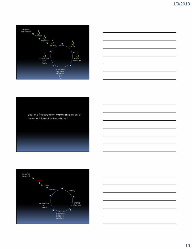
radiographic feature identification
vision cognition
interpretation
▫ the osseous dysplasias are non-neoplastic processes usually confined to the tooth-bearing areas of the jaws or edentulous alveolar processes.
▫ regrettably, osseous dysplasias are sometimes mismanaged.
1/9/2013
4

1/9/2013
5
▫ what radiographic features support a correct interpretation of osseous dysplasia?
reviewer groups
3 general dentists
3 oral
radiologists
▫ 37 osseous dysplasia cases and 13 additional image sets consisting of other similarly-appearing entities:
▫ cementoblastoma▫ ossifying fibroma▫ complex odontoma▫ dense bone island▫ fibrous dysplasia
alsufyani & lam, dmfr 2011

1/9/2013
6
interpretationgeneraldentists
oral radiologists Pa
<0.001correct 38.7% 79.3%incorrect 61.3% 20.7%
a chi-square value = 37.711 df = 1
alsufyani & lam, dmfr 2011
radiographic feature adjusted OR 95.0% CI for exp (β) p
lower upperno root resorption 4.52 1.18 17.30 0.03ant. & post. teeth 3.22 1.42 7.52 0.01
constant 3.45 0.05
logistic regression model for general dentists
alsufyani & lam, dmfr 2011
radiographic feature adjusted OR 95.0% CI for exp (β ) p
lower upperwell-defined periphery 6.67 1.50 28.57 0.01
bilateral 10.23 2.00 52.56 0.01mixed
radiolucent/radiopaque 10.53 2.06 52.63 0.01
no cortical expansion 7.63 1.46 40.00 0.02anterior & posterior teeth 4.34 1.11 17.54 0.04
internal radiolucent periphery 8.28 2.14 32.56 <0.001
constant 14.81 0.01
logistic regression model for oral and maxillofacial radiologists
alsufyani & lam, dmfr 2011

1/9/2013
7
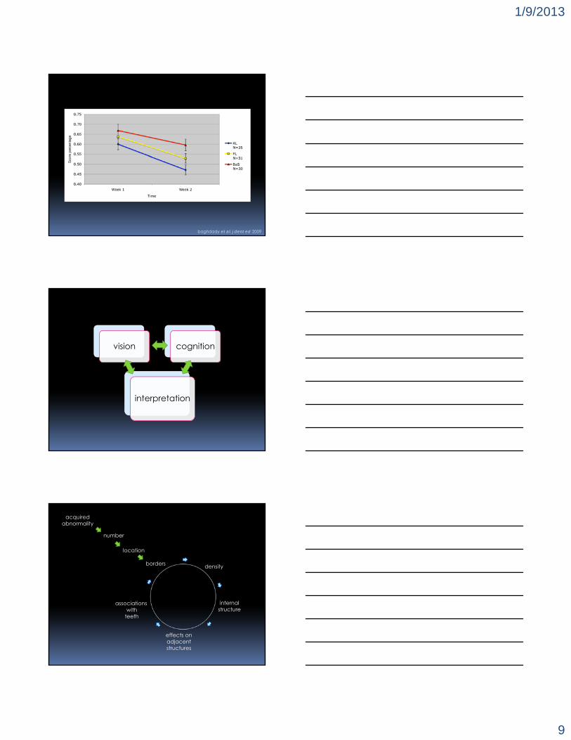
▫ while the identification of key radiographic features may be important in image interpretation, does an understanding of how these features arise enhance the interpretive skills of the novice in oral and maxillofacial radiology?
▫ 96 undergraduate dentistry and dental hygiene students, randomly divided into 3 learning conditions.▫ feature list (unstructured algorithm)▫ structured algorithm▫ basic science
baghdady et al, j dent ed 2009
b dca
a: periapical osseous dysplasiab: complex odontomec: dense bone islandd: sclerosing osteitis
baghdady et al, j dent ed 2009

1/9/2013
8
feature list
Odontomas are the most common odontogenic tumors in the jaws. They often interfere with the eruption of permanent teeth. The lesion has no gender predilection, and most begin forming while the normal dentition is developing. Most odontomas occur in the second decade of life and are found during investigation of delayed eruption of adjacent teeth. 70% of complex odontomas occur in the first and second mandibular molar area. Radiographically, they appear as well-defined corticated lesions with a fibrous capsule internally. The content of these lesions are mixed radiolucent and radiopaque but are largely radiopaque. Complex odontomas contain an irregular mass of calcified tissue. The degree of radiopacity is equivalent to adjacent tooth structure. Odontomas interfere with normal eruption of teeth. 70% of odontomas are associated with abnormalities such as impaction, malpositioning of adjacent teeth, diastema, and devitalization of adjacent teeth.
baghdady et al, j dent ed 2009
structured algorithm
Complex Odontomas are the most common odontogenic tumoursin the jaws.
Location: 70% of complex odontomas occur in the mandibularfirst and second molar region.
Periphery: Odontomas are well defined and have a corticated boarder. Immediately inside and adjacent to the cortical border is a soft tissue capsule appearing as a smooth radiolucent space.
Internal Structure: Complex odontomas contain an irregular mass of calcified tissue. The degree of radiopacity is equivalent to adjacent tooth structure.
Effect on surrounding structures: Odontomas interfere with normal eruption of teeth. 70% of odontomas are associated with abnormalities such as impaction, malpositioning of adjacent teeth, diastema, and devitalization of adjacent teeth.
baghdady et al, j dent ed 2009
basic science
Odontomas are benign tumours that originate from remnants of the dental lamina in the jaws. The histological appearance is characterized by the production of mature enamel, dentin, cementum, and pulp tissue. In complex odontomas the tumor forms nondescript masses of dental tissue. This is manifested radiographically as an irregular radiopaque mass. The degree of radiopacity is equivalent to adjacent tooth structure.
Radiographically, odontomas are well defined with a corticated border, which represents reactive bone. Corticated borders are typically seen in slow growing lesions (i.e. cysts and benign slow growing tumours). Immediately inside and adjacent to the cortical border there is a smooth uniform radiolucent space, which represents the soft tissue fibrous capsule, surrounding the tumor.
Odontomas develop and mature while the related teeth are forming and cease development when the associated teeth complete development. Because of the slow and space-occupying nature of the growth of this tumour, frequently it displaces nearby teeth and obstructs the normal eruption of adjacent teeth.
baghdady et al, j dent ed 2009
1/9/2013
9
baghdady et al, j dent ed 2009
vision cognition
interpretation
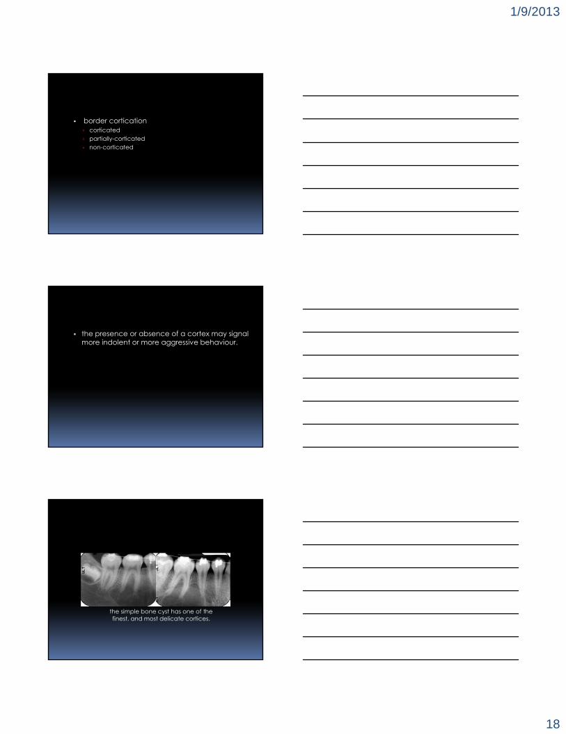
internalstructure
associations with
teeth
effects onadjacentstructures
borders density
number
acquiredabnormality
location
1/9/2013
10
internalstructure
associations with
teeth
effects onadjacentstructures
borders density
number
acquiredabnormality
location
i
i
i
i
i i
i
▫ does the interpretation make sense in light of the other information I may have ?
internalstructure
associations with
teeth
effects onadjacentstructures
borders density
number
acquiredabnormality
location

1/9/2013
11
▫ how many abnormalities do you see? the presence of multiple abnormalities may suggest
underlying systemic involvement.
multiple supernumerary teeth in cleidocranial dysplasia.
multiple cyst-like lesions in nevoid basal cell carcinoma syndrome.
**

1/9/2013
12
multiple well-defined, “punched out” radiolucent areas in multiple myeloma.
internalstructure
associations with
teeth
effects onadjacentstructures
borders density
number
acquiredabnormality
location
▫ where do you see it (or them)? in some instances, pin-pointing a lesion’s growth centre
may enable you to determine its biological origin; whether it is odontogenic or non-odontogenic.

1/9/2013
13
this ameloblastic fibro-odontome arising coronal to the teeth is of odontogenic origin.
the area of rarefying osteitis located at the apex of 1.2 is also of odontogenic origin.

1/9/2013
14
rarefying osteitis at the apex of 1.4 with an unfilled palatal root canal.
but this midline incisive canal cyst is not.
this STAFNE defect is also not odontogenic in origin.

1/9/2013
15
internalstructure
associations with
teeth
effects onadjacentstructures
borders density
number
acquiredabnormality
location
▫ how does the abnormality border what is normal?
border definition well-defined moderately well-defined “punched-out” poorly-defined

1/9/2013
16
how readily can you draw a line around the periphery of the abnormality on a radiograph?
benign, slow-growing lesions, such as this incisive canal cyst typically display a well-defined border.
the same may be said of this dentigerous cyst border.

1/9/2013
17
some malignant lesions such as multiple myeloma may display moderately well-defined, or“punched-out” borders.
in general, malignant lesions have very poorly-defined borders.
lesions that have become secondarily-infectedmay also have poorly-defined borders.
1/9/2013
18
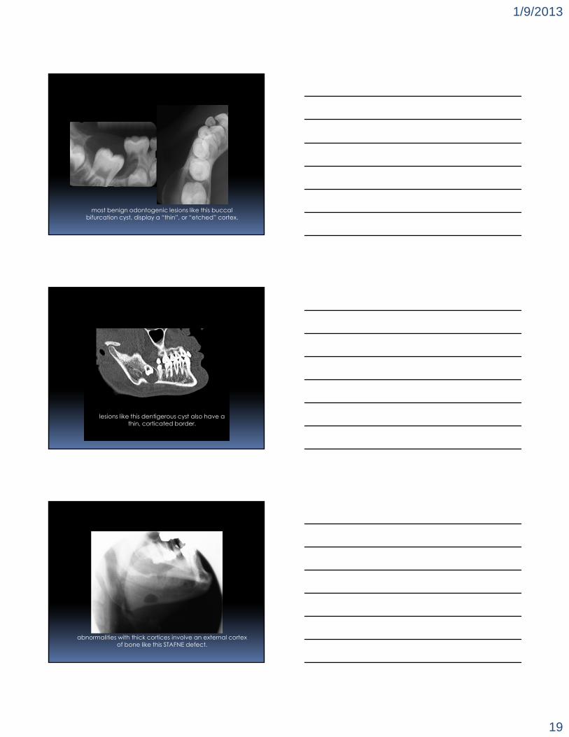
border cortication corticated partially-corticated non-corticated
the presence or absence of a cortex may signal more indolent or more aggressive behaviour.
the simple bone cyst has one of the finest, and most delicate cortices.
1/9/2013
19
most benign odontogenic lesions like this buccalbifurcation cyst, display a “thin”, or “etched” cortex.
lesions like this dentigerous cyst also have athin, corticated border.
abnormalities with thick cortices involve an external cortex of bone like this STAFNE defect.

1/9/2013
20
▫ new bone formation at the bone border periosteal new bone formation
lamellar sun-burst hair-on-end
the “onion skin” effect may arise in response to inflammation.

1/9/2013
21
and osteosarcoma.

1/9/2013
22
metastatic lesions from prostate or breast cancer.
a more “tightly-packed”, hair-on-end appearance to be seen with blood dyscrasias.
internalstructure
associations with
teeth
effects onadjacentstructures
borders density
number
acquiredabnormality
location
1/9/2013
23
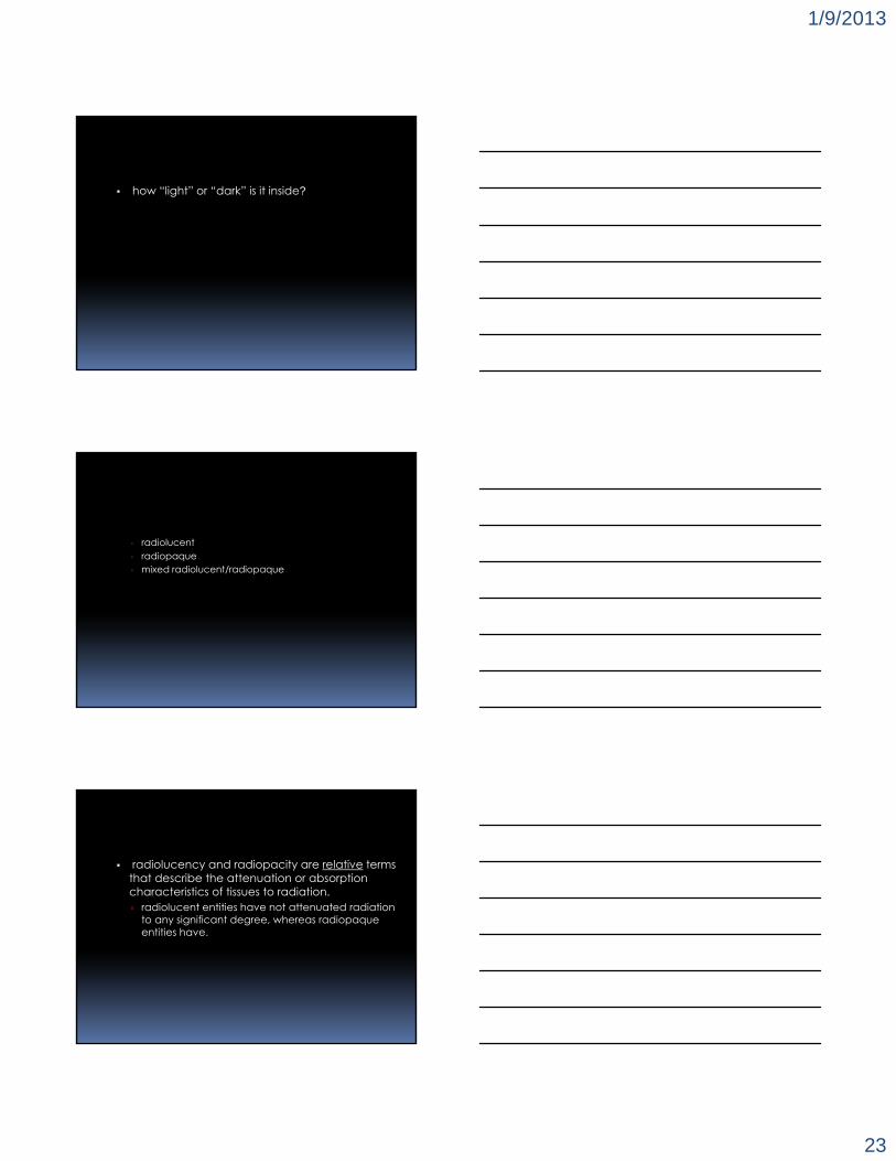
how “light” or “dark” is it inside?
▫ radiolucent▫ radiopaque▫ mixed radiolucent/radiopaque
radiolucency and radiopacity are relative terms that describe the attenuation or absorption characteristics of tissues to radiation. radiolucent entities have not attenuated radiation
to any significant degree, whereas radiopaque entities have.
1/9/2013
24
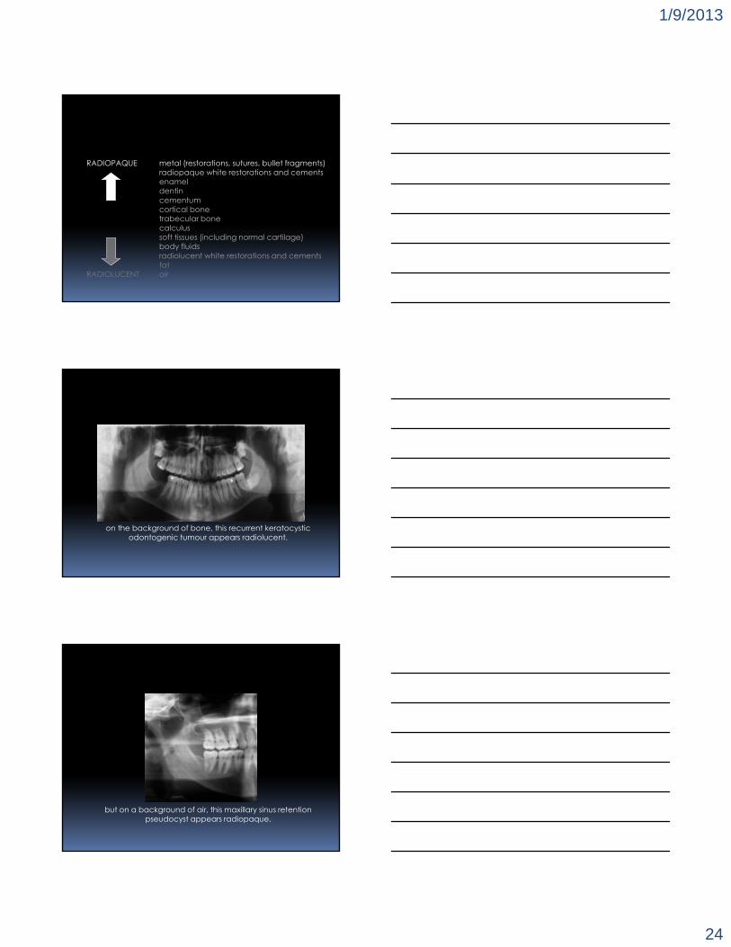
RADIOPAQUE metal (restorations, sutures, bullet fragments)radiopaque white restorations and cementsenameldentincementumcortical bonetrabecular bonecalculussoft tissues (including normal cartilage)body fluidsradiolucent white restorations and cementsfat
RADIOLUCENT air
on the background of bone, this recurrent keratocysticodontogenic tumour appears radiolucent.
but on a background of air, this maxillary sinus retention pseudocyst appears radiopaque.

1/9/2013
25
this dense bone island is also radiopaque.
“mixed” radiolucent/radiopaque lesions are those that show multiple intra-lesional attenuation characteristics. cells within these lesions have the capacity to
produce a mineralized matrix of bone, bone-like, tooth, or tooth-like material, either peripherally or centrally.
the radiopaque areas in florid cemento-osseous dysplasia have a “globular” appearance.

1/9/2013
26
courtesy, DR. M. DAGENAIS
mineralized osteoid matrix deposition in osteosarcoma.
courtesy, DR. M. DAGENAIS

1/9/2013
27
small tooth-like masses may be seen in compound odontomes.
internalstructure
associations with
teeth
effects onadjacentstructures
borders density
number
acquiredabnormality
location
is there anything inside?
1/9/2013
28
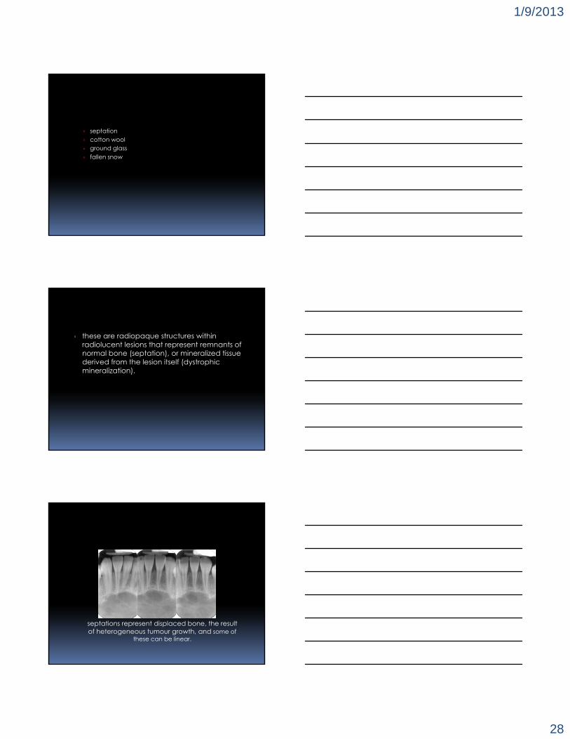
septation cotton wool ground glass fallen snow
▫ these are radiopaque structures within radiolucent lesions that represent remnants of normal bone (septation), or mineralized tissue derived from the lesion itself (dystrophic mineralization).
septations represent displaced bone, the result of heterogeneous tumour growth, and some of
these can be linear.

1/9/2013
29
and some can be curvilinear as in this ameloblastoma.
the so-called cloud, or “cotton wool” appearance is a feature of PAGET’s disease of bone.

1/9/2013
30
the “ground glass”, or “orange peel” pattern may be seen in hyperparathyroidism (bilaterally), and
here, in fibrous dysplasia, unilaterally.
dystrophic tumour calcifications that have the appearance of “fallen snow” may be associated
with the adenomatoid odontogenic tumour.

1/9/2013
31
internalstructure
associations with
teeth
effects onadjacentstructures
borders density
number
acquiredabnormality
location
▫ is anything being pushed around?
other effects of benign lesions when they arise in the maxillae, may include the displacement of adjacent normal anatomical
structures, such as the maxillary sinus floor.

1/9/2013
32
tooth displacement in nevoid basal cell carcinoma syndrome.
extreme tooth displacement in cherubism.
1/9/2013
33
internalstructure
associations with
teeth
effects onadjacentstructures
borders density
number
acquiredabnormality
location
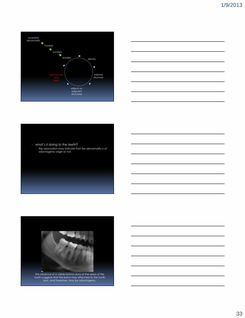
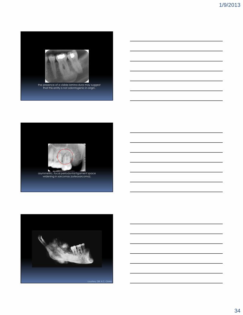
▫ what’s it doing to the teeth? the association may indicate that the abnormality is of
odontogenic origin or not.
the absence of a visible lamina dura at the apex of the tooth suggests that this lesion may attached to the tooth
root, and therefore, may be odontogenic.
1/9/2013
34
the presence of a visible lamina dura may suggest that this entity is not odontogenic in origin.
asymmetric, focal periodontal ligament spacewidening in sarcomas (osteosarcoma).
courtesy, DR. K.C. CHAN

1/9/2013
35
courtesy, DR. K.C. CHAN
tg
b*
p
courtesy, DR. K.C. CHAN
*
▫ MARIAM BAGHDADY, BDS, MSc, FRCD(C)
▫ NICOLE WOODS, PhD
WILSON CENTRE FOR RESEARCH IN EDUCATION
▫ NOURA ALSUFYANI, BDS, MSc, FRCD(C)
▫ BERTHA ROSENSTADT FUND, FACULTY OF DENTISTRY▫ CONNAUGHT FUND, UNIVERSITY OF TORONTO
acknowledgements