Embed Size (px)
Citation preview

The Lichenologisthttp://journals.cambridge.org/LIC
Additional services for The Lichenologist:
Email alerts: Click hereSubscriptions: Click hereCommercial reprints: Click hereTerms of use : Click here
Image analysis for measuring lichen colonization on and within stonework
Claudia GAZZANO, Sergio E. FAVEROLONGO, Enrica MATTEUCCI and Rosanna PIERVITTORI
The Lichenologist / Volume 41 / Issue 03 / May 2009, pp 299 313DOI: 10.1017/S0024282909008366, Published online: 26 May 2009
Link to this article: http://journals.cambridge.org/abstract_S0024282909008366
How to cite this article:Claudia GAZZANO, Sergio E. FAVEROLONGO, Enrica MATTEUCCI and Rosanna PIERVITTORI (2009). Image analysis for measuring lichen colonization on and within stonework. The Lichenologist, 41, pp 299313 doi:10.1017/S0024282909008366
Request Permissions : Click here
Downloaded from http://journals.cambridge.org/LIC, IP address: 171.67.34.69 on 11 Oct 2012

Image analysis for measuring lichen colonization on andwithin stonework
Claudia GAZZANO, Sergio E. FAVERO-LONGO,Enrica MATTEUCCI and Rosanna PIERVITTORI
Abstract: The suitability of image analysis by colour-based pixel classification to quantify lichencolonization on the surface of and within marble, travertine and mortar stonework has been investi-gated. High resolution images of lichenized stonework surfaces were acquired at different field sitesusing a scanner, thus avoiding invasive surveys, and the percentage cover of lichen species wassubsequently measured in the laboratory using dedicated software. Furthermore, microphotographs ofpolished cross-sections of lichenized marble, travertine and mortar, stained using the periodicacid-Schiff (PAS) method to visualize hyphae, were produced by the same software to quantify hyphalspread within the substratum, a parameter which can be used more successfully than the commonlyused depth of hyphal penetration to quantify how much the lichen has affected the conservation of astone substratum. Significant statistical differences in hue, saturation and intensity (HSI) of the lichenthalli and PAS-stained hyphae, with respect to the lithic substrata, allowed the software to discriminateand quantify the lichen species cover on, and hyphal spread within, the three investigated lithotypes.Since such a quantitative approach highlights the volume of influence of lichens on stonework, wherebioweathering processes are likely to develop, it could be used to support decisions on the preservationof our stone cultural heritage.
Key words: biodeterioration, colour model HSI, hyphal penetration component, lichen cover
Introduction
Lichen colonization of stonework has beenextensively investigated (Piervittori et al.1994, 1996, 1998, 2004a, 2004b). Variouseffects, ranging from biodeterioration to bio-protection have been reported, depending onthe weathering ability of the species and onthe physico-chemical features of the litho-type (Adamo & Violante 2000; Chen et al.2000; St. Clair & Seaward 2004). Aesthetic,chemical and/or physical damage by a par-ticular species depends on its morphologyand physiology (Ascaso et al. 2004; de losRìos & Ascaso 2005), but its impact on theconservation of a monument depends on thequantity of the substratum that is colonized
by that species. It is worth noting that lichenscolonize both the surface of rocks, wherethe thalline component (TC, sensu Favero-Longo et al. 2005) influences aesthetics(Nimis et al. 1998; Seaward 2004), and theirinterior, where the hyphal penetration com-ponent (HPC, sensu Favero-Longo et al.2005) induces chemical and physical pro-cesses (Ascaso et al. 2004; de los Rìos &Ascaso 2005).
Analysis of the cover of lichen species inarchaeological, urban and cemetery areascurrently requires time-consuming specialistwork in the field: an expert eye has to exam-ine one relevé after another, recognizespecies and estimate their cover, while at thesame time limiting or avoiding samplingoperations for conservation reasons (e.g.Nimis et al. 1987). On the other hand, analy-sis of the penetrating hyphae, which cur-rently combines microscopy and stainingmethods in the laboratory, mainly focuseson their maximum or average depth of
C. Gazzano, S. E. Favero-Longo, E. Matteucci and R.Piervittori (corresponding author): Department of PlantBiology and Centre of Excellence for Plant and Micro-bial Biosensing (CEBIOVEM), University of Torino,Viale Mattioli 25, 10125 Torino, Italy.Email: [email protected]
The Lichenologist 41(3): 299–313 (2009) © 2009 British Lichen Societydoi:10.1017/S0024282909008366 Printed in the United Kingdom
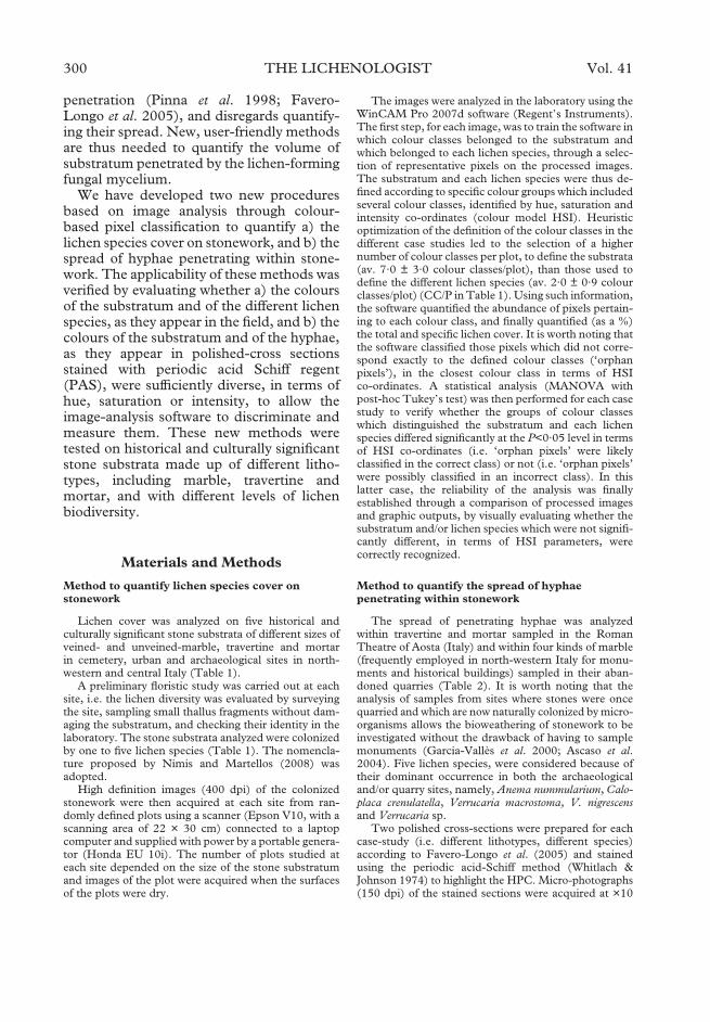
penetration (Pinna et al. 1998; Favero-Longo et al. 2005), and disregards quantify-ing their spread. New, user-friendly methodsare thus needed to quantify the volume ofsubstratum penetrated by the lichen-formingfungal mycelium.
We have developed two new proceduresbased on image analysis through colour-based pixel classification to quantify a) thelichen species cover on stonework, and b) thespread of hyphae penetrating within stone-work. The applicability of these methods wasverified by evaluating whether a) the coloursof the substratum and of the different lichenspecies, as they appear in the field, and b) thecolours of the substratum and of the hyphae,as they appear in polished-cross sectionsstained with periodic acid Schiff regent(PAS), were sufficiently diverse, in terms ofhue, saturation or intensity, to allow theimage-analysis software to discriminate andmeasure them. These new methods weretested on historical and culturally significantstone substrata made up of different litho-types, including marble, travertine andmortar, and with different levels of lichenbiodiversity.
Materials and Methods
Method to quantify lichen species cover onstonework
Lichen cover was analyzed on five historical andculturally significant stone substrata of different sizes ofveined- and unveined-marble, travertine and mortarin cemetery, urban and archaeological sites in north-western and central Italy (Table 1).
A preliminary floristic study was carried out at eachsite, i.e. the lichen diversity was evaluated by surveyingthe site, sampling small thallus fragments without dam-aging the substratum, and checking their identity in thelaboratory. The stone substrata analyzed were colonizedby one to five lichen species (Table 1). The nomencla-ture proposed by Nimis and Martellos (2008) wasadopted.
High definition images (400 dpi) of the colonizedstonework were then acquired at each site from ran-domly defined plots using a scanner (Epson V10, with ascanning area of 22 × 30 cm) connected to a laptopcomputer and supplied with power by a portable genera-tor (Honda EU 10i). The number of plots studied ateach site depended on the size of the stone substratumand images of the plot were acquired when the surfacesof the plots were dry.
The images were analyzed in the laboratory using theWinCAM Pro 2007d software (Regent’s Instruments).The first step, for each image, was to train the software inwhich colour classes belonged to the substratum andwhich belonged to each lichen species, through a selec-tion of representative pixels on the processed images.The substratum and each lichen species were thus de-fined according to specific colour groups which includedseveral colour classes, identified by hue, saturation andintensity co-ordinates (colour model HSI). Heuristicoptimization of the definition of the colour classes in thedifferent case studies led to the selection of a highernumber of colour classes per plot, to define the substrata(av. 7·0 ± 3·0 colour classes/plot), than those used todefine the different lichen species (av. 2·0 ± 0·9 colourclasses/plot) (CC/P in Table 1). Using such information,the software quantified the abundance of pixels pertain-ing to each colour class, and finally quantified (as a %)the total and specific lichen cover. It is worth noting thatthe software classified those pixels which did not corre-spond exactly to the defined colour classes (‘orphanpixels’), in the closest colour class in terms of HSIco-ordinates. A statistical analysis (MANOVA withpost-hoc Tukey’s test) was then performed for each casestudy to verify whether the groups of colour classeswhich distinguished the substratum and each lichenspecies differed significantly at the P<0·05 level in termsof HSI co-ordinates (i.e. ‘orphan pixels’ were likelyclassified in the correct class) or not (i.e. ‘orphan pixels’were possibly classified in an incorrect class). In thislatter case, the reliability of the analysis was finallyestablished through a comparison of processed imagesand graphic outputs, by visually evaluating whether thesubstratum and/or lichen species which were not signifi-cantly different, in terms of HSI parameters, werecorrectly recognized.
Method to quantify the spread of hyphaepenetrating within stonework
The spread of penetrating hyphae was analyzedwithin travertine and mortar sampled in the RomanTheatre of Aosta (Italy) and within four kinds of marble(frequently employed in north-western Italy for monu-ments and historical buildings) sampled in their aban-doned quarries (Table 2). It is worth noting that theanalysis of samples from sites where stones were oncequarried and which are now naturally colonized by micro-organisms allows the bioweathering of stonework to beinvestigated without the drawback of having to samplemonuments (Garcia-Vallès et al. 2000; Ascaso et al.2004). Five lichen species, were considered because oftheir dominant occurrence in both the archaeologicaland/or quarry sites, namely, Anema nummularium, Calo-placa crenulatella, Verrucaria macrostoma, V. nigrescensand Verrucaria sp.
Two polished cross-sections were prepared for eachcase-study (i.e. different lithotypes, different species)according to Favero-Longo et al. (2005) and stainedusing the periodic acid-Schiff method (Whitlach &Johnson 1974) to highlight the HPC. Micro-photographs(150 dpi) of the stained sections were acquired at ×10
300 THE LICHENOLOGIST Vol. 41

magnification under reflected light microscopy using astereomicroscope (Olympus SZH10) equipped with adigital camera (Olympus DP11).
The images acquired were analyzed in the laboratoryusing the WinCAM Pro 2007d software (Regent’sInstruments), training the software in the allocation ofcolours to the substratum and to the PAS-stained pen-etrating hyphae, as described above. A similar numberof colour classes per plot was usually selected to definethe substratum and the stained HPC (CC/P in Table2). Using such information, the software measured theabundance of pixels pertaining to each colour classand quantified the spread of penetrating hyphae
within the substratum (as a percentage) from the partof the rock immediately beneath the TC down to themaximum depth at which the rock was continuouslypenetrated by hyphae (i.e. maximum depth of massivepenetration sensu Favero-Longo et al. 2005). A statisti-cal analysis (MANOVA, Tukey’s test) was performedfor each case study, to verify whether the groups ofcolour classes which distinguished the substratum andthe stained penetrating hyphae differed significantly(P<0·05) in terms of HSI co-ordinates (i.e. ‘orphanpixels’ were classified in the correct class) or not (i.e.‘orphan pixels’ were possibly classified in an incorrectclass).
T 1. Case studies of stonework analyzed for lichen cover
Lithotype (CC/P*) Stonework Plots Site Species (CC/P*)
Unveined pinkmarble-I (12·3)
Column 3 Canopus of Hadrian’sVilla (Tivoli,Central Italy)
Caloplaca aurantia(Pers.) Hellb. (5·0)
Cal.a
Unveined whitemarble-II (6·7)
Windowsill 3 Modern building(Rapallo, NW Italy)
Candelariella aurella(Hoffm.) Zahlbr.(2·0)
Can
Lecanora dispersa(Pers.) Sommerf.(2·0)
Lec.d
cyanobacteria (2·0) cyVeined marble-III
(6·3)Tombstone 3 Santa Maria Assunta
Cemetery (Macra,NW Italy)
Caloplaca flavescens(Huds.)J.R.Laundon (2·7)
Cal.f
Phaeophyscia orbicularis(Neck.) Moberg(1·3)
Pha
sterile brown (2·3) st.bsterile grey (1·0) st.g
Travertine (4·4) Cavea 20 Roman Theatre ofAosta (NW Italy)
Anema nummularium(Durieu & Mont.)Nyl. (1·5)
Ane
Caloplaca crenulatella(Nyl.) H.Olivier(1·6)
Cal.c
Verrucaria macrostomaDC. (1·6)
Ver.m
Mortar (5·3) Cavea 30 Roman Theatre ofAosta (NW Italy)
Anema nummularium(Durieu & Mont.)Nyl. (1·4)
Ane
Caloplaca crenulatella(Nyl.) H.Olivier(2·6)
Cal.c
Candelariella aurella(Hoffm.) Zahlbr.(1·3)
Can
Lecanora hagenii(Ach.) Ach. (1·7)
Lec.h
Verrucaria macrostomaDC. (1·6)
Ver.m
*CC/P: ratio between the number of colour classes used to define the substratum (or the species) and the number ofplots in which the substratum (or the species) occurs.
2009 Image analysis of lichen colonization—Gazzano et al. 301

Results
Measuring lichen species cover onstonework
The high definition images acquired by thescanner allowed observations to be made ofthe lichen colonization similar to those thatcan be obtained using stereomicroscopy at amagnification of around ×5. These imagesalso offered an observation field 35 timeslarger than those available from the stereo-microscope at ×5 (660 cm2 vs. 18 cm2, re-spectively) (Fig. 1).
The graphic input and outputs of theimage analysis performed for the differentcase studies are illustrated in Fig. 2, whichdisplays the original images acquired by thescanner (Fig. 2A–E), how the software rec-ognizes the substratum and the lichen thalliin these images (white and black coloured
areas, respectively; Fig. 2A’–E’), and how itassigns the original pixels to the differentcolour classes defined for the substratum andfor the different lichen species (Fig. 2A”–E”). Black and white graphic outputs offer areliable visualization of the overall lichencover, including species that have con-spicuous, placodioid or foliose thalli withsharp boundaries (e.g. Caloplaca aurantia, C.flavescens, Phaeophyscia orbicularis), continu-ous crustose thalli (e.g. Caloplaca crenulatella,Candelariella aurella, Verrucaria nigrescens),scattered squamulose thalli (e.g. Anemanummularium) and/or inconspicuous crus-tose thalli (e.g. Lecanora dispersa, L. hagenii)(examples highlighted with red arrows inFig. 2A’–E’). The software sometimes mis-took the central decayed parts of well-developed individuals for the substratum,because of their discoloured thalli (examples
T 2. Case studies of stonework analyzed for the spread of hyphae penetrating into the rock (HPC)
Llithotype (CC/P*) Stonework Section Site Species (CC/P*)
Marble (non-porous)(11·5)
– 2 Abandoned marblequarry (PontCanavese, NWItaly)
Verrucariamacrostoma DC.(12·5)
Ver.m
$ ( $ ) (8·0) – 2 Abandoned marblequarry (Ornavasso,NW Italy)
Verrucaria sp. (10·0) Ver.s
$ (porous) (7·5) – 2 Abandoned marblequarry(Chianocco, NWItaly)
Verrucaria nigrescensPers.(8·0)
Ver.n
$ ( $ ) (8·5) – 2 Abandoned marblequarry (Foresto,NW Italy)
Verrucaria nigrescensPers. (10·0)
Ver.n
Travertine (10·3) Cavea 2 Roman Theatre ofAosta (NW Italy)**
Anema nummularium(Durieu & Mont.)Nyl. (7·5)
Ane
2 Caloplaca crenulatella(Nyl.) H. Olivier(5·5)
Cal.c
2 Verrucariamacrostoma DC.(5·0)
Ver.m
Mortar (6·7) Cavea 3 Roman Theatre ofAosta (NW Italy)**
V. macrostoma and A.nummularium (2·7)
Ver.m-Ane
*CC/P: ratio between the number of colour classes used to define the substratum (or the HPC of the differentspecies) and the number of examined plots.**sections were prepared from small lithic fragments already detached from the ruins, which were sampled inmarginal areas of the archaeological site.
302 THE LICHENOLOGIST Vol. 41

highlighted by red circles in Fig. 2A’ and2C’).
At a first glance, the graphic outputs dis-playing colour classes closely resemble theimages acquired by the scanner, the areasoccupied by the different species still beingrecognizable after the software elaboration(examples highlighted with white and black
arrows in Fig. 2A”–E”). Accordingly, thegroups of colour classes used for the lithotypeand the different lichen species in each casestudy, mainly scatter separately on the basisof their HSI co-ordinates (Fig. 3). The dis-tribution width of the colour classes in thediagrams reflects the colour heterogeneity ofthe substratum and lichen species: the
F. 1. High definition image (400 dpi) of the surface of a lichenized mortar, acquired in the field by the scanner andcompared with the same image observed by the naked eye (inset). Scale = 1 cm.
2009 Image analysis of lichen colonization—Gazzano et al. 303

304 THE LICHENOLOGIST Vol. 41

former mainly shows a greater colour hetero-geneity than the latter, with travertine show-ing the largest spread of colour classes.Several groups of colour classes that definelichen species are clearly separated fromthose that define the lithotype (e.g. classesdefining Caloplaca species vs. classes that de-fine each lithotype), while in other cases theyare nested (i.e. Lecanora hagenii vs. mortar)or partially overlapped (e.g. Lecanora dispersavs marble, Verrucaria macrostoma vs. traver-tine and mortar). The groups of colourclasses that define different species in thesame case-study also sometimes overlap,as in the case of Caloplaca crenulatella andCandelariella aurella on mortar, and of Phaeo-physcia orbicularis and the sterile thalli of twoundetermined species on marble-III. In thedifferent case studies, the comparison of thegroups of colour classes defined for the sub-stratum and lichen species, in terms of HSIco-ordinates, highlights that the groups aresignificantly different (P<0·05) for at leastone parameter (H, S or I) in 87 % of thecases. In particular, 94 % and 82 % of thesubstratum/species and the species/speciescomparisons, respectively, show significantdifferences (Table 3). The specific richnessevaluated in the different case studies onlyslightly influences the number of compari-sons which do not show any significant dif-ference in terms of HSI co-ordinates.
The cover values measured in the differentcase studies are shown in Table 4. The reli-ability of the cover data for the species, whichare not significantly different from the sub-stratum or from other species, in terms ofHSI co-ordinates, was established by visuallyevaluating the assignment of the pixels in thecolour classes in the graphic outputs and theuncertain results were treated collectively(only 2 out of 21 data obtained).
Measuring the spread of hyphaepenetrating within stonework
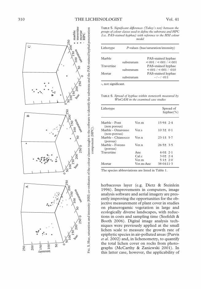
All the samples examined showed a well-developed HPC, that displayed differentpenetration patterns depending on both thelithotype and the lichen species (PAS-stainedhyphae in violet; Fig. 4A–D). The graphicinput and outputs of the image analysesperformed for the different case studies areillustrated in Fig. 4, which shows the micro-photographs obtained under the stereo-microscope (Fig. 4A–D), how the softwarerecognizes the substratum and the HPC inthe micro-photographs (white and blackareas, respectively; Fig. 4A’–D’), and how itassigns the original pixels to the differentcolour classes defined for the substratum andfor the penetrating hyphae (Fig. 4A”–D”).The black and white graphic outputs high-light the spread of the HPC within the sub-stratum, the hyphal structures being clearlyrecognizable in the different kinds of marblesand travertine (Fig. 4A’–C’). In the case ofmortar, the software identified both areasoccupied by hyphal structures and adja-cent areas marked by a violet-halo as HPC(Fig. 4D”). Because of the staining by PAS,the hyphal structures mainly stand outagainst the white and grey colour of the dif-ferent kinds of marbles and travertine, re-spectively. Accordingly, the colour groupsused to define HPC and these substratadiffered significantly for all the HSI co-ordinates (Table 5). In the case of mortar,only the hue values of the colour groupsdefined for the stained hyphae and for theheterogeneous substratum, respectively, dif-fered significantly.
The spread of the hyphae penetratingin the different lithotypes is described inTable 6.
F. 2. Graphic input (A–E) and outputs (A’–E’ & A”–E”) of image analyses through colour-based pixelclassification of lichenized stone surfaces. A, unveined pink marble-I; B, unveined white marble-II; C, veinedmarble-III; D, travertine, E; mortar. A-E, original images acquired in the field by the scanner (details); A’-E’,recognition of the substratum (white areas) and of the lichen thalli (black areas) by the software; A”-E”, assignmentof the original pixels by the software to the different colour classes defined for the substratum and for the differentlichen species. The species abbreviations are listed in Table 1. The arrows highlight a high agreement between inputand output images for the different species. The red circles mark discoloured parts of the thalli which were
consistently not recognized by the software.
2009 Image analysis of lichen colonization—Gazzano et al. 305
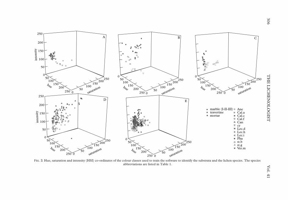
F. 3. Hue, saturation and intensity (HSI) co-ordinates of the colour classes used to train the software to identify the substrata and the lichen species. The speciesabbreviations are listed in Table 1.
306T
HE
LIC
HE
NO
LO
GIS
TV
ol.41

T 3. Significant differences (Tukey’s test) between the groups of colour classes used to define the substrata and lichen species with reference to the HSI colour model
Lithotype P-values (hue/saturation/intensity)
Marble - I Cal.a.substratum - / ·003 / <·001
Marble - II Can Lec.d. cyLec.d. ·013 / ·004 / <·001cy ·001 / <·001 / - - / - / <·001substratum <·001 / <·001 / <·001 - / - / - - / - / <·001
Marble - III Cal.f. Pha st.b st.gPh <·001 / <·001 / -st.b ·002 / <·001 / - - / - / -st.g <·001 / <·001 / - - / - / - ·001 / ·023 / -substratum <·001 / <·001 / <·001 - / - / ·005 - / <·001 / <·001 ·005 / - / <·001
Travertine Ane Cal.c. Ver.mCal.c. - / <·001 / -Ver.m - / - / - - / <·001 / -substratum - / ·039 / <·001 - / ·007 / ·001 - / ·003 / <·001
Mortar Ane Cal.c. Can Lec.h. Ver.mCal.c. ·026 / <·001 / <·001Cand - / <·001 / ·027 - / - / -L.h. - / - / <·001 - / <·001 / ·015 - / <·001 / -Ver.m - / - / ·024 ·011 / 0<·001 / - - / <·001 / - - / - / ·001substratum - / <·001 / <·001 ·010/ <·001 / <·001 - / <·001 / - - / (·050) / - - / ·017 / <·001
-, not significant; the species abbreviations are listed in Table 1.
2009Im
ageanalysis
oflichen
colonization—G
azzanoet
al.307

Discussion
Lichen cover on stonework
Since the small size of lichens makes theiranalysis in the field extremely difficult, obser-vation of stone substratra in the laboratoryfollowing destructive sampling was the mostreliable method for determining speciesdiversity and cover (Roux 1990; Nimis1991). However, as this method is difficult tocarry out on any large scale for environ-mental conservation reasons (Nimis 1991), itis obviously generally inapplicable to thestone cultural heritage, where only conserva-tive approaches are possible.
This study shows that the acquisition ofhigh definition digital images in the fieldbrings information to the laboratory, whichpartially resembles what could previouslyonly be obtained by destructive sampling,but in a way that is sensitive to the needs ofconservation. The digital images do not
facilitate species identification, but theyallow species already identified during a pre-liminary (and necessary!) floristic surveyto be recognized. The cover of the lichenspecies can then be quantified in the labora-tory using an image-analysis approach. Asrelevés can be reduced to the acquisition ofdigital images, i.e. a simple technical activity,fieldwork is less time consuming and doesnot require expert personnel; furthermore,digital ‘relevés’ can be repeated in the longterm and are suitable for random samplingstrategies. Moving the relevé analysis out ofthe field, specialist personnel can then focustheir expertise in the laboratory, and substi-tute the subjective visual estimation ofspecies cover with quantitative image analy-sis procedures with commensurate improve-ment in data quality.
Image analysis has been shown to be auseful tool to quantify plant species cover invegetation studies, mainly concerning the
T 4. Lichen cover of stonework measured by WinCAM in the examined case studies
Lithotype Species Cover (%)
Unveined pink marble (column) Lichen cover 24·4± 4·2Cal.a 24·4± 4·2
Unveined white marble (windowsill) Lichen cover 31·0±11·4Can 26·5±15·7Lec.d 4·6± 4·3
cy 19·4±14·1Veined marble (tombstone) Lichen cover 14·8± 2·0
Cal.f 0·9± 0·2Pha 6·0± 1·5st.b 6·1± 0·5st.g 1·8± 0·2
Travertine (cavea) Lichen cover 54·0±27·3Ane 8·4±13·8Cal.c 1·9± 3·7Ver.m 44·0±27·6
Mortar (cavea) Lichen cover 48·0±15·8Ane 11·1±10·8Cal.c 3·7± 3·6Can 0·1± 0·5
Lec.h 0·3± 1·2Ver.m 36·0±13·5
The use of Italics indicates species identified by classes which do not significantly differ in terms of HSI co-ordinateswith respect to those defined for one or more other species or for the substratum (see Table 3). The reliability of theseanalyses is verified by reconsidering the graphic output files, recognizing the incorrect assignment of the pixelsand finally collectively considering the related measures (crossed analyses). The species abbreviations are listed inTable 1.
308 THE LICHENOLOGIST Vol. 41

F. 4. Graphic input (A–D) and outputs (A’–D’ & A”–D”) of image analyses through colour-based pixelclassification of PAS-stained polished cross sections of lithic substrata colonized by Verrucaria macrostoma (A, C, D)and V. nigrescens (B). A–D, micro-photographs acquired in stereomicroscopy; A’–D’, recognition of the substratum(white areas) and of the hyphal penetration component (black areas) by the software; A”–D”, assignment of theoriginal pixels by the software to the colour classes defined for the substratum (grey) and for the PAS-stained hyphae(violet). A, Pont Canavese non-porous marble; B, Chianocco porous marble; C, travertine; D, mortar. Below thethalline component (TC), analyses deal with the areas of massive development of the hyphal penetration component(HPC; areas delimited by two green lines); areas where the hyphal penetration component develops sporadically(HPC’) were not included in the analyses. Sharp violet-stained structures correspond to hyphae (#), hyphae andbundles of hyphae (*) penetrating the rocks; §, areas showing a pink-violet halo possibly correspond to areas where
extracellular polymeric substances surround lichen hyphae.
2009 Image analysis of lichen colonization—Gazzano et al. 309
herbaceous layer (e.g. Dietz & Steinlein1996). Improvements in computers, imageanalysis software and aerial imagery are pres-ently improving the opportunities for the ob-jective measurement of plant cover in studieson phanerogamic vegetation in large andecologically diverse landscapes, with reduc-tions in costs and sampling time (Seefeldt &Booth 2006). Digital image analysis tech-niques were previously applied at the smalllichen scale to measure the growth rate ofepiphytic species in air-polluted areas (Purviset al. 2002) and, in lichenometry, to quantifythe total lichen cover on rocks from photo-graphs (McCarthy & Zaniewski 2001). Inthis latter case, however, the applicability ofF
.5.
Hue
,sat
urat
ion
and
inte
nsit
y(H
SI)
co-o
rdin
ates
ofth
eco
lour
clas
ses
taug
htto
the
soft
war
eto
iden
tify
the
subs
trat
aan
dth
eP
AS
-sta
ined
hyph
alpe
netr
atio
nco
mpo
nent
(HP
C).
T 5. Significant differences (Tukey’s test) between thegroups of colour classes used to define the substrata and HPC(i.e. PAS-stained hyphae) with reference to the HSI colour
model
Lithotype P-values (hue/saturation/intensity)
Marble PAS-stained hyphaesubstratum <·001 / <·001 / <·001
Travertine PAS-stained hyphaesubstratum <·001 / <·001 / ·010
Mortar PAS-stained hyphaesubstratum - / - / ·011
-, not significant.
T 6. Spread of hyphae within stonework measured byWinCAM in the examined case studies
Lithotype Spread ofhyphae(%)
Marble - Pont(non porous)
Ver.m 15·9± 2·4
Marble - Ornavasso(non-porous)
Ver.s 10·3± 0·1
Marble - Chianocco(porous)
Ver.n 23·1± 5·7
Marble - Foresto(porous)
Ver.n 26·5± 3·5
Travertine Ane 4·0± 2·1Cal.c 3·0± 2·4Ver.m 5·1± 2·0
Mortar Ver.m-Ane 38·0±11·3
The species abbreviations are listed in Table 1.
310 THE LICHENOLOGIST Vol. 41

the technique was limited to uniformly col-oured lithotypes, such as quartzites that areclearly different from the lichens, becauseof limitations imposed by the adopted soft-ware (McCarthy & Zaniewski 2001). In thepresent study, we processed the images ofdifferent lithotypes, showing chromatic vari-ations and different lichen biodiversity,through a colour based pixel classification.We have used our expertise to train the soft-ware as to which colours pertain to the sub-stratum and which pertain to the differentlichen species. It is worth noting that such adefinition of colour classes, the subjectivestep of the method, was performed on imagesacquired by the scanner, which offers a ×5magnification of the surfaces, thus enabling amore detailed observation than that possiblein fieldwork, and a wider observation fieldthan that observed from macro photos orunder stereomicroscopy. Although thalluscolour is an important species specificcharacter, its definition is often problematic(Wirth 1995) and a strong intraspecific varia-bility is often recognized; moreover, differentcolours can characterize different parts ofeach individual such as the vegetative thallusand the reproductive structures. Accord-ingly, several colour classes were used todefine each lichen species (and the substra-tum, which also showed different colours indifferent parts) in each analyzed picture.Graphic outputs offered information on themain critical point concerning each analysisand offered the possibility of checking therecognition of each species by the softwareand of heuristically adjusting the colour classsettings if necessary, thus progressively im-proving the quality of the analysis. Moreover,by analyzing the distribution of the colourclass groups in terms of HSI co-ordinates, itwas possible to evaluate which species wererecognized and which could not be recog-nized and, consequently, which species werecorrectly or wrongly quantified in terms ofcover. In particular, groups of colour classeswhich did not significantly differ in any of theHSI co-ordinates, as they were partially orcompletely overlapped or nested, could leadto an incorrect classification of the orphanpixels.
Species with colours that differ greatlyfrom those of the substratum and from mostother lichen species, such as the brilliantCaloplaca and Candelariella species on thedifferent kinds of marbles examined, weredefined using groups of colour classes whichdiffered significantly from those of the sub-stratum and of other lichen species on thebasis of two or more HSI parameters. Con-sequently, each orphan pixel that pertains tothese species is likely to be correctly recog-nized. On the other hand, species with col-ours which closely resembled those of thesubstratum and/or of other species, such asLecanora dispersa on the unveined whitemarble-II, Caloplaca crenulatella and Cande-lariella aurella on the mortar, and Phaeophys-cia orbicularis and the grey sterile thalli on theveined marble-III, were defined using groupsof colour classes which did not differ signifi-cantly on the basis of any of the three HSIparameters. In these cases, orphan pixelsmight be wrongly or correctly assigned,depending on their dispersion amplitudearound the taught colour classes; the pres-ence of no statistical difference between twocolour groups, in terms of HSI parameters,can thus signal critical cases which have to beverified directly on the images. By reconsid-ering such cases, it was possible to establishthat the software correctly distinguishedLecanora dispersa apothecia with respect totheir substratum and Caloplaca crenulatellaand Candelariella aurella, respectively. Pixelsassigned to Phaeophyscia orbicularis and ster-ile grey thalli were instead assigned to thesame colour classes in the output images,suggesting that the covers of these twospecies should be considered together or thattheir colour classes should be redefined.Such examples not only illustrate that theimage-analysis technique quantifies a lichenspecies’ cover, but that it is also possible tocheck the calculation.
Species overlooked during a preliminaryfloristic examination may be found in theimages using the ×5 magnification and theircover can be quantified. In this case, how-ever, new fieldwork is necessary to takesamples for their identification. On the otherhand, rare or inconspicuous species can be
2009 Image analysis of lichen colonization—Gazzano et al. 311

overlooked, but they are likely to have a lim-ited influence on stonework and could bedisregarded with minimum risk when deci-sions are taken on the preservation of stonecultural heritage.
Spread of hyphae penetrating withinstonework
Light and electron microscopy observationof fractured samples and polished cross sec-tions of lichen-encrusted rocks has allowedus to investigate the lichen-rock interactionsin situ (Ascaso et al. 2004; de los Rìos &Ascaso 2005). Several workers have high-lighted the volume of lichen influence withinstone substrata with reference to the averageor maximum depth of the HPC (e.g. Chenet al. 2000 and references therein; Favero-Longo et al. 2005). On the other hand, to theauthors’ knowledge, the spread of hyphaewithin rocks has never been considered,even though this three-dimensional spread isintuitively a more appropriate measure ofhow much a lichen might affect the rockconservation through physical and chemicalprocesses.
In this study, the development of HPC wasfirst quantified, in terms of hyphal spread,using colour differences between the substra-tum and the stained hyphae to perform imageanalysis based on pixel classification. Imageanalysis of PAS-stained tissues has alreadybeen performed in the medical research field(Becerra et al. 2003; Vielhauer et al. 2004). Inour research, strong differences in colour,that resulted from the HSI analysis, and thecharacteristic hyphal structure prevented anypotential misrecognitions, similar to thosedescribed for cover analyses. Even mortar,with a more composite structure than theother lithotypes, was clearly distinguishedfrom the stained hyphae. Although the hy-phae observed in cross sections beneath athallus probably develop from the thallus, theoccurrence of hyphae of other lichenized orfree living fungi cannot be ruled out (Bjelland& Ekman 2005). The result of the analysismight thus be interpreted as the quantifica-tion of the spread of hyphae associated witha lichen thallus and its complex associated
microcosm, which is increasingly being takeninto account in literature on bioweathering(Banfield et al. 1999; de los Rìos & Ascaso2005). Accordingly, microorganisms associ-ated with the lichen microcosm could beresponsible for the violet halos observed ad-jacent to hyphae within mortar, where PASprobably stains extracellular polymeric sub-stances (Vicente-Garcı́a et al. 2004) whichextend the lichen weathering action.
Since image analysis through colour-basedpixel classification allows a quantification ofboth the lichen species cover on the surface ofstone substrata and of the hyphal spreadwithin them, this technique appears to be thebest suitable to the evaluation of the volumeof influence of lichens on stonework and,consequently, to support decisions on thepreservation of our stone cultural heritage.
The authors express their gratitude to Lorenzo Appolo-nia and Gaetano De Gattis (Soprintendenza per i Beni ele Attività Culturali della Valle d’Aosta) for their kindpermission to carry out fieldwork in the Roman Theatreof Aosta. This research was funded by the CRT Bank –Alfieri Project.
R
Adamo, P. & Violante, P. (2000) Weathering of rocksand neogenesis of minerals associated with lichenactivity. Applied Clay Science 16: 229–256.
Ascaso, C., Garcı́a del Cura, M. A. & de los Rı́os, A.(2004) Microbial biofilms on carbonate rocks froma quarry and monuments in Novelda (Alicante,Spain). In Biodeterioration of Stone Surfaces (L. L.St. Clair & M. R. D. Seaward, eds.): 79–98.Dordrecht: Kluwer Academic Publishers.
Banfield, J. F., Barker, W. W., Welch, S. A. & Taunton,A. (1999) Biological impact on mineral dissolution:Application of the lichen model to understandingmineral weathering in the rhizosphere. Proceedings ofthe National Academy of Sciences 96: 3404–3411.
Becerra, L., Soares, R. V., Bruno, L. S., Siqueira, C.C.,Oppenheim, F. G., Offner, G. D. & Troxler, R. F.(2003) Patterns of secretion of mucins and non-mucin glycoproteins in human submandibular/sublingual secretion. Archives of Oral Biology 48:147–154.
Bjelland, T. & Ekman, S. (2005) Fungal diversity inrock beneath a crustose lichen as revealed by mol-ecular markers. Microbial Ecology 49: 598–603.
Chen, J., Blume, H. P. & Beyer, L. (2000) Weatheringof rocks induced by lichen colonization – a review.Catena 39: 121–146.
de los Rı́os, A. & Ascaso, C. (2005) Contributions ofin situ microscopy to the current understanding ofstone biodeterioration. International Microbiology 8:181–188.
312 THE LICHENOLOGIST Vol. 41

Dietz, H. & Steinlein, T. (1996) Determination of plantspecies cover by means of image analysis. Journal ofVegetation Science 7: 131–136.
Favero-Longo, S. E., Castelli, D., Salvadori, O.,Belluso, E. & Piervittori, R. (2005) Pedogeneticaction of the lichens Lecidea atrobrunnea, Rhizocar-pon geographicum gr. and Sporastatia testudinea onserpentinized ultramafic rocks in an alpine environ-ment. International Biodeterioration and Biodegrada-tion 56: 17–27, 250–251.
McCarthy, D. P. & Zaniewski, K. (2001) Digital analy-sis of lichen cover: a technique for use in lichenom-etry and lichenology. Arctic, Antarctic, and AlpineResearch 31: 107–113.
Nimis, P. L. (1991) Developments in lichen communitystudies. Lichenologist 23: 215–225.
Nimis, P. L., Monte, M. & Tretiach, M. (1987) Flora evegetazione lichenica di aree archeologiche delLazio. Studia Geobotanica 7: 3–161.
Nimis, P. L., Seaward, M. R. D., Ariño, X. & Barreno,E. (1998) Lichen-induced chromatic changes onmonuments: a case-study on the Roman amphi-theater of Italica (S. Spain). Plant Biosystems 132:53–61.
Nimis, P. L. & Martellos, S. (2008) ITALIC – TheInformation System on Italian Lichens. Version 4.0.University of Trieste, Dept. of Biology, IN4.0/1(http://dbiodbs.univ.trieste.it/).
Piervittori, R., Salvadori, O. & Laccisaglia, A. (1994)Literature on lichens and biodeterioration of stone-work. I. Lichenologist 26: 171–192.
Piervittori, R., Salvadori, O. & Laccisaglia, A. (1996)Literature on lichens and biodeterioration of stone-work. II. Lichenologist 28: 471–483.
Piervittori, R., Salvadori, O. & Isocrono, D. (1998)Literature on lichens and biodeterioration of stone-work. III. Lichenologist 30: 263–277.
Piervittori, R., Salvadori, O., Isocrono, D. (2004a) Lit-erature on lichens and biodeterioration of stone-work. IV. Lichenologist 36: 145–157.
Piervittori, R., Salvadori, O. & Seaward, M. R. D.(2004b) Lichens and Monuments: an analyticalbibliography. In Biodeterioration of Stone Surfaces(L. L. St. Clair & M. R. D. Seaward, eds.): 241–282. Dordrecht: Kluwer Academic Publishers.
Pinna, D., Salvadori, O. & Tretiach, M. (1998) Ananatomical investigation of calcicolous endolithiclichens from the Trieste karst (NE Italy). PlantBiosystems 132: 183–195.
Purvis, O. W., Erotokritou, L., Wolseley, P. A.,Williamson, B. & Read, H. (2002) A photographicquadrat recording method employing image analy-sis of lichens as indicator of environmental change.In Monitoring with Lichens – Monitoring Lichens (P.L. Nimis, C. Scheidegger & P. A. Wolseley, eds.):337–341. Dordrecht: Kluwer Academic Publishers.
Roux, C. (1990) Étude écologique et phytosociologiquedes peuplements lichéniques saxicoles-calcicoles duSud-Est de la France. Bibliotheca Lichenologica 15:1–557.
Seaward, M. R. D. (2004) Lichens as subversive agentsof biodeterioration. In Biodeterioration of Stone Sur-faces (L. L. St. Clair & M. R. D. Seaward, eds.):9–18. Dordrecht: Kluwer Academic Publishers.
Seefeldt, S. S. & Booth, D. T. (2006) Measuring plantcover in sagebrush steppe rangelands: a comparisonof methods. Environmental Management 37: 703–711.
St. Clair, L. L. & Seaward, M. R. D. (2004) Bio-deterioration of rock substrata by lichens: progressand problems. In Biodeterioration of Stone Surfaces(L. L. St. Clair & M. R. D. Seaward, eds.): 1–8.Dordrecht: Kluwer Academic Publishers.
Vicente-Garcı́a, V., Rı́os-Leal, E., Calderón-Domı́nguez, G., Cañizares-Villanueva, R. O.,Olvera-Ramı́rez, R. (2004) Detection, isolation,and characterization of exopolysaccharide pro-duced by a strain of Phormidium 94a isolatedfrom an arid zone of Mexico. Biotechnology andBioengineering 85: 306–310.
Vielhauer, V., Berning, E., Eis, V., Kretzler, M.,Segerer, S., Strutz, F., Horuk, R., Gröne, H. J.,Schlöndorff, D. & Anders, H. J. (2004) CCR1blockade reduces interstitial inflammation andfibrosis in mice with glomerulosclerosis and neph-rotic syndrome. Kidney International 66: 2264–2278.
Whitlach, R. B. & Johnson, R. G. (1974) Methodsfor staining organic matter in marine sediments.Journal of Sedimentary Petrology 44: 1310–1312.
Accepted for publication 12 March 2009
2009 Image analysis of lichen colonization—Gazzano et al. 313