Embed Size (px)
Citation preview
2
II. GENERAL AUDIENCE PROJECT SUMMARY
Multicellular animals, such as fruit flies and humans, use DNA sequences known
as enhancers to switch “ON” genes in the correct cells (space), and the correct time
point during life (time). With enhancers sequences consisting of nothing more than a
series of DNA letters (A, C, G, and T), these DNA sequences encode spatial and
temporal instructions for switching genes ON. Moreover, mutations in these sequences
can cause switch activities to differ, which can result in variation between individuals of
the same species, and over evolutionary time an accumulation of such mutations can
result in the adaptation of species to changing environments and the evolution of
altogether new species. Reporter transgenes are an effective tool to look at the switch
like activities of enhancers, where the activity can be seen by detecting the production
of a fluorescent protein. However, comparisons of different enhancers in the same
animal requires multiple fluorescent proteins whose color emission properties are
clearly distinguishable and whose activity at one time point can be distinguished from
those occurring at a later time point. My thesis aims to customize tools for the study of
enhancer functions by comparing a suite of fluorescent proteins and fluorescent timer
proteins in the fruit fly species Drosophila melanogaster.
3
III. PROPOSED THESIS TITLE AND PROPOSED ABSTRACT
Seeing gene expression in space, time, and color: evaluating new fluorescent
proteins for the study of gene regulation in fruit flies
The formation of animal bodies occurs through a continuum of changes in the
spatial locations of gene expression during the time frame of development. The
expression patterns for genes are directed by DNA sequence called enhancers. A major
limitation of contemporary genetics is in understanding how these DNA sequences
encode information specifying when during development and in what cell types a gene
will be expressed. One method to study enhancers is the use of reporter genes, where
an enhancer is coupled with the protein coding sequence for a Fluorescent Protein.
When inserted into animals, the function of an enhancer can be visualized by observing
where and when the Fluorescent Protein is produced. Comparisons of multiple reporter
transgenes with distinct fluorescent proteins in the same animal are limited in utility by
overlaps in the fluorescence emission spectrum for commonly used Fluorescent
Proteins. Compared to the turnover of the endogenous protein regulated by an
enhancer, Fluorescent Proteins linger in cells and thereby preclude a visualization of
any temporal changes in enhancer activity. The aim of this proposal is to evaluate
various Fluorescent Proteins that include diverse and dynamic emission spectra with a
Drosophila melanogaster enhancer that controls a spatially restricted and temporally
changing pattern of expression. The outcomes are likely to identify and adapt an
improved set of Fluorescent Proteins tools for the mechanistic study of gene regulation.
4
IV. Project Description
A. Specific Aims: This project will address two general hypotheses. (1) That blue, red
shifted and far-red shifted Fluorescent Proteins (FPs) can be coupled with Green
Fluorescent Protein for dual labeling experiments. (2) That Fluorescent Timer (FT)
proteins will enable the visualization of temporally changing enhancer activities.
Aim 1: Compare the color emission spectrum outputs of six Fluorescent Proteins
in Drosophila (D.) melanogaster pupae. These are EGFP-NLS, Cerulean-NLS,
DsRed.T4-NLS, mCherry-NLS, dTomato-NLS, and E2-Crimson-NLS.
Aim 2: Test the capabilities of two different Fluorescent Timer proteins to reveal
temporal alterations in enhancer activity during the time course of development.
B. Background and Significance: Embryonic development occurs through the
choreographed expression of thousands of genes in an entity that is constantly
changing in cell number and cell types, and overall morphology. This choreography is
achieved through the process of gene regulation where the expression (making of a
gene’s functional product) patterns for genes are limited to certain cell types and certain
times during development [1]. These expression patterns are encoded in and directed
by DNA sequences known as enhancers. While much remains to be understood about
the encoding of enhancer functions, a general understanding has been reached. In this,
the characteristic expression output specified by an enhancer is encoded by the DNA
sequence as a collection of short motifs that are bound by certain transcription factor
proteins [2]. As these regulatory proteins are limited in expression to certain cell types

5
and times during development, an enhancer can only direct expression in cells where
the right combination of transcription factors are present in the nucleus.
Studies of the human and model organism genomes has revealed that the
enhancers outnumber genes in genomes by several fold [3–5]. This stems from genes
being regulated by multiple enhancers. It is suspected that mutations in enhancers
account for many genetic diseases and disease risk factors, though few causative
mutations have been found [6,7]. Several case studies are known where the evolution
of animal morphology was shaped by changes in enhancer functions [8,9]. Hence, there
is great interest in understanding how an assemblage of “A”, “C”, “G”, and “T” letters –
the primary DNA building blocks – can encode enhancer functions and how changes in
these letters can result in disease, disease risk, or even evolutionary novelty.
One popular method to study enhancer function is to couple an enhancer to the
protein coding sequence for a gene that makes an easily observable product [10]. When
these reporter transgenes are put into a transgenic animal, the activity of the enhancer
can be visualized by observing where the reporter protein is produced. Fluorescent
Proteins (FPs) make excellent reporters as their expression can be detected from
fluorescent light emissions following excitation by light of a specific wavelength. The
most widely used FP is Enhanced Green Fluorescent Protein, or EGFP, that is excited
by 488 nanometer light (nm) and emits light between the 480 and 560 nm.
The use of EGFP has two limitations which this proposal seeks to ameliorate.
First, the emission spectrum for EGFP overlaps with that for other widely used FPs.
This includes DsRed2, which has a spectrum of 540-650 nm and for which newly
translated protein is in an immature green fluorescent state that over time matures to a

6
red state [11]. Due to the spectral overlap, dual-labeling experiments (the use of two
transgenes with different enhancers or a normal and mutant version of the same
enhancer) are problematic as it remains uncertain whether fluorescence is coming from
one, the other, or both FPs. The second limitation has to do with the perdurance of FPs
within cells [12]. This lingering presence prevents the observation of temporal changes
in the activity of an enhancer, notably the cessation of or a change in expression.
The Williams lab studies how gene expression patterns are encoded in enhancer
sequences. The anterior element is an enhancer that drives a robust expression of the
Bab2 protein in the more anterior A3 and A4 segments of the D. melanogaster pupal
abdomen (Appendix 1) [13]. Bab1 expression declines dramatically towards the end of
pupal development, although a comparable declination in enhancer activity cannot be
seen using the EGFP reporter transgene (Appendix 1). I will use the anterior element to
drive the expression of various FPs to find those which eliminate or minimize spectral
overlap with EGFP (Aim 1). I will also use this enhancer with FPs that change color over
their lifespan, and behave like a Fluorescent Timer (FT), to look for changes in anterior
element activity between early and late pupal stages (Aim 2).
C. Research Strategy
Aim 1. Research Methods and Strategy: FASTA sequence files were made with the
coding sequences of the nuclear localization signal (NLS) of the D. melanogaster
transformer gene [14] fused in-frame to the 3’ end of the coding sequences of mCherry
and E2-Crimson proteins. These sequences were synthesized and cloned into the
pUC57 vector (GenScript USA Inc.). Cerulean-NLS, dTomato-NLS, and DsRed.T4-
7
NLS containing vectors were previously constructed [15,16]. I will separately sub-clone
each of these FP sequences into a reporter transgene vector containing the anterior
element and for which the EGFP-NLS sequence was removed. The resulting reporter
transgenes will be integrated into the D. melanogaster attP40 genome site by site-
specific integration methods in order to make transgenic fruit flies [17,18]. Pupa will be
removed from their outer pupariums at 40 hours after puparium formation (hAPF), a
time point of anterior element activity, and mounted on a glass slide. The FP
expression patterns will be determined for blue, green, red, and far-red color spectra by
confocal microscopy (Appendix 2). Detection of green fluorescent light, FP excitation
will be achieved using 488 nm light and emitted light collected between 500-550 nm.
Blue, red and far-red light will respectively be detected using 458, 543 and 633 nm light
with emitted light collected between 450-490, 560-700 and 610-750 nm (Appendix 2).
Possible Outcomes: I anticipate that EGFP fluorescence will only be detected in the
abdomens of pupa when green fluorescence excitation and emission spectrum settings
are used. I suspect that DsRed.T4-NLS, mCherry-NLS, and dTomato-NLS will be best
detected using the red settings and to a lesser extent the far-red settings, whereas the
Cerulean and E2-Crimson-NLS will only be detected using blue and far-red settings
respectively. The outcome sought is the identification of FPs suitable for dual-labeling
studies. I anticipate this outcome is most likely for E2-Crimson-NLS, as its emission
spectrum is most separated from EGFP [19], though it is unclear whether this protein
proceeds through an immature green state as seen for DsRed.T4-NLS (Appendix 2).
Progress: The transgenes with the DsRed.T4-NLS, E2-Crimson-NLS and mCherry-
NLS sequences were made, and are being incorporated into transgenic fruit flies.
8
Aim 2. Research Methods and Strategy: The Timer-1 FT proceeds from an immature
green fluorescent state to a mature red state [20,21]. The Fast-FT timer proceeds from
an immature blue state to a mature red state [22]. I will excise the Timer-1 and Fast-FT
coding sequences respectively from the RIP Timer and Fast-FT vectors. These FT
sequences will be sub-cloned in the place of the EGFP-NLS sequence in the anterior
element containing vector. The resultant reporter transgenes will be incorporated into
transgenic fruit flies as described in Aim 1. Transgenic pupa will be collected for
confocal analysis at early (40 hAPF) and late time points (85 hAPF). Blue, green, and
red fluorescence will be collected using the respective excitation and emission collection
wavelengths: 458 nm and 440-490 nm, 488 and 500-550 nm, and 543 and 560-700 nm.
Possible Outcomes: Bab2 expression, but not anterior element-driven EGFP
expression, declines between early and late pupal stages, presumably by the anterior
element [13]. I suspect that the fluorescence of the FT proteins will recapitulate this
temporal change. For Timer-1, green fluorescence will be robust early and new
expression will cease at a later pupal stage (Appendix 3). This would be evident as a
transition from green (immature) to red (mature) fluorescence. A recent study suggests
that the green form Timer-1 does not progress to the mature red form, and instead is a
dead end product [23]. This would be evident as a persistent pattern of green
fluorescence on top of a later occurring red fluorescence. The blue to red maturation of
Fast-FT proceeds by the maturation of an immature blue fluorescent form. I suspect that
this FT will better document early and late stage activity of anterior element activity as
an absence of blue fluorescence at the later time point (Appendix 3).
Progress – FT Vectors were obtained from the addgene repository (www.addgene.org).
9
V. Timeline
Spring Term 2014 – Complete construction of anterior element reporter transgenes
with DsRed.T4-NLS, mCherry-NLS, and E2-Crimson-NLS (Aim 1). Send the transgenes
to Best Gene Inc. to have transgenic Drosophila melanogaster lines derived.
Summer 2014 – Complete construction of anterior element reporter transgenes with
Cerulean-NLS, dTomato-NLS, Timer-1 and Fast-FT sequences (Aim 2) and have
transgenic lines created. Survey fluorescence expression patterns for Aim 1 transgenes
via confocal microscopy.
Fall 2014 – Survey fluorescent expression patterns at early and late pupal stages for
Aim 2 transgenes via confocal microscopy.
Spring 2015 – During this semester, I will prepare my results for an oral presentation at
the University of Dayton’s honors symposium (March, 2015), and for a poster
presentation at the Stander Symposium (April, 2015).
VI. Bibliography
1. Davidson EH (2006) The Regulatory Genome: Gene Regulatory Networks In Development And Evolution. Burlington, MA: Elsevier Inc. p.
2. Arnone MI, Davidson EH (1997) The hardwiring of development: organization and function of genomic regulatory systems. Development (Cambridge, England) 124: 1851–1864. Available:http://www.ncbi.nlm.nih.gov/pubmed/9169833.
3. Pennisi E (2012) ENCODE Project Writes Eulogy For Junk DNA. Science 337: 1159–1160.
10
4. Nègre N, Brown CD, Ma L, Bristow CA, Miller SW, et al. (2011) A cis-regulatory map of the Drosophila genome. Nature 471: 527–531. Available:http://www.pubmedcentral.nih.gov/articlerender.fcgi?artid=3179250&tool=pmcentrez&rendertype=abstract. Accessed 9 March 2012.
5. Shen Y, Yue F, McCleary DF, Ye Z, Edsall L, et al. (2012) A map of the cis-regulatory sequences in the mouse genome. Nature: 1–5. Available:http://www.nature.com/doifinder/10.1038/nature11243. Accessed 2 July 2012.
6. Visel A, Akiyama JA, Shoukry M, Afzal V, Rubin EM, et al. (2009) Functional autonomy of distant-acting human enhancers. Genomics 93: 509–513. Available:http://www.pubmedcentral.nih.gov/articlerender.fcgi?artid=2683195&tool=pmcentrez&rendertype=abstract. Accessed 3 November 2011.
7. Sethupathy P, Collins FS (2008) MicroRNA target site polymorphisms and human disease. Trends in genetics : TIG 24: 489–497. Available:http://www.ncbi.nlm.nih.gov/pubmed/18778868. Accessed 6 September 2011.
8. Carroll SB (2008) Evo-devo and an expanding evolutionary synthesis: a genetic theory of morphological evolution. Cell 134: 25–36. Available:http://www.ncbi.nlm.nih.gov/pubmed/18614008. Accessed 24 July 2011.
9. Martin A, Orgogozo V (2013) The Loci of repeated evolution: a catalog of genetic hotspots of phenotypic variation. Evolution; international journal of organic evolution 67: 1235–1250. Available:http://www.ncbi.nlm.nih.gov/pubmed/23617905. Accessed 6 August 2013.
10. Rebeiz M, Williams TM (2011) Experimental Approaches to Evaluate the Contributions of Candidate Cis- regulatory Mutations to Phenotypic Evolution. In: Orgogozo V, Rockman M V., editors. Methods in Molecular Biology. Totowa, NJ: Humana Press, Vol. 772. pp. 351–375. Available:http://www.springerlink.com/index/10.1007/978-1-61779-228-1. Accessed 9 November 2011.
11. Baird GS, Zacharias D a, Tsien RY (2000) Biochemistry, mutagenesis, and oligomerization of DsRed, a red fluorescent protein from coral. Proceedings of the National Academy of Sciences of the United States of America 97: 11984–11989. Available:http://www.pubmedcentral.nih.gov/articlerender.fcgi?artid=17281&tool=pmcentrez&rendertype=abstract.
11
12. Tsien RY (2010) Fluorescence readouts of biochemistry in live cells and organisms. In: Weissleder R, Gambhir SS, Rehemtulla A, editors. Molecular Imaging: Principles and Practice. McGraw Hill. pp. 808–828.
13. Williams TM, Selegue JE, Werner T, Gompel N, Kopp A, et al. (2008) The regulation and evolution of a genetic switch controlling sexually dimorphic traits in Drosophila. Cell 134: 610–623. Available:http://www.pubmedcentral.nih.gov/articlerender.fcgi?artid=2597198&tool=pmcentrez&rendertype=abstract. Accessed 4 August 2011.
14. Hedley ML, Amrein H, Maniatis T (1995) An amino acid sequence motif sufficient for subnuclear localization of an arginine/serine-rich splicing factor. Proceedings of the National Academy of Sciences of the United States of America 92: 11524–11528. Available:http://www.pubmedcentral.nih.gov/articlerender.fcgi?artid=40434&tool=pmcentrez&rendertype=abstract.
15. Barolo S, Castro B, Posakony JW (2004) New Drosophila transgenic reporters: insulated P-element vectors expressing fast-maturing RFP. BioTechniques 36: 436–440, 442. Available:http://www.ncbi.nlm.nih.gov/pubmed/15038159.
16. Livet J, Weissman TA, Kang H, Draft RW, Lu J, et al. (2007) Transgenic strategies for combinatorial expression of fluorescent proteins in the nervous system. Nature 450: 56–62. Available:http://www.ncbi.nlm.nih.gov/pubmed/17972876. Accessed 6 August 2013.
17. Markstein M, Pitsouli C, Villalta C, Celniker SE, Perrimon N (2008) Exploiting position effects and the gypsy retrovirus insulator to engineer precisely expressed transgenes. Nature genetics 40: 476–483. Available:http://www.pubmedcentral.nih.gov/articlerender.fcgi?artid=2330261&tool=pmcentrez&rendertype=abstract. Accessed 17 July 2011.
18. Groth AC, Fish M, Nusse R, Calos MP (2004) Construction of Transgenic Drosophila by Using the Site-Specific Integrase From Phage phiC31. Genetics 166: 1775–1782.
19. Strack RL, Hein B, Bhattacharyya D, Hell SW, Keenan RJ, et al. (2009) A rapidly maturing far-red derivative of DsRed-Express2 for whole-cell labeling. Biochemistry 48: 8279–8281. Available:http://www.pubmedcentral.nih.gov/articlerender.fcgi?artid=2861903&tool=pmcentrez&rendertype=abstract. Accessed 7 August 2013.
12
20. Wittkopp PJ, Vaccaro K, Carroll SB (2002) Evolution of yellow gene regulation and pigmentation in Drosophila. Current biology 12: 1547–1556. Available:http://www.ncbi.nlm.nih.gov/pubmed/12372246.
21. Terskikh A, Fradkov A, Ermakova G, Zaraisky A, Tan P, et al. (2000) “Fluorescent Timer”: Protein That Changes Color with Time. Science 290: 1585–1588. Available:http://www.sciencemag.org/cgi/doi/10.1126/science.290.5496.1585. Accessed 7 August 2013.
22. Subach F V, Subach OM, Gundorov IS, Morozova KS, Kiryl D, et al. (2010) report on cellular trafficking. 5: 118–126. doi:10.1038/nchembio.138.Monomeric.
23. Verkhusha V V, Chudakov DM, Gurskaya NG, Lukyanov S, Lukyanov KA (2004) Common Pathway for the Red Chromophore Formation in Fluorescent Proteins and Chromoproteins. Chemistry & Biology 11: 845–854. doi:10.1016/j.

13
VII. BUDGET
Itemized Budget:
Materials/Supplies:
Item Estimated Cost
Gene Synthesis of E2-crimson-NLS and mCherry-NLS coding sequences (GenScript) and
purchase of four reporter gene vectors from addgene repository. $1,000.00
QIAprep Spin Miniprep kit (Qiagen) $250.00
AscI and SbfI restriction enzymes (New England Biolabs) $500.00
T4 DNA ligase (New England Biolabs) $250.00
200 DNA sequencing reactions at $5.00 each (DNA analysis, LTD) $1,000.00
4 cases of Drosophila vials and cotton balls (Fisher Scientific) $100.00
8 transgene injection services (Best Gene Inc, $250 each) $2,000.00
Travel: (If required for Thesis Project)
Mileage X miles X ($0.55/mile) =
*The current UD rate for mileage reimbursement will be used.
Other:
Item Estimated Cost
Other Source(s) of Funding
Source Pending/Secured
Total Amount Requested: $ _1,500.00____
Narrative Budget Justification:
To complete the proposed aims, this project will incur costs that include costs totaling at least $5,500.00. We are having sequences for custom made genes synthesized by a commercial vendor. This project will require the construction of at least 8 transgene vectors. This requires various enzymes, a DNA plasmid preparation kit, and DNA sequencing reactions. Each vector must be injected into Drosophila melanogaster embryos to derive transgenic lines (The Best Gene Inc.). Taking care of transgenic lines will require plastic vials and cotton balls.
14
VIII. LETTER OF SUPPORT
The letter from my advisor, Dr. Thomas Williams an Assistant Professor in the
Department of Biology at the University of Dayton, was emailed to Ramona Speranza
on 3/6/14.
IX. Appendices
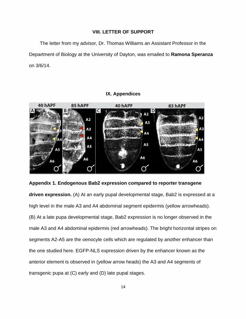
Appendix 1. Endogenous Bab2 expression compared to reporter transgene
driven expression. (A) At an early pupal developmental stage, Bab2 is expressed at a
high level in the male A3 and A4 abdominal segment epidermis (yellow arrowheads).
(B) At a late pupa developmental stage, Bab2 expression is no longer observed in the
male A3 and A4 abdominal epidermis (red arrowheads). The bright horizontal stripes on
segments A2-A5 are the oenocyte cells which are regulated by another enhancer than
the one studied here. EGFP-NLS expression driven by the enhancer known as the
anterior element is observed in (yellow arrow heads) the A3 and A4 segments of
transgenic pupa at (C) early and (D) late pupal stages.
15
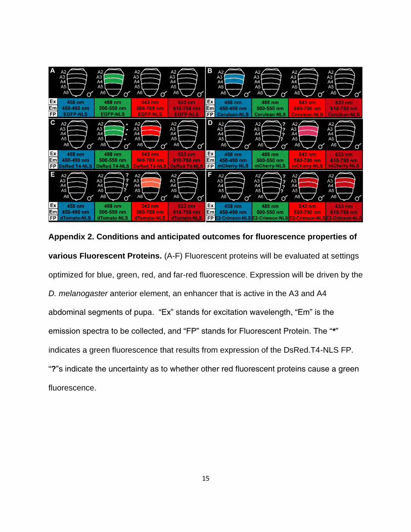
Appendix 2. Conditions and anticipated outcomes for fluorescence properties of
various Fluorescent Proteins. (A-F) Fluorescent proteins will be evaluated at settings
optimized for blue, green, red, and far-red fluorescence. Expression will be driven by the
D. melanogaster anterior element, an enhancer that is active in the A3 and A4
abdominal segments of pupa. “Ex” stands for excitation wavelength, “Em” is the
emission spectra to be collected, and “FP” stands for Fluorescent Protein. The “*”
indicates a green fluorescence that results from expression of the DsRed.T4-NLS FP.
“?”s indicate the uncertainty as to whether other red fluorescent proteins cause a green
fluorescence.
16
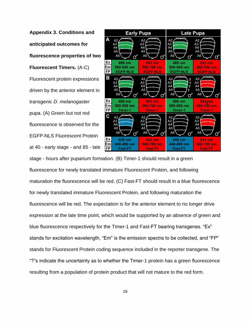
Appendix 3. Conditions and
anticipated outcomes for
fluorescence properties of two
Fluorescent Timers. (A-C)
Fluorescent protein expressions
driven by the anterior element in
transgenic D. melanogaster
pupa. (A) Green but not red
fluorescence is observed for the
EGFP-NLS Fluorescent Protein
at 40 - early stage - and 85 - late
stage - hours after puparium formation. (B) Timer-1 should result in a green
fluorescence for newly translated immature Fluorescent Protein, and following
maturation the fluorescence will be red. (C) Fast-FT should result in a blue fluorescence
for newly translated immature Fluorescent Protein, and following maturation the
fluorescence will be red. The expectation is for the anterior element to no longer drive
expression at the late time point, which would be supported by an absence of green and
blue fluorescence respectively for the Timer-1 and Fast-FT bearing transgenes. “Ex”
stands for excitation wavelength, “Em” is the emission spectra to be collected, and “FP”
stands for Fluorescent Protein coding sequence included in the reporter transgene. The
“?”s indicate the uncertainty as to whether the Timer-1 protein has a green fluorescence
resulting from a population of protein product that will not mature to the red form.