Embed Size (px)
Citation preview

From Dept. Medical Biochemistry and Biophysics
Karolinska Institute, Stockholm, Sweden
IDENTIFYING GENETIC DETERMINANTS
OF T CELL-DEPENDENT
AUTOIMMUNITY USING FORWARD
GENETICS
Gonzalo Fernandez Lahore
Stockholm, August 2021

All previously published papers were reproduced with permission from the publisher.
Published by Karolinska Institutet.
Printed by Universitetsservice US-AB, 2021
© Gonzalo Fernandez Lahore, 2021
ISBN 978-91-8016-294-4
Cover illustration: Genetic predisposition determines rheumatoid arthritis. Illustration by the
author.
IDENTIFYING GENETIC DETERMINANTS OF T CELL-
DEPENDENT AUTOIMMUNITY USING FORWARD
GENETICS
THESIS FOR DOCTORAL DEGREE (Ph.D.)
By
Gonzalo Fernandez Lahore
The thesis will be defended in public at Gustaf Retzius hall, KI, Stockholm, Aug 20, 2021
Principal Supervisor:
Prof. Rikard Holmdahl
Karolinska Institute
Department of Medical Biochemistry and
Biophysics
Division of Medical Inflammation Research
Co-supervisor(s):
Prof. Kutty Selva Nandakumar
Southern Medical University
School of Pharmaceutical Sciences
Dr. Liselotte Bäckdahl
Karolinska Institute
Department of Medical Biochemistry and
Biophysics
Division of Medical Inflammation Research
Opponent:
Prof. Vijay Kuchroo
Evergrande Center for Immunologic Diseases,
Harvard Medical School and Brigham and
Women’s Hospital
Examination Board:
Assoc. Prof. Lisa Westerberg
Karolinska Institute
Department of Microbiology, Tumor and Cell
Biology
Asst. Prof. Jonathan Coquet
Karolinska Institute
Department of Microbiology, Tumor and Cell
Biology
Prof. Göran Andersson
Swedish University of Agricultural Sciences
Department of Animal Breeding and Genetics


Como no estas experimentado en las cosas del mundo, todas las cosas que tienen algo de
dificultad te parecen imposibles. Confía en el tiempo, que suele dar dulces salidas a muchas
amargas dificultades.
Miguel de Cervantes Saavedra —
Don Quijote de la mancha
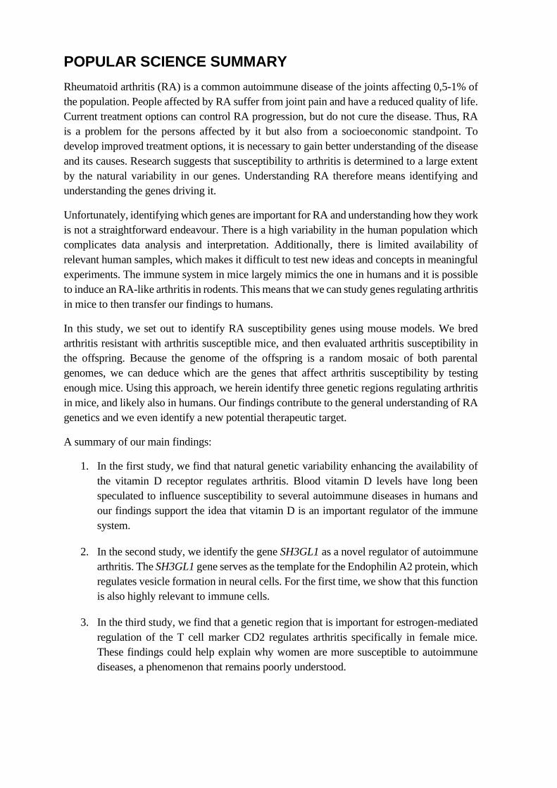
POPULAR SCIENCE SUMMARY
Rheumatoid arthritis (RA) is a common autoimmune disease of the joints affecting 0,5-1% of
the population. People affected by RA suffer from joint pain and have a reduced quality of life.
Current treatment options can control RA progression, but do not cure the disease. Thus, RA
is a problem for the persons affected by it but also from a socioeconomic standpoint. To
develop improved treatment options, it is necessary to gain better understanding of the disease
and its causes. Research suggests that susceptibility to arthritis is determined to a large extent
by the natural variability in our genes. Understanding RA therefore means identifying and
understanding the genes driving it.
Unfortunately, identifying which genes are important for RA and understanding how they work
is not a straightforward endeavour. There is a high variability in the human population which
complicates data analysis and interpretation. Additionally, there is limited availability of
relevant human samples, which makes it difficult to test new ideas and concepts in meaningful
experiments. The immune system in mice largely mimics the one in humans and it is possible
to induce an RA-like arthritis in rodents. This means that we can study genes regulating arthritis
in mice to then transfer our findings to humans.
In this study, we set out to identify RA susceptibility genes using mouse models. We bred
arthritis resistant with arthritis susceptible mice, and then evaluated arthritis susceptibility in
the offspring. Because the genome of the offspring is a random mosaic of both parental
genomes, we can deduce which are the genes that affect arthritis susceptibility by testing
enough mice. Using this approach, we herein identify three genetic regions regulating arthritis
in mice, and likely also in humans. Our findings contribute to the general understanding of RA
genetics and we even identify a new potential therapeutic target.
A summary of our main findings:
1. In the first study, we find that natural genetic variability enhancing the availability of
the vitamin D receptor regulates arthritis. Blood vitamin D levels have long been
speculated to influence susceptibility to several autoimmune diseases in humans and
our findings support the idea that vitamin D is an important regulator of the immune
system.
2. In the second study, we identify the gene SH3GL1 as a novel regulator of autoimmune
arthritis. The SH3GL1 gene serves as the template for the Endophilin A2 protein, which
regulates vesicle formation in neural cells. For the first time, we show that this function
is also highly relevant to immune cells.
3. In the third study, we find that a genetic region that is important for estrogen-mediated
regulation of the T cell marker CD2 regulates arthritis specifically in female mice.
These findings could help explain why women are more susceptible to autoimmune
diseases, a phenomenon that remains poorly understood.
ABSTRACT
Rheumatoid arthritis (RA) affects 0.5-1% of the population and is an important health and
socioeconomic problem. RA has a high degree of heritability. Thus, extensive efforts have been
made to better understand the genetic variability contributing to disease susceptibility.
However, dissecting the genetic component of RA in humans has been difficult due to
heterogeneity in the human population, multiple testing issues, and lack of accessibility to
relevant tissues for proof-of-concept studies. Genetic studies in mouse model systems
circumvent these problems, enhancing the possibility to identify disease regulating genetic
variants. Here, we use a forward genetics approach in mice to identify and characterize genetic
determinants of RA and related autoimmune diseases. First, we have mapped quantitative trait
loci (QTL) regulating experimental arthritis using linkage analysis, and then isolated these QTL
in congenic strains for in depth functional characterization. Using this approach, we make
several important observations.
In the first study, we find that promoter polymorphisms regulating expression of the vitamin D
receptor affect T cell activation and T cell-driven collagen-induced arthritis. These findings are
particularly interesting considering the long-standing association between serum vitamin D and
several autoimmune diseases.
In the second study, we discover a spontaneous insertion of a long terminal repeat which leads
to a deficiency in the SH3GL1 gene (Endophilin A2), protecting mice in several arthritis
models. We are first to identify the immunomodulatory properties of SH3GL1, which may
prove to be a valuable therapeutic target.
In the third study, we identify a polymorphic estrogen receptor binding site that regulates
susceptibility to experimental arthritis and other autoimmune models by interfering with
estrogen regulation of the T cell marker CD2. These results suggest an important role for CD2
and estrogen in shaping the sexually dimorphic immune response. Collectively, our findings
make a significant contribution towards the understanding of RA genetics while demonstrating
the value of animal models.
SCIENTIFIC PAPERS INCLUDED IN THIS THESIS
I. Fernandez Lahore, G, Raposo, B, Lagerquist, M, Ohlsson, C, Sabatier, P, Xu, B, ... &
Holmdahl, R (2020). Vitamin D3 receptor polymorphisms regulate T cells and T cell-
dependent inflammatory diseases. Proceedings of the National Academy of Sciences,
117(40), 24986-24997.
II. Norin, U, Rintisch, C, Meng, L, Forster, F, Ekman, D, Tuncel, J, ... , Fernandez
Lahore, G, … & Holmdahl, R (2021). Endophilin A2 deficiency protects rodents
from autoimmune arthritis by modulating T cell activation. Nature communications,
12(1), 1-11.
III. Fernandez Lahore, G, Förster, M, Johannesson, M, Sabatier, P, Lönnblom, E, Aoun,
M, ... & Holmdahl, R (2021). Polymorphic estrogen receptor binding site causes
CD2-dependent sex bias in the susceptibility to autoimmune diseases. Nature
communications. In press.
CONTRIBUTIONS TO OTHER MANUSCRIPTS
I. Fernandez Lahore, G*, James J*, et al. Redox regulation of LAT drives T cell-
mediated inflammation. Manuscript. * equal contribution.
II. James J, et al. Redox regulation of PTPN22 affects the severity of T cell dependent
autoimmune inflammation. Under review in Redox Biology.
III. Aoun M*, Saxena A*, et al. Bone marrow-selected antigen specific regulatory B
cells. Manuscript. * equal contribution.
IV. Urbonaviciute V, et al. Collagen peptide based vaccine induces T cell-mediated
protection from autoimmune arthritis independent of the cuasative antigen.
Manuscirpt.
CONTENTS
1 INTRODUCTION........................................................................................................... 7
2 LITERATURE REVIEW ............................................................................................... 8
2.1 RHEUMATOID ARTHRITIS .............................................................................. 8
2.2 PATHOPHYSIOLOGY OF ESTABLISHED RA .............................................. 9
2.3 PREDISPOSING FACTORS IN RA ................................................................. 10
2.3.1 Predisposing environmental factors ........................................................ 10
2.3.2 Predisposing genetic factors ................................................................... 11
2.3.3 Limitations of human genetic studies ..................................................... 12
2.4 USING MOUSE MODELS TO STUDY RA GENETICS ............................... 13
2.4.1 Inducing RA-like arthritis in mice .......................................................... 14
2.4.2 Mapping quantitative trait loci in the mouse .......................................... 14
2.4.3 Arriving from QTL to causative gene .................................................... 17
3 RESEARCH AIMS ....................................................................................................... 19
4 METHODS .................................................................................................................... 20
4.1 A brief overview of the animal models applied in this study ............................ 20
5 RESULTS AND DISCUSSION ................................................................................... 21
5.1 Study I - Vitamin D receptor polymorphisms regulate T cells and T cell-
dependent inflammatory diseases. ...................................................................... 21
5.2 Study II - Endophilin A2 deficiency protects rodents from autoimmune
arthritis by modulating T cell activation. ............................................................ 21
5.3 Study III - Polymorphic estrogen receptor binding site causes CD2-
dependent sex bias in the susceptibility to autoimmune diseases ...................... 22
6 CONCLUDING REMARKS ....................................................................................... 23
7 ACKNOWLEDGEMENTS .......................................................................................... 24
8 REFERENCES .............................................................................................................. 25
LIST OF ABBREVIATIONS
(m)Ab (monoclonal) Antibody
APC Antigen presenting cell
ACPA Anti citrullinated protein antibody
CD Cluster of differentiation
CFA Complete freud’s adjuvant
CIA Collagen-induced arthritis
bCII Bovine collagen type II
DC Dendritic cell
E2 17-β-estradiol
EAE Experimental autoimmune encephalomyelitis
ER Estrogen receptor
ERBS Estrogen receptor binding site
GWAS Genome-wide association study
HLA Human leucocyte antigen
IFN Interferon
Ig Immunoglobulin
IL Interleukin
Mbp Mega base pairs
QTL Quantitative trait locus
RA Rheumatoid arthritis
RF Rheumatoid factor
SNP Single nucleotide polymorphism
TCR T cell receptor
Th T helper cell
TNF Tumor necrosis factor
VDR Vitamin D receptor
7
1 INTRODUCTION
The human is in constant contact with commensal and pathogenic microorganisms. The
immune system is an intricate network of specialised cells and mechanisms that provides
protection from possible infections. The immune system can be broadly subclassified in two
groups, innate and adaptive.
The innate immune system is the first line of defence, comprised by a series of non-specific
mechanism that include skin and mucosal barriers, complement proteins, as well as phagocytic
and antigen presenting cells (APCs). Innate cells use a series of somewhat unspecific receptors
to detect, and react to, shared microbial structures such as lipopolysaccharides, proteoglycans,
unmethylated CpG dinucleotides, or double stranded RNA (1). Activated APCs can recruit and
expand adaptive effector T cells to generate highly specific immune responses (2).
Adaptive T and B cells somatically rearrange their receptors during development. In this way,
millions of T and B cell clones with unique receptor combinations are generated. To be
activated, T cells require cognate antigen to be presented by APCs. B cells can be stimulated
by antigen alone or in a T cell-dependent manner to produce antibodies. B cells activated in a
T cell-dependent manner, however, undergo somatic hypermutation of the B cell receptor
variable region, resulting in the generation of mature, highly specific antibodies. This process
results in a flexible and tailored cellular and humoral immune response, but it comes at a cost.
The same process can generate self-reactive clones that attack our own tissue, causing systemic
or organ-specific inflammation and destruction, an autoimmune disease.
Mechanisms are in place to minimize the occurrence of autoreactive events, first at a central,
then at a peripheral level. Developing bone marrow B cells and thymic T cells with autoreactive
potential are deleted, receptor edited, or imparted with anergic or anti-inflammatory properties
during selection (3) (4). Autoreactive cells that escape elimination face obstacles in the
periphery, such as restricted tissue access and low abundance of antigen. Further, autoreactive
B cells lacking essential T cell co-stimulation become anergic (5), and improperly
(co)stimulated autoreactive T cells become apoptotic, anergic or develop regulatory properties
(6).
However, these protective mechanisms are not perfect, and tolerance can be breached under
certain circumstances. Events proposed to break tolerance include exposure of antigen from
immune privileged sites after injuries (7) (8), cross reactivities between exogeneous and
endogenous antigens (9), and reactivity to peripherally modified antigens (neoantigens) (10).
Genetic factors are also critical. For example, rare mutations in single genes affecting central
or peripheral tolerance such as FOXP3 (11) or AIRE (12) can result in autoimmune conditions.
Most often, however, genetic susceptibility is determined by multiple common genetic variants
of low effect sizes with poorly understood and complex underlying mechanisms. Likely, it is
complex interactions between predisposing genetics and environmental factors that determine
the susceptibility to autoimmune diseases.
Today, autoimmune diseases can be treated with different degrees of success but cannot be
cured. These diseases reduce quality of life in affected persons and are a major socioeconomic
problem (13). It is imperative that we improve our knowledge to successfully establish new
therapies and preventive measures. One of the most frequent autoimmune diseases, and the
focus of this work, is rheumatoid arthritis.
8
2 LITERATURE REVIEW
2.1 RHEUMATOID ARTHRITIS
Rheumatoid arthritis (RA) is a multifactorial and chronic autoimmune disease of the joints
affecting 0,5-1% of the population (14). Like many autoimmune diseases, RA is predominant
in females, with a female to male ratio of approximately 3:1 (15). RA patients exhibit
characteristic signs of chronic inflammation in the joints, as evidenced by synovial hyperplasia
and increased signs of angiogenesis (16) (17) (18). Chronic joint inflammation promotes
osteoclast formation and leads to increased bone resorption and erosion. If the inflammation is
not treated, progressive destruction of the joints eventually leads to disability.
RA can be treated with varying degree of success.
Therapies focus on stopping inflammation and
managing pain. Common therapeutic approaches
include the use of so-called disease modifying
antirheumatic drugs (methotrexate,
hydroxychloroquine) in combination with non-
steroidal anti-inflammatory drugs (diclofenac,
ibuprofen) or glucocorticoids, as well as more
recently the use fast-acting biologics such as B
cell-depleting anti-CD20 or TNF blockers.
RA is diagnosed based on clinical manifestations
and the characteristic presence of autoantibodies.
Patients are screened for the presence of
rheumatoid factors (RF) (19) and anti-
citrullinated protein antibodies (ACPA) (20). RF
are polyclonal and germline encoded IgM anti-
IgG antibodies that often occur in the context of
inflammation. ACPAs, on the other hand, are
promiscuous antibodies capable of binding
diverse citrullinated autoantigens (21) that are
highly specific to RA (specificity up to 98%)
(22).
ACPAs are not only the gold-standard for RA
diagnosis but also an important stratification
criterium, as not all patients have these antibodies
(sensitivity up to 77%). ACPA positive and
negative patients differ in e.g. disease progression (23) and genetic risk profiles (24),
suggesting that RA has different underlying aetiologies. It is unclear how or whether ACPAs
or RFs are disease modifiers and, in fact, ACPAs precede onset of symptoms (25). It is likely
that autoimmunity develops in a yearlong subclinical phase before culminating in a chronic
and destructive disease (21). Our current understanding of RA is limited in that it is derived
mostly from established disease.
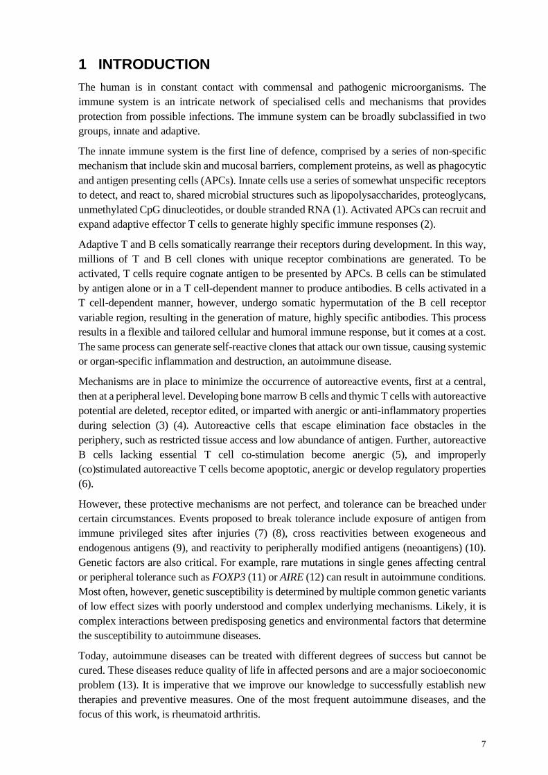
Figure 1: Illustration of typical joint
inflammation in patients with
rheumatoid arthritis. Interphalangeal,
metacarpophalangeal and wrist joints
are most often affected. More chronic
RA may also involve thickening of
metacarpophalangeal joints and ulnar
deviation of the fingers.
9
2.2 PATHOPHYSIOLOGY OF ESTABLISHED RA
Joint inflammation in RA is characterized by the presence leukocyte infiltrates to the joint.
Local inflammation is perpetuated by the presence of effector cytokines such as IL-1, IL-6, and
TNFα (26), which are secreted by, and act on, infiltrating and resident cells alike. Both adaptive
and innate immune cells partake in synovial inflammation (27).
Cells of the adaptive immune system have generated particular interest given the
characteristic presence of B cell-derived autoreactive ACPAs and the genetic risk profile in RA
patients. Many of the genes associated to RA are key players in T cell and B cell activation
(28), together suggesting an underlying adaptive autoimmune response. Indeed, T cell- and B
cell infiltration of the joints is characteristic for RA (29), with both these cell types forming a
large fraction of the synovial infiltrate (30). Infiltrating B and T cells can be found diffusely or
in lymphocytic aggregates (31, 32). Here, they seem to interact productively as ACPAs evolve
over time (33), suggesting a T cell-dependent B cell activation. The relevance of both of these
cell types for pathogenesis is further substantiated by the clinical efficacy of T cell inhibiting
CTLA4Ig (34), and B cell depleting anti-CD20 (35). Interestingly, anti-CD20 treatment does
not effectively deplete antibody producing plasma cells (36), highlighting the importance of
antibody-independent B cell functions. It is known that activated B cells are capable of
secreting proinflammatory cytokines (37) and potently presenting antigen (38), thus supporting
activation of synovial (autoreactive) T cells (39) and other cells. This help in activation seems
reciprocal, as RA T cells also support B cell activation (40).
Synovial T cells in RA display a contracted T cell receptor (TCR) repertoire characterized by
patient-individual clonal expansion (41), some of these clones likely recognizing citrullinated
autoantigens (42). Expanded synovial T cells take on a terminally differentiated and activated
phenotype (40) (43), losing expression of costimulatory molecules such as CD28, while at the
same type gaining other receptors that permit activation in an antigen-independent manner (44).
T cells from RA synovia show increased signs of replicative stress (45) and are, to some extent,
resistant to apoptosis (46). These cells potently produce IFN-ƴ, and to some extent IL-17 (47–
50), enhancing inflammation (51) and joint destruction, for example by promoting
osteoclastogenesis (52). In fact, the frequency of Th1 type cells (producers of IFN-ƴ) correlate
with disease activity (53). Further insight can be extracted from animal models of RA, which
demonstrate that transfer of arthritogenic T cells is enough to elicit the development of
autoimmune arthritis in naïve recipients (54). Thus, although details are still to be sorted out,
current evidence strongly suggests that T cells are important drivers of the autoimmune
response in RA. Therefore, the study of T cell modifiers remains central to understanding RA
pathogenesis.
Perhaps as a consequence to an initially T cell driven autoimmune response, innate type cells
are recruited to the joint. Resident and infiltrating innate immune cells are activated by local
accumulation of proinflammatory cytokines and immune complexes, producing cytokines,
chemokines, prostaglandins, and leukotrienes in response. Activated neutrophils furthermore
have the capacity to produce high levels of reactive oxygen species and secrete nuclear
extracellular traps to promote inflammation (55). Macrophages are present in increased
numbers in the RA synovial membrane and are believed to contribute to inflammation by
presenting antigen and producing cytokines such as TNFα, IL6, IL-1β, IL-12 and IL-23 (56).
10
This recruits monocytes and neutrophils, and has repercussions for osteoclast formation, and T
cell and fibroblast activation. However, recent studies also suggest that resident synovial
membrane macrophages originally serve to shield and protect articular structures from
infiltrating cells (57). Mast cells are also expanded in the arthritic synovial membrane, where
they secrete proteinases (58) and inflammatory mediators (59). Interestingly, they can promote
T cell responses by presenting antigen (60) but are also capable IL-17 producers themselves
(61).
Joint inflammation is not only driven by immune cells, but also by resident cells of
mesenchymal origin. Activated synovial lining fibroblasts are imprinted with an aggressive
phenotype that perseveres for several months of culture, producing proinflammatory cytokines,
chemokines, prostaglandins and matrix-degrading enzymes (62). Similarly, chondrocytes are
activated by IL-1 and TNFα to secrete proteolytic enzymes, leading to extracellular matrix
degradation (63).
Evidently, the rheumatic joint consists of a highly complicated network of proinflammatory
mediators. A recurring theme is the response to -and secretion of- proinflammatory cytokines,
in particular IL-1, IL-6, and TNFα. All these cytokines can be readily detected in RA synovia.
Accordingly, targeting of these pathways presents one of the best therapeutic options in RA
alongside anti-CD20. While current therapeutic approaches successfully manage synovitis, an
estimated 5-20% of patients do not respond well to available treatments (64). Additionally,
biologics carry important added risks such increased susceptibility to infections or
malignancies (65). To develop new targeted therapies, it is paramount to understand which
factors predispose an individual to develop RA.
2.3 PREDISPOSING FACTORS IN RA
Studies have estimated RA heritability at around 50 % (66). The number is not precise, as
estimates vary between 12-68%, but it is clear that RA has a significant genetic component. On
the other hand, this also means that there must be a significant environmental contribution to
RA susceptibility. We will first briefly cover commonly suggested environmental factors to
then dedicate more time to RA genetics.
2.3.1 Predisposing environmental factors
Several environmental factors have been associated to RA. Some of the most frequently
mentioned examples include cigarette smoking (67), infections (68) (69), vitamin D status (70),
use of oral contraceptives (71), and exposure to mineral oils (72).
It has been suggested that infections may trigger cross-reactivities to self through molecular
mimicry. For example, Epstein-Barr (69) viral proteins exhibit similarity to HLA-DR4 (73)
(74) (75). Similarly, smoking (67) and infection with Porphyromonas gingivalis (68), are
suspected to elicit autoimmune responses by promoting the formation of neo-antigens. Special
attention has been given to neo-antigen formation as a result of hypercitrullination, given a
predisposition of RA-associated HLA alleles to bind citrullinated proteins (76), and the
association between polymorphisms of the citrullinating enzyme PADI4 (77) and RA. Both
smoking (78) (79) and periodontitis (80) are suspected to promote citrullination, and fittingly
associate with ACPA seropositive RA (81).
11
A particularly interesting association in the context of study I is that of RA and vitamin D
status. Several studies have reported an association between low serum vitamin D and
incidence of RA (82) (83) (84). In study I we demonstrate that the genetic predisposition to
over and under express the vitamin D receptor affects the immune response in experimental
models of autoimmunity.
Similarly, in study III we provide evidence that may help understand associations such as the
one between RA and use of oral contraceptives (85) (86). A key role for sex hormones in RA
susceptibility has long been suspected, and this notion is further supported by the female
preponderance in RA (87), as well as studies reporting disease modifying effects of hormone
replacement therapy (88) (89). In study III we describe how sex hormones such as estrogen
may directly contribute to the development of RA by regulating the expression of the T cell
costimulatory molecule CD2.
While our studies provide some insight into possible mechanisms fuelling some of the
environmental associations identified to date, in general, associative studies investigating
environmental factors in RA have had limited success. Environmental factors may act years
before RA onset and are thus difficult to identify. Perhaps because of this, studies investigating
environmental factors are scarce and often lack reproducibility (90). In contrast, more energy
has been spent understanding the genetic determinants of RA.
2.3.2 Predisposing genetic factors
The estimated degree of heritability in RA varies depending on the method used. Comparison
of disease discordance between monozygotic and dizygotic twins found heritability estimates
of 12-60%, familial aggregation studies of 50%, and methods considering the cumulative
influence of all SNPs on RA phenotypic variance suggest a heritability of 19-52% (66). While
the heritability estimate may not be exact, it is clear that the contribution of genetics to RA is
significant.
Already in the late 1970s it was observed that PBMCs from RA patients are unusually
compatible with each other in mixed lymphocyte cultures (91), and it was quickly recognized
that this was due to similarities in the HLA alleles of RA patients (92). This association was
soon refined to the HLA-DRB1 gene and remains the strongest genetic association to RA to
date (93), accounting for around 30% of the total estimated heritability (94). It has been
proposed that several of the HLA-DRB1 susceptibility alleles share a common amino acid
sequence in the third hypervariable region of DRβ1 (shared epitope hypothesis (92)). This
shared motif may favour binding of arthritogenic peptides although a main candidate remains
to be singled out.
The emergence of high throughput sequencing and genotyping methods has enabled genome
wide association studies that helped clarify the genetic landscape of RA by further identifying
hundreds of non-MHC risk loci. Interestingly, many of the non-MHC associations are to genes
relevant to T cell biology and activation, further cementing the importance of T cells for RA
pathology. Some of the most cited findings include PTPN22, STAT4, TRAF1-C5, IL2RA,
CD28, and CTLA4 (95), as well as more recently Th17-related CD26 and CCR6 (96) among
others. T cell-unrelated genes with other relevant functions, such as the citrullinating enzyme
PADI4 (95), have also been detected. Some of these associations can be ethnic group-specific,
12
such as is the case for HLA-DRB1 alleles or for Caucasian-specific PTPN22, but most
reproducible associations are shared across populations (97). In contrast, seropositive and
seronegative RA seem to have a different genetic risk factors (24). For example, HLA-DRB1
and PTPN22 seem to associate primarily with seropositive RA (98) (99).
Despite the considerable progress made by modern genetic association studies, it should be
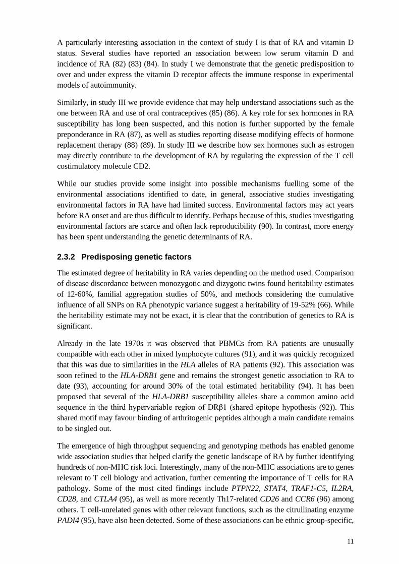
noted that the newfound loci only make small individual contributions to explaining the overall
heritability of RA (figure 2). Cumulatively, the current genetic findings explain an estimated
20% of the total phenotypic variance (97) as opposed to approximately 40% estimated by
familial and twin studies. This gap has been appropriately termed missing heritability and
remains an important gap in the genetic understanding of RA.
Figure 2. Cumulative proportion of the total phenotypic variance explained by findings of
current genome wide association studies. Adapted from Viatte and Barton, 2017 (66). CC-BY
4.0.
2.3.3 Limitations of human genetic studies
The missing heritability can be explained by an over-estimation of the heritability in twin
studies due to limited sample numbers, but also by failure of modern genome wide association
studies to capture all genetic variation due to technical limitations. For example, genome wide
scans are designed to detect common variants and are blind to low-frequency variants (100).
This is problematic because RA likely cannot be explained only through common genetic
variation (101). Further, key but structurally complex genetic regions can be excluded from
analysis due to technical difficulties (102). It should also be considered that genotyping markers
may be in incomplete linkage disequilibrium with causative variants (103). Also problematic
is the analysis of higher order interactions to be able to account for the likely very relevant
contribution of gene-gene (epistasis) and gene-environment interactions. Work from model
organisms such a Drosophila melanogaster has made clear that complex traits are shaped by
many genetic variants of moderate effect size that act in concert with each other and with the
environment (104).
13
Last but not least, human genetic association studies also suffer from statistical limitations.
They lack sensitivity towards loci of weak effect size (105) given the heterogeneity in the
human population and high statistical thresholds imposed by multiple testing issues (106)
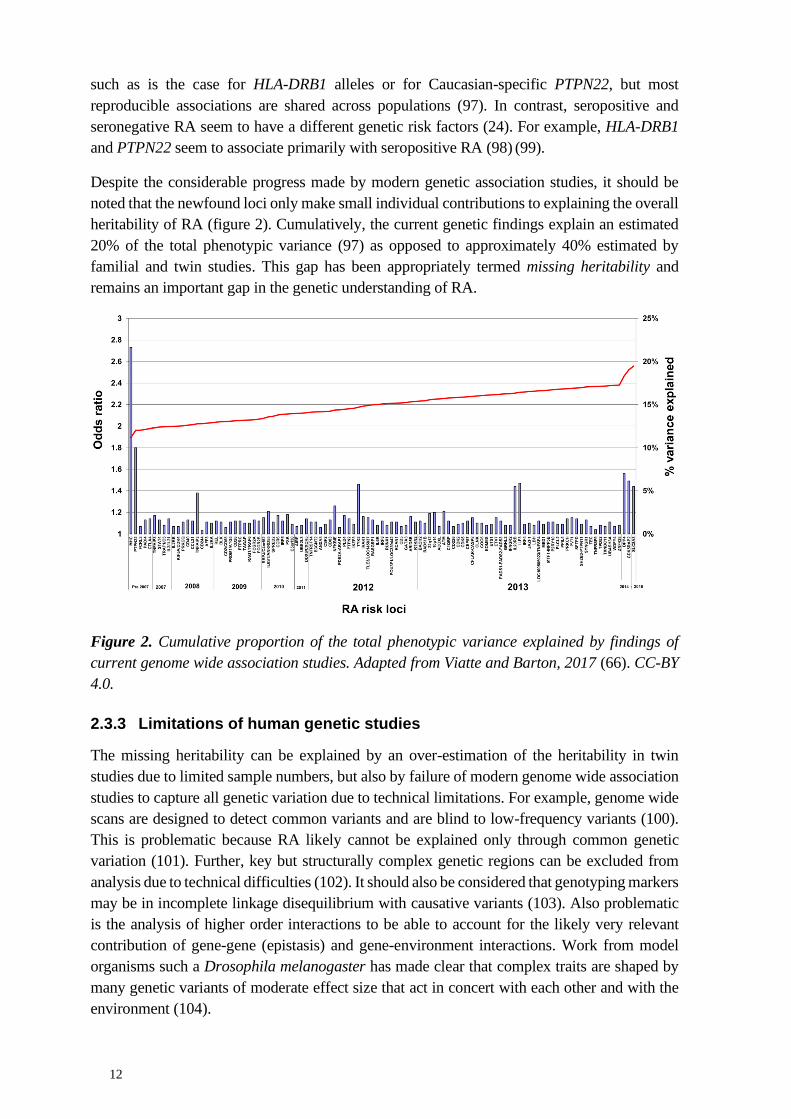
(107). As a result, it is likely that several risk loci remain to be identified. Figure 3 visualizes
the astounding differences in power when trying to detect strong versus moderate-to-weak RA
susceptibility loci. This is unfortunate, as we demonstrate in the present work that
polymorphisms in genes with moderate effect size are likely critical co-determinants of disease
development and perpetuation. We find that polymorphisms in genes such as VDR, CD2, or
SH3GL1 can be critical to fine-tuning T cell responses and inflammation.
Figure 3: Regional association plots showing strength of association with RA across the HLA-
DRB1, PTPN22, and CD2 locus. Black bars give an approximation of the respective gene. The
genome-wide significance threshold is indicated by the lower black line. GWAS summary
statistics were extracted from the IEU open GWAS project (108).
Once a candidate risk locus is identified, further resistance comes in the form of functional
interpretation. Unfortunately, most identified variants in genetic association studies concentrate
in non-coding regions (109) (probably of regulatory nature (110)) and are thus difficult to
interpret. Often, non-coding variants have regulatory properties, affecting regulation of nearby
genes (111) (112). However, clarifying these mechanisms is time and resource consuming, a
process further impeded by cell type or general context specificity of the effects of some
variants (113). Additional difficulty is given by the fact that a sequenced SNP can be in high
linkage disequilibrium to actual causative variants.
Evidently, disentangling the genetic component of RA is complicated. Complementary
approaches to human genetic association studies are needed to broaden our understanding of
genetic variants contributing to disease susceptibility. Using animal models of arthritis, we
demonstrate in study I and study III how non-coding variants can control gene expression to
regulate immune phenotypes.
2.4 USING MOUSE MODELS TO STUDY RA GENETICS
Contrary to human samples, work with animal models provides stable experimental conditions,
genetic uniformity, accessibility to disease-relevant tissues, and a broad genetic toolkit for
functional characterization of candidate genes.
Mice and human shared a common ancestor 60-80 million years ago, but identity and
arrangement of genes have largely stayed the same (114). The mouse and human genome are
14
highly conserved (115). This is also true for immune biology, although it should be recognized
that discrete differences exist between species (116) (117) (118). Thus, rodents are an excellent
model system to identify and characterize genetic factors regulating immune, as well as other,
traits. In theory, it is possible to dissect the genetic component of a complex trait using only a
few hundred mice (119), whereas in humans the same effort may require tens of thousands of
individuals (120). Optimally, of course, human and mouse studies can be integrated, with
findings from each species informing the biology of the other. To generate relevant and
translatable findings, mouse genetic studies require well characterised model systems closely
resembling the human trait or pathological condition under study.
2.4.1 Inducing RA-like arthritis in mice
Several models exist to induce RA-like autoimmune arthritis in mice and rats. Many of these
models consist in immunizing mice with common cartilage proteins in adjuvant. Some
examples include collagen type II (121) and type XI (122), as well as cartilage oligomeric
matrix protein (123). Nevertheless, it is also possible to induce arthritis using systemically
expressed antigens such as glucose-6-phosphate isomerase (124) or using mineral oils, such as
is the case in pristane-induced arthritis (125). This last model is particularly interesting
considering the association between RA and exposure to mineral oils (72). Not all arthritis
models need to be induced. Mice with transgenic overexpression of TNF-α (126) or mice with
a spontaneous mutation in the T cell signalling ZAP-70 protein (127) develop spontaneous
arthritis.
In the present work, we have mostly employed the collagen-induced arthritis (CIA) model
(121), as it is a well characterized and widely used rodent model of RA with several parallels
to the human autoimmune condition. In this model, an autoimmune arthritis is triggered by
immunizing mice with heterologous bovine collagen type II (bCII) in Complete Freund’s
Adjuvant (128). This elicits an autoimmune response driven by CII autoreactive T cells (129)
(130) (131) and characterized by the development of arthritogenic autoantibodies (132). A
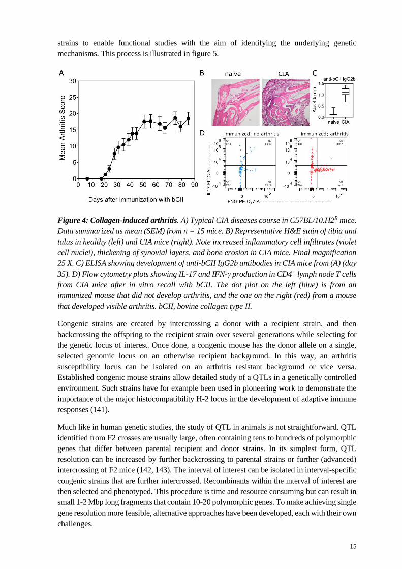
typical CIA experiment is depicted in figure 4. Much like in RA, CIA mice exhibit progressive
and severe joint lesions with T cell infiltrates (133), cartilage injury, bone remodelling (134),
and synovial hyperplasia (135). Similarly, several key RA cytokines are also involved in CIA
immune pathology, including IL-17 (136), IFN-ƴ (137) , IL-6 (138), TNFα (139), or IL-1β
(140).
CIA is a convenient screening model, as it is well characterized, incidence is high in susceptible
strains, and arthritis development can be easily monitored by scoring visible inflammation of
front and hind paws. Using this model, our research group has set out to identify genetic
determinants of autoimmune arthritis in a forward genetics-based approach. ‘Forward’ in this
case describes the direction of the workflow, starting from a phenotype and working towards
the unknown underlying genetic mechanism. Importantly, this method is hypothesis-free.
2.4.2 Mapping quantitative trait loci in the mouse
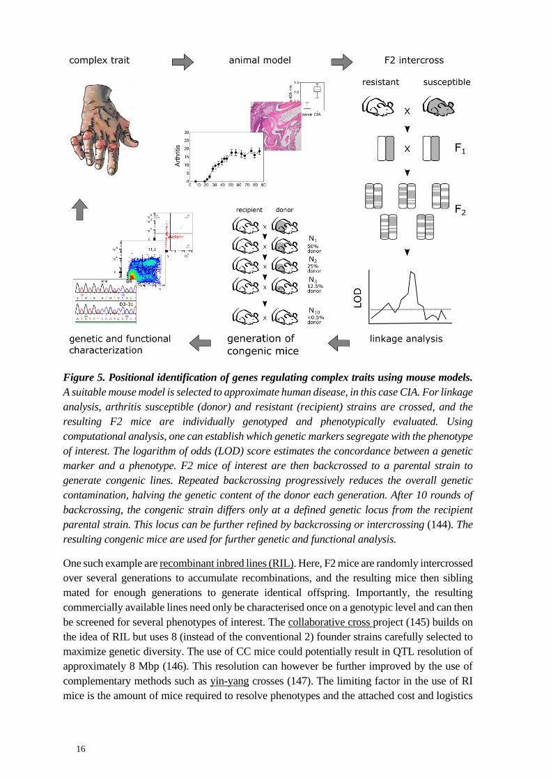
The positional identification of risk genes consists of two phases. First, genetic loci affecting a
quantitative phenotype of interest (quantitative trait loci, QTL) are roughly mapped by linkage
analysis, or in genetic association studies. Then, the identified QTL are isolated in congenic
15
strains to enable functional studies with the aim of identifying the underlying genetic
mechanisms. This process is illustrated in figure 5.
Figure 4: Collagen-induced arthritis. A) Typical CIA diseases course in C57BL/10.H2R mice.
Data summarized as mean (SEM) from n = 15 mice. B) Representative H&E stain of tibia and
talus in healthy (left) and CIA mice (right). Note increased inflammatory cell infiltrates (violet
cell nuclei), thickening of synovial layers, and bone erosion in CIA mice. Final magnification
25 X. C) ELISA showing development of anti-bCII IgG2b antibodies in CIA mice from (A) (day
35). D) Flow cytometry plots showing IL-17 and IFN-γ production in CD4+ lymph node T cells
from CIA mice after in vitro recall with bCII. The dot plot on the left (blue) is from an
immunized mouse that did not develop arthritis, and the one on the right (red) from a mouse
that developed visible arthritis. bCII, bovine collagen type II.
Congenic strains are created by intercrossing a donor with a recipient strain, and then
backcrossing the offspring to the recipient strain over several generations while selecting for
the genetic locus of interest. Once done, a congenic mouse has the donor allele on a single,
selected genomic locus on an otherwise recipient background. In this way, an arthritis
susceptibility locus can be isolated on an arthritis resistant background or vice versa.
Established congenic mouse strains allow detailed study of a QTLs in a genetically controlled
environment. Such strains have for example been used in pioneering work to demonstrate the
importance of the major histocompatibility H-2 locus in the development of adaptive immune
responses (141).
Much like in human genetic studies, the study of QTL in animals is not straightforward. QTL
identified from F2 crosses are usually large, often containing tens to hundreds of polymorphic
genes that differ between parental recipient and donor strains. In its simplest form, QTL
resolution can be increased by further backcrossing to parental strains or further (advanced)
intercrossing of F2 mice (142, 143). The interval of interest can be isolated in interval-specific
congenic strains that are further intercrossed. Recombinants within the interval of interest are
then selected and phenotyped. This procedure is time and resource consuming but can result in
small 1-2 Mbp long fragments that contain 10-20 polymorphic genes. To make achieving single
gene resolution more feasible, alternative approaches have been developed, each with their own
challenges.
16
Figure 5. Positional identification of genes regulating complex traits using mouse models.
A suitable mouse model is selected to approximate human disease, in this case CIA. For linkage
analysis, arthritis susceptible (donor) and resistant (recipient) strains are crossed, and the
resulting F2 mice are individually genotyped and phenotypically evaluated. Using
computational analysis, one can establish which genetic markers segregate with the phenotype
of interest. The logarithm of odds (LOD) score estimates the concordance between a genetic
marker and a phenotype. F2 mice of interest are then backcrossed to a parental strain to
generate congenic lines. Repeated backcrossing progressively reduces the overall genetic
contamination, halving the genetic content of the donor each generation. After 10 rounds of
backcrossing, the congenic strain differs only at a defined genetic locus from the recipient
parental strain. This locus can be further refined by backcrossing or intercrossing (144). The
resulting congenic mice are used for further genetic and functional analysis.
One such example are recombinant inbred lines (RIL). Here, F2 mice are randomly intercrossed
over several generations to accumulate recombinations, and the resulting mice then sibling
mated for enough generations to generate identical offspring. Importantly, the resulting
commercially available lines need only be characterised once on a genotypic level and can then
be screened for several phenotypes of interest. The collaborative cross project (145) builds on
the idea of RIL but uses 8 (instead of the conventional 2) founder strains carefully selected to
maximize genetic diversity. The use of CC mice could potentially result in QTL resolution of
approximately 8 Mbp (146). This resolution can however be further improved by the use of
complementary methods such as yin-yang crosses (147). The limiting factor in the use of RI
mice is the amount of mice required to resolve phenotypes and the attached cost and logistics
17
of maintaining thousands of lines. Furthermore, RIL often are bad breeders due to extensive
inbreeding.
Breeding problems can be circumvented by using outbred heterogeneous stock (HS) mice
(148)(149). HS mice result from advanced intercross of several inbred lines bred in pseudo-
random manner to minimize loss of genetic variability. Recombinations accumulate over
generations so that it becomes technically feasible to map QTL with sub-Mbp accuracy. Every
mouse is unique and must be individually genotyped in a genome wide scan much like in
human studies. The disadvantages of genome-wide analysis using HS mice lies in the
complexities of the analysis, need for large cohorts (thousands of mice), high-density
genotyping required (>6000 markers), possibility of false positives, and multiple testing issues.
Finally, it is possible to avoid breeding work altogether by mapping QTL in silico. Since
common inbred mouse strains have shared ancestry, it is possible to identify regions identical
by descent affecting quantitative traits. Here, statistical power can be low, as there is only a
limited number of inbred lines available, and assumptions need to be made regarding
conservation and inheritance pattern of identified haplotype blocks between inbred strains and
original founders. Further, a general limitation from genetic associations studies are false
positives (150). Regardless of the method used for mapping QTL, ultimately linkage analysis
and further functional studies are needed to conclusively prove findings from association
studies.
2.4.3 Arriving from QTL to causative gene
Distilling a causative genetic variant from a QTL is a challenging endeavour (151) (152).
Usually, the driving genes or variants in a QTL cannot be exactly positioned but instead need
to be inferred. For example, polymorphic genes within a QTL may be prioritized if they are
immediately relevant to the investigated trait, especially in the presence of non-synonymous
polymorphisms (153) (154) (155). Likewise of interest, may be polymorphic genes exhibiting
altered expression levels (156), for example as a consequence of non-coding polymorphisms
in promoters or enhancers. Once a candidate gene is singled out, the hypothesis is tested by
complementary approaches, to demonstrate functional relevance of the proposed candidate.
Popular strategies to demonstrate functional relevance include knockout, inhibition, or
overexpression of the candidate transcript in a relevant setting (157), as well as stimulation or
inhibition of the target protein using agonists or antagonists, respectively. A particularly elegant
method is quantitative complementation, where congenic mice are crossed to a null allele of
the candidate gene to compare the ability of the congenic and wildtype alleles to compensate
for the deficiency of the gene under investigation (158). Finally, the identified pathway should
also be explored in humans to corroborate overall relevance of the findings obtained in mice.
While systematic mutagenesis or systematic knockout may seem like more straightforward
alternatives to genetic mapping studies, these approaches have their own limitations (159). An
approximated 15% of genes are essential and are thus not optimal target for knockout strategies.
Further, knockouts approaches result in complete loss of gene function, which does not reflect
the physiological differences generated by natural variation. As a result, knockouts may elicit
compensatory mechanisms or disrupt important genetic or protein interactions, resulting in
artificial phenotypes that may not mirror the effects of common genetic variation. Therefore,

18
established congenic strains and linkage analysis remain a valuable tool as they not only allow
the conclusive identification of important genetic determinants, but also provide a
physiological context to study biological mechanisms in a meaningful manner.
19
3 RESEARCH AIMS
The genetic component of complex autoimmune diseases such as RA remains poorly
understood. To aid in this process, we previously set out to identify QTLs critical to the
development of autoimmune arthritis in mouse and rat model systems. The aim of this study
was to identify and characterize the genetic polymorphisms and molecular pathways
underlying several previously reported QTL.
Specifically, the aims were as follows:
• Identify the genetic and molecular mechanisms driving Cia37 (142)
• Identify the genetic and molecular mechanisms driving Cia21 (160)
• Establish the molecular mechanisms underlying the spontaneous Pia34 QTL (161)
20
4 METHODS
4.1 A brief overview of the animal models applied in this study
Collagen-induced arthritis. CIA is induced by immunizing mice intradermally with
heterologous collagen type II in CFA. Susceptible mice develop joint inflammation
characterised by synovitis, pannus formation, and cartilage and bone damage. CIA is MHC, T
cell, and B cell dependent. This model is described in more detail under 2.4.1.
Pristane-induced arthritis. Rats immunized with a single intradermal pristane injection
develop severe and chronic arthritis with symmetrical joint inflammation. PIA is MHC and T
cell dependent and indeed T cells infiltrates can be found in PIA joints. However, the exact
mechanism remains unclear as possible immunodominant T cell or B cell epitopes are not
known. In contrast to CIA, B cells are considered less important in PIA because serum does
not transfer disease (162).
Glucose-6-phosphate isomerase-induced arthritis. Transgenic K/BxN mice develop
antibodies against the GPI protein, causing spontaneous arthritis. GIA can also be induced by
immunizing mice with GPI full protein or immunodominant peptide hGPI325-339. Mice
develop a monophasic arthritis that is T cell and B cell dependent (163). Anti-GPI
autoantibodies can be found in a subset of RA patients (164).
Collagen antibody-induced arthritis. Arthritis is induced through intraperitoneal
injection of a cocktail of monoclonal arthritogenic antibodies targeting different epitope of the
collagen type II protein. Mice develop a short lived monophasic arthritis that is mediated by
neutrophils and macrophages activated by the presence of antibody aggregates on the cartilage.
In this context, complement and FcγRs seem to play important roles. CAIA is T and B cell
independent, and a useful model to study the effector phase of CIA (165).
Delayed-type hypersensitivity. The DTH model is, in essence, an in vivo antigen recall
assay driven by Th1-type cells infiltrating the skin (166). Mice are sensitized by immunizing
with antigen in CFA, and antigen-specific T cells are allowed to expand for 10 days. Thereafter,
mice are challenged with antigen intradermally and tissue swelling is measured after 24-72hrs
using a calliper to evaluate T cell responses to the antigen.
Experimental autoimmune encephalomyelitis. EAE is a model of multiple sclerosis.
Mice are immunized intradermally with myelin derived antigen emulsified in CFA. Pertussis
toxin can be administered to increase susceptibility. Mice can be immunized with myelin
oligodendrocyte protein, proteolipid protein, myelin basic protein, or spinal cord homogenate.
The result is a progressive, ascending paralysis of the extremities that begins at the tail.
Paralysis is caused by monocytes and T cells infiltrating the central nervous system and causing
axonal demyelination. CD4+ T cells are central for EAE with contributions from Th1 and Th17-
type cells chiefly producing IFN-γ, IL-17, and GM-CSF. Both myelin-reactive CD4+ and CD8+
T cells can transfer EAE, and are required to induce it. B cell-derived autoantibodies seem less
critical, with B cell acting mainly to support T cell responses (167).
DSS-induced colitis. DSS-induced colitis is a quick, 10 day model of inflammatory
bowel disease. Administration of dextran sodium sulfate (DSS) in the drinking water induces
severe colitis in susceptible mice by damaging the intestinal epithelium. Disease activity is
evaluated by following rectal bleeding, weight loss, and stool consistency. This model is driven
by innate cells like neutrophils and monocytes, and is T and B cell independent (168).
21
5 RESULTS AND DISCUSSION
5.1 Study I - Vitamin D receptor polymorphisms regulate T cells and T cell-
dependent inflammatory diseases.
In this study we positionally identify polymorphisms in the vitamin D receptor (VDR) promoter
as regulators of T cells in autoimmunity. We find that mice that overexpress VDR in activated
autoreactive T cells develop more severe autoimmune arthritis and that physiological levels of
vitamin D are required for normal T cell biology and activation. Due to the physiological nature
of our model, we can rule out calcaemic confounding factors. We also show that lymphocytes
in rheumatic joints highly upregulate VDR, suggesting that our finding are relevant to human
RA. In conclusion, our results suggest an inflammatory role for VDR in activated T cells.
The immunomodulatory properties of vitamin D were first recognized when it became evident
that cod liver oil, which is high in vitamin D, arrested development of tuberculosis (169) (170).
Further studies then showed that vitamin D availability is critical to innate immune responses
and bacterial clearance. At the same time, however, vitamin D deficiency has been associated
to several autoimmune conditions (171) (172), and together with in vitro studies fuelled the
idea that vitamin D may have a key immune dampening -rather than activating- role during
adaptive responses (173) (174) (175) (176). Naturally, the concept of vitamin D as a dietary
and inexpensive supplement to help restrain inflammation generated great interest.
However, whether vitamin D has meaningful immunoregulatory properties in a biological
context has remained questionable, as the beneficiary effects of vitamin D supplementation in
double-blinded clinical trials are not entirely convincing (177) (178) (179). Moreover, studies
often demonstrate immunomodulatory properties of vitamin D using supraphysiological doses
that provoke undesired calcaemic side effects. Our study brings a unique perspective to this
issue by exploiting naturally conserved genetic variation to investigate the immunomodulatory
effects of vitamin D and VDR in a physiological setting. Our findings indicate that VDR in a
physiological range supports T cell activation, and, in a broader sense, help cement the
importance of vitamin D for immune system biology.
5.2 Study II - Endophilin A2 deficiency protects rodents from autoimmune
arthritis by modulating T cell activation.
This work originated from the observation that a subset of rats in the otherwise highly arthritis-
susceptible DA strain inexplicably became resistant to arthritis. Using linkage analysis, we
found this protective phenotype to be driven by a QTL on chromosome 9 (Pia43). Sequence
analysis revealed a spontaneous insertion of a viral long terminal repeat in the first intron of
the SH3GL1 gene, resulting in silenced transcription of SH3GL1. We speculated that SH3GL1
deficiency was protecting rats from arthritis, and indeed could confirm this observation in
SH3GL1 knockout mice. Functional characterization revealed that SH3GL1 affected T cell
activation by interfering with TCR internalization. Finally, we showed that SH3GL1 is
overexpressed in PBMCs from RA patients, suggesting a role for SH3GL1 in the pathogenesis
of autoimmune arthritis.
Genetic and immunological studies strongly suggest that aberrant activation of autoreactive T
cells is key to the chronicity and lack of resolution in autoimmune diseases. Although
22
autoreactive T cells are promising therapeutic targets, development of (antigen-specific) T cell-
based immunotherapies has proven challenging. Monoclonal antibody-based targeting of broad
T cell populations (180) has had limited success, and more specific strategies are either still
under development (vaccination-type strategies) (181) or have proven ineffective (oral type II
collagen) (182) (183). Thus, there is a need to broaden our understanding of T cell regulation
with the aim of improving T cell-based therapies. Our results contribute to this by identifying
SH3GL1 as a key regulator of T cells and a promising new therapeutic pathway to target
(autoreactive) T cells.
5.3 Study III - Polymorphic estrogen receptor binding site causes CD2-
dependent sex bias in the susceptibility to autoimmune diseases
In this study we identify a polymorphic estrogen receptor binding site (ERBS) leading to sex-
dependent differences in several models of autoimmunity. This ERBS orchestrated expression
of surrounding genes in an estrogen-dependent manner, including the T cell costimulatory
molecule CD2. As a result, polymorphisms in this ERBS interfered with hormone-dependent
regulation of gene expression. Sex-specific changes in CD2 expression then culminated in
sexually dimorphic T cell responses. Our findings have several implications for human
autoimmunity.
First, we provide functional evidence that polymorphisms in the CD2 locus are relevant to RA
susceptibility. The CD2 locus has been associated to RA before (184, 185), however
association data is limited and functional evidence lacking. We demonstrate that
polymorphisms in the CD2 locus are important modulators of autoreactive T cell responses and
autoimmune disease in mice. Further, we show that CD2 expression is increase in RA synovia
and that CD2 expression correlates with disease activity, suggesting that CD2 polymorphisms
affecting CD2 expression are likely relevant to the development or perpetuation of human
autoimmunity.
Secondly, this study gives insight into the mechanisms driving sex differences in the immune
response. RA not only occurs predominantly in females, but its symptoms often recede during
pregnancy (186) (187) and have even been coupled to the menstrual cycle (188). Further, oral
contraceptives (85) (86) and HRT (88) associate positively with RA (although contradictory
reports exist (189)). Together, these data suggest an important immune modulatory function of
sex hormones. Indeed, both CD4+ T cell and humoral responses are sex biased (190) and often
stronger in women. ERs are expressed in CD4+ T cells (191), and likely play important
regulatory functions during immune responses. This is supported by studies showing that E2
promotes T cell responses at physiological concentrations (192) (193) (194). In the present
study, we show that E2 shapes the sexually dimorphic immune response by regulating the
expression of the T cell activation marker CD2.
Finally, this study also demonstrates the importance of sex-genotype interactions for regulating
(auto)immune responses. Sex-genotype interactions are an attractive mechanism of action to
explain the sexual dimorphism in autoimmunity, but it has been challenging to collect
conclusive evidence beyond associative findings. Our study not only shows that genotype-sex
interactions shape (auto)immune phenotypes, but also provides a tangible mechanism by
demonstrating how polymorphisms can act in a sex-specific manner by interfering with binding
of key hormone receptors.
23
6 CONCLUDING REMARKS
There is a clear need to further understand the substantial genetic component in RA and in other
autoimmune diseases. Technological advances in sequencing and high throughput genotyping
made the full characterization of complex genetic diseases such as RA a tangible goal.
However, this has been bottlenecked by unexpected degrees of complexity. While the
expectation to genetic associations studies was to find a discrete number of moderate effect
size-QTL with single underlying genes, complex diseases instead turned out to be governed by
a sea of difficult to interpret and interacting low effect size QTL (<5%) (146) (160) (195), with
only a few dominant QTL of strong effect size. A reason for this structure may be high selective
pressure on variants with large effect sizes.
Cloning QTL with small effect size is fundamentally problematic because it means identifying
and understanding small and interweaving molecular and genetic effects. The problem of
understanding complex traits is, at its core, the problem of understanding small genetic effects.
This is only aggravated by genotype-environment interactions. To successfully study complex
traits, model systems are required that reduce environmental and genetic variability while
enabling thorough functional analysis. Mouse models meet these requirements while
accurately recapitulating human (patho)physiology. Even in mouse models, however,
achieving single gene resolution from large QTLs is laborious. QTNs and causative genes need
not only be identified, but also functionally interpreted. Only a few studies using congenic
strains have been able to achieve this (155) (196). Historically, cloned QTL have had
exceptional effect sizes. Of over 2000 reported QTL, only 1% have arrived at a single gene
(146).
The results we present here are the culmination of more than a decade of successful genetic
work to identify and characterize key genetic determinants of autoimmune arthritis in mice.
Our findings aid in our understanding of the genetic architecture of complex autoimmune
diseases, providing functional evidence for the relevance of non-coding genetic variants in the
development of complex traits. The importance of our findings is not limited to RA, as complex
autoimmune diseases share many mechanistic features. This study thus helps clarify recurring
questions in the field of inflammation and autoimmunity, such as the contribution of vitamin
D receptor polymorphisms (197) or the importance of sex-genotype interactions for the
development and perpetuation of autoimmune diseases (198). Additionally, we identify a novel
immune regulator which may prove to be therapeutically useful (199).

24
7 ACKNOWLEDGEMENTS
I would like to start by thanking my main supervisor Rikard Holmdahl for the
opportunity to do my PhD in his research group and for being optimistic in every situation. I
will dearly miss your hands-off supervision style.
I also have to thank my co-supervisor Kutty Selva Nandakumar for originally recruiting
me as a PhD student and securing initial funding. As well as my other co-supervisor Liselotte
Bäckdahl for being generally supportive.
Further, I would like to thank colleagues that shaped me on a scientific level: Bruno R for
teaching me the basics of experimental immunology, providing the groundwork for the VDR
project, and guidance during manuscript writing. Michael F for doing important groundwork
in the CD2/ER project and providing advice when writing the manuscript. Martina J for
introducing me to genetics. Jonathan T and Sabrina H for being excellent scientist role
models. Min Y for teaching me to be disciplined. Johan B for teaching me to be thorough and
use common sense. Simon G for teaching me to focus. Vilma U for showing me that good
science also needs time and patience. Amit S for teaching me to spend more time thinking.
Susanne W for teaching me to be critical of everything. Changrong G for inspiring me to dive
into bioinformatics. Bingze X for inspiring me to be positive and work harder.
On a more personal note, I would like to thank: Mike A and Clara MH for being good
friends and excellent companions during my PhD journey. Jaime J for being in many ways
like me. Yibo H for fun breaks and sharing my passion for technology. Ulrika N for all the fun
and your ability to make the day-to-day a rollercoaster. Alexander K for overshadowing
everything with your offbeat personality. Michael B for not mincing words and
uncompromisingly defending your interests. Ana C for sharing my views on fashion and taste
in mediocre pop music. Laura RC for gossips in Spanish and your endless and contagious
energy to pursue extracurricular activities no matter how hard the work day. Erik L for having
memorized -and being able to recite- seemingly all existing epidemiological literature.
Danielle V for your unapologetic laugh. Angel YM for everything, from warmly welcoming
me when I arrived, to always fixing my salary during my prolonged studies. Zhongwei X for
being a friendly person and troubleshooting everything. Lei C for being the politest person.
Weiwei C for funny and unexpected comebacks. Huqiao L for always being professional and
willing to help. Xiaojie C for saying things how they are. Taotao L for keeping the lab in shape
and being my biggest fan. Biborka BV for fun moments and your quirky character. Outi S for
being cheerful and laughing at my (mediocre) comments. Betül T for your love for mechanical
engineering. Zeynep S for your deadpan humour. Rajan KP for being a nice guy and
motivated postdoc. Jianghong Z for showing it to everyone. Meng L for being friendly and
always smiling. Guanzhi for your cool style. Carlos P for bringing closer my South American
home. Pierre S for being forthcoming, helping with proteomics, and being a great tennis
partner. Christian B for constant wittiness. Amirata SD for having anecdotes to every topic.
Magali, Parisa, Arian and Oksana for providing entertainment and laughs to balance work
over many dinners. Aitor, Maria, Caitrin, Sunjay, Fadwa, Amo, and Yildiz for being a first
surrogate family when I arrived in Sweden on a dark October.
And finally, I thank my family, for continuous support in everything and bringing much
needed light-heartedness to every situation.
25
8 REFERENCES
1. S. E. Turvey, D. H. Broide, Innate immunity. Journal of Allergy and Clinical
Immunology (2010) https:/doi.org/10.1016/j.jaci.2009.07.016.
2. S. J. Gaudino, P. Kumar, Cross-talk between antigen presenting cells and T cells
impacts intestinal homeostasis, bacterial infections, and tumorigenesis. Frontiers in
Immunology (2019) https:/doi.org/10.3389/fimmu.2019.00360.
3. L. Klein, E. A. Robey, C. S. Hsieh, Central CD4 + T cell tolerance: deletion versus
regulatory T cell differentiation. Nature Reviews Immunology (2019)
https:/doi.org/10.1038/s41577-018-0083-6.
4. D. Nemazee, Mechanisms of central tolerance for B cells. Nature Reviews
Immunology (2017) https:/doi.org/10.1038/nri.2017.19.
5. Y. Yarkoni, A. Getahun, J. C. Cambier, Molecular underpinning of B-cell anergy.
Immunological Reviews (2010) https:/doi.org/10.1111/j.1600-065X.2010.00936.x.
6. M. A. ElTanbouly, R. J. Noelle, Rethinking peripheral T cell tolerance: checkpoints
across a T cell’s journey. Nature Reviews Immunology (2021)
https:/doi.org/10.1038/s41577-020-00454-2.
7. Z. Yang, et al., Autoimmunity and Traumatic Brain Injury. Current Physical Medicine
and Rehabilitation Reports (2017) https:/doi.org/10.1007/s40141-017-0146-9.
8. V. M. Stoecklein, A. Osuka, J. A. Lederer, Trauma equals danger--damage control by
the immune system. Journal of Leukocyte Biology (2012)
https:/doi.org/10.1189/jlb.0212072.
9. M. Rojas, et al., Molecular mimicry and autoimmunity. Journal of Autoimmunity
(2018) https:/doi.org/10.1016/j.jaut.2018.10.012.
10. S. Becart, et al., The role of posttranslational modifications in generating neo-epitopes
that bind to rheumatoid arthritis-associated HLA-DR alleles and promote autoimmune
T cell responses. PLoS ONE (2021) https:/doi.org/10.1371/journal.pone.0245541.
11. C. L. Bennett, et al., The immune dysregulation, polyendocrinopathy, enteropathy, X-
linked syndrome (IPEX) is caused by mutations of FOXP3. Nature Genetics (2001)
https:/doi.org/10.1038/83713.
12. J. Aaltonen, et al., An autoimmune disease, APECED, caused by mutations in a novel
gene featuring two PHD-type zinc-finger domains. Nature Genetics (1997)
https:/doi.org/10.1038/ng1297-399.
13. E. Grip, K. Carlsson, Kostnader för reumatoid artrit i sverige år 2014 (2016).
14. M. Cross, et al., The global burden of rheumatoid arthritis: Estimates from the Global
Burden of Disease 2010 study. Annals of the Rheumatic Diseases (2014)
https:/doi.org/10.1136/annrheumdis-2013-204627.
15. S. T. Ngo, F. J. Steyn, P. A. McCombe, Gender differences in autoimmune disease.
Frontiers in Neuroendocrinology (2014) https:/doi.org/10.1016/j.yfrne.2014.04.004.
16. H. G. Fassbender, M. Simmling‐Annefeld, The potential aggressiveness of synovial
tissue in rheumatoid arthritis. The Journal of Pathology (1983)
https:/doi.org/10.1002/path.1711390314.
17. A. E. Koch, et al., Vascular endothelial growth factor. A cytokine modulating
endothelial function in rheumatoid arthritis. Journal of immunology (Baltimore, Md. :
1950) (1994).
18. M. Rooney, et al., Analysis of the histologic variation of synovitis in rheumatoid
arthritis. Arthritis & Rheumatism (1988) https:/doi.org/10.1002/art.1780310803.
19. E. Waaler, On the occurrence of a factor in human serum activating the specific
agglutination of sheep blood corpuscles. APMIS (2007) https:/doi.org/10.1111/j.1600-
0463.2007.apm_682a.x.
20. E. Ossipova, et al., Affinity purified anti-citrullinated protein/peptide antibodies target
antigens expressed in the rheumatoid joint. Arthritis Research and Therapy 16 (2014).
26
21. C. Ge, R. Holmdahl, The structure, specificity and function of anti-citrullinated protein
antibodies. Nature Reviews Rheumatology (2019) https:/doi.org/10.1038/s41584-019-
0244-4.
22. K. Nishimura, et al., Meta-analysis: Diagnostic accuracy of anti-cyclic citrullinated
peptide antibody and rheumatoid factor for rheumatoid arthritis. Annals of Internal
Medicine (2007) https:/doi.org/10.7326/0003-4819-146-11-200706050-00008.
23. K. Forslind, M. Ahlmén, K. Eberhardt, I. Hafström, B. Svensson, Prediction of
radiological outcome in early rheumatoid arthritis in clinical practice: Role of
antibodies to citrullinated peptides (anti-CCP). Annals of the Rheumatic Diseases
(2004) https:/doi.org/10.1136/ard.2003.014233.
24. L. Padyukov, et al., A genome-wide association study suggests contrasting
associations in ACPA-positive versus ACPA-negative rheumatoid arthritis. Annals of
the Rheumatic Diseases (2011) https:/doi.org/10.1136/ard.2009.126821.
25. M. M. J. Nielen, et al., Specific Autoantibodies Precede the Symptoms of Rheumatoid
Arthritis: A Study of Serial Measurements in Blood Donors. Arthritis and Rheumatism
(2004) https:/doi.org/10.1002/art.20018.
26. P. P. Tak, et al., Analysis of the synovial cell infiltrate in early rheumatoid synovial
tissue in relation to local disease activity. Arthritis and Rheumatism (1997)
https:/doi.org/10.1002/art.1780400206.
27. P. P. Tak, et al., Analysis of the synovial cell infiltrate in early rheumatoid synovial
tissue in relation to local disease activity. Arthritis and Rheumatism (1997)
https:/doi.org/10.1002/art.1780400206.
28. Y. Okada, S. Eyre, A. Suzuki, Y. Kochi, K. Yamamoto, Genetics of rheumatoid
arthritis: 2018 status. Annals of the Rheumatic Diseases (2019)
https:/doi.org/10.1136/annrheumdis-2018-213678.
29. D. Baeten, et al., Comparative study of the synovial histology in rheumatoid arthritis,
spondyloarthropathy, and osteoarthritis: Influence of disease duration and activity.
Annals of the Rheumatic Diseases (2000) https:/doi.org/10.1136/ard.59.12.945.
30. J. A. van Boxel, S. A. Paget, Predominantly T-Cell Infiltrate in Rheumatoid Synovial
Membranes. New England Journal of Medicine (1975)
https:/doi.org/10.1056/nejm197509112931101.
31. A. E. Schröder, A. Greiner, C. Seyfert, C. Berek, Differentiation of B cells in the
nonlymphoid tissue of the synovial membrane of patients with rheumatoid arthritis.
Proceedings of the National Academy of Sciences of the United States of America
(1996) https:/doi.org/10.1073/pnas.93.1.221.
32. S. Takemura, et al., Lymphoid Neogenesis in Rheumatoid Synovitis. The Journal of
Immunology (2001) https:/doi.org/10.4049/jimmunol.167.2.1072.
33. H. U. Scherer, T. W. J. Huizinga, G. Krönke, G. Schett, R. E. M. Toes, The B cell
response to citrullinated antigens in the development of rheumatoid arthritis. Nature
Reviews Rheumatology (2018) https:/doi.org/10.1038/nrrheum.2018.10.
34. J. M. Kremer, et al., Treatment of Rheumatoid Arthritis by Selective Inhibition of T-
Cell Activation with Fusion Protein CTLA4Ig. New England Journal of Medicine
(2003) https:/doi.org/10.1056/nejmoa035075.
35. J. C. W. Edwards, et al., Efficacy of B-Cell–Targeted Therapy with Rituximab in
Patients with Rheumatoid Arthritis. New England Journal of Medicine (2004)
https:/doi.org/10.1056/nejmoa032534.
36. R. M. Thurlings, et al., Synovial tissue response to rituximab: Mechanism of action
and identification of biomarkers of response. Annals of the Rheumatic Diseases 67
(2008).
37. M. E. Duddy, A. Alter, A. Bar-Or, Distinct Profiles of Human B Cell Effector
Cytokines: A Role in Immune Regulation? The Journal of Immunology 172 (2004).
27
38. S. L. Constant, B lymphocytes as antigen-presenting cells for CD4+ T cell priming in
vivo. Journal of immunology (Baltimore, Md. : 1950) (1999).
39. S. Takemura, P. A. Klimiuk, A. Braun, J. J. Goronzy, C. M. Weyand, T Cell
Activation in Rheumatoid Synovium Is B Cell Dependent. The Journal of Immunology
167 (2001).
40. R. Thomas, M. McIlraith, L. S. Davis, P. E. Lipsky, Rheumatoid synovium is enriched
in CD45RBdim mature memory T cells that are potent helpers for B cell
differentiation. Arthritis & Rheumatism (1992) https:/doi.org/10.1002/art.1780351209.
41. P. L. Klarenbeek, et al., Inflamed target tissue provides a specific niche for highly
expanded T-cell clones in early human autoimmune disease. Annals of the Rheumatic
Diseases (2012) https:/doi.org/10.1136/annrheumdis-2011-200612.
42. E. A. James, et al., Citrulline-specific Th1 cells are increased in rheumatoid arthritis
and their frequency is influenced by disease duration and therapy. Arthritis and
Rheumatology (2014) https:/doi.org/10.1002/art.38637.
43. N. Matthews, P. Emery, D. Pilling, A. Akbar, M. Salmon, Subpopulations of primed t
helper cells in rheumatoid arthritis. Arthritis & Rheumatism (1993)
https:/doi.org/10.1002/art.1780360505.
44. K. J. Warrington, S. Takemura, J. J. Goronzy, C. M. Weyand, CD4+,CD28- T cells in
rheumatoid arthritis patients combine features of the innate and adaptive immune
systems. Arthritis and Rheumatism (2001) https:/doi.org/10.1002/1529-
0131(200101)44:1<13::AID-ANR3>3.0.CO;2-6.
45. A. P. Cope, H. Schulze-Koops, M. Aringer, The central role of T cells in rheumatoid
arthritis. Clinical and Experimental Rheumatology (2007).
46. K. Raza, et al., Synovial fluid leukocyte apoptosis is inhibited in patients with very
early rheumatoid arthritis. Arthritis Research and Therapy 8 (2006).
47. R. J. E. M. Dolhain, A. N. van der Heiden, N. T. ter Haar, F. C. Breedveld, A. M. M.
Miltenburg, Shift toward T lymphocytes with a T helper 1 cytokine-secretion profile in
the joints of patients with rheumatoid arthritis. Arthritis and Rheumatism 39 (1996).
48. S. Yang, C. Smith, J. M. Prahl, X. Luo, H. F. DeLuca, Vitamin D Deficiency
Suppresses Cell-Mediated Immunity in Vivo. Archives of Biochemistry and Biophysics
(1993) https:/doi.org/10.1006/abbi.1993.1260.
49. M. Chabaud, et al., Human interleukin-17: A T cell-derived proinflammatory cytokine
produced by the rheumatoid synovium. Arthritis and Rheumatism (1999)
https:/doi.org/10.1002/1529-0131(199905)42:5<963::AID-ANR15>3.0.CO;2-E.
50. H. Yamada, et al., Th1 but not Th17 cells predominate in the joints of patients with
rheumatoid arthritis. Annals of the Rheumatic Diseases 67 (2008).
51. P. A. Klimiuk, H. Yang, J. J. Goronzy, C. M. Weyand, Production of cytokines and
metalloproteinases in rheumatoid synovitis is T cell dependent. Clinical Immunology
(1999) https:/doi.org/10.1006/clim.1998.4618.
52. S. Kotake, et al., IL-17 in synovial fluids from patients with rheumatoid arthritis is a
potent stimulator of osteoclastogenesis. Journal of Clinical Investigation (1999)
https:/doi.org/10.1172/JCI5703.
53. M. Takeshita, et al., Multi-dimensional analysis identified rheumatoid arthritis-driving
pathway in human T cell. Annals of the Rheumatic Diseases 78 (2019).
54. J. Tuncel, et al., Self-reactive T cells induce and perpetuate chronic relapsing arthritis.
Arthritis Research and Therapy (2020) https:/doi.org/10.1186/s13075-020-2104-7.
55. I. Cecchi, et al., Neutrophils: Novel key players in Rheumatoid Arthritis. Current and
future therapeutic targets. Autoimmunity Reviews 17 (2018).
56. I. A. Udalova, A. Mantovani, M. Feldmann, Macrophage heterogeneity in the context
of rheumatoid arthritis. Nature Reviews Rheumatology 12 (2016).
28
57. S. Culemann, et al., Locally renewing resident synovial macrophages provide a
protective barrier for the joint. Nature (2019) https:/doi.org/10.1038/s41586-019-1471-
1.
58. K. Shin, et al., Mast Cells Contribute to Autoimmune Inflammatory Arthritis via Their
Tryptase/Heparin Complexes. The Journal of Immunology 182 (2009).
59. F. Rivellese, F. W. Rossi, M. R. Galdiero, C. Pitzalis, A. de Paulis, Mast cells in early
rheumatoid arthritis. International Journal of Molecular Sciences 20 (2019).
60. J. Suurmond, et al., Communication between human mast cells and CD4+ T cells
through antigen-dependent interactions. European Journal of Immunology 43 (2013).
61. A. J. Hueber, et al., Cutting Edge: Mast Cells Express IL-17A in Rheumatoid Arthritis
Synovium. The Journal of Immunology 184 (2010).
62. B. Bartok, G. S. Firestein, Fibroblast-like synoviocytes: Key effector cells in
rheumatoid arthritis. Immunological Reviews 233 (2010).
63. C. C. Tseng, et al., Dual role of chondrocytes in rheumatoid arthritis: The chicken and
the egg. International Journal of Molecular Sciences 21 (2020).
64. N. M. T. Roodenrijs, et al., Characteristics of difficult-to-treat rheumatoid arthritis:
Results of an international survey. Annals of the Rheumatic Diseases (2018)
https:/doi.org/10.1136/annrheumdis-2018-213687.
65. T. Bongartz, et al., Anti-TNF antibody therapy in rheumatoid arthritis and the risk of
serious infections and malignancies: Systematic review and meta-analysis of rare
harmful effects in randomized controlled trials. Journal of the American Medical
Association (2006) https:/doi.org/10.1001/jama.295.19.2275.
66. S. Viatte, A. Barton, Genetics of rheumatoid arthritis susceptibility, severity, and
treatment response. Seminars in Immunopathology (2017)
https:/doi.org/10.1007/s00281-017-0630-4.
67. L. Klareskog, V. Malmström, K. Lundberg, L. Padyukov, L. Alfredsson, Smoking,
citrullination and genetic variability in the immunopathogenesis of rheumatoid
arthritis. Seminars in Immunology 23, 92–98 (2011).
68. N. Wegner, et al., Peptidylarginine deiminase from Porphyromonas gingivalis
citrullinates human fibrinogen and α-enolase: Implications for autoimmunity in
rheumatoid arthritis. Arthritis & Rheumatism 62, 2662–2672 (2010).
69. M. M. Newkirk, K. N. W. Duffy, J. Leclerc, N. Lambert, J. B. Shiroky, Detection of
cytomegalovirus, epstein-barr virus and herpes virus-6 in patients with rheumatoid
arthritis with or without sjögren’s syndrome. Rheumatology (1994)
https:/doi.org/10.1093/rheumatology/33.4.317.
70. S. R. Harrison, D. Li, L. E. Jeffery, K. Raza, M. Hewison, Vitamin D, Autoimmune
Disease and Rheumatoid Arthritis. Calcified Tissue International (2019)
https:/doi.org/10.1007/s00223-019-00577-2.
71. C. Orellana, et al., Oral contraceptives, breastfeeding and the risk of developing
rheumatoid arthritis: Results from the Swedish EIRA study. Annals of the Rheumatic
Diseases (2017) https:/doi.org/10.1136/annrheumdis-2017-211620.
72. B. Sverdrup, et al., Association between occupational exposure to mineral oil and
rheumatoid arthritis: results from the Swedish EIRA case-control study. Arthritis
research & therapy 7, R1296-303 (2005).
73. N. Balandraud, J. Roudier, Epstein-Barr virus and rheumatoid arthritis. Joint Bone
Spine (2018) https:/doi.org/10.1016/j.jbspin.2017.04.011.
74. J. Roudier, J. Petersen, G. H. Rhodes, J. Luka, D. A. Carson, Susceptibility to
rheumatoid arthritis maps to a T-cell epitope shared by the HLA-Dw4 DR β-1 chain
and the Epstein-Barr virus glycoprotein gp110. Proceedings of the National Academy
of Sciences of the United States of America (1989)
https:/doi.org/10.1073/pnas.86.13.5104.
29
75. J. Roudier, J. Petersen, G. H. Rhodes, J. Luka, D. A. Carson, Susceptibility to
rheumatoid arthritis maps to a T-cell epitope shared by the HLA-Dw4 DR β-1 chain
and the Epstein-Barr virus glycoprotein gp110. Proceedings of the National Academy
of Sciences of the United States of America (1989)
https:/doi.org/10.1073/pnas.86.13.5104.
76. S. W. Scally, et al., A molecular basis for the association of the HLA-DRB1 locus,
citrullination, and rheumatoid arthritis. Journal of Experimental Medicine (2013)
https:/doi.org/10.1084/jem.20131241.
77. Y. Kochi, et al., PADI4 polymorphism predisposes male smokers to rheumatoid
arthritis. Annals of the Rheumatic Diseases (2011)
https:/doi.org/10.1136/ard.2010.130526.
78. D. Makrygiannakis, et al., Smoking increases peptidylarginine deiminase 2 enzyme
expression in human lungs and increases citrullination in BAL cells. Annals of the
Rheumatic Diseases (2008) https:/doi.org/10.1136/ard.2007.075192.
79. E. B. Lugli, et al., Expression of citrulline and homocitrulline residues in the lungs of
non-smokers and smokers: Implications for autoimmunity in rheumatoid arthritis.
Arthritis Research and Therapy (2015) https:/doi.org/10.1186/s13075-015-0520-x.
80. W. Nesse, et al., The periodontium of periodontitis patients contains citrullinated
proteins which may play a role in ACPA (anti-citrullinated protein antibody)
formation. Journal of Clinical Periodontology (2012) https:/doi.org/10.1111/j.1600-
051X.2012.01885.x.
81. K. Eriksson, et al., Seropositivity combined with smoking is associated with increased
prevalence of periodontitis in patients with rheumatoid arthritis. Annals of the
Rheumatic Diseases (2018) https:/doi.org/10.1136/annrheumdis-2017-212091.
82. I. Kostoglou-Athanassiou, P. Athanassiou, A. Lyraki, I. Raftakis, C. Antoniadis,
Vitamin D and rheumatoid arthritis. Therapeutic Advances in Endocrinology and
Metabolism (2012) https:/doi.org/10.1177/2042018812471070.
83. L. A. Merlino, et al., Vitamin D Intake Is Inversely Associated With Rheumatoid
Arthritis: Results From the Iowa Women’s Health Study. Arthritis and Rheumatism
50, 72–77 (2004).
84. G. G. Song, S. C. Bae, Y. H. Lee, Association between vitamin D intake and the risk
of rheumatoid arthritis: A meta-analysis. Clinical Rheumatology (2012)
https:/doi.org/10.1007/s10067-012-2080-7.
85. M. Pedersen, et al., Environmental risk factors differ between rheumatoid arthritis with
and without auto-antibodies against cyclic citrullinated peptides. Arthritis Research
and Therapy (2006) https:/doi.org/10.1186/ar2022.
86. M. Pedersen, et al., Strong combined gene-environment effects in anti-cyclic
citrullinated peptide-positive rheumatoid arthritis: A nationwide case-control study in
Denmark. Arthritis and Rheumatism (2007) https:/doi.org/10.1002/art.22597.
87. S. T. Ngo, F. J. Steyn, P. A. McCombe, Gender differences in autoimmune disease.
Frontiers in Neuroendocrinology 35, 347–369 (2014).
88. L. A. Merlino, J. R. Cerhan, L. A. Criswell, T. R. Mikuls, K. G. Saag, Estrogen and
Other Female Reproductive Risk Factors Are Not Strongly Associated With the
Development of Rheumatoid Arthritis in Elderly Women. Seminars in Arthritis and
Rheumatism (2003) https:/doi.org/10.1016/S0049-0172(03)00084-2.
89. G. M. Hall, M. Daniels, E. C. Huskisson, T. D. Spector, A randomised controlled trial
of the effect of hormone replacement therapy on disease activity in postmenopausal
rheumatoid arthritis. Annals of the Rheumatic Diseases (1994)
https:/doi.org/10.1136/ard.53.2.112.
90. K. P. Liao, L. Alfredsson, E. W. Karlson, Environmental influences on risk for
rheumatoid arthritis. Current opinion in rheumatology 21, 279–83 (2009).
30
91. P. Stastny, Mixed lymphocyte cultures in rheumatoid arthritis. Journal of Clinical
Investigation (1976) https:/doi.org/10.1172/JCI108382.
92. P. K. Gregersen, J. Silver, R. J. Winchester, The Shared Epitope Hypothesis. Arthritis
and Rheumatism (1987).
93. Y. Okada, S. Eyre, A. Suzuki, Y. Kochi, K. Yamamoto, Genetics of rheumatoid
arthritis: 2018 status. Annals of the Rheumatic Diseases (2019)
https:/doi.org/10.1136/annrheumdis-2018-213678.
94. K. McAllister, S. Eyre, G. Orozco, Genetics of rheumatoid arthritis: GWAS and
beyond. Open Access Rheumatology: Research and Reviews (2011)
https:/doi.org/10.2147/OARRR.S14725.
95. Y. Okada, S. Eyre, A. Suzuki, Y. Kochi, K. Yamamoto, Genetics of rheumatoid
arthritis: 2018 status. Annals of the Rheumatic Diseases (2019)
https:/doi.org/10.1136/annrheumdis-2018-213678.
96. L. Jiang, et al., Novel risk loci for rheumatoid arthritis in han chinese and congruence
with risk variants in europeans. Arthritis and Rheumatology (2014)
https:/doi.org/10.1002/art.38353.
97. S. Viatte, A. Barton, Genetics of rheumatoid arthritis susceptibility, severity, and
treatment response. Seminars in Immunopathology (2017)
https:/doi.org/10.1007/s00281-017-0630-4.
98. D. Van Der Woude, et al., Quantitative heritability of anti-citrullinated protein
antibody-positive and anti-citrullinated protein antibody-negative rheumatoid arthritis.
Arthritis and Rheumatism (2009) https:/doi.org/10.1002/art.24385.
99. F. Kurreeman, et al., Genetic basis of autoantibody positive and negative rheumatoid
arthritis risk in a multi-ethnic cohort derived from electronic health records. American
Journal of Human Genetics (2011) https:/doi.org/10.1016/j.ajhg.2010.12.007.
100. P. L. Auer, G. Lettre, Rare variant association studies: Considerations, challenges and
opportunities. Genome Medicine (2015) https:/doi.org/10.1186/s13073-015-0138-2.
101. T. A. Manolio, et al., Finding the missing heritability of complex diseases. Nature 461,
747–753 (2009).
102. L. M. Olsson, et al., A single nucleotide polymorphism in the NCF1 gene leading to
reduced oxidative burst is associated with systemic lupus erythematosus. Annals of the
Rheumatic Diseases (2017) https:/doi.org/10.1136/annrheumdis-2017-211287.
103. J. Yang, et al., Common SNPs explain a large proportion of the heritability for human
height. Nature Genetics (2010) https:/doi.org/10.1038/ng.608.
104. T. F. C. Mackay, R. R. H. Anholt, Of flies and man: Drosophila as a model for human
complex traits. Annual Review of Genomics and Human Genetics (2006)
https:/doi.org/10.1146/annurev.genom.7.080505.115758.
105. L. Bomba, K. Walter, N. Soranzo, The impact of rare and low-frequency genetic
variants in common disease. Genome Biology 18, 77 (2017).
106. O. A. Panagiotou, et al., What should the genome-wide significance threshold be?
Empirical replication of borderline genetic associations. International Journal of
Epidemiology (2012) https:/doi.org/10.1093/ije/dyr178.
107. P. C. Sham, S. M. Purcell, Statistical power and significance testing in large-scale
genetic studies. Nature Reviews Genetics 15, 335–346 (2014).
108. B. Elsworth, et al., The MRC IEU OpenGWAS data infrastructure. bioRxiv (2020)
https:/doi.org/10.1101/2020.08.10.244293.
109. S. L. Edwards, J. Beesley, J. D. French, A. M. Dunning, Beyond GWASs: illuminating
the dark road from association to function. American journal of human genetics 93,
779–97 (2013).
110. M. T. Maurano, et al., Systematic localization of common disease-associated variation
in regulatory DNA. Science (New York, N.Y.) 337, 1190–5 (2012).
31
111. K. Musunuru, et al., From noncoding variant to phenotype via SORT1 at the 1p13
cholesterol locus. Nature 466, 714–9 (2010).
112. O. Harismendy, et al., 9p21 DNA variants associated with coronary artery disease
impair interferon-γ signalling response. Nature 470, 264–8 (2011).
113. A. M. Walsh, et al., Integrative genomic deconvolution of rheumatoid arthritis GWAS
loci into gene and cell type associations. Genome biology 17, 79 (2016).
114. K. Paigen, A miracle enough: The power of mice. Nature Medicine (1995)
https:/doi.org/10.1038/nm0395-215.
115. R. H. Waterston, et al., Initial sequencing and comparative analysis of the mouse
genome. Nature (2002) https:/doi.org/10.1038/nature01262.
116. J. Mestas, C. C. W. Hughes, Of Mice and Not Men: Differences between Mouse and
Human Immunology. The Journal of Immunology (2004)
https:/doi.org/10.4049/jimmunol.172.5.2731.
117. K. Takao, T. Miyakawa, Genomic responses in mouse models greatly mimic human
inflammatory diseases. Proceedings of the National Academy of Sciences of the United
States of America (2015) https:/doi.org/10.1073/pnas.1401965111.
118. Junhee Seok, et al., Genomic responses in mouse models poorly mimic human
inflammatory diseases. Proceedings of the National Academy of Sciences of the United
States of America (2013) https:/doi.org/10.1073/pnas.1222878110.
119. D. T. Burke, et al., Dissection of complex adult traits in a mouse synthetic population.
Genome research 22, 1549–57 (2012).
120. J. Flint, E. Eskin, Genome-wide association studies in mice. Nature reviews. Genetics
13, 807–17 (2012).
121. A.-K. B. Lindqvist, et al., Mouse models for rheumatoid arthritis. Trends in Genetics
18, S7–S13 (2002).
122. S. Lu, S. Carlsen, A. S. Hansson, R. Holmdahl, Immunization of rats with homologous
type XI collagen leads to chronic and relapsing arthritis with different genetics and
joint pathology than arthritis induced with homologous type II collagen. Journal of
Autoimmunity (2002) https:/doi.org/10.1006/jaut.2001.0581.
123. S. Carlsén, A. S. Hansson, H. Olsson, D. Heinegård, R. Holmdahl, Cartilage
oligomeric matrix protein (COMP)-induced arthritis in rats. Clinical and Experimental
Immunology (1998) https:/doi.org/10.1046/j.1365-2249.1998.00739.x.
124. K. Iwanami, et al., Arthritogenic T cell epitope in glucose-6-phosphate isomerase-
induced arthritis. Arthritis Research & Therapy 10, R130 (2008).
125. C. Vingsbo, et al., Pristane-induced arthritis in rats: A new model for rheumatoid
arthritis with a chronic disease course influenced by both major histocompatibility
complex and non-major histocompatibility complex genes. American Journal of
Pathology (1996).
126. P. Li, E. M. Schwarz, The TNF-α transgenic mouse model of inflammatory arthritis.
Springer Seminars in Immunopathology (2003) https:/doi.org/10.1007/s00281-003-
0125-3.
127. N. Sakaguchi, et al., Altered thymic T-cell selection due to a mutation of the ZAP-70
gene causes autoimmune arthritis in mice. Nature 426, 454–460 (2003).
128. D. D. Brand, K. A. Latham, E. F. Rosloniec, Collagen-induced arthritis. Nature
Protocols (2007) https:/doi.org/10.1038/nprot.2007.173.
129. T. J. Goldschmidt, R. Holmdahl, Anti‐T cell receptor antibody treatment of rats with
established autologous collagen‐induced arthritis: suppression of arthritis without
reduction of anti‐type II collagen autoantibody levels. European Journal of
Immunology (1991) https:/doi.org/10.1002/eji.1830210536.
130. K. M. Kadowaki, H. Matsuno, H. Tsuji, I. Tunru, CD4+ T cells from collagen-induced
arthritic mice are essential to transfer arthritis into severe combined immunodeficient
32
mice. Clinical and Experimental Immunology (1994) https:/doi.org/10.1111/j.1365-
2249.1994.tb06070.x.
131. A. Corthay, A. Johansson, M. Vestberg, R. Holmdahl, Collagen-induced arthritis
development requires alpha beta T cells but not gamma delta T cells: studies with T
cell-deficient (TCR mutant) mice. International immunology 11, 1065–73 (1999).
132. K. S. Nandakumar, J. Bäcklund, M. Vestberg, R. Holmdahl, Collagen type II (CII)-
specific antibodies induce arthritis in the absence of T or B cells but the arthritis
progression is enhanced by CII-reactive T cells. Arthritis research & therapy 6, R544–
R550 (2004).
133. S. H. Park, et al., BATF regulates collagen-induced arthritis by regulating T helper cell
differentiation. Arthritis Research and Therapy (2018) https:/doi.org/10.1186/s13075-
018-1658-0.
134. P. Caplazi, et al., Mouse Models of Rheumatoid Arthritis. Veterinary Pathology 52,
819–826 (2015).
135. J. S. Courtenay, M. J. Dallman, A. D. Dayan, A. Martin, B. Mosedale, Immunisation
against heterologous type II collagen induces arthritis in mice [13]. Nature (1980)
https:/doi.org/10.1038/283666a0.
136. S. Nakae, A. Nambu, K. Sudo, Y. Iwakura, Suppression of Immune Induction of
Collagen-Induced Arthritis in IL-17-Deficient Mice. The Journal of Immunology
(2003) https:/doi.org/10.4049/jimmunol.171.11.6173.
137. M. ‐C Boissier, et al., Biphasic effect of interferon‐γ in murine collagen‐induced
arthritis. European Journal of Immunology (1995)
https:/doi.org/10.1002/eji.1830250508.
138. T. Alonzi, et al., Interleukin 6 is required for the development of collagen-induced
arthritis. Journal of Experimental Medicine (1998)
https:/doi.org/10.1084/jem.187.4.461.
139. D. M. Butler, et al., DBA/1 mice expressing the human TNF-alpha transgene develop
a severe, erosive arthritis: characterization of the cytokine cascade and cellular
composition. Journal of immunology (Baltimore, Md. : 1950) (1997).
140. W. B. van den Berg, L. A. B. Joosten, M. Helsen, F. A. J. van de Loo, Amelioration of
established murine collagen-induced arthritis with anti- IL-1 treatment. Clinical and
Experimental Immunology (1994) https:/doi.org/10.1111/j.1365-2249.1994.tb06517.x.
141. H. O. Mcdevitt, A. Chinitz, Genetic control of the antibody response: Relationship
between immune response and histocompatibility (H-2) type. Science (1969)
https:/doi.org/10.1126/science.163.3872.1207.
142. E. Ahlqvist, R. Bockermann, R. Holmdahl, Fragmentation of two quantitative trait loci
controlling collagen-induced arthritis reveals a new set of interacting subloci. Journal
of immunology (Baltimore, Md. : 1950) 178, 3084–90 (2007).
143. M. Johannesson, et al., Gene expression profiling of arthritis using a QTL chip reveals
a complex gene regulation of the Cia5 region in mice. Genes and immunity 6, 575–83
(2005).
144. A. Darvasi, Experimental strategies for the genetic dissection of complex traits in
animal models. Nature Genetics (1998) https:/doi.org/10.1038/ng0198-19.
145. G. A. Churchill, et al., The Collaborative Cross, a community resource for the genetic
analysis of complex traits. Nature Genetics (2004) https:/doi.org/10.1038/ng1104-
1133.
146. J. Flint, W. Valdar, S. Shifman, R. Mott, Strategies for mapping and cloning
quantitative trait genes in rodents. Nature Reviews Genetics 6, 271–286 (2005).
147. S. Shifman, A. Darvasi, Mouse inbred strain sequence information and yin-yang
crosses for quantitative trait locus fine mapping. Genetics (2005)
https:/doi.org/10.1534/genetics.104.032474.
33
148. W. Valdar, et al., Genome-wide genetic association of complex traits in heterogeneous
stock mice. Nature Genetics (2006) https:/doi.org/10.1038/ng1840.
149. G. A. Churchill, D. M. Gatti, S. C. Munger, K. L. Svenson, The Diversity Outbred
mouse population. Mammalian Genome (2012) https:/doi.org/10.1007/s00335-012-
9414-2.
150. G. Manenti, et al., Mouse genome-wide association mapping needs linkage analysis to
avoid false-positive loci. PLoS Genetics (2009)
https:/doi.org/10.1371/journal.pgen.1000331.
151. D. Vaartjes, et al., Genetic dissection of a major haplotype associated with arthritis
reveal FcγR2b and FcγR3 to act additively. European Journal of Immunology (2020)
https:/doi.org/10.1002/eji.202048605.
152. N. R. Drinkwater, et al., The Long Path from QTL to Gene. PLoS Genetics 8,
e1002975 (2012).
153. S. T. Qureshi, et al., Endotoxin-tolerant mice have mutations in toll-like receptor 4
(Tlr4). Journal of Experimental Medicine (1999)
https:/doi.org/10.1084/jem.189.4.615.
154. A. A. Perelygin, et al., Positional cloning of the murine flavivirus resistance gene.
Proceedings of the National Academy of Sciences of the United States of America
(2002) https:/doi.org/10.1073/pnas.142287799.
155. P. Olofsson, et al., Positional identification of Ncf1 as a gene that regulates arthritis
severity in rats. Nature Genetics (2003) https:/doi.org/10.1038/ng1058.
156. J. R. Shorter, et al., Quantitative trait mapping in Diversity Outbred mice identifies
two genomic regions associated with heart size. Mammalian Genome (2018)
https:/doi.org/10.1007/s00335-017-9730-7.
157. U. C. Rogner, P. Avner, Congenic mice: cutting tools for complex immune disorders.
Nature Reviews Immunology 3, 243–252 (2003).
158. T. L. Turner, Fine-mapping natural alleles: Quantitative complementation to the
rescue. Molecular Ecology (2014) https:/doi.org/10.1111/mec.12719.
159. K. E. Noll, M. T. Ferris, M. T. Heise, The Collaborative Cross: A Systems Genetics
Resource for Studying Host-Pathogen Interactions. Cell Host and Microbe (2019)
https:/doi.org/10.1016/j.chom.2019.03.009.
160. M. Johannesson, et al., Identification of epistasis through a partial advanced intercross
reveals three arthritis loci within the Cia5 QTL in mice. Genes and Immunity 6, 175–
185 (2005).
161. U. Norin, et al., Endophilin A2 deficiency protects rodents from autoimmune arthritis
by modulating T cell activation. Nature Communications (2021)
https:/doi.org/10.1038/s41467-020-20586-2.
162. C. Vingsbo, et al., Pristane-induced arthritis in rats: A new model for rheumatoid
arthritis with a chronic disease course influenced by both major histocompatibility
complex and non-major histocompatibility complex genes. American Journal of
Pathology (1996).
163. A. Pizzolla, F. Laulund, K. Wing, R. Holmdahl, A new model of arthritis induced by a
glucose-6-phosphate isomerase peptide: immunological requirements and peptide
characterisation. Annals of the Rheumatic Diseases 71, A83.2-A83 (2012).
164. M. Schaller, D. R. Burton, H. J. Ditzel, Autoantibodies to GPI in rheumatoid arthritis:
linkage between an animal model and human disease. Nature immunology 2, 746–53
(2001).
165. K. S. Nandakumar, R. Holmdahl, Collagen antibody induced arthritis. Methods in
molecular medicine (2007) https:/doi.org/10.1007/978-1-59745-402-5_16.
166. Y. Luo, M. E. Dorf, Delayed-type hypersensitivity. Current protocols in immunology /
edited by John E. Coligan ... [et al.] Chapter 4, Unit 4.5 (2001).
34
167. S. Glatigny, E. Bettelli, Experimental Autoimmune Encephalomyelitis (EAE) as
Animal Models of Multiple Sclerosis (MS). Cold Spring Harbor perspectives in
medicine (2018) https:/doi.org/10.1101/cshperspect.a028977.
168. B. Chassaing, J. D. Aitken, M. Malleshappa, M. Vijay-Kumar, Dextran sulfate sodium
(DSS)-induced colitis in mice. Current protocols in immunology / edited by John E.
Coligan ... [et al.] 104, Unit 15.25. (2014).
169. M. Green, Cod liver oil and tuberculosis. BMJ (Online) (2011)
https:/doi.org/10.1136/bmj.d7505.
170. P. T. Liu, et al., Toll-like receptor triggering of a vitamin D-mediated human
antimicrobial response. Science (2006) https:/doi.org/10.1126/science.1123933.
171. T. Skaaby, L. L. N. Husemoen, B. H. Thuesen, A. Linneberg, Prospective population-
based study of the association between vitamin D status and incidence of autoimmune
disease. Endocrine (2015) https:/doi.org/10.1007/s12020-015-0547-4.
172. Y. H. Lee, S. C. Bae, S. J. Choi, J. D. Ji, G. G. Song, Associations between vitamin D
receptor polymorphisms and susceptibility to rheumatoid arthritis and systemic lupus
erythematosus: A meta-analysis. Molecular Biology Reports (2011)
https:/doi.org/10.1007/s11033-010-0477-4.
173. G. B. Ferreira, et al., 1,25-Dihydroxyvitamin D 3 Promotes Tolerogenic Dendritic
Cells with Functional Migratory Properties in NOD Mice . The Journal of Immunology
(2014) https:/doi.org/10.4049/jimmunol.1302350.
174. W. F. C. Rigby, T. Stacy, M. W. Fanger, Inhibition of T lymphocyte mitogenesis by
1,25-dihydroxyvitamin D3 (calcitriol). Journal of Clinical Investigation (1984)
https:/doi.org/10.1172/JCI111557.
175. L. E. Jeffery, et al., 1,25-Dihydroxyvitamin D3 and IL-2 Combine to Inhibit T Cell
Production of Inflammatory Cytokines and Promote Development of Regulatory T
Cells Expressing CTLA-4 and FoxP3. The Journal of Immunology (2009)
https:/doi.org/10.4049/jimmunol.0803217.
176. D. Bruce, S. Yu, J. H. Ooi, M. T. Cantorna, Converging pathways lead to
overproduction of IL-17 in the absence of vitamin D signaling. International
Immunology (2011) https:/doi.org/10.1093/intimm/dxr045.
177. K. E. Hansen, C. M. Bartels, R. E. Gangnon, A. N. Jones, J. Gogineni, An evaluation
of high-dose vitamin D for rheumatoid arthritis. Journal of clinical rheumatology :
practical reports on rheumatic & musculoskeletal diseases 20, 112–4 (2014).
178. M. Salesi, Z. Farajzadegan, Efficacy of Vitamin D in patients with active rheumatoid
arthritis receiving methotrexate therapy. Rheumatology International 32, 2129–2133
(2012).
179. W. Dankers, E. M. Colin, J. P. van Hamburg, E. Lubberts, Vitamin D in
Autoimmunity: Molecular Mechanisms and Therapeutic Potential. Frontiers in
immunology 7, 697 (2016).
180. E. C. Keystone, Abandoned therapies and unpublished trials in rheumatoid arthritis.
Current Opinion in Rheumatology (2003) https:/doi.org/10.1097/00002281-
200305000-00012.
181. K. S. Rosenthal, R. Carambula, D. H. Zimmerman, Why Don’t We Have a Vaccine
Against Autoimmune Diseases? - A Review. Journal of clinical & cellular
immunology (2019) https:/doi.org/10.4172/2155-9899.1000574.
182. M. L. Barnett, et al., Treatment of rheumatoid arthritis with oral type II collagen:
Results of a multicenter, double-blind, placebo-controlled trial. Arthritis and
Rheumatism (1998) https:/doi.org/10.1002/1529-0131(199802)41:2<290::AID-
ART13>3.0.CO;2-R.
183. M. Cazzola, et al., Oral type II collagen in the treatment of rheumatoid arthritis. A six-
month double blind placebo-controlled study. Clinical and Experimental
Rheumatology (2000).
35
184. D. Diogo, et al., Rare, low-frequency, and common variants in the protein-coding
sequence of biological candidate genes from GWASs contribute to risk of rheumatoid
arthritis. American Journal of Human Genetics (2013)
https:/doi.org/10.1016/j.ajhg.2012.11.012.
185. S. Raychaudhuri, et al., Genetic variants at CD28, PRDM1 and CD2/CD58 are
associated with rheumatoid arthritis risk. Nature Genetics (2009)
https:/doi.org/10.1038/ng.479.
186. M. Østensen, L. Fuhrer, R. Mathieu, M. Seitz, P. M. Villiger, A prospective study of
pregnant patients with rheumatoid arthritis and ankylosing spondylitis using validated
clinical instruments. Annals of the Rheumatic Diseases (2004)
https:/doi.org/10.1136/ard.2003.016881.
187. P. Dieppe, F. A. Wollheim, H. R. Schumacher, “The ameliorating effect of pregnancy
on chronic atrophic (infectious rheumatoid) arthritis, fibrositis, and intermittent
hydrarthrosis” in Classic Papers in Rheumatology, (2021)
https:/doi.org/10.3109/9780203214237-49.
188. N. S. Latman, Relation of menstrual cycle phase to symptoms of rheumatoid arthritis.
The American Journal of Medicine (1983) https:/doi.org/10.1016/0002-
9343(83)90789-1.
189. G. M. Hall, M. Daniels, E. C. Huskisson, T. D. Spector, A randomised controlled trial
of the effect of hormone replacement therapy on disease activity in postmenopausal
rheumatoid arthritis. Annals of the Rheumatic Diseases (1994)
https:/doi.org/10.1136/ard.53.2.112.
190. I. F. Cook, Sexual dimorphism of humoral immunity with human vaccines. Vaccine
(2008) https:/doi.org/10.1016/j.vaccine.2008.04.054.
191. K. L. Phiel, R. A. Henderson, S. J. Adelman, M. M. Elloso, Differential estrogen
receptor gene expression in human peripheral blood mononuclear cell populations.
Immunology Letters (2005) https:/doi.org/10.1016/j.imlet.2004.10.007.
192. W. Gilmore, L. P. Weiner, J. Correale, Effect of estradiol on cytokine secretion by
proteolipid protein-specific T cell clones isolated from multiple sclerosis patients and
normal control subjects. Journal of immunology (Baltimore, Md. : 1950) (1997).
193. A. Maret, et al., Estradiol enhances primary antigen-specific CD4 T cell responses and
Th1 development in vivo. Essential role of estrogen receptor α expression in
hematopoietic cells. European Journal of Immunology (2003)
https:/doi.org/10.1002/immu.200310027.
194. A. Andersson, et al., Estrogen regulates T helper 17 phenotype and localization in
experimental autoimmune arthritis. Arthritis research & therapy 17, 32 (2015).
195. P. L. Podolin, et al., Localization of two insulin-dependent diabetes (Idd) genes to the
Idd10 region on mouse Chromosome 3. Mammalian Genome (1998)
https:/doi.org/10.1007/s003359900749.
196. M. Bagot, et al., CD101 is expressed by skin dendritic cells. Role in T-lymphocyte
activation. Tissue antigens 50, 439–48 (1997).
197. G. Fernandez Lahore, et al., Vitamin D3 receptor polymorphisms regulate T cells and
T cell-dependent inflammatory diseases. Proceedings of the National Academy of
Sciences (2020) https:/doi.org/10.1073/pnas.2001966117.
198. G. Fernandez Lahore, et al., Polymorphic estrogen receptor binding site causes CD2-
dependent sex bias in the susceptibility to autoimmune diseases. Nature Portfolio
(2021) https:/doi.org/10.21203/rs.3.rs-337166/v1.
199. U. Norin, et al., Endophilin A2 deficiency protects rodents from autoimmune arthritis
by modulating T cell activation. Nature Communications (2021)
https:/doi.org/10.1038/s41467-020-20586-2.