Embed Size (px)
Citation preview

812 Research Article
IntroductionIn addition to the well-characterized role of phosphatidylinositol-3-phosphate (PtdIns3P) in endocytosis, recent evidence hasuncovered a crucial requirement for this lipid in macroautophagy(autophagy) (Axe et al., 2008; Obara et al., 2008a; Petiot et al.,2000). Autophagy occurs constitutively in nearly all cells tomaintain homeostasis, but is further activated in response tostress as a survival or adaptive mechanism. During autophagy, adouble-membrane phagophore forms and elongates aroundportions of cytosol, matures into an enclosed autophagosome,and delivers its contents to the lysosome for degradation(Klionsky, 2007). Basic biochemical components (i.e. aminoacids and fatty acids) are exported from the lysosome and recycledby the cell, representing an energetically favorable alternative tode novo synthesis. In mammalian systems, the lipid kinase Vps34forms a complex with several proteins, including Vps15, Beclin1,Atg14L, UVRAG and Bif1 to generate PtdIns3P on autophagicvesicles (Itakura et al., 2008; Zhong et al., 2009). PtdIns3P thenrecruits and tethers effector proteins, such as WIPI-1 (Atg18),which are required for proper membrane formation (Obara et al.,2008b; Proikas-Cezanne et al., 2004). The crucial requirementfor PtdIns3P in this process is evidenced by the fact thatautophagy is ablated in mutant Vps34 yeast strains and geneticVps34 knockouts in Drosophila (Juhasz et al., 2008; Kihara etal., 2001). Despite knowledge of PtdIns3P production, theantagonistic phosphatases that regulate PtdIns3P duringautophagy have remained elusive. Two myotubularin-relatedphosphatases, MTMR3 and MTMR14 (hJumpy), have recentlybeen shown to dephosphorylate autophagic PtdIns3P in variouscontexts (Taguchi-Atarashi et al., 2010; Vergne et al., 2009).
However, given the complexity of autophagy execution and thenumber of proteins in the autophagy network, we predict thatadditional protein phosphatases exist to regulate this process.Accordingly, we performed a high-content cell-based RNAi screenusing a fluorescent PtdIns3P sensor to identify proteinphosphatases that function upstream of PtdIns3P duringautophagy.
ResultsRNAi screening identifies PTPs as a modulator ofPtdIns3P signalingFYVE (Fab1, YOTB, Vac1 and EEA1) domains are cysteine-richzinc-finger binding motifs that specifically recognize and bindPtdIns3P (Gaullier et al., 1998). An EGFP molecule fused to twotandem FYVE domains, termed EGFP–2xFYVE, serves as aneffective cellular sensor of PtdIns3P (Gillooly et al., 2000). Weanalyzed U2OS cells stably expressing this construct by fluorescentmicroscopy and found that PtdIns3P predominantly localized topunctate, often perinuclear, vesicles when cultured in complete growthmedium with full nutrients (Fig. 1A, supplementary material Movie1). RNAi-mediated knockdown of Vps34 reduced cellular PtdIns3Pcontent and resulted in a diffuse cytosolic distribution of EGFP–2xFYVE (Fig. 1B,F, supplementary material Fig. S1A). By contrast,a redistribution of EGFP–2xFYVE to small abundant autophagicvesicles occurred when cells were deprived of amino acids to potentlyinduce autophagy (Fig. 1C, supplementary material Movie 2).
To identify genes that downregulate PtdIns3P signaling, weused several siRNAs targeting over 200 known and putativehuman phosphatases. The siRNAs were introduced into U2OSEGFP–2xFYVE cells, and the cells were subsequently monitored
SummaryMacroautophagy is a dynamic process whereby portions of the cytosol are encapsulated in double-membrane vesicles and deliveredto the lysosome for degradation. Phosphatidylinositol-3-phosphate (PtdIns3P) is concentrated on autophagic vesicles and recruitseffector proteins that are crucial for this process. The production of PtdIns3P by the class III phosphatidylinositol 3-kinase Vps34, hasbeen well established; however, protein phosphatases that antagonize this early step in autophagy remain to be identified. To identifysuch enzymes, we screened human phosphatase genes by RNA interference and found that loss of PTPs, a dual-domain proteintyrosine phosphatase (PTP), increases levels of cellular PtdIns3P. The abundant PtdIns3P-positive vesicles conferred by loss of PTPsstrikingly phenocopied those observed in cells starved of amino acids. Accordingly, we discovered that loss of PTPs hyperactivatesboth constitutive and induced autophagy. Finally, we found that PTPs localizes to PtdIns3P-positive membranes in cells, and thisvesicular localization is enhanced during autophagy. We therefore describe a novel role for PTPs and provide insight into theregulation of autophagy. Mechanistic knowledge of this process is crucial for understanding and targeting therapies for several humandiseases, including cancer and Alzheimer’s disease, in which abnormal autophagy might be pathological.
Key words: FYVE, PTPs, PtdIns3P, RNAi, Autophagy, Phosphatase
Accepted 9 November 2010Journal of Cell Science 124, 812-819 © 2011. Published by The Company of Biologists Ltddoi:10.1242/jcs.080341
Identification of PTPs as an autophagic phosphataseKatie R. Martin1,2, Yong Xu3, Brendan D. Looyenga1, Ryan J. Davis1, Chia-Lun Wu4, Michel L. Tremblay4,H. Eric Xu3 and Jeffrey P. MacKeigan1,*1Laboratory of Systems Biology, Van Andel Research Institute, Grand Rapids, MI 49503, USA2Cell and Molecular Biology Graduate Program, Michigan State University, East Lansing, MI 48824-4320, USA3Laboratory of Structural Sciences, Van Andel Research Institute, Grand Rapids, MI 49503, USA4Goodman Cancer Centre, Department of Biochemistry, McGill University, Montreal, QC H3A 1A3, Canada*Author for correspondence ([email protected])
Jour
nal o
f Cel
l Sci
ence

for PtdIns3P signaling. We identified several genes whoseknockdown significantly increased the abundance of cellularEGFP–2xFYVE punctae (Fig. 1E, supplementary material TableS1). Most notably, PtdIns3P was substantially increased followingknockdown of the myotubularin family member MTMR6(supplementary material Fig. S1B,C), as well as the dual-domainprotein tyrosine phosphatase PTPRS (PTPs) (Fig. 1D,E).Although reduced expression of MTMR6 was characterized bythe appearance of enlarged perinuclear vesicles, the siRNAstargeting PTPs caused a dramatic accumulation of abundantsmaller vesicles throughout the cytosol, which phenocopied resultsobserved during amino acid starvation (Fig. 1C,D, supplementarymaterial Movie 3). Quantification of PtdIns3P-positive vesiclesrevealed a 3.5-fold increase in abundance during starvation-induced autophagy and a nearly 5-fold increase caused byknockdown of PTPs alone (Fig. 1F). This phenotype wasconfirmed using four unique siRNA sequences targeting PTPs(supplementary material Fig. S1D-K).
To validate a physiological increase in PtdIns3P followingknockdown of PTPs, phospholipids were radiolabeled with[32P]orthophosphate in vivo, extracted, and resolved by thin-layerchromatography. Indeed, PtdIns3P levels were specifically elevatedin the absence of PTPs, whereas other lipid species remainedunchanged (Fig. 1G). To determine the identity of the PtdIns3P-
813PTPs regulates autophagy
positive vesicles formed by knockdown of PTPs, weimmunostained cells with well-established markers of earlyendosomes [anti-EEA1 (early endosomal antigen 1)] andautophagic vesicles (AVs) [anti-LC3 (light chain 3)]. We foundthat knockdown of PTPs had no substantial effect on the presenceof EEA1-positive endosomes (Fig. 1H, supplementary materialFig. S2A), but significantly increased the abundance of LC3-positive vesicles (Fig. 1I). From this, we hypothesized that PTPsfunctions during autophagy and focused our attention on thisenzyme as a candidate autophagic phosphatase.
Loss of PTPs hyperactivates constitutive and inducedautophagyThe striking resemblance of PtdIns3P-positive vesicles induced byPTPs knockdown to AVs formed during amino acid starvation ledus to propose that autophagy is hyperactivated in the absence ofPTPs, despite the presence of nutrients. To test this, autophagywas analyzed in U2OS cells by evaluating two ubiquitin-likeproteins, Atg12 and LC3 (Atg8), which become conjugated to AVsduring autophagy. Following phagophore nucleation, Atg12covalently binds Atg5 and oligomerizes with Atg16L at theautophagic membrane (Klionsky, 2007). To measure vesicleabundance at this step, we immunostained cells for endogenousAtg12 and quantified Atg12-positive punctae. We found that
Fig. 1. A cell-based siRNA screen identifies PTPs as a modulator of PtdIns3P. (A–D) U2OS EGFP–2xFYVE cells transfected with control siRNA (A), VSP34siRNA (B), starved of amino acids for 3 hours (C), or transfected with PTPRS siRNA (D), were fixed and imaged at 60� magnification by fluorescent microscopy(green, PtdIns3P, EGFP–2xFYVE; blue, nuclei). Insets show 2� magnifications of small EGFP–2xFYVE vesicles. Scale bars: 10m. (E)Following knockdownof phosphatases, EGFP–2xFYVE-positive punctae were scored from –100 (decreased from control cells) to +100 (increased). Phosphatases are ranked and plottedby decreasing score (left to right) with genes whose loss increased EGFP–2xFYVE fluorescence highlighted in green and whose loss caused decreases highlightedin blue. PTPRS is identified. (F)Mean EGFP–2xFYVE-positive punctate were quantified from cells under the conditions indicated using image analysis software.Bars represent s.e.m.; *P<0.05. (G)Phospholipids were radiolabeled in vivo, extracted, resolved by TLC, and visualized by autoradiography following transfectionwith control or PTPRS siRNA. A radiolabeled PtdIns3P standard was resolved adjacent to extracted lipids. (H,I)Endosomes were labeled by immunostaining withanti-EEA1 antibodies (H) and autophagic vesicles were labeled with anti-LC3B antibodies (I) following transfection with control or PTPRS siRNA (red, EEA1;green, LC3B; blue, nuclei). Insets show 2� magnifications of LC3-positive vesicles. Scale bars: 10m.Jo
urna
l of C
ell S
cien
ce

knockdown of PTPs increased AV abundance three- or fivefoldover the control when cells were cultured with rapamycin, a potentmTOR inhibitor and autophagy inducer, or full nutrients,respectively (Fig. 2A, supplementary material Fig. S2B).
The membrane-bound Atg5–Atg12–Atg16L complex permitslipidation of LC3, which is a classic marker for AVs (Hanada etal., 2007). LC3 is unique among the autophagy proteins in that aportion of it remains membrane bound and is degraded in thelysosome along with vesicle cargo. Therefore, lysosomal functioncan be inhibited [i.e. with bafilomycin A1 (Baf-A1) or chloroquinetreatment] and LC3 accumulation used as a measure of autophagicflux (Tanida et al., 2005). We found that both knockdown of PTPsand amino acid starvation increased the abundance of LC3-II, theAV-lipidated form of LC3, when lysates were probed withendogenous antibodies (Fig. 2B). Similarly, we observed anincreased number of EGFP–LC3-positive punctae when PTPsexpression was reduced under normal growth conditions, and thesestructures accumulated substantially when cells were cultured withBaf-A1, indicating their functionality (Fig. 2C–F). Knockdown ofPTPs caused an even greater increase in EGFP–LC3 punctaeabove the control level when cells were treated with both Baf-A1and rapamycin (Fig. 2G,H). Similar results were obtained whenAVs were quantified from cells immunostained for endogenousLC3 (supplementary material Fig. S2C).
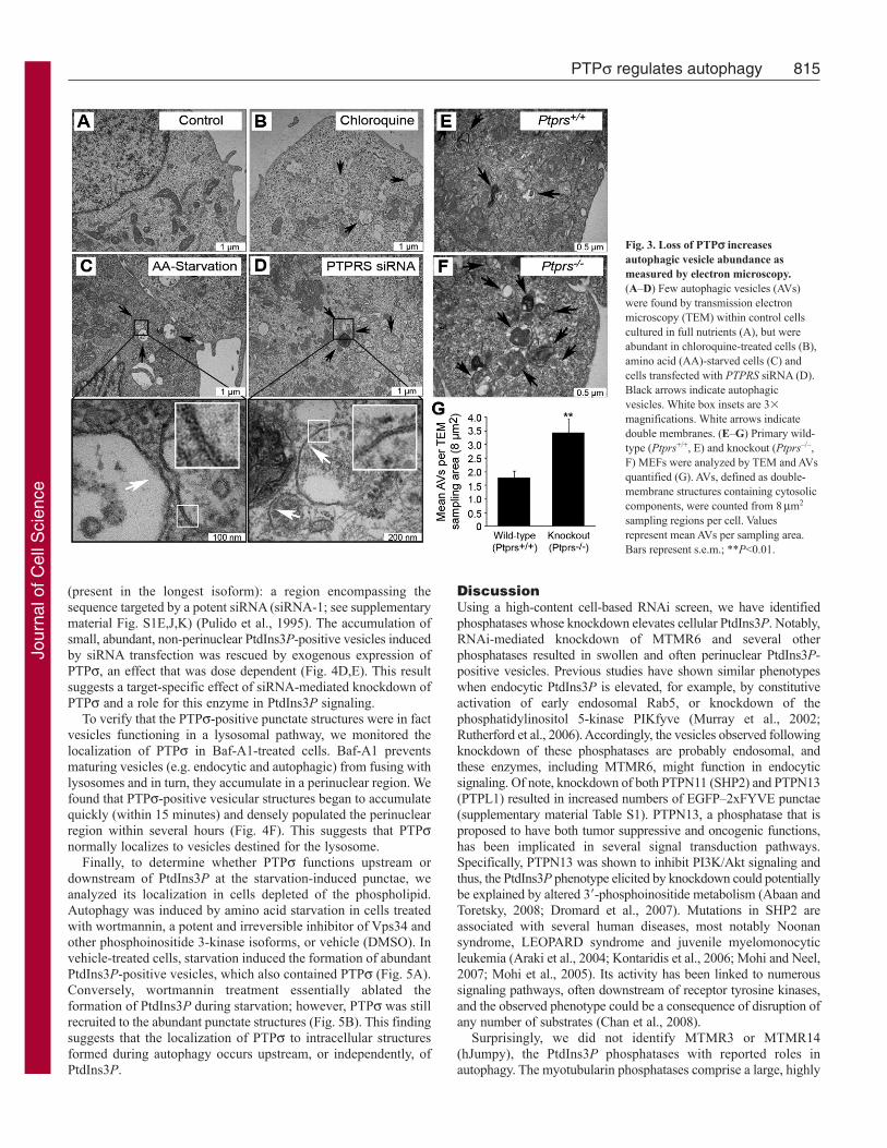
To confirm hyperactive autophagy in the absence of PTPsindependently of fluorescent markers, we detected AVs bytransmission electron microscopy (TEM). AVs are hallmarked byunique double membranes and by the presence of engulfed cytosoliccontent: features that allow them to be easily detected by TEM.Although control cells contained very few AVs, chloroquinetreatment increased their abundance, most of which appeared to beautolysosomal, as expected (Fig. 3A,B). Similarly, cells deprivedof amino acids for 1 hour harbored elevated numbers of double-membrane AVs, as did cells transfected with siRNAs against PTPs,but cultured in full nutrients (Fig. 3C,D). To establish this phenotypeindependently of RNAi, we examined autophagy during PTPs lossusing wild-type (Ptprs+/+) and PTPs knockout (Ptprs–/–) murineembryonic fibroblasts (MEFs). We have previously generated
814 Journal of Cell Science 124 (5)
Ptprs–/– mice by inserting a selectable neomycin resistance geneinto the D1 phosphatase (catalytic) domain. From these mice, wegenerated MEFs that lack both Ptprs transcript and protein, asmeasured by northern blot and western blot, respectively (Elcheblyet al., 1999). TEM analysis showed that both Ptprs+/+ and Ptprs–/–
primary MEFs contained a basal level of AVs; however, they weretwice as abundant in Ptprs–/– MEFs (Fig. 3E-G). Collectively,these results demonstrate that loss of PTPs, by RNAi and geneticdeletion, increases both constitutive and induced autophagy.
PTPs localizes to PtdIns3P-positive vesicles and rescuesthe siRNA phenotypeGiven the robust PtdIns3P response elicited by knockdown ofPTPs, we hypothesized that PTPs regulates autophagy byfunctioning at the level of PtdIns3P-positive vesicles. PTPs is abulky receptor-like PTP with an extracellular segment and twotandem cytosolic phosphatase domains (termed D1 and D2).Complex processing events have been reported for PTPs andrelated enzymes, including ectodomain shedding and internalizationfrom the cell surface (Aicher et al., 1997; Ruhe et al., 2006). Todetermine the localization of PTPs phosphatase domains, untaggedfull-length protein (FL-PTPs) was transiently expressed in U2OSEGFP–2xFYVE cells and detected by fluorescent microscopy usingD1-targeted monoclonal antibodies. We found that in addition toits presence at the plasma membrane, PTPs localized to theperinuclear region and to numerous intracellular-vesicle-likestructures, many of which were positive for PtdIns3P (Fig. 4A).Strikingly, autophagy induction by amino acid starvation induceda redistribution of PTPs to smaller vesicles, which were abundantthroughout the cytosol and were almost entirely PtdIns3P positive(Fig. 4B,C). In support of the notion that this localization wasautophagic, we discovered that PTPs was also capable of localizingto mRFP–LC3-positive punctae in the context of both basal andinduced autophagy (supplementary material Fig. S3B).
We further used exogenous PTPs expression in an RNAi rescueexperiment to demonstrate the specificity of phenotype induced byPTPRS siRNA. The naturally occurring isoform of PTPs used inour studies lacks the fourth through seventh fibronectin domains
Fig. 2. Loss of PTPs hyperactivates autophagy. (A)ATG12-positive punctae were quantified from 60� magnification imagesof cells transfected with control (black) or PTPRS siRNAs (white)and treated for 1 hour with normal growth medium (full nutrients)or 50 nM rapamycin. Values represent relative ATG12-positivepunctae per cell following normalization to control cells culturedwith full nutrients. Bars represent s.e.m.; ***P<0.001. (B)LC3-Iand LC3-II were analyzed by western blot using whole celllysates from cells transfected with control or PTPRS siRNA orstarved of amino acids. -tubulin was probed as a loading control.(C–H) EGFP–LC3-positive punctae were visualized in U2OScells transfected with control (C,E,G) or PTPRS (D,F,H) siRNAfollowing treatment for 2 hours with normal growth medium (fullnutrients; C,D), 100 nM bafilomycin A1 (Baf-A1; E,F), or 50 nMrapamycin and 100 nM Baf-A1 (G,H) (green, EGFP-LC3; blue,nuclei). Insets are 2� magnifications of EGFP–LC3-positiveAVs. Scale bars: 10m.
Jour
nal o
f Cel
l Sci
ence
(present in the longest isoform): a region encompassing thesequence targeted by a potent siRNA (siRNA-1; see supplementarymaterial Fig. S1E,J,K) (Pulido et al., 1995). The accumulation ofsmall, abundant, non-perinuclear PtdIns3P-positive vesicles inducedby siRNA transfection was rescued by exogenous expression ofPTPs, an effect that was dose dependent (Fig. 4D,E). This resultsuggests a target-specific effect of siRNA-mediated knockdown ofPTPs and a role for this enzyme in PtdIns3P signaling.
To verify that the PTPs-positive punctate structures were in factvesicles functioning in a lysosomal pathway, we monitored thelocalization of PTPs in Baf-A1-treated cells. Baf-A1 preventsmaturing vesicles (e.g. endocytic and autophagic) from fusing withlysosomes and in turn, they accumulate in a perinuclear region. Wefound that PTPs-positive vesicular structures began to accumulatequickly (within 15 minutes) and densely populated the perinuclearregion within several hours (Fig. 4F). This suggests that PTPsnormally localizes to vesicles destined for the lysosome.
Finally, to determine whether PTPs functions upstream ordownstream of PtdIns3P at the starvation-induced punctae, weanalyzed its localization in cells depleted of the phospholipid.Autophagy was induced by amino acid starvation in cells treatedwith wortmannin, a potent and irreversible inhibitor of Vps34 andother phosphoinositide 3-kinase isoforms, or vehicle (DMSO). Invehicle-treated cells, starvation induced the formation of abundantPtdIns3P-positive vesicles, which also contained PTPs (Fig. 5A).Conversely, wortmannin treatment essentially ablated theformation of PtdIns3P during starvation; however, PTPs was stillrecruited to the abundant punctate structures (Fig. 5B). This findingsuggests that the localization of PTPs to intracellular structuresformed during autophagy occurs upstream, or independently, ofPtdIns3P.
815PTPs regulates autophagy
DiscussionUsing a high-content cell-based RNAi screen, we have identifiedphosphatases whose knockdown elevates cellular PtdIns3P. Notably,RNAi-mediated knockdown of MTMR6 and several otherphosphatases resulted in swollen and often perinuclear PtdIns3P-positive vesicles. Previous studies have shown similar phenotypeswhen endocytic PtdIns3P is elevated, for example, by constitutiveactivation of early endosomal Rab5, or knockdown of thephosphatidylinositol 5-kinase PIKfyve (Murray et al., 2002;Rutherford et al., 2006). Accordingly, the vesicles observed followingknockdown of these phosphatases are probably endosomal, andthese enzymes, including MTMR6, might function in endocyticsignaling. Of note, knockdown of both PTPN11 (SHP2) and PTPN13(PTPL1) resulted in increased numbers of EGFP–2xFYVE punctae(supplementary material Table S1). PTPN13, a phosphatase that isproposed to have both tumor suppressive and oncogenic functions,has been implicated in several signal transduction pathways.Specifically, PTPN13 was shown to inhibit PI3K/Akt signaling andthus, the PtdIns3P phenotype elicited by knockdown could potentiallybe explained by altered 3�-phosphoinositide metabolism (Abaan andToretsky, 2008; Dromard et al., 2007). Mutations in SHP2 areassociated with several human diseases, most notably Noonansyndrome, LEOPARD syndrome and juvenile myelomonocyticleukemia (Araki et al., 2004; Kontaridis et al., 2006; Mohi and Neel,2007; Mohi et al., 2005). Its activity has been linked to numeroussignaling pathways, often downstream of receptor tyrosine kinases,and the observed phenotype could be a consequence of disruption ofany number of substrates (Chan et al., 2008).
Surprisingly, we did not identify MTMR3 or MTMR14(hJumpy), the PtdIns3P phosphatases with reported roles inautophagy. The myotubularin phosphatases comprise a large, highly
Fig. 3. Loss of PTPs increasesautophagic vesicle abundance asmeasured by electron microscopy.(A–D) Few autophagic vesicles (AVs)were found by transmission electronmicroscopy (TEM) within control cellscultured in full nutrients (A), but wereabundant in chloroquine-treated cells (B),amino acid (AA)-starved cells (C) andcells transfected with PTPRS siRNA (D).Black arrows indicate autophagicvesicles. White box insets are 3�
magnifications. White arrows indicatedouble membranes. (E–G) Primary wild-type (Ptprs+/+, E) and knockout (Ptprs–/–,F) MEFs were analyzed by TEM and AVsquantified (G). AVs, defined as double-membrane structures containing cytosoliccomponents, were counted from 8m2
sampling regions per cell. Valuesrepresent mean AVs per sampling area.Bars represent s.e.m.; **P<0.01.
Jour
nal o
f Cel
l Sci
ence

conserved family of enzymes whose members have been shown tofunction as heteromeric partners (Lorenzo et al., 2006). As oneexample, MTMR3 and MTMR4, both FYVE-domain containingphosphatases, have been demonstrated to interact with one anotherand inhibit PtdIns3P (Lorenzo et al., 2006). Accordingly, gene-by-gene loss-of-function analysis of this family might not revealphenotypes if compensation within the family occurs. Furthermore,these enzymes may serve cell- or context-specific functions thatare not revealed in this study.
816 Journal of Cell Science 124 (5)
The most striking result from this study was the presence ofabundant PtdIns3P-positive vesicles following knockdown ofPTPs, which phenocopied that of an autophagic cell. We confirmedhyperactive autophagy in the absence of PTPs through the use ofseveral autophagy markers, as well as electron microscopy. Atg12-and LC3-positive autophagic vesicles were substantially moreabundant in the absence of PTPs when cells were cultured in fullnutrients (constitutive AVs) or treated with rapamycin (inducedAVs). These autophagic vesicles accumulated upon treatment with
Fig. 4. Exogenous PTPs localizes to PtdIns3P vesicles and rescues the siRNA phenotype. (A,B)FL-PTPs was transiently expressed in U2OS EGFP–2xFYVEcells and PtdIns3P and PTPs imaged by fluorescent microscopy following incubation for 2 hours with full nutrient medium (A) or amino acid starvation medium(B) [green, PtdIns3P; red, anti-PTPs (D1-targeted antibodies); blue, nuclei]. Insets are 2� magnifications of boxed regions. Scale bars: 10m. (C)U2OS EGFP–2xFYVE cells transfected with FL-PTPs and amino acid starved for 2 hours were imaged using D1-targeted PTPs antibodies. A Z-stack of 0.25m incrementswas captured using sequential channel acquisition and confocal microscopy, with the third slice displayed and Z-stacks through the X and Y planes shown at theborder. Insets are 2� magnifications of boxed regions. Scale bar: 10m (green, PtdIns3P, EGFP–2xFYVE; red, anti-PTPs; yellow, colocalization). (D,E)U2OSEGFP–2xFYVE cells were transfected with PTPRS siRNA-1 for 48 hours, after which FL-PTPs (which lacks the sequence targeted by siRNA-1) was introducedfor an additional 24 hours. PtdIns3P and PTPs were imaged as previously described. The presence of siRNA-induced phenotype (abundant, non-perinuclearEGFP–2xFYVE-positive vesicles; indicated by white arrows in D) was determined for cells expressing no, low or high levels of FL-PTPs (E). Scale bars: 10m.(F)FL-PTPs-positive vesicular structures accumulate when lysosomal fusion is inhibited. U2OS cells expressing FL-PTPs for 24 hours were treated with 100 nMBaf-A1 in full nutrient medium for 0, 15, 60 or 240 minutes and FL-PTPs imaged using D1-targeted PTPs antibodies (red). Nuclei were stained with Hoechst33342 (blue). Scale bars: 10m.
Fig. 5. Localization of PTPs to vesicular structures does notrequire PtdIns3P. (A,B)U2OS cells expressing FL-PTPs weretreated with vehicle (DMSO; A) or 100 nM wortmannin (PI3Kinhibitor; B) for 2 hours while cultured in amino acid starvationmedium and PtdIns3P and PTPs was imaged by fluorescentmicroscopy [green, PtdIns3P, EGFP–2xFYVE; red, anti-PTPs(D1-targeted antibodies); blue, nuclei]. Insets are 2�
magnifications of boxed regions. Scale bars: 10m.
Jour
nal o
f Cel
l Sci
ence

the lysosomal inhibitors, Baf-A1 and chloroquine, demonstratingthat they were functional and destined for lysosomal degradation.This phenotype suggests that PTPs regulates an early step inautophagy induction, and its loss results in increased autophagicvesicle generation. This is consistent with the fact that PtdIns3P isgenerated on early phagophores and is required for properautophagic vesicle formation. A role for PTPs in autophagyinduction and more specifically in PtdIns3P regulation, is supportedby our findings that PTPs localizes to PtdIns3P-positive vesiclesthat increase in number during autophagy.
It remains to be addressed how PTPs is targeted to autophagicvesicles. PTPs is expressed at the cell surface in a two-subunitcomplex comprised of a large ectodomain and a membrane-spanning intracellular domain. Accordingly, it is implicated in cell–cell and cell–ECM interactions, and it is a crucial regulator of axonhomeostasis and neuronal development (Aicher et al., 1997;Elchebly et al., 1999; Uetani et al., 2006; Wallace et al., 1999).Relevant to our own work, ectodomain shedding and internalizationof a membrane-bound C-terminal fragment has been demonstratedpreviously (Aicher et al., 1997). Through immunofluorescenceanalysis of PTPs using D1-domain-specific antibodies, we placeintracellular PTPs on PtdIns3P-positive autophagic vesicles.Autophagosomes frequently fuse with endosomes during theirmaturation, forming hybrid organelles called amphisomes,establishing the possibility that PTPs is internalized by endocytosisto arrive at autophagic vesicles (Klionsky, 2007). Furthermore, theclose relative of PTPs LAR (PTPRF), undergoes an additionalproteolytic event whereby a soluble intracellular domain is formedand targeted inside the cell, similarly to the Notch receptor (Ruheet al., 2006). PTPs contains similar cleavage residues to LAR,making it therefore plausible that PTPs is targeted from the plasmamembrane to autophagic vesicles through a series of proteolyticevents in response to autophagic stimuli. Thus, this phosphatasemight serve several unique functions during various cellularconditions that are governed by its subcellular localization.
An important finding presented here is the recruitment of PTPsto vesicular structures during amino acid starvation, which occurseven in the absence of PtdIns3P generation. This finding, togetherwith the hyperactivation of autophagy elicited by knockdown ofPTPs (as measured by PtdIns3P, Atg12 and LC3), suggests thatPTPs regulates autophagy at an early step upstream of this lipid.In further support of this, we found that although almost all PTPs-positive vesicles are also positive for PtdIns3P (EGFP–2xFYVEpresence), fewer harbored LC3, a marker that is incorporated intoAVs at later stages of maturation.
There are several potential mechanisms by which PTPs mightfunction to regulate autophagy. First, it is possible that PTPsdirectly dephosphorylates PtdIns3P following recruitment to AVs.We tested the activity of recombinant PTPs in vitro, and althoughwe could not detect PtdIns3P phosphatase activity, it cannot beentirely excluded that PtdIns3P does not serve as a direct substratein vivo (supplementary material Fig. S4). It is also possible thatPTPs uses its robust protein phosphatase activity to regulate thefunction of a PtdIns3P-modifying enzyme, such as a PtdIns3Pphosphatase or a phosphoinositide 4- or 5-kinase. Alternatively,PTPs could control the activity of Vps34, which contains at leastone phosphotyrosine site, or another component within the largerVps34 complex (Imami et al., 2008). Finally, PTPs mightcontribute to the regulation of autophagy at the earliest initiationstep, which is executed by a complex of autophagy proteins,namely ULK1 (Atg1) and Atg13. The functional formation of this
817PTPs regulates autophagy
complex, which permits the generation of the PtdIns3P-positivephagophore, was recently found to be tightly regulated byphosphorylation events (Chang and Neufeld, 2009; Ganley et al.,2009; Hosokawa et al., 2009; Jung et al., 2009). The aim of futurework will be to determine the precise mechanism by which PTPsfunctions to regulate autophagy.
Materials and MethodssiRNA screen and validationU2OS EGFP–2xFYVE cells were seeded on 96-well plates (2000 cells per well) inMcCoy’s 5A medium (Invitrogen, Carlsbad, CA) supplemented with 10% fetalbovine serum (FBS, Invitrogen) at 37°C for 24 hours. Four siRNA molecules perphosphatase gene (phosphatase siRNA library version 2.0, Qiagen, Valencia, CA)were transfected per well at a final concentration of 25 nM using 0.2 l HiPerfecttransfection reagent (Qiagen) per well. After 48 hours, cells were fixed with 3.7%formaldehyde and nuclei were stained with Hoechst 33342 (Invitrogen). Cells werevisualized at 40� magnification on a Zeiss LSM 510 Meta confocal microscope(Oberkochen, Germany) and EGFP–2xFYVE fluorescence was compared with thatof control siRNA transfected cells within each plate. Triplicate wells from eachgene were qualitatively scored by two independent scorers on a scale from –100(decreased EGFP–2xFYVE signal and distribution) to +100 (increased) and meanscores were determined. Twenty-seven phosphatase genes whose knockdownincreased EGFP–2xFYVE fluorescence in the primary screen were used in asecondary screen, where four siRNAs were individually transfected to eliminateoff-target hits. The primary score was multiplied by a binned secondary screenscore (score of 1.0 for 3/4 or 4/4 siRNAs yielding a phenotype; 0.75 for 2/4siRNAs; and 0 for 0/4 or 1/4 siRNAs). Quantitative real-time PCR (qRT-PCR)assays with SYBR green dye (Roche, Basel, Switzerland) and gene-specific primersconfirmed that siRNAs effectively reduced mRNA expression of target genes. Forimaging, cells were cultured on number 1.5 coverglass, transfections repeated asabove, cells fixed, nuclei stained, and coverglass inverted into microslides withmounting gel. A control siRNA transfected well was cultured for 3 hours in aminoacid starvation medium [Dulbecco’s phosphate-buffered saline (DPBS) with 10%FBS and 1 g/l D-glucose]. Cells were imaged using a 60� oil objective on a NikonTE300 fluorescent microscope (Tokyo, Japan). EGFP–2xFYVE-positive vesicleswere quantified using image analysis software.
Phospholipid labeling, extraction and thin-layer chromatographyU2OS cells were seeded in McCoy’s 5A with 10% FBS at 200,000 cells per well ofsix-well tissue culture plates. After 24 hours, control or PTPRS siRNAs weretransfected at a final concentration of 25 nM using 2 l HiPerfect transfectionreagent per ml medium. Control siRNA was All-star Negative Control (Qiagen) andPTPRS siRNAs were two unique sequences (SI02759288, SI03056284, Qiagen).After 48 hours of transfection, the medium was replaced with phosphate-free DMEM(Invitrogen) supplemented with 10% phosphate-free FBS for 30 minutes. [32P]O4(0.25 mCi) was added per ml of medium for an additional 2 hours (Perkin Elmer,Waltham, MA). Radiolabeling was quenched with ice-cold TCA (10% finalconcentration) and cells incubated on ice for 1 hour. Cells were scraped, pelleted andlipids extracted via an acidified Bligh and Dyer method (Bird, 1994). Lipids werelyophilized, resuspended in chloroform:methanol (1:1), spotted on 20 cm � 20 cmsilica gel TLC plates (Whatman, Maidstone, UK), and resolved in a chamber usingboric acid buffer (Walsh et al., 1991). A PtdIns3P standard was generated byincubating synthetic phosphatidylinositol (diC16 PtdIns; Echelon, Salt Lake City,UT) with immunoprecipitated PI3K (using anti-p85, Cell Signaling, Danvers, MA)and [32P]ATP (Perkin Elmer). The TLC plate was exposed to film for 20 hours at –80°C and developed.
Fluorescent microscopy and western blot analyses of autophagy markersU2OS cells were seeded at a density of 35,000 cells per well in McCoy’s 5A mediumwith 10% FBS on number 1.5 coverglasses in 24-well tissue culture plates (forfluorescent imaging) or 150,000 cells per well on six-well dishes (for western blot).After 24 hours, cells were transfected with control or PTPRS siRNAs for 48 hours,as described above. Following, cells were treated for 1–2 hours in amino acidstarvation medium or with 50 nM rapamycin (Calbiochem, San Diego, CA), 25 Mchloroquine (Sigma, St Louis, MO), 100 nM Baf-A1 (A.G. Scientific, San Diego,CA) or normal growth medium (full nutrients; McCoy’s 5A with 10% FBS), asindicated. For western blots, cells were lysed [in 10 mM KPO4, 1 mM EDTA, 10mM MgCl2, 5 mM EGTA, 50 mM bis-glycerophosphate, 0.5% NP40, 0.1% Brij35,0.1% sodium deoxycholate, 1 mM NaVO4, 5 mM NaF, 2 mM DTT, and completeprotease inhibitors (Sigma)] and 20 g of total protein was resolved by SDS-PAGE.Proteins were transferred to PVDF membranes and probed with primary antibodies(LC3B, Cell Signaling Technologies; anti--tubulin, Sigma) for 16 hours at 4°Cfollowed by secondary antibodies (HRP-linked rabbit or mouse IgG, GE, Piscataway,NJ) for 1 hour at room temperature. Proteins were detected by enhancedchemiluminescence. For EGFP–LC3 imaging, U2OS cells stably expressing ptfLC3(Addgene plasmid 21074) (Kimura et al., 2007) were fixed with 3.7% formaldehydeand nuclei stained with Hoechst 33342 (2 g/ml). Coverglasses were inverted onto
Jour
nal o
f Cel
l Sci
ence

818 Journal of Cell Science 124 (5)
microslides using mounting gel and cells imaged using a 100� oil-immersionobjective on a Nikon Eclipse Ti fluorescence microscope. For immunofluorescence,cells were fixed with 3.7% formaldehyde, permeabilized with 0.2% Triton-X 100,and blocked with 3% bovine serum albumin (BSA) in PBS. Antibodies (LC3B,ATG12, EEA1; Cell Signaling Technologies) were added for 16 hours at 4°Cfollowed by Alexa-Fluor-488-conjugated anti-rabbit IgG (Invitrogen) for 1 hour atroom temperature. Nuclei were counterstained with Hoechst 33342, coverglasseswere inverted onto microslides using mounting gel and cells were imaged using 60�or 100� oil-immersion objectives on a Nikon TE300 fluorescence microscope (LC3,ATG12) or a 63� water-immersion objective on a Zeiss LSM510 Meta confocalmicroscope (EEA1). For quantification, punctae were counted using image analysissoftware after establishing an intensity threshold.
Transmission electron microscopyU2OS cells in 10 cm dishes were transfected with control or PTPRS siRNAs for 48hours as described above. Cells were briefly trypsinized, pelleted, rinsed andresuspended in 2% glutaraldehyde fixative (Sigma). Cell pellets were embedded in2% agarose, postfixed in osmium tetroxide, and dehydrated with an acetone series.Samples were infiltrated and embedded in Poly/Bed 812 resin and polymerized at60°C for 24 hours. Ultrathin sections (70 nm) were generated with a Power TomeXL (Boeckeler Instruments, Tucson, Arizona) and placed on copper grids. Cellswere examined using a JEOL 100C� Transmission Electron Microscope at 100 kV(Tokyo, Japan). Autophagic structures were quantified from images encompassingapproximately 8.5 m2 of cell area each. Electron microscopy services wereperformed by the Michigan State University Center for Advanced Microscopy (EastLansing, MI).
Exogenous PTPs expression and immunofluorescenceU2OS EGFP–2xFYVE cells were seeded at a density of 20,000 cells per well inMcCoy’s 5A medium with 10% FBS on number 1.5 coverglasses in 24-well tissueculture dishes. Full-length PTPRS cDNA (BC104812) was inserted into pRK7 byEcoRI digestion and ligation to yield FL-PTPs-pRK7 (FL-PTPs). DNA wastransfected at 0.15 g per well using 0.45 l FuGeneHD transfection reagent (Roche,Mannheim, Germany) in 50 l Optimem and 450 l McCoy’s 5A with 10% FBS for24 hours. For 2 hours, cells were cultured with full nutrient medium or starved ofamino acids (Fig. 4A-C), or amino acid starved while treated with DMSO or 100nM wortmannin (Sigma) (Fig. 5, supplementary material Fig. S3). Alternatively,cells were treated with Baf-A1 (100 nM in full nutrient medium) for 0, 15, 60 or 240minutes (Fig. 4E). Cells were then fixed with 3.7% formaldehyde, permeabilizedwith 0.2% Triton X-100, blocked in 3% BSA, and stained with antibodies targetingthe D1 domain of PTPs for 2 hours at room temperature. AF-546-conjugated anti-mouse-IgG (Invitrogen) were incubated for an additional hour at room temperatureand nuclei were stained with Hoechst 33342. Cells were imaged using oil-immersionobjectives at 60� on a Nikon TE3000 or 100� on an Eclipse Ti fluorescentmicroscope. For Fig. 4C, cells were treated as above and imaged using a 63� water-immersion objective on a Zeiss LSM510 Meta microscope. Red (AF-546, FL-PTPs)and green (EGFP–2xFYVE, PtdIns3P) channels were captured with confocalitythrough the Z-plane using 16 increments of 0.25 m. Stacks through the indicated Xand Y planes are shown at the border of an image of the third Z-plane. Forsupplementary material Fig. S3B, U2OS cells stably expressing mRFP–LC3(Addgene plasmid 21075) (Kimura et al., 2007) were seeded, transfected, treated(full nutrient or amino acid starvation media for 2 hours), and stained as above.Images were captured at 100� using an oil-immersion objective on an Eclipse Tifluorescent microscope.
Rescue of siRNA phenotypeU2OS EGFP–2xFYVE cells were seeded on number 1.5 coverglasses in 24-welldishes at 20,000 cells per well in McCoy’s 5A medium with 10% FBS. 24 hourslater, PTPRS siRNA-1 (CACGGCATCAGGCGTGCACAA; Qiagen) was transfectedat a concentration of 25 nM using 1 l oligofectamine per well per 500 l McCoy’swith 10% FBS (Invitrogen). FL-PTPs-pRK7 plasmid DNA was transfected 24 hourslater at a concentration of 0.15 g DNA per well using 0.45 l FuGeneHD transfectionreagent in 50 l Optimem and 450 l McCoy’s 5A with 10% FBS for an additional24 hours. Cells were fixed and immunostained as described above. Cells wereimaged using a 60� oil objective on a Nikon TE300 fluorescent microscope forEGFP–2xFYVE phenotype (green) and FL-PTPs-pRK7 expression (red). FL-PTPs-pRK7 expression levels were categorized as high or low. The presence or absenceof a robust PTPRS siRNA-induced EGFP–2xFYVE phenotype was determined(phenotype defined as the presence of small, abundant, non-perinuclear EGFP–2xFYVE-positive vesicles; indicated with white arrows in Fig. 4D) for 30–40 cellseach of low and high FL-PTPs-expressing cells, as well as cells transfected withPTPRS siRNA-1 but not FL-PTPs-pRK7.
In vitro phosphatase assaysGST-tagged recombinant PTPs containing all residues C-terminal to thetransmembrane domain (BC104812 cDNA; aa 883–1501) was generated in pGEXKG(Guan and Dixon, 1991). GST-tagged full-length MTMR6 (NM_004685.2) wasgenerated in pGEXKG and GST-tagged recombinant PTP1B was purchased (Upstate,Billerica, MA). Proteins were purified from BL21 Escherichia coli after isopropyl
b-D-1-thiogalactopyranoside (IPTG) induction and used in phosphatase assays. ForPtdIns3P phosphatase reactions, 1 g protein was suspended in 50 l assay buffer(50 mM sodium acetate, 25 mM Tris-HCl, 10 mM DTT, pH 6.5) with 0, 25, 50 or200 M diC8-PtdIns3P and reactions carried out at 37°C for 25 minutes. For Tyr-Pphosphatase assays, reactions were carried out as above using 0, 10, 25 or 100 MTyr-P peptide (TSTEPQpYQPGENL; Upstate) at 37°C for 15 minutes. Releasedphosphates were detected with malachite green (Upstate) and absorbance measuredat 650 nm. Background levels from enzyme-only and substrate-only (Tyr-P orPtdIns3P) reactions were subtracted and absorbance converted to picomoles freephosphate released per minute using a standard curve.
We thank the Van Andel Institute Systems Biology lab for advice,analysis, and reagents. This work was supported by the Department ofDefense Prostate Cancer Research Program of the Office ofCongressionally Directed Medical Research Programs PC081089 toJ.P.M. J.P.M. is also supported by Award Number R01CA138651 fromthe National Cancer Institute. Deposited in PMC for release after 12months.
Supplementary material available online athttp://jcs.biologists.org/cgi/content/full/124/5/812/DC1
ReferencesAbaan, O. D. and Toretsky, J. A. (2008). PTPL1: a large phosphatase with a split
personality. Cancer Metastasis Rev. 27, 205-214.Aicher, B., Lerch, M. M., Muller, T., Schilling, J. and Ullrich, A. (1997). Cellular
redistribution of protein tyrosine phosphatases LAR and PTPsigma by inducibleproteolytic processing. J. Cell Biol. 138, 681-696.
Araki, T., Mohi, M. G., Ismat, F. A., Bronson, R. T., Williams, I. R., Kutok, J. L.,Yang, W., Pao, L. I., Gilliland, D. G., Epstein, J. A. et al. (2004). Mouse model ofNoonan syndrome reveals cell type- and gene dosage-dependent effects of Ptpn11mutation. Nat. Med. 10, 849-857.
Axe, E. L., Walker, S. A., Manifava, M., Chandra, P., Roderick, H. L., Habermann,A., Griffiths, G. and Ktistakis, N. T. (2008). Autophagosome formation from membranecompartments enriched in phosphatidylinositol 3-phosphate and dynamically connectedto the endoplasmic reticulum. J. Cell Biol. 182, 685-701.
Bird, I. M. (1994). Analysis of cellular phosphoinositides and phosphoinositols byextraction and simple analytical procedures. Methods Mol. Biol. 27, 227-248.
Chan, G., Kalaitzidis, D. and Neel, B. G. (2008). The tyrosine phosphatase Shp2(PTPN11) in cancer. Cancer Metastasis Rev. 27, 179-192.
Chang, Y. Y. and Neufeld, T. P. (2009). An Atg1/Atg13 complex with multiple roles inTOR-mediated autophagy regulation. Mol. Biol. Cell 20, 2004-2014.
Dromard, M., Bompard, G., Glondu-Lassis, M., Puech, C., Chalbos, D. and Freiss,G. (2007). The putative tumor suppressor gene PTPN13/PTPL1 induces apoptosisthrough insulin receptor substrate-1 dephosphorylation. Cancer Res. 67, 6806-6813.
Elchebly, M., Wagner, J., Kennedy, T. E., Lanctot, C., Michaliszyn, E., Itie, A.,Drouin, J. and Tremblay, M. L. (1999). Neuroendocrine dysplasia in mice lackingprotein tyrosine phosphatase sigma. Nat. Genet. 21, 330-333.
Ganley, I. G., Lam du, H., Wang, J., Ding, X., Chen, S. and Jiang, X. (2009).ULK1.ATG13.FIP200 complex mediates mTOR signaling and is essential for autophagy.J. Biol. Chem. 284, 12297-12305.
Gaullier, J. M., Simonsen, A., D’Arrigo, A., Bremnes, B., Stenmark, H. and Aasland,R. (1998). FYVE fingers bind PtdIns(3)P. Nature 394, 432-433.
Gillooly, D. J., Morrow, I. C., Lindsay, M., Gould, R., Bryant, N. J., Gaullier, J. M.,Parton, R. G. and Stenmark, H. (2000). Localization of phosphatidylinositol 3-phosphate in yeast and mammalian cells. EMBO J. 19, 4577-4588.
Guan, K. L. and Dixon, J. E. (1991). Eukaryotic proteins expressed in Escherichia coli:an improved thrombin cleavage and purification procedure of fusion proteins withglutathione S-transferase. Anal. Biochem. 192, 262-267.
Hanada, T., Noda, N. N., Satomi, Y., Ichimura, Y., Fujioka, Y., Takao, T., Inagaki, F.and Ohsumi, Y. (2007). The Atg12-Atg5 conjugate has a novel E3-like activity forprotein lipidation in autophagy. J. Biol. Chem. 282, 37298-37302.
Hosokawa, N., Hara, T., Kaizuka, T., Kishi, C., Takamura, A., Miura, Y., Iemura, S.,Natsume, T., Takehana, K., Yamada, N. et al. (2009). Nutrient-dependent mTORC1association with the ULK1-Atg13-FIP200 complex required for autophagy. Mol. Biol.Cell 20, 1981-1991.
Imami, K., Sugiyama, N., Kyono, Y., Tomita, M. and Ishihama, Y. (2008). Automatedphosphoproteome analysis for cultured cancer cells by two-dimensional nanoLC-MSusing a calcined titania/C18 biphasic column. Anal. Sci. 24, 161-166.
Itakura, E., Kishi, C., Inoue, K. and Mizushima, N. (2008). Beclin 1 forms two distinctphosphatidylinositol 3-kinase complexes with mammalian Atg14 and UVRAG. Mol.Biol. Cell 19, 5360-5372.
Juhasz, G., Hill, J. H., Yan, Y., Sass, M., Baehrecke, E. H., Backer, J. M. and Neufeld,T. P. (2008). The class III PI(3)K Vps34 promotes autophagy and endocytosis but notTOR signaling in Drosophila. J. Cell Biol. 181, 655-666.
Jung, C. H., Jun, C. B., Ro, S. H., Kim, Y. M., Otto, N. M., Cao, J., Kundu, M. andKim, D. H. (2009). ULK-Atg13-FIP200 complexes mediate mTOR signaling to theautophagy machinery. Mol. Biol. Cell 20, 1992-2003.
Kihara, A., Noda, T., Ishihara, N. and Ohsumi, Y. (2001). Two distinct Vps34phosphatidylinositol 3-kinase complexes function in autophagy and carboxypeptidaseY sorting in Saccharomyces cerevisiae. J. Cell Biol. 152, 519-530.
Jour
nal o
f Cel
l Sci
ence

819PTPs regulates autophagy
Kimura, S., Noda, T. and Yoshimori, T. (2007). Dissection of the autophagosomematuration process by a novel reporter protein, tandem fluorescent-tagged LC3.Autophagy 3, 452-460.
Klionsky, D. J. (2007). Autophagy: from phenomenology to molecular understanding inless than a decade. Nat. Rev. Mol. Cell Biol. 8, 931-937.
Kontaridis, M. I., Swanson, K. D., David, F. S., Barford, D. and Neel, B. G. (2006).PTPN11 (Shp2) mutations in LEOPARD syndrome have dominant negative, notactivating, effects. J. Biol. Chem. 281, 6785-6792.
Lorenzo, O., Urbe, S. and Clague, M. J. (2006). Systematic analysis of myotubularins:heteromeric interactions, subcellular localisation and endosome related functions. J.Cell Sci. 119, 2953-2959.
Mohi, M. G. and Neel, B. G. (2007). The role of Shp2 (PTPN11) in cancer. Curr. Opin.Genet. Dev. 17, 23-30.
Mohi, M. G., Williams, I. R., Dearolf, C. R., Chan, G., Kutok, J. L., Cohen, S.,Morgan, K., Boulton, C., Shigematsu, H., Keilhack, H. et al. (2005). Prognostic,therapeutic, and mechanistic implications of a mouse model of leukemia evoked byShp2 (PTPN11) mutations. Cancer Cell 7, 179-191.
Murray, J. T., Panaretou, C., Stenmark, H., Miaczynska, M. and Backer, J. M.(2002). Role of Rab5 in the recruitment of hVps34/p150 to the early endosome. Traffic3, 416-427.
Obara, K., Noda, T., Niimi, K. and Ohsumi, Y. (2008a). Transport of phosphatidylinositol3-phosphate into the vacuole via autophagic membranes in Saccharomyces cerevisiae.Genes Cells 13, 537-547.
Obara, K., Sekito, T., Niimi, K. and Ohsumi, Y. (2008b). The Atg18-Atg2 complex isrecruited to autophagic membranes via phosphatidylinositol 3-phosphate and exerts anessential function. J. Biol. Chem. 283, 23972-23980.
Petiot, A., Ogier-Denis, E., Blommaart, E. F., Meijer, A. J. and Codogno, P. (2000).Distinct classes of phosphatidylinositol 3�-kinases are involved in signaling pathwaysthat control macroautophagy in HT-29 cells. J. Biol. Chem. 275, 992-998.
Proikas-Cezanne, T., Waddell, S., Gaugel, A., Frickey, T., Lupas, A. and Nordheim,A. (2004). WIPI-1alpha (WIPI49), a member of the novel 7-bladed WIPI proteinfamily, is aberrantly expressed in human cancer and is linked to starvation-inducedautophagy. Oncogene 23, 9314-9325.
Pulido, R., Serra-Pages, C., Tang, M. and Streuli, M. (1995). The LAR/PTP delta/PTPsigma subfamily of transmembrane protein-tyrosine-phosphatases: multiple humanLAR, PTP delta, and PTP sigma isoforms are expressed in a tissue-specific manner andassociate with the LAR-interacting protein LIP.1. Proc. Natl. Acad. Sci. USA 92, 11686-11690.
Ruhe, J. E., Streit, S., Hart, S. and Ullrich, A. (2006). EGFR signaling leads todownregulation of PTP-LAR via TACE-mediated proteolytic processing. Cell. Signal.18, 1515-1527.
Rutherford, A. C., Traer, C., Wassmer, T., Pattni, K., Bujny, M. V., Carlton, J. G.,Stenmark, H. and Cullen, P. J. (2006). The mammalian phosphatidylinositol 3-phosphate 5-kinase (PIKfyve) regulates endosome-to-TGN retrograde transport. J. CellSci. 119, 3944-3957.
Taguchi-Atarashi, N., Hamasaki, M., Matsunaga, K., Omori, H., Ktistakis, N. T.,Yoshimori, T. and Noda, T. (2010). Modulation of local PtdIns3P levels by the PIphosphatase MTMR3 regulates constitutive autophagy. Traffic 11, 468-478.
Tanida, I., Minematsu-Ikeguchi, N., Ueno, T. and Kominami, E. (2005). Lysosomalturnover, but not a cellular level, of endogenous LC3 is a marker for autophagy.Autophagy 1, 84-91.
Uetani, N., Chagnon, M. J., Kennedy, T. E., Iwakura, Y. and Tremblay, M. L. (2006).Mammalian motoneuron axon targeting requires receptor protein tyrosine phosphatasessigma and delta. J. Neurosci. 26, 5872-5880.
Vergne, I., Roberts, E., Elmaoued, R. A., Tosch, V., Delgado, M. A., Proikas-Cezanne,T., Laporte, J. and Deretic, V. (2009). Control of autophagy initiation byphosphoinositide 3-phosphatase jumpy. EMBO J. 28, 2244-2258.
Wallace, M. J., Batt, J., Fladd, C. A., Henderson, J. T., Skarnes, W. and Rotin, D.(1999). Neuronal defects and posterior pituitary hypoplasia in mice lacking the receptortyrosine phosphatase PTPsigma. Nat. Genet. 21, 334-338.
Walsh, J. P., Caldwell, K. K. and Majerus, P. W. (1991). Formation ofphosphatidylinositol 3-phosphate by isomerization from phosphatidylinositol 4-phosphate. Proc. Natl. Acad. Sci. USA 88, 9184-9187.
Zhong, Y., Wang, Q. J., Li, X., Yan, Y., Backer, J. M., Chait, B. T., Heintz, N. and Yue,Z. (2009). Distinct regulation of autophagic activity by Atg14L and Rubicon associatedwith Beclin 1-phosphatidylinositol-3-kinase complex. Nat. Cell Biol. 11, 468-476.
Jour
nal o
f Cel
l Sci
ence