Embed Size (px)
Citation preview
IDENTIFICATION OF MIRNA TARGETS
What are miRNAs?
miRNAs or miRs are small (~22nt), non-coding RNAs that regulate the expression of their target genes. They do so either by causing degradation, or by inhibiting the translation of their target mRNAs. They act as “guides” to target mRNA degradation, or translation inhibition protein complexes to specific mRNAs. A single miR, therefore, can regulate the expression of many genes, as long as these genes have sequences that are complementary to the miR.
Why are they important?
Today, miRs are known to be important in a number of different contexts. By regulating sets of genes, they can play a role in normal development, and misregulation of miRs are involved in a number of disease conditions, including a myriad of cancers. Therefore, miRs are being investigates as both therapeutic agents, and as targets for drugs. A key aspect to all of this is the ability to identify the targets of a given miR, so we can better understand its role in health and disease.
What’s the problem with identifying miR targets?
It turns out that just having a complementary sequence does not guarantee that an mRNA will be the target of a specific miR. Further, an mRNA could have only a few nucleotides of
complementarity with a given miR, and still be a target of that miR. All of this makes the computational prediction of miR targets extremely difficult, and requires manual validation of all candidates.
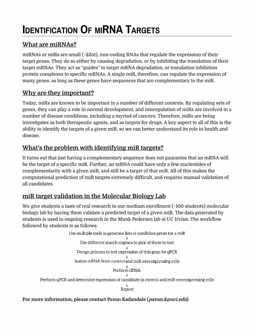
miR target validation in the Molecular Biology Lab
We give students a taste of real research in our medium enrollment (~100 students) molecular biology lab by having them validate a predicted target of a given miR. The data generated by students is used in ongoing research in the Munk Pedersen lab at UC Irvine. The workflow followed by students is as follows:
For more information, please contact Pavan Kadandale ([email protected])
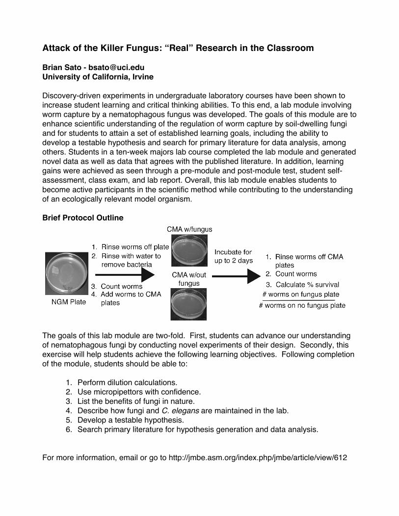
Attack of the Killer Fungus: “Real” Research in the Classroom Brian Sato - [email protected] University of California, Irvine Discovery-driven experiments in undergraduate laboratory courses have been shown to increase student learning and critical thinking abilities. To this end, a lab module involving worm capture by a nematophagous fungus was developed. The goals of this module are to enhance scientific understanding of the regulation of worm capture by soil-dwelling fungi and for students to attain a set of established learning goals, including the ability to develop a testable hypothesis and search for primary literature for data analysis, among others. Students in a ten-week majors lab course completed the lab module and generated novel data as well as data that agrees with the published literature. In addition, learning gains were achieved as seen through a pre-module and post-module test, student self-assessment, class exam, and lab report. Overall, this lab module enables students to become active participants in the scientific method while contributing to the understanding of an ecologically relevant model organism. Brief Protocol Outline
The goals of this lab module are two-fold. First, students can advance our understanding of nematophagous fungi by conducting novel experiments of their design. Secondly, this exercise will help students achieve the following learning objectives. Following completion of the module, students should be able to:
1. Perform dilution calculations. 2. Use micropipettors with confidence. 3. List the benefits of fungi in nature. 4. Describe how fungi and C. elegans are maintained in the lab. 5. Develop a testable hypothesis. 6. Search primary literature for hypothesis generation and data analysis.
For more information, email or go to http://jmbe.asm.org/index.php/jmbe/article/view/612
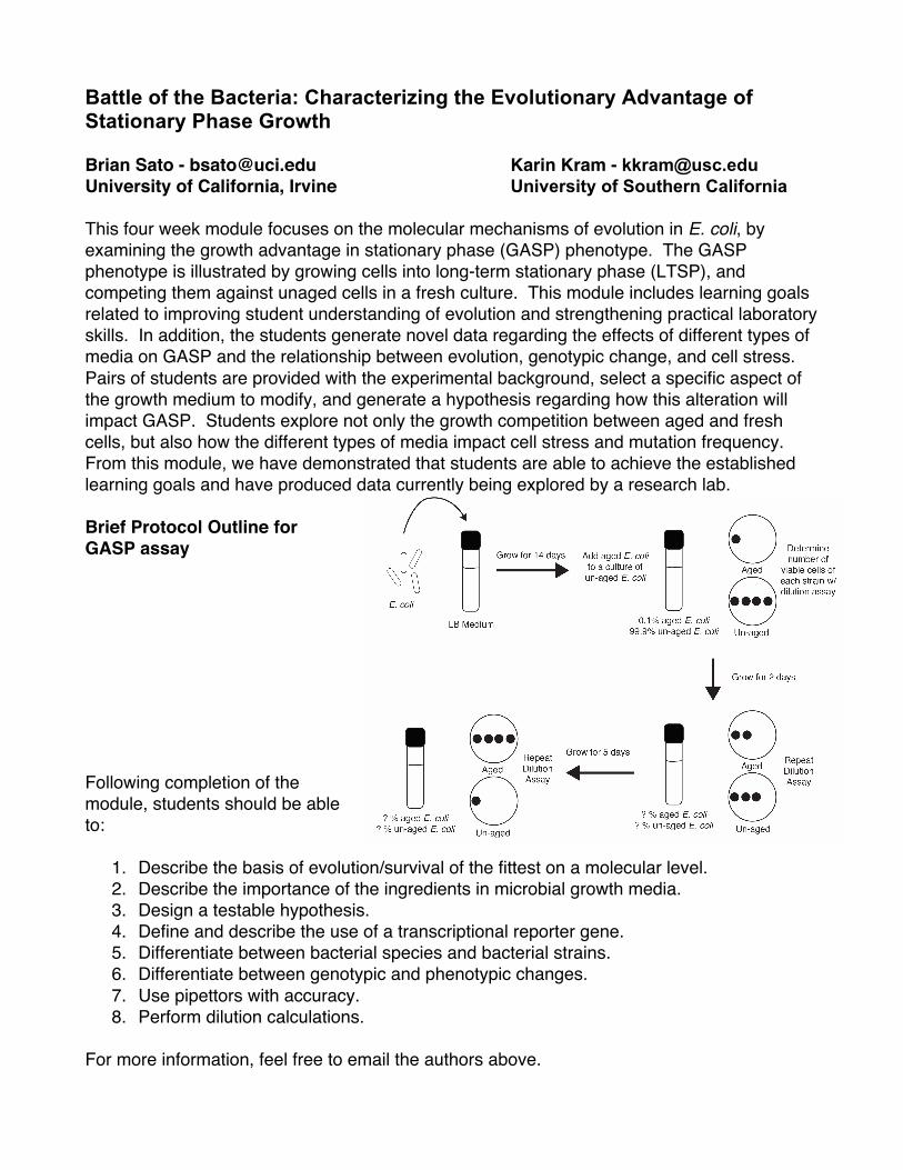
Battle of the Bacteria: Characterizing the Evolutionary Advantage of Stationary Phase Growth Brian Sato - [email protected] Karin Kram - [email protected] University of California, Irvine University of Southern California This four week module focuses on the molecular mechanisms of evolution in E. coli, by examining the growth advantage in stationary phase (GASP) phenotype. The GASP phenotype is illustrated by growing cells into long-term stationary phase (LTSP), and competing them against unaged cells in a fresh culture. This module includes learning goals related to improving student understanding of evolution and strengthening practical laboratory skills. In addition, the students generate novel data regarding the effects of different types of media on GASP and the relationship between evolution, genotypic change, and cell stress. Pairs of students are provided with the experimental background, select a specific aspect of the growth medium to modify, and generate a hypothesis regarding how this alteration will impact GASP. Students explore not only the growth competition between aged and fresh cells, but also how the different types of media impact cell stress and mutation frequency. From this module, we have demonstrated that students are able to achieve the established learning goals and have produced data currently being explored by a research lab. Brief Protocol Outline for GASP assay Following completion of the module, students should be able to:
1. Describe the basis of evolution/survival of the fittest on a molecular level. 2. Describe the importance of the ingredients in microbial growth media. 3. Design a testable hypothesis. 4. Define and describe the use of a transcriptional reporter gene. 5. Differentiate between bacterial species and bacterial strains. 6. Differentiate between genotypic and phenotypic changes. 7. Use pipettors with accuracy. 8. Perform dilution calculations.
For more information, feel free to email the authors above.