Embed Size (px)
Citation preview
Identification of Host-Immune Response ProteinCandidates in the Sera of Human Oral Squamous CellCarcinoma PatientsYeng Chen1,2*, Siti Nuraishah Azman3, Jesinda P. Kerishnan1, Rosnah Binti Zain2,4, Yu Nieng Chen5, Yin-
Ling Wong1, Subash C. B. Gopinath1
1Department of Oral Biology & Biomedical Sciences, Faculty of Dentistry, University of Malaya, Kuala Lumpur, Malaysia, 2Oral Cancer Research and Coordinating Center,
Faculty of Dentistry, University of Malaya, Kuala Lumpur, Malaysia, 3 Institute for Research in Molecular Medicine, Universiti Sains Malaysia, Georgetown, Penang, Malaysia,
4Department of Oro-Maxillofacial and Medical Science, Faculty of Dentistry, University of Malaya, Kuala Lumpur, Malaysia, 5Chen Dental Specialist Clinic, Kueh Hock Kui
Commercial Centre, Jalan Tun Ahmad Zaidi Adruce, Kuching, Sarawak, Malaysia
Abstract
One of the most common cancers worldwide is oral squamous cell carcinoma (OSCC), which is associated with a significantdeath rate and has been linked to several risk factors. Notably, failure to detect these neoplasms at an early stage representsa fundamental barrier to improving the survival and quality of life of OSCC patients. In the present study, serum samplesfrom OSCC patients (n = 25) and healthy controls (n = 25) were subjected to two-dimensional gel electrophoresis (2-DE) andsilver staining in order to identify biomarkers that might allow early diagnosis. In this regard, 2-DE spots corresponding tovarious up- and down-regulated proteins were sequenced via high-resolution MALDI-TOF mass spectrometry and analyzedusing the MASCOT database. We identified the following differentially expressed host-specific proteins within sera fromOSCC patients: leucine-rich a2-glycoprotein (LRG), alpha-1-B-glycoprotein (ABG), clusterin (CLU), PRO2044, haptoglobin(HAP), complement C3c (C3), proapolipoprotein A1 (proapo-A1), and retinol-binding protein 4 precursor (RBP4). Moreover,five non-host factors were detected, including bacterial antigens from Acinetobacter lwoffii, Burkholderia multivorans,Myxococcus xanthus, Laribacter hongkongensis, and Streptococcus salivarius. Subsequently, we analyzed the immunogenicityof these proteins using pooled sera from OSCC patients. In this regard, five of these candidate biomarkers were found to beimmunoreactive: CLU, HAP, C3, proapo-A1 and RBP4. Taken together, our immunoproteomics approach has identifiedvarious serum biomarkers that could facilitate the development of early diagnostic tools for OSCC.
Citation: Chen Y, Azman SN, Kerishnan JP, Zain RB, Chen YN, et al. (2014) Identification of Host-Immune Response Protein Candidates in the Sera of Human OralSquamous Cell Carcinoma Patients. PLoS ONE 9(10): e109012. doi:10.1371/journal.pone.0109012
Editor: Qing-Yi Wei, Duke Cancer Institute, United States of America
Received June 10, 2014; Accepted September 6, 2014; Published October 1, 2014
Copyright: � 2014 Chen et al. This is an open-access article distributed under the terms of the Creative Commons Attribution License, which permitsunrestricted use, distribution, and reproduction in any medium, provided the original author and source are credited.
Data Availability: The authors confirm that all data underlying the findings are fully available without restriction. All relevant data are within the paper.
Funding: This study was supported by University of Malaya (UM)-High Impact Research(HIR)-the Ministry of Education (MoE) Grant UM/C/625/1/HIR/MOE/DENT/9 and UM.C/625/1/HIR/MOE/DENT/20. The funders had no role in study design, data collection and analysis, decision to publish, or preparation of the manuscript.
Competing Interests: The authors have declared that no competing interests exist.
* Email: [email protected]
Introduction
Oral cancer represents the sixth most prevalent cancer in the
world. Among the different types of oral cancer, oral squamous
cell carcinoma (OSCC) arising from the oral mucosa accounts for
more than 90% of these malignancies. Thus, OSCC is the most
common malignancy affecting the head and neck region. Notably,
there are nearly 300,000 new cases of oral cancer reported
annually [1,2], and it was estimated that approximately 128,000
oral cancer patients died worldwide in 2008 [3]. Therefore,
despite recent advances in the diagnosis and treatment of oral
cancer (e.g., chemotherapy, radiotherapy, and surgical therapy)
the survival rate of OSCC patients has remained less than 60%
[4,5]. A fundamental barrier in improving the survival of OSCC
patients is the fact that these malignancies often remain undetected
until the later stages. In this regard, it was reported that a several-
month delay in diagnosis could reduce the chance of survival from
80% to 40% [6]. Thus, in order to prevent the high OSCC-related
mortality rate, recent attention has been focused on identifying
potential diagnostic molecular markers (e.g., cell cycle regulators)
that might represent biological predictors of oral cancer [7].
Moreover, OSCC has been linked to several risk factors, including
various bacterial pathogens [8], which might be useful for
detecting OSCC.
Recent studies have indicated that early diagnosis, lifestyle
modification, and effective treatment can prevent more than two-
thirds of OSCC-related mortalities. However, currently available
diagnostic methods do not allow for the detection of oral cancer in
the early stages. To visualize malignant lesions in the oral cavity,
different microscopic methods are available, which make use of
various techniques, including autofluorescence, chemilumines-
cence, or dye-based tissue staining. However, due to the low
sensitivity and specificity of these diagnostic strategies, clinicians
generally use biopsies to detect OSCC [9]. Nevertheless, successful
diagnosis through tissue biopsy is highly dependent on acquiring
whole and complete tissue samples from patients for examination.
In this regard, biopsies harvested from oral cancer patients are
often associated with the soft tissues that surround the cancer
PLOS ONE | www.plosone.org 1 October 2014 | Volume 9 | Issue 10 | e109012
tissue. In addition, oral cancer frequently involves the develop-
ment of multiple primary tumors. Indeed, the occurrence of a
second primary tumor is 3–7% higher per year in oral cancer
when compared to other malignancies [10]. Therefore, the
identification of suitable and reliable OSCC biomarkers is essential
for achieving early detection and treatment, which can reduce
mortality rates in OSCC patients. In this respect, antibody-based
diagnostic tests that recognize specific tumor-associated antigens in
cancer sera might represent a valid methodology [11].
Proteomic analysis allows the identification and quantification
of proteins and peptides in biological samples [12]. However,
through this approach, numerous post-translational forms of
protein regulation, including regulating enzymes and low abun-
dance proteins may remain undetected. Thus, in the present
investigation, we employed an immunoproteomic approach and
pooled human antibodies to detect host-specific response proteins
in OSCC patients. Specifically, we used a well-characterized
analytical platform combining two-dimensional gel electrophoresis
(2-DE) and mass spectrometry (MS) to identify biomarkers in
unfractionated sera from OSCC patients and normal controls.
This immunoproteomics approach can be used to identify antigens
targeted by the immune system in sera during disease progression.
In addition, the immune responses are known to be involved in the
mechanism of carcinogenesis [13]. Therefore, our comparative
analyses revealed distinct OSCC biomarkers that might promote
the development of specific diagnostic tests for early detection of
oral cancer.
Materials and Methods
Serum samplesTwenty-five serum samples from OSCC patients were obtained
from the Oral Cancer Research and Coordinating Center
(OCRCC) at the University of Malaya (Kuala Lumpur). Addi-
tionally, 25 control serum samples were acquired from healthy
individuals. All samples were collected with the verbal consent of
patients, and the Dental Faculty at the University of Malaya and
the Universiti Sains Malaysia Medical Ethics Committee (Ref:
USMKK/PPP/JEPeM [213.3(09)]) approve this consent proce-
dure. We obtained the permission from the above committee and
they have cleared all the approval and having the record. This
study was also conducted in accordance with International
Conference on Harmonisation–Good Clinical Practice (ICH–
GCP) guidelines and the Declaration of Helsinki.
Two-dimensional gel electrophoresis (2-DE)We performed 2-DE as previously described [14]. Briefly,
unfractionated human serum samples (10 ml) were lysed,
rehydrated in lysis buffer (2M thiourea, 8M urea, 4% CHAPS,
1% dithreitol, and 2% pharmalyte), and subjected to isoelectric
focusing in 13-cm rehydrated precast immobilized dry strips
(pH 4–7; GE Healthcare, Sweden). Sodium dodecyl sulfate–
polyacrylamide gel electrophoresis (SDS–PAGE) was performed
using 8–18% gradient polyacrylamide gels in the presence of SDS
for the second dimension separation. Gel silver staining was
performed as previously described [15]. Silver staining and
Coomassie Brilliant Blue staining for MS were conducted using
slightly modified published methods [16].
Differential image acquisition and statistical analysisThe ImageScanner III (GE Healthcare, Sweden) was used to
capture and store 2-DE gel images. PD-Quest 2-D gel analysis
software (version 8.0.1, Bio-Rad) was employed to evaluate the
differentially expressed protein spots. Identical spots were matched
in the serial gels and normalized by correcting for spot
quantification values and gel-to-gel variation unrelated to expres-
sion changes. For the normalization method, we used total
densities from the gel images (i.e., raw quantity of each gel spot
was divided by the total quantity of all spots within the gel). All
protein concentration values are presented as means of percentage
volume (% volume) 6 standard deviations (SD). The student’s t-
test and one-way analysis of variance (ANOVA) were used to
analyze differences between patients and controls. P-values less
than 0.05 (p,0.05) were considered as statistically significant.
Mass spectrometry analysis and database searchSpots of interest were excised and subjected to in-gel tryptic
digestion using a commercially available kit (Calbiochem,
Germany). MS analysis and database searches were performed
at the Proteomic Center within the Faculty of Biological Sciences
at the National University of Singapore. Digested peptides were
mixed with 1.2 ml of CHCA matrix solution (5 mg/ml of cyano-4-
hydroxy-cinnamic acid in 0.1% trifluoroacetic acid [TFA] and
50% acetonitrile [ACN]) and spotted onto MALDI target plates.
An ABI 4800 Proteomics Analyzer MALDI-TOF/TOF Mass
Spectrometer was used for spectra analysis (Applied Biosystems,
USA), and the MASCOT search engine (version 2.1; Matrix
Science, UK) was employed for database searches. In addition,
GPS Explorer software (version 3.6; Applied Biosystems, USA)
was utilized along with MASCOT to identify peptides and
proteins. Search parameters allowed for N-terminal acetylation,
C-terminal carbamidomethylation of cysteine (fixed modification),
and methionine oxidation (variable modification). Peptide and
fragment mass tolerance were set to 100 ppm and 60.2 Da,
respectively. The peptide mass fingerprinting (PMF) parameters
were set as follows: one missed cleavage allowed in trypsin digest;
monoisotropic mass value; 60.1 Da peptide mass tolerance; and
1+ peptide charge state.
Peptides were initially identified using the ProteinPilot proteo-
mics software on the Mass Spectrometer (Applied Biosystems,
USA). A score reflecting the relationship between theoretically and
experimentally determined masses was calculated and assigned.
Analyses were conducted using the International Protein Index
(http://www.ebi.ac.uk/IPI) and NCBI Unigene human databases
(version 3.38). A total of 100,907 entries were searched, and a
score of.82 was considered as significant in the MASCOT NCBI
database.
ImmunoblottingOur 2-DE gel immunoblotting protocol was organized into four
categories: (1) normal sera probed with normal sera, (2) normal
sera probed with OSCC sera, (3) OSCC sera probed with normal
sera, and (4) OSCC sera probed with OSCC sera. After running
the 2-DE gels, they were transferred onto nitrocellulose mem-
branes using the Multiphor II Novablot semi-dry system (GE
Healthcare, Sweden). Membranes were blocked with SuperBlock
(Pierce, USA) and washed three times with Tris-buffered saline
(TBS)–Tween-20. The membranes were subsequently incubated
overnight (4uC) with pooled sera from patients or healthy subjects
that contains the primary antibodies against various targets (1:200
dilution). Following washing, membranes were incubated for 1
hour at room temperature with horseradish peroxidase (HRP)-
linked monoclonal anti-human immunoglobulin M (IgM) (1:5000;
Invitrogen, USA). The membranes were again washed and then
visualized using chemiluminescence substrate (Pierce, USA) and
18 cm 6 24 cm films (Kodak, USA).
Host-Immune Response Proteins in OSCC Patients
PLOS ONE | www.plosone.org 2 October 2014 | Volume 9 | Issue 10 | e109012
Functional annotation and protein interaction analysesFunctional annotation analysis was performed using DAVID
v6.7 (Database for Annotation, Visualization, and Integrated
Discovery), which provides a comprehensive set of functional
annotation tools to understand the biological significance associ-
ated with large lists of genes or proteins [17]. This functional
categorization is considered significant when a p-value of less than
0.05 (p,0.05) is obtained. STRING v9.1 (Search Tool for the
Retrieval of Interacting Genes) was used to examine protein–
protein interaction networks [18].
Results and Discussion
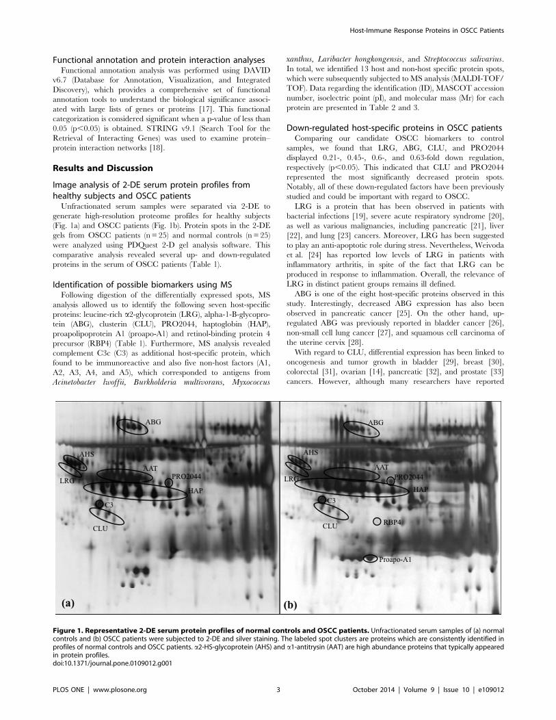
Image analysis of 2-DE serum protein profiles fromhealthy subjects and OSCC patientsUnfractionated serum samples were separated via 2-DE to
generate high-resolution proteome profiles for healthy subjects
(Fig. 1a) and OSCC patients (Fig. 1b). Protein spots in the 2-DE
gels from OSCC patients (n = 25) and normal controls (n = 25)
were analyzed using PDQuest 2-D gel analysis software. This
comparative analysis revealed several up- and down-regulated
proteins in the serum of OSCC patients (Table 1).
Identification of possible biomarkers using MSFollowing digestion of the differentially expressed spots, MS
analysis allowed us to identify the following seven host-specific
proteins: leucine-rich a2-glycoprotein (LRG), alpha-1-B-glycopro-
tein (ABG), clusterin (CLU), PRO2044, haptoglobin (HAP),
proapolipoprotein A1 (proapo-A1) and retinol-binding protein 4
precursor (RBP4) (Table 1). Furthermore, MS analysis revealed
complement C3c (C3) as additional host-specific protein, which
found to be immunoreactive and also five non-host factors (A1,
A2, A3, A4, and A5), which corresponded to antigens from
Acinetobacter lwoffii, Burkholderia multivorans, Myxococcus
xanthus, Laribacter hongkongensis, and Streptococcus salivarius.In total, we identified 13 host and non-host specific protein spots,
which were subsequently subjected to MS analysis (MALDI-TOF/
TOF). Data regarding the identification (ID), MASCOT accession
number, isoelectric point (pI), and molecular mass (Mr) for each
protein are presented in Table 2 and 3.
Down-regulated host-specific proteins in OSCC patientsComparing our candidate OSCC biomarkers to control
samples, we found that LRG, ABG, CLU, and PRO2044
displayed 0.21-, 0.45-, 0.6-, and 0.63-fold down regulation,
respectively (p,0.05). This indicated that CLU and PRO2044
represented the most significantly decreased protein spots.
Notably, all of these down-regulated factors have been previously
studied and could be important with regard to OSCC.
LRG is a protein that has been observed in patients with
bacterial infections [19], severe acute respiratory syndrome [20],
as well as various malignancies, including pancreatic [21], liver
[22], and lung [23] cancers. Moreover, LRG has been suggested
to play an anti-apoptotic role during stress. Nevertheless, Weivoda
et al. [24] has reported low levels of LRG in patients with
inflammatory arthritis, in spite of the fact that LRG can be
produced in response to inflammation. Overall, the relevance of
LRG in distinct patient groups remains ill defined.
ABG is one of the eight host-specific proteins observed in this
study. Interestingly, decreased ABG expression has also been
observed in pancreatic cancer [25]. On the other hand, up-
regulated ABG was previously reported in bladder cancer [26],
non-small cell lung cancer [27], and squamous cell carcinoma of
the uterine cervix [28].
With regard to CLU, differential expression has been linked to
oncogenesis and tumor growth in bladder [29], breast [30],
colorectal [31], ovarian [14], pancreatic [32], and prostate [33]
cancers. However, although many researchers have reported
Figure 1. Representative 2-DE serum protein profiles of normal controls and OSCC patients. Unfractionated serum samples of (a) normalcontrols and (b) OSCC patients were subjected to 2-DE and silver staining. The labeled spot clusters are proteins which are consistently identified inprofiles of normal controls and OSCC patients. a2-HS-glycoprotein (AHS) and a1-antitrysin (AAT) are high abundance proteins that typically appearedin protein profiles.doi:10.1371/journal.pone.0109012.g001
Host-Immune Response Proteins in OSCC Patients
PLOS ONE | www.plosone.org 3 October 2014 | Volume 9 | Issue 10 | e109012
increased CLU expression during tumorigenesis, we have
observed significantly down-regulated CLU levels in sera from
OSCC patients. Nevertheless, other reports have also described a
similar loss of CLU expression in various tumors, including
prostate cancer [34], pancreatic cancer [32], esophageal squamous
cell carcinoma [35] and neuroblastoma [36]. Notably, these
discrepancies may stem from the fact that there are differentially
expressed CLU isoforms in human tissues and fluids that may
exhibit distinct functions in tumors [14,37]. Even though it has
been suggested that CLU down regulation could be associated
with disease progression [32,38], this may depend on the type of
cancer [39,40]. Notably, the true function of CLU has remained
elusive despite extensive investigation. So far, CLU has been
proposed to participate in the immediate cellular response to
stress, which regulates cell growth and survival [41]. In this regard,
its function appears isoform dependent, with both proapoptotic
and anti-apoptotic forms [34].
Finally, our analysis revealed that PRO2044 (the C-terminal
fragment of albumin, ALB) was the most down-regulated protein
in the sera of OSCC patients. Interestingly, Kawakami et al. [22]
observed that PRO2044 was also down regulated in hepatocellular
carcinoma patients following curative radiofrequency ablation. In
contrast, Jin et al. [42] reported that PRO2044 levels were
increased in the cerebrospinal fluid of patients with Guillain–Barre
syndrome, an acute inflammatory autoimmune disorder of the
peripheral nervous system.
Up-regulated host-specific proteins in OSCC patientsIn addition to down-regulated proteins, up-regulated host-
specific antigens were also identified in OSCC patients, including
HAP, proapo-A1, and RBP4. Compared to the control samples,
these proteins displayed 1.47-, 1.82-, and 2.66-fold increases,
respectively (p,0.05). RBP4 was found to be the most up-
regulated.
Notably, a correlation between HAP expression and malignan-
cies has been reported [43,44]. Indeed, similar to a previous study
by Lai et al. [45], we found that HAP levels were significantly
increased in sera from OSCC patients. Furthermore, since HAP is
primarily hepatocyte-produced, a rise in its levels may indicate the
occurrence of an acute phase response in OSCC [44]. Moreover,
HAP has also been reported to participate in cell migration,
extracellular matrix degradation, and arterial restructuring,
suggesting its possible role in cancer [46]. In addition, HAP
might act as an angiogenic agent that contributes to endothelial
cell growth and differentiation [47].
With regard to proapo-A1 (or apo-A1), our findings are
consistent with studies that have found elevated expression in
various malignancies, including breast [48], colorectal [49], non-
small cell lung [50] pancreatic [51], and hepatocellular [52]
cancers. It is possible that increases in proapo-A1 might stem from
reduced activity of proapo-A1 cleaving enzyme or higher turnover
of apo-A1 [50,53].
We also observed an elevation of RBP4 levels in OSCC. It has
been suggested that RBP4 over expression in cancer cells could
result from an inhibition of phosphatidylinositol-3 kinase (PI3K)
activity [54,55]. Moreover, RBP4 expression might be related to
retinoid depletion, which is a common feature in cancer patients
[56–58]. In addition, RBP4 levels can be influenced by
transthyretin, which could reduce renal clearance of RBP4 [59].
Non-host specific proteins detected in OSCC patientsIn addition to the above down- and up-regulated self-antigens,
we also identified five non-host markers in the proteomic profiles
of OSCC patients. These proteins were derived from various
bacteria, including A. lwoffii, B. multivorans, M. xanthus, L.hongkogenisis, and S. salivarius. Although our detection of these
factors could have resulted from nosocomial infections [60,61],
heightened risk of malignancy has been linked to viruses, bacteria,
and schistosomes [62,63]. In fact, the relationship between
bacterial infections and cancer has been discussed for decades
[64,65]. In this regard, there are several possible mechanisms by
which bacteria could be oncogenic. For example, altered host
responses during bacterial infection (e.g., chronic inflammation,
antigen-driven lymphoproliferation, and hormone induction that
promotes epithelial cell proliferation) have been suggested to
influence oncogenesis [66]. Also, bacterial infection can lead to the
production of toxin and/or carcinogenic metabolites that enhance
oncogenesis [67]. In contrast, bacterial infections have also been
suggested to play a protective role by altering host physiology and
reducing cancer risk [68].
Oral cancer is considered to be a multi-factorial disease, as it
can stem from exposure to several types of carcinogens, including
microbial factors [69]. Indeed, several pieces of evidence have
supported the association of microbial infection with oncogenesis.
For instance, Helicobacter pylori has been linked to gastric cancer
[70] and categorized by the World Health Organization
International Agency for Research on Cancer (IARC) as a
carcinogenic factor in humans [71–73]. Similarly, Chlamydophilapneumoniae has been associated with malignant lymphoma and
lung cancer in males [74,75], whereas Candida albicans and
Streptococcus anginosus have been linked to oral carcinoma [70–
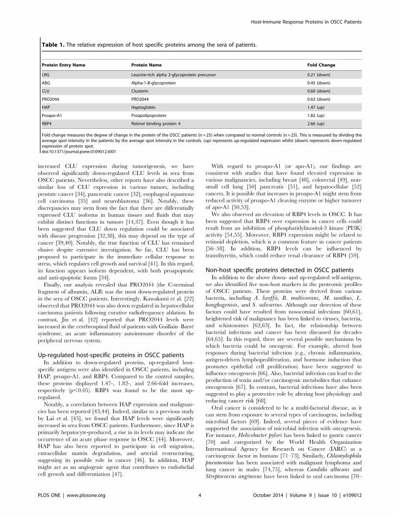
Table 1. The relative expression of host specific proteins among the sera of patients.
Protein Entry Name Protein Name Fold Change
LRG Leucine-rich alpha 2-glycoprotein precursor 0.21 (down)
ABG Alpha-1-B-glycoprotein 0.45 (down)
CLU Clusterin 0.60 (down)
PRO2044 PRO2044 0.63 (down)
HAP Haptoglobin 1.47 (up)
Proapo-A1 Proapolipoprotein 1.82 (up)
RBP4 Retinol binding protein 4 2.66 (up)
Fold change measures the degree of change in the protein of the OSCC patients (n = 25) when compared to normal controls (n= 25). This is measured by dividing theaverage spot intensity in the patients by the average spot intensity in the controls. (up) represents up-regulated expression whilst (down) represents down-regulatedexpression of protein spot.doi:10.1371/journal.pone.0109012.t001
Host-Immune Response Proteins in OSCC Patients
PLOS ONE | www.plosone.org 4 October 2014 | Volume 9 | Issue 10 | e109012
Table
2.Massspectrometric
identificationofhost-specificprotein
spots
from
serum
protein
profilesusingMASC
OTsearch
enginean
dtheNCBIdatab
ase.
Pro
tein
Name
MASCOTaccession
number
pI
Theorectical
mass
Sequence
coverage
Search
score
Queries
match
Expected
value
Alpha-1-B-glycoprotein
–human
[Homosapiens]
gi|69990
5.65
52479
40%
824
24
9.1e-078
Leucine-richalpha-2-glycoprotein
precursor[Homosapiens]
gi|1641846
6.45
38382
40%
601
18
1.8e-055
Clusterin[Homosapiens]
gi|2666585
5.60
16267
11%
39
429
Retinolbindingprotein
4[Homosapiens]
gi|1808832
5.76
23371
48%
276
14
2.9e-021
PRO2044[Homosapiens]
gi|6650826
6.97
39984
45%
355
17
3.7e-029
Hap
toglobin
[Homosapiens]
gi|3337390
6.14
38722
32%
264
11
4.6e-020
Proap
ollipoprotein
[Homosapiens]
gi|178775
5.45
28944
53%
503
20
1.1e-045
ChainB,Human
ComplementComponentC3[Homosapines]
gi|7810126
5.55
114238
20%
469
24
1.5e-040
doi:10.1371/journal.pone.0109012.t002
Table
3.Massspectrometric
identificationofnon-host
specificprotein
spots
from
serum
protein
profilesusingMASC
OTsearch
enginean
dtheNCBIdatab
ase.
Pro
tein
Name
MASCOTaccession
number
pI
Theorectical
mass
Sequence
coverage
Search
score
Queries
match
Expected
value
(A1)Predictedprotein
[Acinetobacter
lwoffiiSH
145]
gi|262375905
8.89
9185
83%
56
12
30
(A2)Hyp
otheticalprotein
BURMUCGD2M_4365[Burkholderia
multivoransCGD2M]
gi|221195969
8.31
4546
92%
51
71e+0
02
(A3)Hyp
otheticalprotein
MXAN_1050[M
yxococcusxanthus
DK1622]
gi|108761930
5.05
10984
36%
52
882
(A4)Hyp
otheticalprotienLH
K_003399[Laribacter
hongkongensisHLH
K9]
gi|226939330
9.50
6340
68%
44
64.2e+0
02
(A5)HemolysinA[StreptococcussalivariusSK
126]
gi|228476878
5.47
30333
20%
50
10
1.2e+0
02
doi:10.1371/journal.pone.0109012.t003
Host-Immune Response Proteins in OSCC Patients
PLOS ONE | www.plosone.org 5 October 2014 | Volume 9 | Issue 10 | e109012
76]. It has also been demonstrated that OSCC patients possess
significantly elevated concentrations of certain bacteria in their
saliva. Thus, changes in salivary microflora could represent a non-
invasive diagnostic tool for predicting oral cancer [70].
Confirmation of host-specific proteins by westernblottingImmunoglobulin M (IgM) antibodies are present in the
circulation of normal humans and other mammalian species.
IgM is initially secreted by B cells upon primary antigen
stimulation [77,78] and participates in natural defenses against
foreign pathogens as well as neoplastic cells and tumors [79]. In
fact, autoantibodies against specific cancer antigens have been
identified for several types of tumors, including colon, breast, lung,
ovary, prostate, and head and neck. These antibodies have been
found to recognize several overexpressed (e.g., Her2), mutated
(e.g., p53), or tissue-restricted (e.g., testis-cancer antigens) proteins,
which are produced by cancer cells and elicit immune responses
[6]. Therefore, detection of such autoantibodies in patient sera
could be exploited as a means of cancer diagnosis. Indeed, the
specificity and sensitivity of the antibody response to low antigen
levels make it an ideal screening/diagnostic tool for early
identification of cancer biomarkers in serum-based assays.
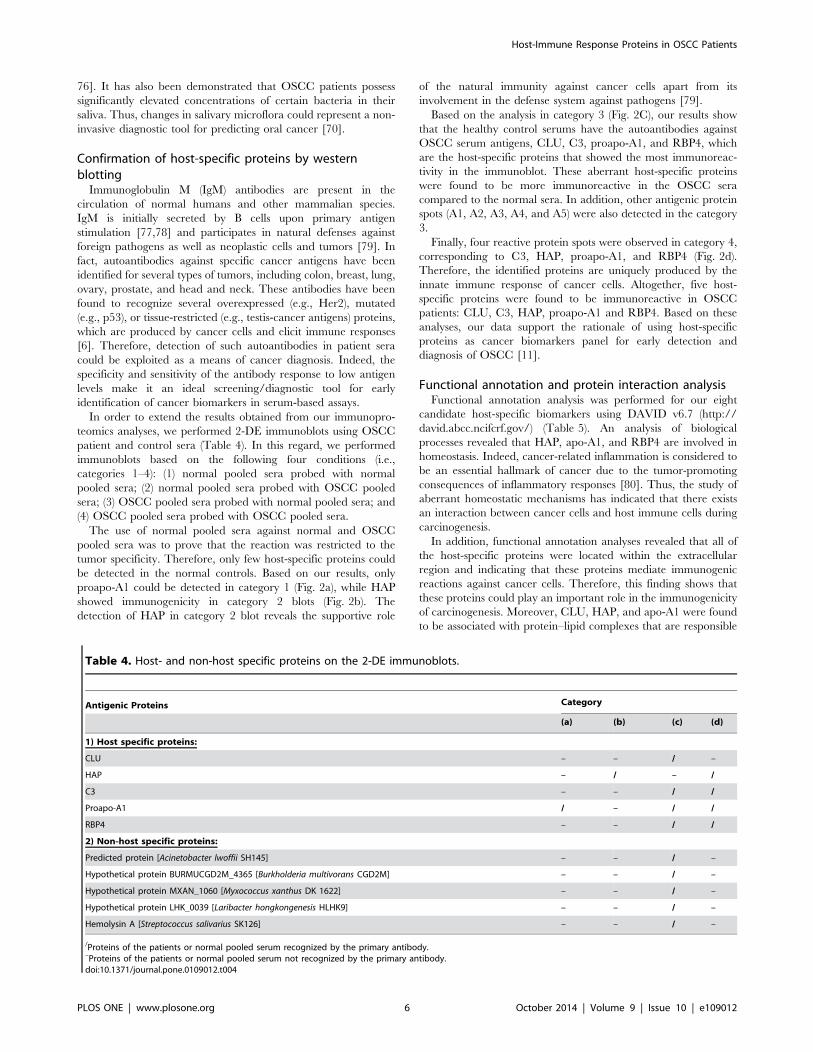
In order to extend the results obtained from our immunopro-
teomics analyses, we performed 2-DE immunoblots using OSCC
patient and control sera (Table 4). In this regard, we performed
immunoblots based on the following four conditions (i.e.,
categories 1–4): (1) normal pooled sera probed with normal
pooled sera; (2) normal pooled sera probed with OSCC pooled
sera; (3) OSCC pooled sera probed with normal pooled sera; and
(4) OSCC pooled sera probed with OSCC pooled sera.
The use of normal pooled sera against normal and OSCC
pooled sera was to prove that the reaction was restricted to the
tumor specificity. Therefore, only few host-specific proteins could
be detected in the normal controls. Based on our results, only
proapo-A1 could be detected in category 1 (Fig. 2a), while HAP
showed immunogenicity in category 2 blots (Fig. 2b). The
detection of HAP in category 2 blot reveals the supportive role
of the natural immunity against cancer cells apart from its
involvement in the defense system against pathogens [79].
Based on the analysis in category 3 (Fig. 2C), our results show
that the healthy control serums have the autoantibodies against
OSCC serum antigens, CLU, C3, proapo-A1, and RBP4, which
are the host-specific proteins that showed the most immunoreac-
tivity in the immunoblot. These aberrant host-specific proteins
were found to be more immunoreactive in the OSCC sera
compared to the normal sera. In addition, other antigenic protein
spots (A1, A2, A3, A4, and A5) were also detected in the category
3.
Finally, four reactive protein spots were observed in category 4,
corresponding to C3, HAP, proapo-A1, and RBP4 (Fig. 2d).
Therefore, the identified proteins are uniquely produced by the
innate immune response of cancer cells. Altogether, five host-
specific proteins were found to be immunoreactive in OSCC
patients: CLU, C3, HAP, proapo-A1 and RBP4. Based on these
analyses, our data support the rationale of using host-specific
proteins as cancer biomarkers panel for early detection and
diagnosis of OSCC [11].
Functional annotation and protein interaction analysisFunctional annotation analysis was performed for our eight
candidate host-specific biomarkers using DAVID v6.7 (http://
david.abcc.ncifcrf.gov/) (Table 5). An analysis of biological
processes revealed that HAP, apo-A1, and RBP4 are involved in
homeostasis. Indeed, cancer-related inflammation is considered to
be an essential hallmark of cancer due to the tumor-promoting
consequences of inflammatory responses [80]. Thus, the study of
aberrant homeostatic mechanisms has indicated that there exists
an interaction between cancer cells and host immune cells during
carcinogenesis.
In addition, functional annotation analyses revealed that all of
the host-specific proteins were located within the extracellular
region and indicating that these proteins mediate immunogenic
reactions against cancer cells. Therefore, this finding shows that
these proteins could play an important role in the immunogenicity
of carcinogenesis. Moreover, CLU, HAP, and apo-A1 were found
to be associated with protein–lipid complexes that are responsible
Table 4. Host- and non-host specific proteins on the 2-DE immunoblots.
Antigenic Proteins Category
(a) (b) (c) (d)
1) Host specific proteins:
CLU – – / –
HAP – / – /
C3 – – / /
Proapo-A1 / – / /
RBP4 – – / /
2) Non-host specific proteins:
Predicted protein [Acinetobacter lwoffii SH145] – – / –
Hypothetical protein BURMUCGD2M_4365 [Burkholderia multivorans CGD2M] – – / –
Hypothetical protein MXAN_1060 [Myxococcus xanthus DK 1622] – – / –
Hypothetical protein LHK_0039 [Laribacter hongkongenesis HLHK9] – – / –
Hemolysin A [Streptococcus salivarius SK126] – – / –
/Proteins of the patients or normal pooled serum recognized by the primary antibody.–Proteins of the patients or normal pooled serum not recognized by the primary antibody.doi:10.1371/journal.pone.0109012.t004
Host-Immune Response Proteins in OSCC Patients
PLOS ONE | www.plosone.org 6 October 2014 | Volume 9 | Issue 10 | e109012
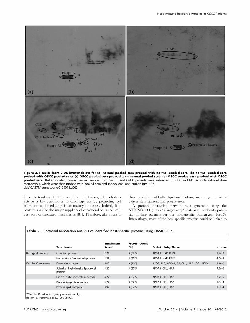
for cholesterol and lipid transportation. In this regard, cholesterol
acts as a key contributor to carcinogenesis by promoting cell
migration and mediating inflammatory processes. Indeed, lipo-
proteins may be the major suppliers of cholesterol to cancer cells
via receptor-mediated mechanisms [81]. Therefore, alterations in
these proteins could alter lipid metabolism, increasing the risk of
cancer development and progression.
A protein interaction network was generated using the
STRING v9.1 (http://string-db.org/) database to identify poten-
tial binding partners for our host-specific biomarkers (Fig. 3).
Interestingly, most of the host-specific proteins could be linked to
Figure 2. Results from 2-DE immunoblots for (a) normal pooled sera probed with normal pooled sera, (b) normal pooled seraprobed with OSCC pooled sera, (c) OSCC pooled sera probed with normal pooled sera, (d) OSCC pooled sera probed with OSCCpooled sera. Unfractionated, pooled serum samples from control and OSCC patients were subjected to 2-DE and blotted onto nitrocellulosemembranes, which were then probed with pooled sera and monoclonal anti-human IgM-HRP.doi:10.1371/journal.pone.0109012.g002
Table 5. Functional annotation analysis of identified host-specific proteins using DAVID v6.7.
Term NameEnrichmentScore+
Protein Count(%) Protein Entry Name p value
Biological Process Chemical process 2.28 3 (37.5) APOA1, HAP, RBP4 1.9e-2
Homeostasis/Hemostasisprocess 2.28 3 (37.5) APOA1, HAP, RBP4 4.0e-2
Cellular Component Extracellular region 5.05 8 (100) A1BG, ALB, APOA1, C3, CLU, HAP, LRG1, RBP4 2.4e-6
Spherical high-density lipoproteinparticle
4.22 3 (37.5) APOA1, CLU, HAP 7.2e-6
High-density lipoprotein particle 4.22 3 (37.5) APOA1, CLU, HAP 7.7e-5
Plasma lipoprotein particle 4.22 3 (37.5) APOA1, CLU, HAP 1.5e-4
Protein-lipid complex 3.92 3 (37.5) APOA1, CLU, HAP 1.5e-4
+The classification stringency was set to high.doi:10.1371/journal.pone.0109012.t005
Host-Immune Response Proteins in OSCC Patients
PLOS ONE | www.plosone.org 7 October 2014 | Volume 9 | Issue 10 | e109012
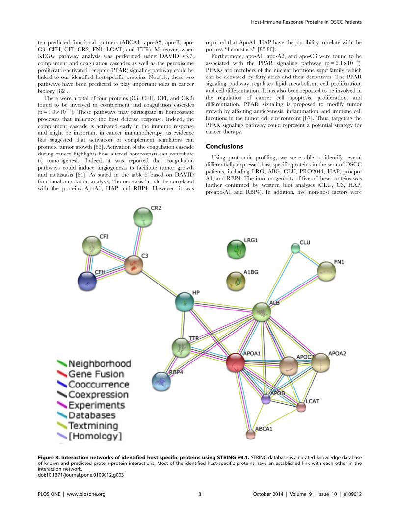
ten predicted functional partners (ABCA1, apo-A2, apo-B, apo-
C3, CFH, CFI, CR2, FN1, LCAT, and TTR). Moreover, when
KEGG pathway analysis was performed using DAVID v6.7,
complement and coagulation cascades as well as the peroxisome
proliferator-activated receptor (PPAR) signaling pathway could be
linked to our identified host-specific proteins. Notably, these two
pathways have been predicted to play important roles in cancer
biology [82].
There were a total of four proteins (C3, CFH, CFI, and CR2)
found to be involved in complement and coagulation cascades
(p = 1.961024). These pathways may participate in homeostatic
processes that influence the host defense response. Indeed, the
complement cascade is activated early in the immune response
and might be important in cancer immunotherapy, as evidence
has suggested that activation of complement regulators can
promote tumor growth [83]. Activation of the coagulation cascade
during cancer highlights how altered homeostasis can contribute
to tumorigenesis. Indeed, it was reported that coagulation
pathways could induce angiogenesis to facilitate tumor growth
and metastasis [84]. As stated in the table 5 based on DAVID
functional annotation analysis, ‘‘homeostasis’’ could be correlated
with the proteins ApoA1, HAP and RBP4. However, it was
reported that ApoA1, HAP have the possibility to relate with the
process ‘‘hemostasis’’ [85,86].
Furthermore, apo-A1, apo-A2, and apo-C3 were found to be
associated with the PPAR signaling pathway (p = 6.161024).
PPARs are members of the nuclear hormone superfamily, which
can be activated by fatty acids and their derivatives. The PPAR
signaling pathway regulates lipid metabolism, cell proliferation,
and cell differentiation. It has also been reported to be involved in
the regulation of cancer cell apoptosis, proliferation, and
differentiation. PPAR signaling is proposed to modify tumor
growth by affecting angiogenesis, inflammation, and immune cell
functions in the tumor cell environment [87]. Thus, targeting the
PPAR signaling pathway could represent a potential strategy for
cancer therapy.
Conclusions
Using proteomic profiling, we were able to identify several
differentially expressed host-specific proteins in the sera of OSCC
patients, including LRG, ABG, CLU, PRO2044, HAP, proapo-
A1, and RBP4. The immunogenicity of five of these proteins was
further confirmed by western blot analyses (CLU, C3, HAP,
proapo-A1 and RBP4). In addition, five non-host factors were
Figure 3. Interaction networks of identified host specific proteins using STRING v9.1. STRING database is a curated knowledge databaseof known and predicted protein-protein interactions. Most of the identified host-specific proteins have an established link with each other in theinteraction network.doi:10.1371/journal.pone.0109012.g003
Host-Immune Response Proteins in OSCC Patients
PLOS ONE | www.plosone.org 8 October 2014 | Volume 9 | Issue 10 | e109012
detected, including proteins from A. lwoffii, B. multivorans, M.xanthus, L. hongkogenisis, and S. salivarius. As previously
suggested [88], combined proteomic and serological approaches,
such as the one used in the present study, can reflect numerous
events occurring in vivo simultaneously due to the fact that patient
serum is complex and consists of many proteins. Our data indicate
that immunoproteomics approach could be a promising applica-
tion for biomarker discovery and disease progression. Our study
could be used as a landmark in a more comprehensive study and
can be applied to individual patient serums or to a larger sample
size. Using these methods, we have identified distinct serum
biomarkers that might facilitate the development of early
diagnostic tools for OSCC and promote further understanding
the ‘host responses’ that occur in OSCC patients.
Acknowledgments
The authors acknowledged the Oral Cancer Research and Coordinating
Centre (OCRCC), University of Malaya (UM) for providing serum
samples.
Author Contributions
Conceived and designed the experiments: YC SNA. Performed the
experiments: YC SNA JPK. Analyzed the data: YC RBZ YNC YLW
SCBG. Contributed reagents/materials/analysis tools: YC. Contributed to
the writing of the manuscript: YC SNA JPK YLW SCBG.
References
1. Parkin DM, Bray F, Ferlay J, Pisani P (2005) Global cancer statistics, 2002. CA
Cancer J Clin 55: 74–108.
2. Arellano-Garcia ME, Li R, Liu X, Xie Y, Yan X, et al. (2010) Identification of
tetranectin as a potential biomarker for metastatic oral cancer. Int J Mol Sci 11:
3106–3121.
3. Jemal A, Bray F, Center MM, Ferlay J, Ward E, et al. (2011) Global cancer
statistics. CA Cancer J Clin 61: 69–90.
4. Neville BW, Day TA (2002) Oral cancer and precancerous lesions. CA
Cancer J Clin 52: 195–215.
5. Forastiere AA, Goepfert H, Maor M, Pajak TF, Weber R, et al. (2003)
Concurrent chemotherapy and radiotherapy for organ preservation in advanced
laryngeal cancer. N Engl J Med 349: 2091–2098.
6. Lin HS, Talwar HS, Tarca AL, Ionan A, Chatterjee M, et al. (2007)
Autoantibody approach for serum-based detection of head and neck cancer.
Cancer Epidemiol Biomarkers Prev 16: 2396–2405.
7. da Silva SD, Ferlito A, Takes RP, Brakenhoff RH, Valentin MD, et al. (2011)
Advances and applications of oral cancer basic research. Oral Oncol 47: 783–
791.
8. Whitmore SE, Lamont RJ (2014) Oral bacteria and cancer. PLoS Pathog 10:
e1003933.
9. Lingen MW, Kalmar JR, Karrison T, Speight PM (2008) Critical evaluation of
diagnostic aids for the detection of oral cancer. Oral Oncol 44: 10–22.
10. Day GL, Blot WJ (1992) Second primary tumors in patients with oral cancer.
Cancer 70: 14–19.
11. Mou Z, He Y, Wu Y (2009) Immunoproteomics to identify tumor-associated
antigens eliciting humoral response. Cancer Lett 278: 123–129.
12. Arnott D, Emmert-Buck MR (2010) Proteomic profiling of cancer–opportuni-
ties, challenges, and context. J Pathol 222: 16–20.
13. Goldszmid RS, Dzutsev A, Trinchieri G (2014) Host immune response to
infection and cancer: unexpected commonalities. Cell Host Microbe 15: 295–
305.
14. Chen Y, Lim BK, Peh SC, Abdul-Rahman PS, Hashim OH (2008) Profiling of
serum and tissue high abundance acute-phase proteins of patients with epithelial
and germ line ovarian carcinoma. Proteome Sci 6: 20.
15. Heukeshoven J, Dernick R (1988) Improved silver staining procedure for fast
staining in PhastSystem Development Unit. I. Staining of sodium dodecyl sulfate
gels. Electrophoresis 9: 28–32.
16. Shevchenko A, Wilm M, Vorm O, Mann M (1996) Mass spectrometric
sequencing of proteins silver-stained polyacrylamide gels. Anal Chem 68: 850–
858.
17. Huang da W, Sherman BT, Lempicki RA (2009) Systematic and integrative
analysis of large gene lists using DAVID bioinformatics resources. Nat Protoc 4:
44–57.
18. Franceschini A, Szklarczyk D, Frankild S, Kuhn M, Simonovic M, et al. (2013)
STRING v9.1: protein-protein interaction networks, with increased coverage
and integration. Nucleic Acids Res 41: D808–815.
19. Bini L, Magi B, Marzocchi B, Cellesi C, Berti B, et al. (1996) Two-dimensional
electrophoretic patterns of acute-phase human serum proteins in the course of
bacterial and viral diseases. Electrophoresis 17: 612–616.
20. Chen JH, Chang YW, Yao CW, Chiueh TS, Huang SC, et al. (2004) Plasma
proteome of severe acute respiratory syndrome analyzed by two-dimensional gel
electrophoresis and mass spectrometry. Proc Natl Acad Sci U S A 101: 17039–
17044.
21. Kakisaka T, Kondo T, Okano T, Fujii K, Honda K, et al. (2007) Plasma
proteomics of pancreatic cancer patients by multi-dimensional liquid chroma-
tography and two-dimensional difference gel electrophoresis (2D-DIGE): up-
regulation of leucine-rich alpha-2-glycoprotein in pancreatic cancer. J Chroma-
togr B Analyt Technol Biomed Life Sci 852: 257–267.
22. Kawakami T, Hoshida Y, Kanai F, Tanaka Y, Tateishi K, et al. (2005)
Proteomic analysis of sera from hepatocellular carcinoma patients after
radiofrequency ablation treatment. Proteomics 5: 4287–4295.
23. Okano T, Kondo T, Kakisaka T, Fujii K, Yamada M, et al. (2006) Plasmaproteomics of lung cancer by a linkage of multi-dimensional liquid chromatog-
raphy and two-dimensional difference gel electrophoresis. Proteomics 6: 3938–3948.
24. Weivoda S, Andersen JD, Skogen A, Schlievert PM, Fontana D, et al. (2008)ELISA for human serum leucine-rich alpha-2-glycoprotein-1 employing
cytochrome c as the capturing ligand. J Immunol Methods 336: 22–29.
25. Li C, Zolotarevsky E, Thompson I, Anderson MA, Simeone DM, et al. (2011) A
multiplexed bead assay for profiling glycosylation patterns on serum proteinbiomarkers of pancreatic cancer. Electrophoresis 32: 2028–2035.
26. Kreunin P, Zhao J, Rosser C, Urquidi V, Lubman DM, et al. (2007) Bladdercancer associated glycoprotein signatures revealed by urinary proteomic
profiling. J Proteome Res 6: 2631–2639.
27. Liu Y, Luo X, Hu H, Wang R, Sun Y, et al. (2012) Integrative proteomics and
tissue microarray profiling indicate the association between overexpressed serumproteins and non-small cell lung cancer. PLoS One 7: e51748.
28. Jeong DH, Kim HK, Prince AE, Lee DS, Kim YN, et al. (2008) Plasmaproteomic analysis of patients with squamous cell carcinoma of the uterine
cervix. J Gynecol Oncol 19: 173–180.
29. Stejskal D, Fiala RR (2006) Evaluation of serum and urine clusterin as a
potential tumor marker for urinary bladder cancer. Neoplasma 53: 343–346.
30. Redondo M, Villar E, Torres-Munoz J, Tellez T, Morell M, et al. (2000)Overexpression of clusterin in human breast carcinoma. Am J Pathol 157: 393–
399.
31. Rodriguez-Pineiro AM, de la Cadena MP, Lopez-Saco A, Rodriguez-Berrocal
FJ (2006) Differential expression of serum clusterin isoforms in colorectal cancer.
Mol Cell Proteomics 5: 1647–1657.
32. Xie MJ, Motoo Y, Su SB, Mouri H, Ohtsubo K, et al. (2002) Expression ofclusterin in human pancreatic cancer. Pancreas 25: 234–238.
33. Zellweger T, Chi K, Miyake H, Adomat H, Kiyama S, et al. (2002) Enhancedradiation sensitivity in prostate cancer by inhibition of the cell survival protein
clusterin. Clin Cancer Res 8: 3276–3284.
34. Scaltriti M, Brausi M, Amorosi A, Caporali A, D’Arca D, et al. (2004) Clusterin
(SGP-2, ApoJ) expression is downregulated in low- and high-grade humanprostate cancer. Int J Cancer 108: 23–30.
35. Zhang LY, Ying WT, Mao YS, He HZ, Liu Y, et al. (2003) Loss of clusterin bothin serum and tissue correlates with the tumorigenesis of esophageal squamous
cell carcinoma via proteomics approaches. World J Gastroenterol 9: 650–654.
36. Santilli G, Aronow BJ, Sala A (2003) Essential requirement of apolipoprotein J
(clusterin) signaling for IkappaB expression and regulation of NF-kappaBactivity. J Biol Chem 278: 38214–38219.
37. Wei L, Xue T, Wang J, Chen B, Lei Y, et al. (2009) Roles of clusterin inprogression, chemoresistance and metastasis of human ovarian cancer.
Int J Cancer 125: 791–806.
38. Wu J, Xie X, Nie S, Buckanovich RJ, Lubman DM (2013) Altered expression of
sialylated glycoproteins in ovarian cancer sera using lectin-based ELISA assayand quantitative glycoproteomics analysis. J Proteome Res 12: 3342–3352.
39. Lourda M, Trougakos IP, Gonos ES (2007) Development of resistance to
chemotherapeutic drugs in human osteosarcoma cell lines largely depends on
up-regulation of Clusterin/Apolipoprotein J. Int J Cancer 120: 611–622.
40. Redondo M, Rodrigo I, Alcaide J, Tellez T, Roldan MJ, et al. (2010) Clusterin
expression is associated with decreased disease-free survival of patients withcolorectal carcinomas. Histopathology 56: 932–936.
41. Trougakos IP, Lourda M, Agiostratidou G, Kletsas D, Gonos ES (2005)
Differential effects of clusterin/apolipoprotein J on cellular growth and survival.
Free Radic Biol Med 38: 436–449.
42. Jin T, Hu LS, Chang M, Wu J, Winblad B, et al. (2007) Proteomic identificationof potential protein markers in cerebrospinal fluid of GBS patients. Eur J Neurol
14: 563–568.
43. Kuhajda FP, Katumuluwa AI, Pasternack GR (1989) Expression of haptoglobin-
related protein and its potential role as a tumor antigen. Proc Natl Acad
Sci U S A 86: 1188–1192.
Host-Immune Response Proteins in OSCC Patients
PLOS ONE | www.plosone.org 9 October 2014 | Volume 9 | Issue 10 | e109012
44. Ahmed N, Barker G, Oliva KT, Hoffmann P, Riley C, et al. (2004) Proteomic-
based identification of haptoglobin-1 precursor as a novel circulating biomarkerof ovarian cancer. Br J Cancer 91: 129–140.
45. Lai CH, Chang NW, Lin CF, Lin CD, Lin YJ, et al. (2010) Proteomics-based
identification of haptoglobin as a novel plasma biomarker in oral squamous cellcarcinoma. Clin Chim Acta 411: 984–991.
46. Zhao C, Annamalai L, Guo C, Kothandaraman N, Koh SC, et al. (2007)Circulating haptoglobin is an independent prognostic factor in the sera of
patients with epithelial ovarian cancer. Neoplasia 9: 1–7.
47. Ye B, Cramer DW, Skates SJ, Gygi SP, Pratomo V, et al. (2003) Haptoglobin-alpha subunit as potential serum biomarker in ovarian cancer: identification and
characterization using proteomic profiling and mass spectrometry. Clin CancerRes 9: 2904–2911.
48. Huang HL, Stasyk T, Morandell S, Dieplinger H, Falkensammer G, et al. (2006)Biomarker discovery in breast cancer serum using 2-D differential gel
electrophoresis/MALDI-TOF/TOF and data validation by routine clinical
assays. Electrophoresis 27: 1641–1650.49. Yu B, Li SY, An P, Zhang YN, Liang ZJ, et al. (2004) Comparative study of
proteome between primary cancer and hepatic metastatic tumor in colorectalcancer. World J Gastroenterol 10: 2652–2656.
50. Huang LJ, Chen SX, Huang Y, Luo WJ, Jiang HH, et al. (2006) Proteomics-
based identification of secreted protein dihydrodiol dehydrogenase as a novelserum markers of non-small cell lung cancer. Lung Cancer 54: 87–94.
51. Mikuriya K, Kuramitsu Y, Ryozawa S, Fujimoto M, Mori S, et al. (2007)Expression of glycolytic enzymes is increased in pancreatic cancerous tissues as
evidenced by proteomic profiling by two-dimensional electrophoresis and liquidchromatography-mass spectrometry/mass spectrometry. Int J Oncol 30: 849–
855.
52. Wang HY (2007) Laser capture microdissection in comparative proteomicanalysis of hepatocellular carcinoma. Methods Cell Biol 82: 689–707.
53. Harn HJ, Chen YL, Lin PC, Cheng YL, Lee SC, et al. (2010) Exploration ofPotential Tumor Markers for Lung Adenocarcinomas by Two-Dimensional Gel
Electrophoresis Coupled with Nano-LC/MS/MS. Journal of the Chinese
Chemical Society 57: 180–188.54. Kuppumbatti YS, Rexer B, Nakajo S, Nakaya K, Mira-y-Lopez R (2001) CRBP
suppresses breast cancer cell survival and anchorage-independent growth.Oncogene 20: 7413–7419.
55. Farias EF, Marzan C, Mira-y-Lopez R (2005) Cellular retinol-binding protein-Iinhibits PI3K/Akt signaling through a retinoic acid receptor-dependent
mechanism that regulates p85-p110 heterodimerization. Oncogene 24: 1598–
1606.56. Kuppumbatti YS, Bleiweiss IJ, Mandeli JP, Waxman S, Mira YLR (2000)
Cellular retinol-binding protein expression and breast cancer. J Natl Cancer Inst92: 475–480.
57. Reynolds CP, Matthay KK, Villablanca JG, Maurer BJ (2003) Retinoid therapy
of high-risk neuroblastoma. Cancer Lett 197: 185–192.58. Lorkova L, Pospisilova J, Lacheta J, Leahomschi S, Zivny J, et al. (2012)
Decreased concentrations of retinol-binding protein 4 in sera of epithelialovarian cancer patients: a potential biomarker identified by proteomics. Oncol
Rep 27: 318–324.59. Kotnik P, Fischer-Posovszky P, Wabitsch M (2011) RBP4: a controversial
adipokine. Eur J Endocrinol 165: 703–711.
60. Forster DH, Daschner FD (1998) Acinetobacter species as nosocomialpathogens. Eur J Clin Microbiol Infect Dis 17: 73–77.
61. Velasco E, Byington R, Martins CS, Schirmer M, Dias LC, et al. (2004)Bloodstream infection surveillance in a cancer centre: a prospective look at
clinical microbiology aspects. Clin Microbiol Infect 10: 542–549.
62. Pisani P, Parkin DM, Munoz N, Ferlay J (1997) Cancer and infection: estimatesof the attributable fraction in 1990. Cancer Epidemiol Biomarkers Prev 6: 387–
400.63. de Martel C, Ferlay J, Franceschi S, Vignat J, Bray F, et al. (2012) Global burden
of cancers attributable to infections in 2008: a review and synthetic analysis.
Lancet Oncol 13: 607–615.
64. Parsonnet J (1995) Bacterial infection as a cause of cancer. Environ Health
Perspect 103 Suppl 8: 263–268.
65. Beebe JL, Koneman EW (1995) Recovery of uncommon bacteria from blood:
association with neoplastic disease. Clin Microbiol Rev 8: 336–356.
66. Chang AH, Parsonnet J (2010) Role of bacteria in oncogenesis. Clin Microbiol
Rev 23: 837–857.
67. Mager DL (2006) Bacteria and cancer: cause, coincidence or cure? A review.
J Transl Med 4: 14.
68. Francois F, Roper J, Goodman AJ, Pei Z, Ghumman M, et al. (2008) The
association of gastric leptin with oesophageal inflammation and metaplasia. Gut
57: 16–24.
69. Ogbureke KU, Bingham C (2012) Overview of Oral Cancer. In: Ogbureke KU,
editor. Oral Cancer. Croatia: Intech. 3–20.
70. Hooper SJ, Wilson MJ, Crean SJ (2009) Exploring the link between
microorganisms and oral cancer: a systematic review of the literature. Head
Neck 31: 1228–1239.
71. Bjorkholm B, Falk P, Engstrand L, Nyren O (2003) Helicobacter pylori:
resurrection of the cancer link. J Intern Med 253: 102–119.
72. Correa P, Houghton J (2007) Carcinogenesis of Helicobacter pylori. Gastroen-
terology 133: 659–672.
73. Peek RM Jr, Blaser MJ (2002) Helicobacter pylori and gastrointestinal tract
adenocarcinomas. Nat Rev Cancer 2: 28–37.
74. Anttila TI, Lehtinen T, Leinonen M, Bloigu A, Koskela P, et al. (1998)
Serological evidence of an association between chlamydial infections and
malignant lymphomas. Br J Haematol 103: 150–156.
75. Kocazeybek B (2003) Chronic Chlamydophila pneumoniae infection in lung
cancer, a risk factor: a case-control study. J Med Microbiol 52: 721–726.
76. Sasaki M, Yamaura C, Ohara-Nemoto Y, Tajika S, Kodama Y, et al. (2005)
Streptococcus anginosus infection in oral cancer and its infection route. Oral Dis
11: 151–156.
77. Tchoudakova A, Hensel F, Murillo A, Eng B, Foley M, et al. (2009) High level
expression of functional human IgMs in human PER.C6 cells. MAbs 1: 163–
171.
78. Zouali M (2002) Antibodies. eLS.
79. Brandlein S, Pohle T, Ruoff N, Wozniak E, Muller-Hermelink HK, et al. (2003)
Natural IgM antibodies and immunosurveillance mechanisms against epithelial
cancer cells in humans. Cancer Res 63: 7995–8005.
80. Hanahan D, Weinberg RA (2011) Hallmarks of cancer: the next generation. Cell
144: 646–674.
81. Cruz PM, Mo H, McConathy WJ, Sabnis N, Lacko AG (2013) The role of
cholesterol metabolism and cholesterol transport in carcinogenesis: a review of
scientific findings, relevant to future cancer therapeutics. Front Pharmacol 4:
119.
82. Krupp M, Maass T, Marquardt JU, Staib F, Bauer T, et al. (2011) The
functional cancer map: a systems-level synopsis of genetic deregulation in cancer.
BMC Med Genomics 4: 53.
83. Kolev M, Towner L, Donev R (2011) Complement in cancer and cancer
immunotherapy. Arch Immunol Ther Exp (Warsz) 59: 407–419.
84. Rickles FR, Patierno S, Fernandez PM (2003) Tissue factor, thrombin, and
cancer. Chest 124: 58S–68S.
85. Talens S, Leebeek FW, Demmers JA, Rijken DC (2012) Identification of fibrin
clot-bound plasma proteins. PLoS One 7: e41966.
86. Li D, Weng S, Yang B, Zander DS, Saldeen T, et al. (1999) Inhibition of arterial
thrombus formation by ApoA1 Milano. Arterioscler Thromb Vasc Biol 19: 378–
383.
87. Michalik L, Wahli W (2008) PPARs Mediate Lipid Signaling in Inflammation
and Cancer. PPAR Res 2008: 134059.
88. Yeng C, Osman E, Mohamed Z, Noordin R (2010) Detection of immunogenic
parasite and host-specific proteins in the sera of active and chronic individuals
infected with Toxoplasma gondii. Electrophoresis 31: 3843–3849.
Host-Immune Response Proteins in OSCC Patients
PLOS ONE | www.plosone.org 10 October 2014 | Volume 9 | Issue 10 | e109012