Embed Size (px)
Citation preview
Tropical Life Sciences Research, 29(2), 187–199, 2018
© Penerbit Universiti Sains Malaysia, 2018. This work is licensed under the terms of the Creative Commons Attribution (CC BY) (http://creativecommons.org/licenses/by/4.0/).
SHORT COMMUNICATION
Identification of Cultivable Bacteria from Tropical Marine Sponges and Their Biotechnological Potentials
1Tan Suet May Amelia, 2,3,4Al-Ashraf Abdullah Amirul, 1,5Jasnizat Saidin and 1,3,5Kesaven Bhubalan*
1School of Marine and Environmental Sciences, Universiti Malaysia Terengganu, 21030 Kuala Nerus, Terengganu, Malaysia2School of Biological Sciences, Universiti Sains Malaysia, 11800 USM Pulau Pinang, Malaysia3Malaysian Institute of Pharmaceuticals and Nutraceuticals, National Institutes of Biotechnology Malaysia (NIBM), Ministry of Science, Technology and Innovation, 11700 Gelugor, Pulau Pinang, Malaysia4Centre of Chemical Biology, Universiti Sains Malaysia, 11900 Bayan Lepas, Pulau Pinang, Malaysia5Institute of Marine Biotechnology, Universiti Malaysia Terengganu, 21030 Kuala Nerus, Terengganu, Malaysia
Published Online: 6 July 2018To cite this article: Tan Suet May Amelia, Al-Ashraf Abdullah Amirul, Jasnizat Saidin and Kesaven Bhubalan. (2018). Identification of cultivable bacteria from tropical marine sponges and their biotechnological potentials. Tropical Life Sciences Research 29(2): 187–199. https://doi.org/10.21315/tlsr2018.29.2.13To link to this article: https://doi.org/10.21315/tlsr2018.29.2.13
Abstrak: Span laut diakui sebagai titik panas bakteria di bioma samudera. Bakteria akuatik sedang diselidiki secara komprehensif untuk kompleks bioaktif dan metabolit sekunder. Bakteria yang boleh dikulturkan dikenal pasti dari spesies span laut berlainan di perairan Laut China Selatan berdekatan Pulau Bidong, Terengganu. Pengenalpastian molekul dicapai dengan pengklonan dan penjujukan gen 16S rRNA. Empat belas spesies bakteria telah dikenalpasti dan hubungan filogenetik mereka telah dianalisis dengan membina pokok neighbour-joining menggunakan Molecular Evolutionary Genetics Analysis 6. Spesies yang dikenal pasti merangkumi empat kelas bakteria yang pernah dikaitkan dengan span, iaitu kelas Firmikuta, Aktinobakteria, Alfaproteobakteria dan Gammaproteobakteria. Aplikasi bioteknologi bakteria yang dikenal pasti telah dibandingkan dan diulaskan berdasarkan kajian lepas yang berkaitan. Fungsi bioteknologi 14 isolat kultivar telah dilaporkan sebelum ini, justeru meyakinkan bahawa span adalah sumber kaya dengan bahan-bahan yang penting dari segi saintifik. Kewujudan bakteria psikotoleran Psychrobacter celer di perairan tropika yang hangat memiliki prospek ketara untuk penyelidikan masa depan.
Kata kunci: Span Laut, Bakteria Span Laut, Bioteknologi, Laut China Selatan, Pulau Bidong
*Corresponding author: [email protected]
Tan Suet May Amelia et al.
188
Abstract: Marine sponges are acknowledged as bacterial hotspots in the oceanic biome. Aquatic bacteria are being investigated comprehensively for bioactive complexes and secondary metabolites. Cultivable bacteria associated with different species of sea sponges in South China Sea waters adjacent to Bidong Island, Terengganu were identified. Molecular identification was accomplished using 16S rRNA gene cloning and sequencing. Fourteen bacterial species were identified and their phylogenetic relationships were analysed by constructing a neighbour-joining tree with Molecular Evolutionary Genetics Analysis 6. The identified species encompassed four bacterial classes that were Firmicutes, Actinobacteria, Alphaproteobacteria and Gammaproteobacteria known to have been associated with sponges. The potential biotechnological applications of the identified bacteria were compared and reviewed based on relevant past studies. The biotechnological functions of the 14 cultivable isolates have been previously reported, hence reinforcing that bacteria associated with sponges are an abundant resource of scientifically essential compounds. Resilience of psychrotolerant bacteria, Psychrobacter celer, in warm tropical waters holds notable prospects for future research.
Keywords: Marine Sponge, Marine Sponge-Associated Bacteria, Biotechnology, South China Sea, Bidong Island
Progressively known as a wealthy source of significant bioactive compounds, bacteria associated with sea sponges are receiving attention from the scientific community for novel secondary metabolites with desirable economical, pharmaceutical and cosmetic values (Thomas et al. 2010). In this study, we identified 14 cultivable bacterial species related with sea sponges from adjacent waters of Bidong Island, Terengganu. Three different marine sponges namely Xestospongia sp., Haliclona sp. and Aaptos sp. were collected and identified. Bacteria associated with these sponges were identified and their biotechnological functions were addressed based on literature and previous researches.
A portion of the sponge surface (approximately 3 cm2 × 0.3 cm depth), which included the pinacoderm and mesohyl, was sampled using a sterile scalpel. The sponge tissue was divided into smaller pieces (2 mm3) using a sterile scalpel. Each 2 mm3 fragment was transferred into an Eppendorf tube. The sponge tissue fragments were washed with 1 mL of 0.22 µm filtered sterile natural seawater by gently shaking for 1 min at room temperature (25 ± 1°C), to remove loosely attached bacteria (Saidin et al. 2017). The bacterial isolates went through primary screening based on colonial morphology observation on Zobell marine agar 2216 and Gram (1884) staining. The strains were isolated from the tissues of two Xestospongia sp., two Haliclona sp., and one Aaptos sp. marine sponges. The 16S ribosomal ribonucleic acid (16S rRNA) gene was amplified via direct colony polymerase chain reaction (PCR) by adding magnesium chloride (MgCl2), deoxyribonucleotide triphosphate (dNTP) mix, PCR buffer, forward primer 63F (5'-CAGGCCTAACACATGCAAGTC-3') and reverse primer 1389R (5'-ACGGGCGGTGTGTACAAG-3'), Taq polymerase, and sterile distilled water into each tube. A single colony as template was added into each tube using sterilised toothpick (Riviere et al. 2013). The Applied Biosystems® Veriti® 96-Well Thermal Cycler (Thermo Fisher Scientific Corp., California, United States) was set with pre-denaturation at 95°C for 5 min, followed by 26 cycles of denaturation, annealing
Marine Sponge-Associated Bacteria
189
and extension at 95°C for 1 min, 50°C for 30 s, and 70°C for 90 s respectively before proceeding with final extension at 72°C for 5 min (Turner et al. 1999).
Subsequently, an agarose gel was prepared by dissolving 0.7% agarose powder in 1x tris- acetate-EDTA (TAE) buffer. The DNA ladder and PCR products with 6X loading dye were loaded into the wells. The loaded gel was run in an electrophoresis machine at a voltage, current and time of 90 V, 35 mA and 75 min respectively using Bio-Rad PowerPac™ Basic Power Supply gel electrophoresis machine (Bio-Rad Laboratories Inc., California, United States) (Sambrook & Russell 2006). The fluorescent Promega Diamond™ Nucleic Acid Dye was used to stain and view the gel using a Gel Doc XR+ Imaging System, which helped to determine the sizes of PCR DNA fragments.
After purification with Promega Wizard® Genomic DNA Purification Kit (Catalog No. A1120, Promega Corp., Wisconsin, United States), the PCR DNA was sequenced at First BASE Laboratories Sdn. Bhd. The DNA sequence for each isolate was identified using Standard Nucleotide BLAST program (BLASTN) to search the nucleotide databases using a nucleotide query, which is one of the programs under the BLAST® (Basic Local Alignment Search Tool, National Library of Medicine, Maryland, USA) program. The sequences were aligned using ClustalW sequence alignment program. A phylogenetic tree of the identified bacteria and their bacterial evolutionary relationships was inferred via Molecular Evolutionary Genetics Analysis version 6 (MEGA6) (Tamura et al. 2013).
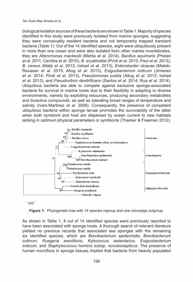
Fourteen bacteria species were identified by closest similarity with sequence databases using BLAST (Fig. 1). The identified sponge-associated bacteria species were Alteromonas macleodii, Bacillus aquimaris, B. aryabhattai, B. cereus, Brevibacterium epidermidis, B. iodinum, Enterobacter cloacae, Exiguobacterium indicum, Kytococcus sedentarius, Pseudomonas putida, Pseudovibrio denitrificans, Psychrobacter celer, Ruegeria arenilitoris, Staphylococcus hominis subsp. novobiosepticus. The phylogenetic tree in Figure 1 was inferred using the Neighbour-Joining method (Saitou & Nei 1987) with software MEGA6. The percentages of replicate trees where the associated taxa clustered together in the bootstrap test (10,000 replicates) are shown above the branches (Felsenstein 1985). The tree was drawn to scale, with branch lengths in the same units as those of the evolutionary distances used to infer the phylogenetic tree. The evolutionary distances were computed using the Maximum Composite Likelihood method (Tamura et al. 2004) and are in the units of the number of base substitutions per site. All positions containing gaps and missing data were eliminated. The phylogenetic tree analysed 15 nucleotide sequences: an ingroup comprising of 14 identified bacteria species and an outgroup of one microalga species.
Fig. 1 shows a phylogenetic tree with the identified bacteria categorised into four bacteria groups, which are the phylum Firmicutes as well as the classes Actinobacteria, Alphaproteobacteria, and Gammaproteobacteria. Bacteria from these similar groups have been previously isolated from marine sponges in geographically disparate areas such as the coastal waters of India, China, Ireland and Brazil (Webster & Taylor 2012). The previously reported geographic and
Tan Suet May Amelia et al.
190
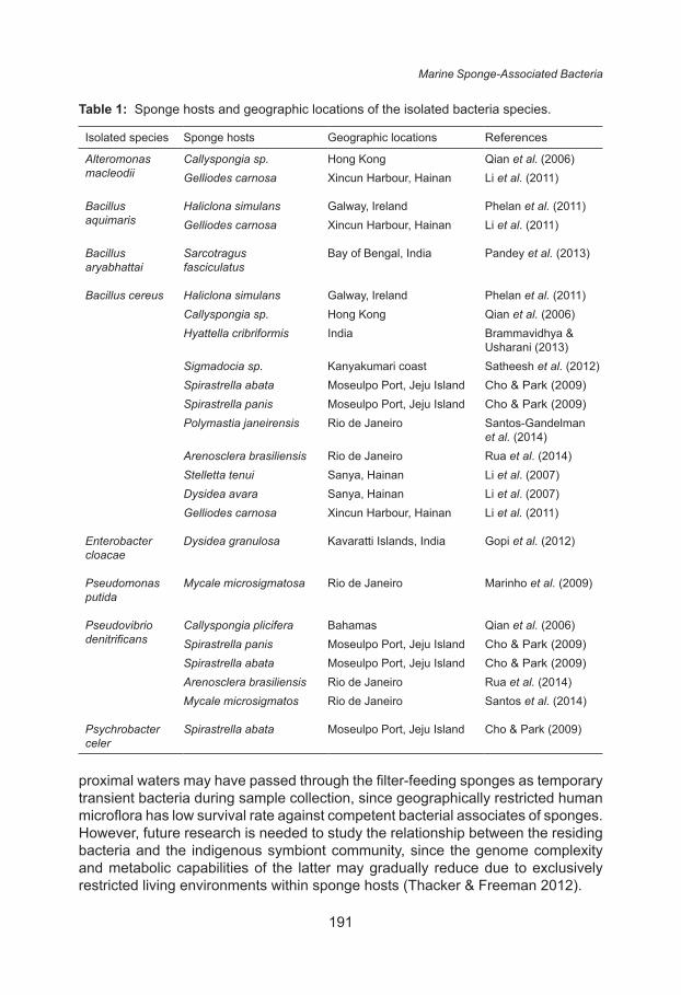
biological isolation sources of these bacteria are shown in Table 1. Majority of species identified in this study were previously isolated from marine sponges, suggesting they were conceivably resident bacteria and not temporarily trapped transient bacteria (Table 1). Out of the 14 identified species, eight were ubiquitously present in more than one ocean and were also isolated from other marine invertebrates, they are Alteromonas macleodii (Mehta et al. 2014), Bacillus aquimaris (Phelan et al. 2011, Cerritos et al. 2010), B. aryabhattai (Pindi et al. 2013; Paul et al. 2013), B. cereus (Maliji et al. 2013; Irshad et al. 2013), Enterobacter cloacae (Maleki-Ravasan et al. 2015; Altug et al. 2013), Exiguobacterium indicum (Jimenez et al. 2014; Pindi et al. 2013), Pseudomonas putida (Altug et al. 2013; Irshad et al. 2013), and Pseudovibrio denitrificans (Santos et al. 2014; Rua et al. 2014). Ubiquitous bacteria are able to compete against exclusive sponge-associated bacteria for survival in marine hosts due to their flexibility in adapting to diverse environments, namely by exploiting resources, producing secondary metabolites and bioactive compounds, as well as tolerating broad ranges of temperature and salinity (Ivars-Martinez et al. 2008). Consequently, the presence of competent ubiquitous bacteria within sponge larvae promotes the survivability of the latter when both symbiont and host are dispersed by ocean current to new habitats lacking in optimum physical parameters or symbionts (Thacker & Freeman 2012).
Figure 1: Phylogenetic tree with 14 species ingroup and one microalga outgroup.
As shown in Table 1, 8 out of 14 identified species were previously reported to have been associated with sponge hosts. A thorough search of relevant literature yielded no previous records that associated sea sponges with the remaining six identified species, which are Brevibacterium epidermidis, Brevibacterium iodinum, Ruegeria arenilitoris, Kytococcus sedentarius, Exiguobacterium indicum, and Staphylococcus hominis subsp. novobiosepticus. The presence of human microflora in sponge tissues implied that bacteria from heavily populated
Marine Sponge-Associated Bacteria
191
Table 1: Sponge hosts and geographic locations of the isolated bacteria species.
Isolated species Sponge hosts Geographic locations References
Alteromonas macleodii
Callyspongia sp. Hong Kong Qian et al. (2006)Gelliodes carnosa Xincun Harbour, Hainan Li et al. (2011)
Bacillus aquimaris
Haliclona simulans Galway, Ireland Phelan et al. (2011)Gelliodes carnosa Xincun Harbour, Hainan Li et al. (2011)
Bacillus aryabhattai
Sarcotragus fasciculatus
Bay of Bengal, India Pandey et al. (2013)
Bacillus cereus Haliclona simulans Galway, Ireland Phelan et al. (2011)Callyspongia sp. Hong Kong Qian et al. (2006)Hyattella cribriformis India Brammavidhya &
Usharani (2013)Sigmadocia sp. Kanyakumari coast Satheesh et al. (2012)Spirastrella abata Moseulpo Port, Jeju Island Cho & Park (2009)Spirastrella panis Moseulpo Port, Jeju Island Cho & Park (2009)Polymastia janeirensis Rio de Janeiro Santos-Gandelman
et al. (2014)Arenosclera brasiliensis Rio de Janeiro Rua et al. (2014)Stelletta tenui Sanya, Hainan Li et al. (2007)Dysidea avara Sanya, Hainan Li et al. (2007)Gelliodes carnosa Xincun Harbour, Hainan Li et al. (2011)
Enterobacter cloacae
Dysidea granulosa Kavaratti Islands, India Gopi et al. (2012)
Pseudomonas putida
Mycale microsigmatosa Rio de Janeiro Marinho et al. (2009)
Pseudovibrio denitrificans
Callyspongia plicifera Bahamas Qian et al. (2006)Spirastrella panis Moseulpo Port, Jeju Island Cho & Park (2009)Spirastrella abata Moseulpo Port, Jeju Island Cho & Park (2009)Arenosclera brasiliensis Rio de Janeiro Rua et al. (2014)Mycale microsigmatos Rio de Janeiro Santos et al. (2014)
Psychrobacter celer
Spirastrella abata Moseulpo Port, Jeju Island Cho & Park (2009)
proximal waters may have passed through the filter-feeding sponges as temporary transient bacteria during sample collection, since geographically restricted human microflora has low survival rate against competent bacterial associates of sponges. However, future research is needed to study the relationship between the residing bacteria and the indigenous symbiont community, since the genome complexity and metabolic capabilities of the latter may gradually reduce due to exclusively restricted living environments within sponge hosts (Thacker & Freeman 2012).
Tan Suet May Amelia et al.
192
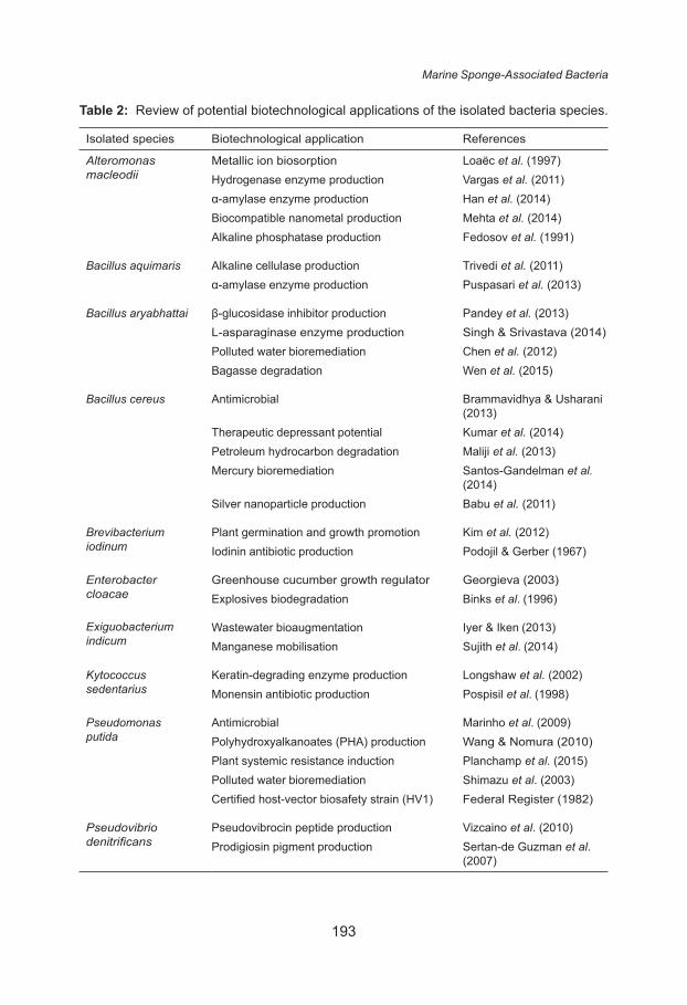
Table 2 displays the biotechnological applications or potentials recorded for the species recognised in this study. Some were studied for the synthesis of enzymes and pharmaceutical or medical properties (Table 2). Among the identified species, the most common biotechnological function was associated with bioremediation, while subsequent common capabilities were plant growth promotion, plant systemic resistance, and biological syntheses of biocompatible nanometal and biodegradable polymers. We would like to note several interesting species that harbour unique abilities or products, namely lead, cadmium and zinc biosorption mechanism in A. macleodii (Loaëc et al. 1997); halotolerant and psychrotolerant α-amylase with broad pH tolerance in A. macleodii (Han et al. 2014); skeletal muscle relaxation properties and central nervous system (CNS) depressants in B. cereus (Kumar et al. 2014); antileukaemic and antineoplastic L-asparaginase in B. aryabhattai (Singh & Srivastava 2014); polyhydroxyalkanoate (PHA) synthesis in P. putida (Wang & Nomura 2010); biodegradation of highly explosive pentaerythritol tetranitrate (PETN) by E. cloacae (Binks et al. 1996); as well as antibacterial, immunosuppressive and anticancer prodigiosin in P. denitrificans (Sertan-de Guzman et al. 2007).
The commonly recorded capability of bioremediation in some of the identified ubiquitous bacteria is explicable, for the degradation and utilisation of surrounding resource allowed these adaptable bacteria to tolerate severe environments by exploiting nutrients, producing secondary metabolites and bioactive compounds, and tolerating broad ranges of temperature and salinity (Ivars- Martinez et al. 2008), hence eventually settling as competent symbionts of marine invertebrates. Moreover, it should be brought to attention that the water bioremediation capacity of Pseudomonas putida (Table 2) was not an innate ability but expressed via ice-nucleation protein (INP) surface anchor, which revealed a ten-fold catalytic reaction compared to preceding research on Escherichia coli (Shimazu et al. 2003). Additional common biotechnological aptitude was the biosynthesis of nanometals that has been earlier reported for two identified species in this study, which are A. macleodii and B. cereus. Bacteria that biosynthesise nanometal particles are resistant to heavy metals, which could be found in ports and waters polluted with heavy metal effluents (Ramanathan et al. 2013).
A lack of previous reports were available for the biotechnological applications of B. epidermidis, R. arenilitoris, P. celer, and S. hominis subsp. novobiosepticus. Among the four strains, B. epidermidis is characteristically found on human skin as an ordinary human microflora. Although research on B. epidermidis is deficient, human microflora may harbour unknown dermatological prospects, such as callous treatment discovered in a human microflora recognised as Kytococcus sedentarius (Longshaw et al. 2002). Furthermore, P. celer is of a psychrophilic or psychrotolerant genus, Psychrobacter sp., whereby its ability to thrive in both warm and cold regions may be a desirable trait for industrial research purposes (Rodrigues et al. 2009). R. arenilitoris is a relatively recent species requiring research on its biotechnological prospects (Park & Yoon 2012). Although the recognised species were chiefly ubiquitous bacteria whereby they were neither exclusive nor specific to sea sponges, the results of this research nevertheless
Marine Sponge-Associated Bacteria
193
Table 2: Review of potential biotechnological applications of the isolated bacteria species.
Isolated species Biotechnological application References
Alteromonas macleodii
Metallic ion biosorption Loaëc et al. (1997)Hydrogenase enzyme production Vargas et al. (2011)α-amylase enzyme production Han et al. (2014)Biocompatible nanometal production Mehta et al. (2014)Alkaline phosphatase production Fedosov et al. (1991)
Bacillus aquimaris Alkaline cellulase production Trivedi et al. (2011)α-amylase enzyme production Puspasari et al. (2013)
Bacillus aryabhattai β-glucosidase inhibitor production Pandey et al. (2013)L-asparaginase enzyme production Singh & Srivastava (2014)Polluted water bioremediation Chen et al. (2012)Bagasse degradation Wen et al. (2015)
Bacillus cereus Antimicrobial Brammavidhya & Usharani (2013)
Therapeutic depressant potential Kumar et al. (2014)Petroleum hydrocarbon degradation Maliji et al. (2013)Mercury bioremediation Santos-Gandelman et al.
(2014)Silver nanoparticle production Babu et al. (2011)
Brevibacterium iodinum
Plant germination and growth promotion Kim et al. (2012)Iodinin antibiotic production Podojil & Gerber (1967)
Enterobacter cloacae
Greenhouse cucumber growth regulator Georgieva (2003)Explosives biodegradation Binks et al. (1996)
Exiguobacterium indicum
Wastewater bioaugmentation Iyer & Iken (2013)Manganese mobilisation Sujith et al. (2014)
Kytococcus sedentarius
Keratin-degrading enzyme production Longshaw et al. (2002)Monensin antibiotic production Pospisil et al. (1998)
Pseudomonas putida
Antimicrobial Marinho et al. (2009)Polyhydroxyalkanoates (PHA) production Wang & Nomura (2010)Plant systemic resistance induction Planchamp et al. (2015)Polluted water bioremediation Shimazu et al. (2003)Certified host-vector biosafety strain (HV1) Federal Register (1982)
Pseudovibrio denitrificans
Pseudovibrocin peptide production Vizcaino et al. (2010)Prodigiosin pigment production Sertan-de Guzman et al.
(2007)
Tan Suet May Amelia et al.
194
correspond with past studies that sponges are abundant with microbes capable of pharmaceutical and biotechnological applications (Schippers et al. 2012).
The biotechnological uses shown in Table 2 show ample research prospects among the identified cultivable symbionts associated with sponges. However, we would like to note that the same bacteria species does not necessarily yield similar products due to the mutation or adaptation of strains in respect to their surrounding environment. Majority of the identified species were ubiquitous with proficient survival mechanisms that promote adaptability in harsh environments, thus opportunistically outgrowing adjacent bacteria in marine sponges and residing as a part of the symbiont community in marine sponges. Furthermore, ten identified species were recorded in past studies to have harboured at least two biotechnological purposes, illuminating sponges as a markedly wealthy mine of microbes that are medically and biotechnologically substantial. However, cultivable bacteria under laboratory conditions occupy less than 1% of the total sponge microbial community (Amann et al. 1995), suggesting an uncharted existence of valuable research prospects within the putative 99% of uncultivated sponge bacteria. As a result, research is essential on growth media that foster the development of sponge symbionts, and on progressive metagenomic approaches that access the environmental metagenome devoid of isolation on complex culture media.
REFERENCES
Altug G, Cardak M, Ciftci P S and Gurun S. (2013). First records and microgeographical variations of culturable heterotrophic bacteria in an inner sea (the Sea of Marmara) between the Mediterranean and the Black Sea, Turkey. Turkish Journal of Biology 37(2):184–190. https://doi.org/10.3906/biy-1112-21
Amann R I, Ludwig W and Schleifer K H. (1995). Phylogenetic identification and in-situ detection of individual microbial cells without cultivation. Microbiological Reviews 59(1):143–169.
Babu M M G, Sridhar J and Gunasekaran P. (2011). Global transcriptome analysis of Bacillus cereus ATCC 14579 in response to silver nitrate stress. Journal of Nanobiotechnology 9: 49. https://doi.org/10.1186/1477-3155-9-49
Binks P R, French C E, Nicklin S and Bruce N C. (1996). Degradation of pentaerythritol tetranitrate by Enterobacter cloacae PB2. Applied and Environmental Microbiology 62(4): 1214–1219.
Brammavidhya S and Usharani G. (2013). Bioactive potential of sponge associated Bacillus cereus SBS02 isolated from Hyattela cribriformis. International Journal of Research in Environmental Science and Technology 3(2): 61–64.
Cerritos R, Eguiarte L E, Avitia M, Siefert J, Travisano M, Verdugo A R and Souza V. (2010). Diversity of culturable thermo-resistant aquatic bacteria along an environmental gradient in Cuatro Cienegas, Coahuila, Mexico. Antonie Van Leeuwenhoek 99(2): 303–318. https://doi.org/10.1007/s10482-010-9490-9
Chen W M, Tang Y Q, Kazuhiro M and Wu X L. (2012). Distribution of culturable endophytic bacteria in aquatic plants and their potential for bioremediation in polluted waters. Aquatic Biology 15(2): 99–110. https://doi.org/10.3354/ab00422
Marine Sponge-Associated Bacteria
195
Cho C W and Park S H. (2009). Comparative analysis of the community of culturable bacteria associated with sponges, Spirastrella abata and Spirastrella panis by 16S rDNA-RFLP. The Korean Journal of Microbiology 45(2): 155–162.
Federal Register. (1982). Appendix E: Certified host-vector systems. Federal Register 47(77): 17197.
Fedosov Y V, Mikhailov V V, Zhigalina I I, Ivanova E P, Kozhemyako V B, Onoprienko N B, Rasskazov V A and Elyakov G B. (1991). Doklady Akademii Nauk SSSR, 320(2): 485–487.
Felsenstein J. (1985). Confidence limits on phylogenies: An approach using the bootstrap. Evolution 39(4): 783–791. https://doi.org/10.1111/j.1558-5646.1985.tb00420.x
Georgieva O. (2003). Enterobacter cloacae bacterium as a growth regulator in greenhouse cucumbers (Cucumis sativus L.). Cucurbit Genetics Cooperative Report 26(2): 4–6.
Gopi M, Kumaran S, Kumar T T A, Deivasigamani B, Alagappan K and Prasad S G. (2012). Antibacterial potential of sponge endosymbiont marine Enterobacter sp at Kavaratti Island, Lakshadweep archipelago. Asian Pacific Journal of Tropical Medicine 5(2): 142–146. https://doi.org/10.1016/S1995-7645(12)60013-3
Gram H C. (1884). Uber die isolierte farbung der schizomyceten in schnitt-und trockenpraparaten. Fortschritte Der Medizin 2: 185–189.
Han X, Lin B, Ru G, Zhang Z, Liu Y and Hu Z. (2014). Gene cloning and characterization of an α- amylase from Alteromonas macleodii B7 for enteromorpha polysaccharide degradation. Journal of Microbiology and Biotechnology 24(2): 254–263. https://doi.org/10.4014/jmb.1304.04036
Irshad A, Ahmad I and Kim S B. (2013). Isolation, characterization and antimicrobial activity of halophilic bacteria in foreshore soils. African Journal of Microbiology Research 7(3): 164–173. https://doi.org/10.5897/AJMR12.1004
Ivars-Martinez E, Dauria G, Rodriguez-Valera F, Sanchez-Porro C, Ventosa A, Joint I and Muhling M. (2008). Biogeography of the ubiquitous marine bacterium Alteromonas macleodii determined by multilocus sequence analysis. Molecular Ecology 17(18): 4092–4106. https://doi.org/10.1111/j.1365-294X.2008.03883.x
Iyer R and Iken B. (2013). Identification of water-borne bacterial isolates for potential remediation of organophosphate contamination. Advances in Biological Chemistry 3(1): 146–152. https://doi.org/10.4236/abc.2013.31018
Jimenez E, Sanchez B, Farina A, Margolles A and Rodriguez J M. (2014). Characterization of the bile and gall bladder microbiota of healthy pigs. Microbiology Open 3(6): 937–949. https://doi.org/10.1002/mbo3.218
Kim K, Hwang S, Saravanan V S and Sa T. (2012). Effect of Brevibacterium iodinum RS16 and Methylobacterium oryzae CBMB20 inoculation on seed germination and early growth of maize and sorghum-sudangrass hybrid seedling under different salinity levels. Korean Journal of Soil Science and Fertilizer 45(1): 51–58. https://doi.org/10.7745/KJSSF.2012.45.1.051
Kumar M L V, Thippeswamy B and Kuppust I J. (2014). Evaluation of Bacillus cereus and Bacillus pumilus metabolites for CNS depressant and anticonvulsant activities. International Journal of Pharmacy and Pharmaceutical Sciences 6(2): 510–514.
Li Z Y, Hu Y, Huang Y Q and Huang Y. (2007). Isolation and phylogenetic analysis of the biologically active bacteria associated with three South China Sea sponges. Microbiology 76(4): 494–499. https://doi.org/10.1134/S0026261707040169
Tan Suet May Amelia et al.
196
Li C, Liu W, Zhu P, Yang J and Cheng K. (2011). Phylogenetic diversity of bacteria associated with the marine sponge Gelliodes carnosa collected from the Hainan Island coastal waters of the South China Sea. Microbial Ecology 62(4): 800–812. https://doi.org/10.1007/s00248-011-9896-6
Loaëc M, Olier R and Guezennec J. (1997). Uptake of lead, cadmium and zinc by a novel bacterial exopolysaccharide. Water Research 31(5): 1171–1179. https://doi.org/10.1016/S0043-1354(96)00375-2
Longshaw C M, Wright J D, Farrell A M and Holland K T. (2002). Kytococcus sedentarius, the organism associated with pitted keratolysis, produces two keratin-degrading enzymes. Journal of Applied Microbiology 93(5): 810–816. https://doi.org/10.1046/j.1365- 2672.2002.01742.x
Maleki-Ravasan N, Oshaghi M A, Afshar D, Arandian M H, Hajikhani S, Akhavan A A, Yakhchali B, Shirazi M H, Rassi Y, Jafari R, Aminian K, Fazeli-Varzaneh R A and Durvasula R. (2015). Aerobic bacterial flora of biotic and abiotic compartments of a hyperendemic Zoonotic Cutaneous Leishmaniasis (ZCL) focus. Parasites & Vectors 8: 63. https://doi.org/10.1186/s13071-014-0517-3
Maliji D, Olama Z and Holail H. (2013). Environmental studies on the microbial degradation of oil hydrocarbons and its application in Lebanese oil polluted coastal and marine ecosystem. International Journal of Current Microbiology and Applied Sciences 2(6): 1–18.
Marinho P R, Moreira A P B, Pellegrino E L P C, Muricy G, de Freire Bastos M D C, dos Santos K R N, Giambiagi-deMarval M, Laport M S. (2009). Marine Pseudomonas putida: A potential source of antimicrobial substances against antibiotic-resistant bacteria. Memórias do Instituto Oswaldo Cruz 104(5): 678–682. https://doi.org/10.1590/S0074-02762009000500002
Mehta A, Sidhu C, Pinnaka A K, Choudhury A R. (2014). Extracellular polysaccharide production by a novel osmotolerant marine strain of Alteromonas macleodii and its application towards biomineralization of silver. PLoS ONE 9(6): e98798. https://doi.org/10.1371/journal.pone.0098798
Pandey S, Sree A, Dash S S, Sethi D P and Chowdhury L. (2013). Diversity of marine bacteria producing beta-glucosidase inhibitors. Microbial Cell Factories 12: 35. https://doi.org/10.1186/1475-2859-12-35
Park S and Yoon J. (2012). Ruegeria arenilitoris sp. nov., isolated from the seashore sand around a seaweed farm. Antonie van Leeuwenhoek 102(4): 581–589. https://doi.org/10.1007/s10482-012-9753-8
Paul N C, Ji S H, Deng J X, Yu S H. (2013). Assemblages of endophytic bacteria in chilli pepper (Capsicum annuum L.) and their antifungal activity against phytopathogens in vitro. Plant Omics Journal 6(6): 441–448.
Phelan R W, O’Halloran J A, Kennedy J, Morrissey J P, Dobson A D W, O’Gara F and Barbosa T M. (2011). Diversity and bioactive potential of endospore-forming bacteria cultured from the marine sponge Haliclona simulans. Journal of Applied Microbiology 112(1): 65–78. https://doi.org/10.1111/j.1365-2672.2011.05173.x
Pindi P K, Yadav P R and Shanker A S. (2013). Identification of opportunistic pathogenic bacteria in drinking water samples of different rural health centers and their clinical impacts on humans. BioMed Research International Article ID 348250: 10. https://doi.org/10.1155/2013/348250
Planchamp C, Glauser G and Mauch-Mani B. (2015). Root inoculation with Pseudomonas putida KT2440 induces transcriptional and metabolic changes and systemic resistance in maize plants. Frontiers in Plant Science 5: 719. https://doi.org/10.3389/fpls.2014.00719
Marine Sponge-Associated Bacteria
197
Podojil M and Gerber N N. (1967). The biosynthesis of 1,6-phenazinediol 5,10-dioxide (iodinin) by Brevibacterium iodinum. Biochemistry 6(9): 2701–2705. https://doi.org/10.1021/bi00861a009
Pospisil S, Benada O, Kofronova O, Petricek M, Janda L and Havlicek V. (1998). Kytococcus sedentarius (formerly Micrococcus sedentarius) and Dermacoccus nishinomiyaensis (formerly Micrococcus nishinomiyaensis) produce monensins, typical Streptomycescinnamonensis metabolites. Canadian Journal of Microbiology 44(10): 1007–1011. https://doi.org/10.1139/w98-081
Puspasari F, Radjasa O K, Noer A S, Nurachman Z, Syah Y M, van der Maarel M, Dijkhuizen L, Janecek S and Natalia D. (2013). Raw starch-degrading α-amylase from Bacillus aquimaris MKSC 6.2: Isolation and expression of the gene, bioinformatics and biochemical characterization of the recombinant enzyme. Journal of Applied Microbiology 114(1): 108–120. https://doi.org/10.1111/jam.12025
Qian P Y, Dobrestov S, Dahms H U and Pawlik J. (2006). Antifouling activity and microbial diversity of two congeneric sponges Callyspongia spp. from Hong Kong and the Bahamas. Marine Ecology Progress Series 324(11): 151–165. https://doi.org/10.3354/meps324151
Ramanathan R, Field M R, O’Mullane A P, Smooker P M, Bhargava S K and Bansal V. (2013). Aqueous phase synthesis of copper nanoparticles: A link between heavy metal resistance and nanoparticle synthesis ability in bacterial systems. Nanoscale 5(6): 2300–2306. https://doi.org/10.1039/C2NR32887A
Riviere M L, Roumagnac M, Garrabou J and Bally M. (2013). Transient shifts in bacterial communities associated with the temperate gorgonian Paramuricea clavata in the Northwestern Mediterranean Sea. PLoS ONE 8(2): e57385. https://doi.org/10.1371/journal.pone.0057385
Rodrigues D F, Jesus E C, Ayala-del-Río H L, Pellizari V H, Gilichinsky D, Sepulveda-Torres L and Tiedje J M. (2009). Biogeography of two cold-adapted genera: Psychrobacter and Exiguobacterium. The International Society for Microbial Ecology Journal 3(6): 658–665. https://doi.org/10.1038/ismej.2009.25
Rua C P J, Trindade-Silva A E, Appolinario L R, Venas T M, Garcia G D, Carvalho L S, Lima A, Kruger R, Pereira R C, Berlinck R G S, Valle R A B, Thompson C C and Thompson F. (2014). Diversity and antimicrobial potential of culturable heterotrophic bacteria associated with the endemic marine sponge Arenosclera brasiliensis. PeerJ 2: e419. https://doi.org/10.7717/peerj.419
Saidin J B, Wahid M E A and Pennec G L. (2017). Characterization of the in vitro production of N-acyl homoserine lactones by cultivable bacteria inhabiting the sponge Suberites domuncula. Journal of the Marine Biological Association of the United Kingdom 97(1): 119–127. https://doi.org/ 10.1017/S0025315416000151
Saitou N and Nei M. (1987). The neighbor-joining method: A new method for reconstructing phylogenetic trees. Molecular Biology and Evolution 4(4): 406–425.
Sambrook J and Russell D W. (2006). Agarose gel electrophoresis. Cold Spring Harbor Protocols 1(1): pdb.prot4020. https://doi.org/10.1101/pdb.prot4020
Santos O C S, Soares A R, Machado F L S, Romanos M T V, Muricy G, Giambiagi-deMarval M and Laport M S. (2014). Investigation of biotechnological potential of sponge-associated bacteria collected in Brazilian coast. Letters in Applied Microbiology 60(2): 140–147. https://doi.org/10.1111/lam.12347
Santos-Gandelman J F, Cruz K, Crane S, Muricy G, Giambiagi-deMarval M, Barkay T, Laport M S. (2014). Potential application in mercury bioremediation of a marine sponge-isolated Bacillus cereus strain Pj1. Current Microbiology 69(3): 374–380. https://doi.org/10.1007/s00284-014-0597-5
Tan Suet May Amelia et al.
198
Satheesh S, Soniamby A R, Shankar C V S and Punitha S M J. (2012). Antifouling activities of marine bacteria associated with sponge (Sigmadocia sp.). Journal of Ocean University of China 11(3): 354–360. https://doi.org/10.1007/s11802-012-1927-5
Schippers K J, Sipkema D, Osinga R, Smidt H, Pomponi S A, Martens D E and Wijffels R H. (2012). Cultivation of sponges, sponge cells and symbionts: Achievements and future prospects. Advances in Marine Biology 62(2): 273–337. https://doi.org/10.1016/B978-0-12-394283- 8.00006-0
Sertan-de Guzman A A, Predicala R Z, Bernardo E B, Neilan B A, Elardo S P, Mangalindan G C, Tasdemir D, Ireland C M, Barraquio W L and Concepcion G P. (2007). Pseudovibriodenitrificans strain Z143-1, a heptylprodigiosin producing bacterium isolated from a Philippine tunicate. FEMS Microbiology Letters 277(2): 188–196. https://doi.org/10.1111/j.1574-6968.2007.00950.x
Shimazu M, Nguyen A, Mulchandani A and Chen W. (2003). Cell surface display of organophosphorus hydrolase in Pseudomonas putida using an ice-nucleation protein anchor. Biotechnology Progress 19(5): 1612–1614. https://doi.org/10.1021/bp0340640
Singh Y and Srivastava S K. (2014). Performance improvement of Bacillus aryabhattai ITBHU02 for high-throughput production of a tumor-inhibitory L-asparaginase using a kinetic model based approach. Journal of Chemical Technology and Biotechnology 89(1): 117–127. https://doi.org/10.1002/jctb.4121
Sujith P P, Mourya B S, Krishnamurthi S, Meena R M and Loka Bharathi P A. (2014). Mobilization of manganese by basalt associated Mn(II)-oxidizing bacteria from the Indian Ridge System. Chemosphere 95(2): 486–495. https://doi.org/10.1016/j.chemosphere.2013.09.103
Tamura K, Nei M and Kumar S. (2004). Prospects for inferring very large phylogenies by using the neighbor-joining method. Proceedings of the National Academy of Sciences (USA) 101(30): 11030–11035. https://doi.org/10.1073/pnas.0404206101
Tamura K, Stecher G, Peterson D, Filipski A and Kumar S. (2013). MEGA6: Molecular evolutionary genetics analysis version 6.0. Molecular Biology and Evolution 30(12): 2725–2729. https://doi.org/10.1093/molbev/mst197
Thacker R W and Freeman C J. (2012). Sponge-microbe symbioses: Recent advances and new directions. Advances in Marine Biology 62(2): 57–111. https://doi.org/10.1016/B978-0-12-394283-8.00002-3
Thomas T R, Kavlekar D P and LokaBharathi P A. (2010). Marine drugs from sponge-microbe association: A review. Marine Drugs 8(4): 1417–1468. https://doi.org/ 10.3390/md8041417
Trivedi N, Gupta V, Kumar M, Kumari P, Reddy C R K and Jha B. (2011). Solvent tolerant marine bacterium Bacillus aquimaris secreting organic solvent stable alkaline cellulase. Chemosphere 83(5): 706–712. https://doi.org/10.1016/j.chemosphere.2011.02.006
Turner S, Pryer K M, Miao V P W and Palmer J D. (1999). Investigating deep phylogenetic relationships among cyanobacteria and plastids by small subunit rRNA sequence analysis. Journal of Eukaryotic Microbiology 46(4): 327–338. https://doi.org/ 10.1111/j.1550-7408.1999.tb04612.x
Vargas W A, Weyman P D, Tong Y, Smith H O and Xu Q. (2011). [NiFe] Hydrogenase from Alteromonas macleodii with unusual stability in the presence of oxygen and high temperature. Applied and Environmental Microbiology 77(6): 1990–1998. https://doi.org/10.1128/AEM.01559-10
Marine Sponge-Associated Bacteria
199
Vizcaino M I, Johnson W R, Kimes N E, Williams K, Torralba M, Nelson K E, Smith G W, Weil E, Moeller P D and Morris P J. (2010). Antimicrobial resistance of the coral pathogen Vibrio coralliilyticus and Caribbean sister phylotypes isolated from a diseased octocoral. Microbial Ecology 59(4): 646–57. https://doi.org/10.1007/s00248-010-9644-3
Wang Q and Nomura C T. (2010). Monitoring differences in gene expression levels and polyhydroxyalkanoate (PHA) production in Pseudomonas putida KT2440 grown on different carbon sources. Journal of Bioscience and Bioengineering 110(6): 653–659. https://doi.org/10.1016/j.jbiosc.2010.08.001
Webster N S and Taylor M W. (2012). Marine sponges and their microbial symbionts: Love and other relationships. Environmental Microbiology 14(2): 335–346. https://doi.org/10.1111/j.1462- 2920.2011.02460.x
Wen J, Ren C, Huan N, Liu Y and Zeng R. (2015). Draft genome of bagasse-degrading bacteria Bacillus aryabhattai GZ03 from deep sea water. Marine Genomics 19(1): 13–14. https://doi.org/10.1016/j.margen.2014.11.004