Embed Size (px)
Citation preview
Identification of Critical Host Mitochondrion-Associated Genes duringEhrlichia chaffeensis Infections
Tonia Von Ohlen,a,b Alison Luce-Fedrow,a M. Teresa Ortega,a Roman R. Ganta,b and Stephen K. Chapesa
Kansas State University, Division of Biology, Manhattan, Kansas, USA,a and Kansas State University, Department of Diagnostic Medicine and Pathobiology, Manhattan,Kansas, USAb
Ehrlichia chaffeensis is an obligate intracellular bacterium that causes human monocytic ehrlichiosis (HME). To determine whathost components are important for bacterial replication, we performed microarray analysis on Drosophila melanogaster S2 cellsby comparing host gene transcript levels between permissive and nonpermissive conditions for E. chaffeensis growth. Five-hun-dred twenty-seven genes had increased transcript levels unique to permissive growth conditions 24 h postinfection. We screenedadult flies that were mutants for several of the “permissive” genes for the ability to support Ehrlichia replication. Three addi-tional D. melanogaster fly lines with putative mutations in pyrimidine metabolism were also tested. Ten fly lines carrying muta-tions in the genes CG6479, separation anxiety, chitinase 11, CG6364 (Uck2), CG6543 (Echs1), withered (whd), CG15881 (Ccdc58),CG14806 (Apop1), CG11875 (Nup37), and dumpy (dp) had increased resistance to infection with Ehrlichia. Analysis of RNA byquantitative real-time reverse transcription-PCR (qRT-PCR) confirmed that the bacterial load was decreased in these mutantflies compared to wild-type infected control flies. Seven of these genes (san, Cht11, Uck2, Echs1, whd, Ccdc58, and Apop1) en-coded proteins that had mitochondrial functions or could be associated with proteins with mitochondrial functions. Treatmentof THP-1 cells with double-stranded RNA to silence the human UCK2 gene indicates that the disruption of the uridine-cytidinekinase affects E. chaffeensis replication in human macrophages. Experiments with cyclopentenyl cytosine (CPEC), a CTP synthe-tase inhibitor and cytosine, suggest that the nucleotide salvage pathway is essential for E. chaffeensis replication and that it maybe important for the provision of CTP, uridine, and cytidine nucleotides.
Ehrlichia chaffeensis is the causative agent of human monocyticehrlichiosis (HME). There were 1,429 cases of HME in 2010
and 2011 (14). This represents a significant increase in the inci-dence of the disease since 2003 and qualifies HME as an emerginginfectious disease (25). In addition to being reported in the UnitedStates, HME has also been documented in Africa, Europe, China,and Brazil (9, 16, 42, 67). E. chaffeensis is an obligate intracellularbacterium. However, little is known about the parasitized-hostrequirements for bacterial replication.
Drosophila melanogaster has been used to study a variety ofintracellular pathogens. In particular, it has been successfully ma-nipulated for the identification of genes involved in host-patho-gen interactions. These pathogens include Listeria monocytogenes(1, 2), Chlamydia trachomatis (20), Mycobacterium marinum (1,18, 30, 48), Francisella tularensis (51, 64), and the protozoan par-asite Plasmodium gallinaceum (8, 53).
We previously demonstrated that E. chaffeensis is capable ofinfecting, completing its life cycle, and maintaining its pathoge-nicity in both Drosophila S2 cells (39) and adult flies (40). We havealso identified growth conditions that were nonpermissive for thegrowth of E. chaffeensis infection in Drosophila S2 cells (39).Therefore, we hypothesized that a transcriptional microarrayanalysis of permissively infected and nonpermissively infected S2cells would reveal host genes that contribute to the replication ofEhrlichia. We used the Affymetrix Drosophila 2.0 array to identifya subset of genes that were exclusively expressed during E.chaffeensis infection in infected S2 cells under permissive growthconditions. We screened adult flies carrying mutations in severalof the candidate genes, 10 of which change the fly response to E.chaffeensis compared to wild-type flies. Gene products from 7 ofthe 10 genes identified are associated with mitochondrial functionand/or location. We also describe follow-up experiments to inves-
tigate how Uck2 might function and its relevance to mammalianinfections.
MATERIALS AND METHODSMaintenance of cell lines and E. chaffeensis infections. The canine mac-rophage cell line DH82 (ATCC CRL-10389) was cultured in Eagle’s min-imal essential medium supplemented with 7% fetal bovine serum (FBS)(Atlanta Biologicals, Atlanta, GA) (EMEM7). THP-1 (ATCC TIB-202)cells were cultured in RPMI 1640 medium supplemented with 10% FBS.Drosophila S2 cells, obtained from the Drosophila RNAi Screening Center(Harvard Medical School, Boston, MA), were cultivated at 25°C in Sch-neider’s Drosophila medium (Invitrogen, Carlsbad, CA) supplementedwith 10% fetal bovine serum. For experiments, S2 cells were seeded at aconcentration of 1 � 106 cells/well in 6-well plates.
The E. chaffeensis Arkansas isolate was continuously cultivated inDH82 cells, as described previously, at 37°C, 8% CO2 in EMEM7 (40).Purified bacteria were used to reinfect DH82, THP-1, and S2 cells.
Infections in permissive and nonpermissive Drosophila S2 cells. Tomake S2 cells nonpermissive to E. chaffeensis infection, the cells were al-lowed to adhere for 30 min prior to adding sonicated lipopolysaccharide(LPS) from Salmonella enterica serovar Minnesota (Sigma, St. Louis, MO)at a concentration of 10 �g per ml. The S2 cells plus LPS were incubated
Received 20 June 2012 Returned for modification 18 July 2012Accepted 23 July 2012
Published ahead of print 30 July 2012
Editor: R. P. Morrison
Address correspondence to Stephen K. Chapes, [email protected].
This is Kansas Agriculture Experiment Station publication 12-447-J.
Supplemental material for this article may be found at http://iai.asm.org/.
Copyright © 2012, American Society for Microbiology. All Rights Reserved.
doi:10.1128/IAI.00670-12
3576 iai.asm.org Infection and Immunity p. 3576–3586 October 2012 Volume 80 Number 10
on February 11, 2018 by guest
http://iai.asm.org/
Dow
nloaded from
for 5 h prior to infection with E. chaffeensis. The Ehrlichia-infected S2 cellswere monitored at 24 and 96 h postinfection (p.i.). Additionally, unin-fected S2 cells, inactivated and activated with LPS, were used as controls.S2 cells were centrifuged at 300 � g for 5 min prior to RNA isolation fromcell pellets using 1 ml of TriReagent (Molecular Research Center, Inc.,Cincinnati, OH). The TriReagent-cell mixture was transferred to 2.0-mlHeavy Phase Lock Gel tubes (5 Prime, Westbury, NY). Chloroform (200�l) was added, and the mixture was gently mixed for 15 s. After centrifu-gation at 12,000 � g for 10 min at 4°C, the aqueous phase was poured into1.5-ml tubes. The RNA was precipitated and washed with isopropanoland ethanol according to the manufacturer’s instructions. The RNA pelletwas suspended in 50 �l of nuclease-free water, and the RNA concentra-tion was determined spectrophotometrically (NanoDrop Technologies,Wilmington, DE) before and after DNA digestion using a Turbo DNA-free kit (Ambion Inc., Austin, TX).
Determination of infection by RT-PCR and quantitative real-timeRT-PCR (qRT-PCR). For the microarray, infections were confirmed byreverse transcriptase (RT) PCR using the Promega Access One-Step RT-PCR kit (Madison, WI). Total RNA (750 ng) was used for each 25-�lreaction mixture, which contained the following reagents: 1� buffer, 0.2mM deoxynucleoside triphosphates (dNTPs), 2 �M forward primer, 2�M reverse primer, 1.5 mM MgSO4, 1 U/�l DNA polymerase, 1 U/�lreverse transcriptase, and nuclease-free water. RT-PCRs were performedin a thermocycler (Eppendorf Mastercycler Gradient, Hauppauge, NY).Primers and probes were designed with Primer Quest software (Inte-grated DNA Technologies, Coralville, IA) and data from NCBI referencesequences (see Table S1 in the supplemental material). RT-PCR condi-tions for the 16S rRNA of E. chaffeensis were 48°C for 45 min, 94°C for 4min, and then 35 cycles of 94°C for 30 s, 52°C for 30 s, and 72°C for 1 min(21). The ribosomal protein 49 gene (rp49) was used as a housekeepinggene in RT-PCR experiments with Drosophila S2 cells. The RT-PCR con-ditions for rp49 gene amplification were 48°C for 45 min, 94°C for 2 min,and then 35 cycles of 94°C for 45 s, 50°C for 1 min, and 72°C for 1.5 min.No-template RT-PCR controls were also included. RT-PCR productbands were identified on a ChemiImager (Protein Simple, Santa Clara,CA) after electrophoresis in 2% agarose gels and staining with ethidiumbromide.
Flies were anesthetized using CO2 prior to homogenization with dis-posable pestles (Kimble Chase, Vineland, NJ) in 1 ml of TriReagent. Thehomogenates were transferred to 2.0 ml Heavy Phase Lock Gel tubes andprocessed as described above for RNA isolation.
Ehrlichia quantification in S2 cells and fly experiments was estimatedusing TaqMan-based qRT-PCR, as previously described (40, 55), usingDrosophila ribosomal protein 15a as the housekeeping gene with primersdescribed in Table S1 in the supplemental material. E. chaffeensis wasdetected as described previously (40, 55). The qRT-PCRs were performedin the Cepheid SmartCycler System (Sunnyvale, CA).
Analysis of gene expression based on qRT-PCR results was performedusing the method described by Pfaffl (47). In short, primer efficiencieswere calculated from standard dilution curves plotting threshold cycle(CT) values versus log RNA using the following equation: efficiency �10
(�1/slope of standard curve)
.The change in CT values for both genes of interest and housekeeping
controls was determined by calculating the ��CT using the followingequation: (efficiency of gene of interestGene of interest:�CT control � treated)/(efficiency of housekeeping geneHousekeeping gene:�CT control � treated).
Microarray analysis. Microarray analysis was performed at the Uni-versity of Kansas Medical Center Microarray Facility (Kansas City, KS)using Affymetrix (Santa Clara, CA) Drosophila 2.0 Gene chips accordingto the manufacturer’s specifications. The analysis was performed on fourtreatment groups (each submitted in triplicate at 24 h p.i.): (i) S2 cellsinfected with E. chaffeensis, (ii) S2 cells incubated with LPS and theninfected with E. chaffeensis, (iii) S2 cells incubated with LPS, and (iv)untreated/uninfected S2 cells. CHP files were analyzed using GeneSpring7.3 software and normalized by “per gene: normalize to median.” The
“filter on volcano plot” was applied at 1.5-fold change and one-way anal-ysis of variance (ANOVA) at a significance level (�) of 0.05. Genes up-regulated 1.5-fold or more above basal expression levels were identifiedunder both permissive and nonpermissive conditions compared to unin-fected controls at 24 h p.i. These gene sets were then compared, and thoseupregulated exclusively under either permissive or nonpermissive condi-tions at 24 h p.i. were identified. Microarray MIAMI-compliant data areavailable at the publicly accessible database (http://bioinformatics.kumc.edu/mdms/login.php) by accessing the experiment entitled “DifferentialGene Expression in Ehrlichia chaffeensis-infected S2 cells.”
D. melanogaster. Flies were maintained on standard dextrose-molasses-yeast medium at 18 to 29°C. For all experiments, flies with the appropriatebackground were used as wild-type (WT) controls. w;Hemese-Gal4 UASGFPflies (GFPHeme) (from Michael J. Williams, Umea Centre for MolecularPathogenesis, Umea University, Umea, Sweden), yellow-white (yw) (main-tained in our stock collection at Kansas State University), and/or white ocelli(wo1) (stock number 634, from the Bloomington Drosophila Stock Center atIndiana University, Bloomington, IN) were used as the WT in these experi-ments. withered (whd1) (FBgn0004012; stock number 441), dumpy (dpov1)(FBgn0053196; stock number 276), and tilt (tt1wo1) (FBgn0003868; stocknumber 623) mutants are all the result of spontaneous mutations (35, 44, 65)and were obtained from the Bloomington Drosophila Stock Center at IndianaUniversity, Bloomington, IN. The stock numbers, genotypes, and associatedgenes of adult Drosophila flies screened by microinjection are listed in TableS2 in the supplemental material.
Adult Drosophila Infections. Flies were transferred to fresh food atleast 24 h prior to injection/infection. For injection/infection, adult maleand female flies were anesthetized with CO2 (for no longer than 15 min ata time). Flies were injected with approximately 50 nl of sterile PBS or with�5,000 bacteria, using pulled glass capillary needles. Injections were madein the abdomen of the fly, close to the junction of the thorax and ventral tothe junction between the dorsal and ventral cuticles. Following injection,the flies were maintained in clean bottles with molasses caps that werechanged every other day throughout the course of the experiments. Sur-vival of the flies was monitored daily.
CPEC treatment of S2 cells. Cyclopentenylcytosine (CPEC) was ob-tained from the National Cancer Institute (Drug Synthesis and ChemistryBranch, Developmental Therapeutics Program, Division of Cancer Treat-ment and Diagnosis, National Cancer Institute, Bethesda, MD) through aMaterials Transfer Agreement. The CPEC was prepared to a final concen-tration of 15.06 mM using sterile water containing 1% dimethyl sulfoxide(DMSO). Two different infection protocols were used for testing the effectof CPEC on the growth of E. chaffeensis in the S2 cells. For the first pro-tocol, CPEC was added to S2 cells at final concentrations of 100, 10, 1, or0.1 �M/well; E. chaffeensis was added to the treated cells 2 days later, andRNA was extracted from the cells 2 days postinfection. For the secondprotocol, S2 cells were infected with E. chaffeensis for 2 days, and thenvarious concentrations of CPEC were then added and RNA extractionswere performed 2 days following the CPEC treatment. S2 cells treated withdiluent only and infected with Ehrlichia and uninfected S2 cells were usedas controls. A qRT-PCR assay was used to analyze transcript levels ofEhrlichia 16S rRNA and Drosophila ribosomal protein 15a, as describedabove.
Addition of exogenous cytosine to Drosophila S2 cells. Cytosine wasobtained from Sigma-Aldrich Co. The cytosine was dissolved in 500 mMhydrochloric acid and then in sterile water to 100 mM, according to themanufacturer’s recommendation. For infection experiments, S2 cellswere incubated for 24 h prior to cytosine addition to a final concentrationof 25 mM; the cells were further incubated for 24 h. E. chaffeensis wasadded to the cytosine-treated cells, to untreated S2 cells, and to S2 cellstreated with the cytosine diluent. Untreated, uninfected S2 cells were usedas a negative control. RNA was extracted from the cells at 24, 48, 72, and 96h p.i. qRT-PCR was used to measure transcript levels of Ehrlichia 16SrRNA and Drosophila ribosomal protein 15a, as described above.
Host Factors in E. chaffeensis Growth
October 2012 Volume 80 Number 10 iai.asm.org 3577
on February 11, 2018 by guest
http://iai.asm.org/
Dow
nloaded from
THP-1 cell differentiation and cell cultures. THP-1 human macro-phages were cultured as described above. For differentiation, THP-1 cells(6 � 104 THP-1 cells/well; 24-well plates) were incubated with phorbol12-myristate 13-acetate (PMA) (Sigma-Aldrich Chemical Co., St. Louis,MO) at a final concentration of 50 �M. The cells were cultured for 24 h toallow final differentiation into macrophages in a cell culture incubator at37°C with 8% CO2.
Measurement of transfection efficiency and UCK2 knockdown. Weselected the small interfering RNA (siRNA) transfection conditions basedon the transfection efficiency in the cells of a fluorescently labeled trans-fection control siRNA duplex conjugated with the fluorescent markerTYE 563. Transiently transfected cells were analyzed for transfection effi-ciency using fluorescence microscopy.
UCK2 transcript expression was knocked down in THP-1 cells usingsiRNA specific to the human UCK2 transcript. We used the TriFecta RNAinterference (RNAi) kit with predesigned oligonucleotide sets from IDT.The siRNA predesigned oligonucleotide query was done using the UCK2RNA coding sequence (CDS) from NCBI reference sequenceXM_846154.2 for Homo sapiens. We also used scrambled siRNA and hu-man hypoxanthine phosphoribosyltransferase (HPRT) siRNAs as nega-tive and positive controls, respectively, to confirm transfection and siRNAknockdown efficiency. All siRNA duplexes were dissolved in nuclease-freewater (pH 7.2) to a final concentration of 2 nM. Three UCK2-specific, anHPRT-specific, and scrambled siRNA duplexes were transfected into dif-ferentiated THP-1 cells using the Lipofectamine transfection reagent (In-vitrogen) following the manufacturer’s protocol.
THP-1 cells were infected with E. chaffeensis 48 h posttransfection.Transfected and Ehrlichia-infected THP-1 cells were analyzed for UCK2,HPRT knockdown efficiency and the level of bacterial infection by qRT-PCR 48 h postinfection using primers and probes described in Table S1 inthe supplemental material, as explained above.
TEM. Transmission electron microscopy (TEM) analysis of unin-fected and E. chaffeensis-infected DH82 cultures was performed as de-scribed previously (17).
Statistics. Data are presented as means and standard errors of themean (SEM). Differences in means were determined using the Mann-Whitney two-tailed rank-sum statistical test, which is independent of theunderlying population distribution (StatMost statistical package; DataXIOM, Los Angeles, CA). Survival data were analyzed for significanceusing the log rank test of Kaplan-Meier plots and Prism (La Jolla, CA)Graphpad software. P values of � 0.05 were considered significant.
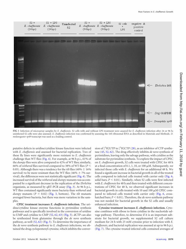
RESULTSMicroarray “permissive-exclusive” genes after infection with E.chaffeensis. To identify host genes that are necessary for E.chaffeensis infection, we examined transcript levels of DrosophilaS2 cells under four different conditions at 24 h p.i. to assess thehost factors that might be needed to initiate the infection. Theyincluded (i) uninfected S2 cells, (ii) uninfected S2 cells activatedwith LPS, (iii) S2 cells activated with LPS and then infected with E.chaffeensis (nonpermissive conditions; this experiment, per-formed as in previous studies, demonstrated the inhibition of E.chaffeensis replication in LPS-activated S2 cells [43]), and (iv) S2cells infected with E. chaffeensis (permissive conditions). E.chaffeensis replicated only under permissive conditions and notunder nonpermissive conditions for as long as 96 h p.i. (Fig. 1).Uninfected cells were used to discern the basal transcript levels inthe S2 cells, and comparisons across the different conditions werebased on this basal level of expression. In order to understandwhich gene transcripts were specific to nonpermissive S2 cells, weused the S2 cells that were treated only with LPS as a comparison.Cells that received only LPS treatment revealed activation-specificgene transcripts. We compared those genes to the genes that hadincreased transcript levels in cells infected under nonpermissive
conditions for bacterial growth to deduce transcripts that wereupregulated exclusively under nonpermissive conditions. We alsodetermined the gene transcripts that were upregulated 1.5-fold ormore exclusively under permissive infection conditions for E.chaffeensis growth, and 2,128 genes were upregulated 1.5-foldmore than in uninfected controls. Under nonpermissive growthconditions, 1,742 of the genes were upregulated 1.5-fold morethan in uninfected S2 cells. When we compared transcripts thatwere upregulated under permissive growth conditions to tran-scripts that were upregulated under nonpermissive growth condi-tions, we identified 527 genes unique to permissive conditions(see Table S3 in the supplemental material). Of these, 210 hadpreviously been ascribed some function and had some character-ization. The functions of the remaining 307 genes had yet to bedefined and had “CG” (computed gene) gene designations. How-ever, a number of these genes had orthologs with some describedproperties. These ortholog gene descriptions were used to ascribefunctions for our analyses. Viable and fertile adult fly mutantstocks were available for 118 of the 527 genes (37 stocks for definedgenes and 81 stocks for undefined genes). These flies had appro-priate mutations in coding exons in the genes of interest and al-lowed testing of how the absence of a functional gene would affectfly survival and/or bacterial replication in vivo. Our screen of mu-tant fly lines was loosely based on descriptions of genes that con-trolled functions that we guessed might be important for bacterialgrowth or were random choices of CG-designated genes.
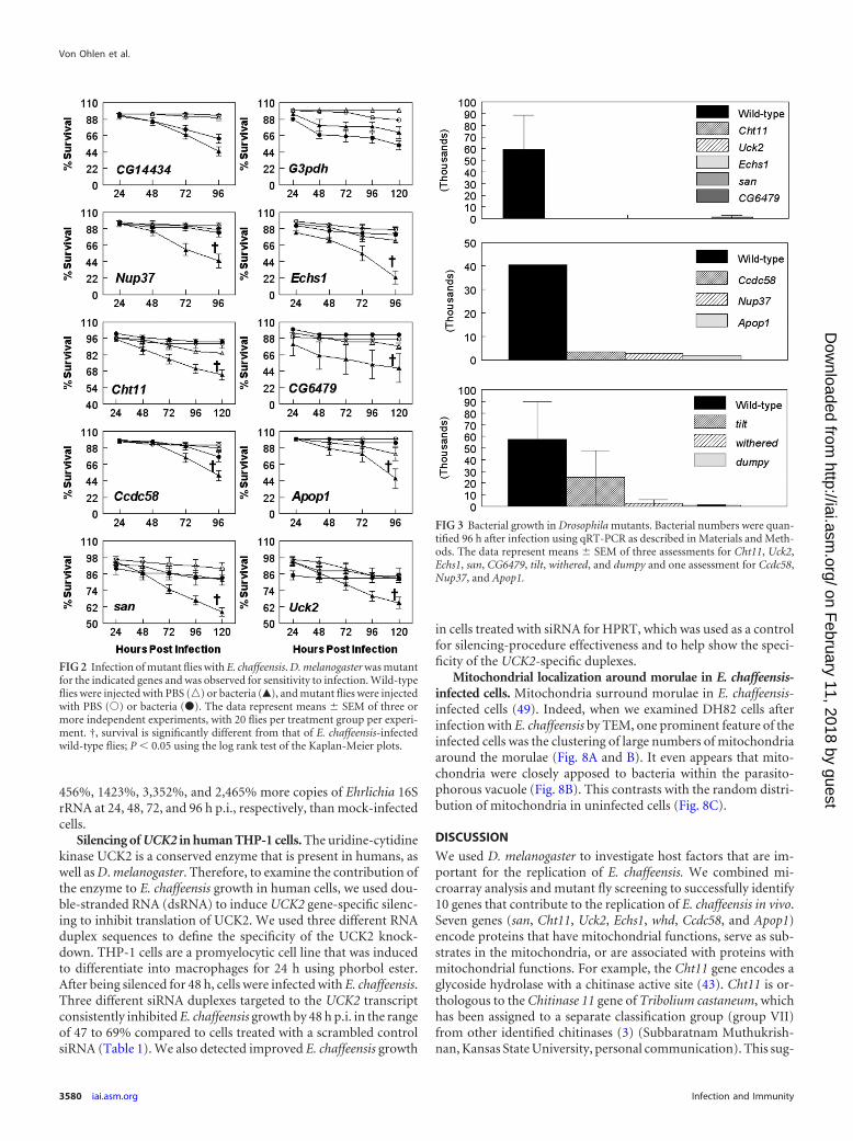
E. chaffeensis infection in selected mutant Drosophila flylines. We screened 19 Drosophila lines with mutations in the genesof interest (see Table S2 in the supplemental material) for bacterialreplication and fly survival. Flies (wild type and mutant) wereinjected with cell-free E. chaffeensis or sterile PBS and monitoredfor survival for 96 to 120 h p.i. (20 flies per treatment group perexperiment; the experiment was repeated independently at least 3times). We looked for the mutants that displayed increased sur-vival of the flies after bacterial challenge compared to the wild-type flies. Our hypothesis was that the gene affected in a mutantallowed increased survival because its expression was needed for E.chaffeensis replication. We found that the flies that had mutationsin Nup37, Echs1, Cht11, CG6479, Ccdc58, Apop1, san, and Uck2displayed significantly increased survival compared to wild-typeflies after infection with E. chaffeensis (Fig. 2). In contrast, severalmutants did not show increased resistance to E. chaffeensis infec-tions. They included three G3pdh mutants (stock 1124, shown inFig. 2) and CG14434 (Fig. 2), as well as CG10672, CG4743,CG9300, tsp3A, CG10992, and Gap69C (data not shown).
To confirm that Nup37, Echs1, Cht11, CG6479, Ccdc58, Apop1,san, and Uck2 gene mutations affected E. chaffeensis infections, wealso estimated bacterial replication as measured by qRT-PCR (Fig.3). Experimental infection of flies with disruptions in all 8 genesresulted in a significant drop in the numbers of bacteria comparedto those observed in wild-type flies infected with the organism(Fig. 3).
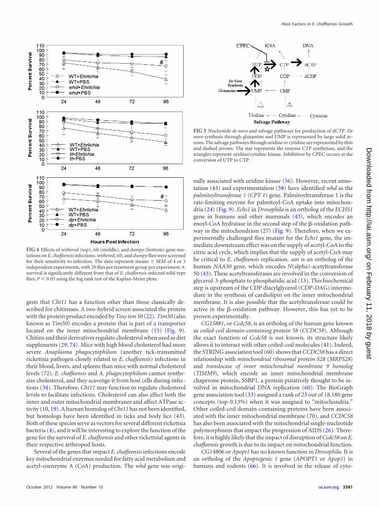
Uridine/cytidine kinase mutations affect fly survival andbacterial replication. To understand the physiological relevanceof disruption of gene function to E. chaffeensis growth in the host,we focused on Uck2, as an ortholog for this gene has been identi-fied in mammalian species (43). Stroman (56) previously reportedthat the Drosophila dumpy, tilt, and withered stocks carried a mu-tation(s) that affects uridine/cytidine kinase function. To confirmthe impact of Uck2 on E. chaffeensis growth, the three fly lines with
Von Ohlen et al.
3578 iai.asm.org Infection and Immunity
on February 11, 2018 by guest
http://iai.asm.org/
Dow
nloaded from
putative defects in uridine/cytidine kinase function were infectedwith E. chaffeensis and assessed for bacterial replication. Two ofthese fly lines were significantly more resistant to E. chaffeensischallenge than WT flies (Fig. 4). For example, at 96 h p.i., 61% ofthe dumpy flies were alive compared to 42% of WT flies; similarly,66% of withered flies survived compared to 38% of WT flies (P �0.05). Although there was a tendency for the tilt flies (60% 16%survival) to be more resistant than the WT flies (46% 7% sur-vival), the differences were not statistically significant (Fig. 4). Theincreased survival of the withered and dumpy mutants was accom-panied by a significant decrease in the replication of the Ehrlichiaorganisms, as measured by qRT-PCR assay (Fig. 3). At 96 h p.i.,WT flies contained significantly more bacteria than withered anddumpy mutants (P � 0.02) (Fig. 3, bottom). The tilt mutantsaveraged fewer bacteria, but there was more variation in the sam-ples.
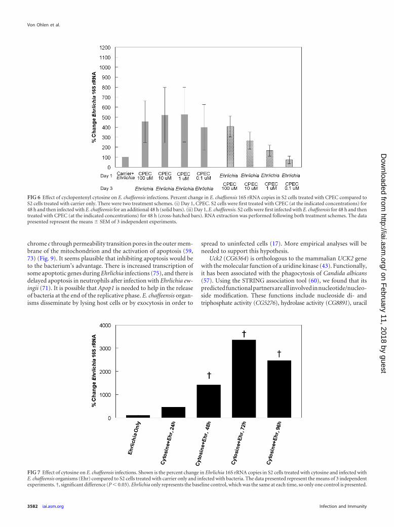
CPEC treatment increases E. chaffeensis infection. The uri-dine/cytidine kinase enzyme functions in pyrimidine synthesispathways and is specifically involved in the conversion of uridineto UMP and cytidine to CMP (52, 62, 63) (Fig. 5). dCTP can alsobe synthesized from glutamine through the de novo synthesispathway, as well (52, 62) (Fig. 5). To determine the importance ofthe de novo synthesis pathway to E. chaffeensis infections, we ob-tained the drug cyclopentenyl cytosine, which inhibits the conver-
sion of [3H]UTP to [3H]CTP (28), as an inhibitor of CTP synthe-tase (45, 52, 62). The drug effectively inhibits de novo synthesis ofpyrimidines, leaving only the salvage pathway, with cytidine as thesubstrate for pyrimidine synthesis. To explore the impact of CPECon E. chaffeensis growth, S2 cells were treated with CPEC for 48 hat a final concentration of 0.1, 1, 10, or 100 �M. Subsequently, weinfected those cells with E. chaffeensis for an additional 48 h. Wefound a significant increase in bacterial growth in all of the treatedcells compared to infected cells treated with carrier only (Fig. 6,solid bars; P � 0.01). Similarly, when S2 cells were first infectedwith E. chaffeensis for 48 h and then treated with different concen-trations of CPEC for 48 h, we observed significant increases inbacterial growth in cells treated with 10 and 100 �M CPEC com-pared to infected cells treated with carrier only (Fig. 6, cross-hatched bars; P � 0.01). Therefore, the de novo synthesis pathwaywas not needed for bacterial growth in the S2 cells and usuallyenhanced infection.
Cytosine treatment increases E. chaffeensis infection. Cyto-sine combines with ribose to form cytidine in the nucleotide sal-vage pathway. Therefore, to determine if it is an important sub-strate for bacterial growth, we supplemented S2 cell culturemedium for 24 h with 25 mM cytosine prior to infection with E.chaffeensis, and bacterial replication was assessed at up to 96 h p.i.(Fig. 7). The cytosine-treated infected cells contained averages of
FIG 1 Infection of microarray samples by E. chaffeensis. S2 cells with and without LPS treatment were assayed for E. chaffeensis infection after 24 or 96 h;uninfected S2 cells were also assessed. E. chaffeensis infection was confirmed by assessing the 16S ribosomal RNA as described in Materials and Methods. D.melanogaster rp49 transcript was used as a loading control.
Host Factors in E. chaffeensis Growth
October 2012 Volume 80 Number 10 iai.asm.org 3579
on February 11, 2018 by guest
http://iai.asm.org/
Dow
nloaded from
456%, 1423%, 3,352%, and 2,465% more copies of Ehrlichia 16SrRNA at 24, 48, 72, and 96 h p.i., respectively, than mock-infectedcells.
Silencing of UCK2 in human THP-1 cells. The uridine-cytidinekinase UCK2 is a conserved enzyme that is present in humans, aswell as D. melanogaster. Therefore, to examine the contribution ofthe enzyme to E. chaffeensis growth in human cells, we used dou-ble-stranded RNA (dsRNA) to induce UCK2 gene-specific silenc-ing to inhibit translation of UCK2. We used three different RNAduplex sequences to define the specificity of the UCK2 knock-down. THP-1 cells are a promyelocytic cell line that was inducedto differentiate into macrophages for 24 h using phorbol ester.After being silenced for 48 h, cells were infected with E. chaffeensis.Three different siRNA duplexes targeted to the UCK2 transcriptconsistently inhibited E. chaffeensis growth by 48 h p.i. in the rangeof 47 to 69% compared to cells treated with a scrambled controlsiRNA (Table 1). We also detected improved E. chaffeensis growth
in cells treated with siRNA for HPRT, which was used as a controlfor silencing-procedure effectiveness and to help show the speci-ficity of the UCK2-specific duplexes.
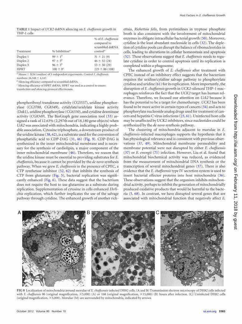
Mitochondrial localization around morulae in E. chaffeensis-infected cells. Mitochondria surround morulae in E. chaffeensis-infected cells (49). Indeed, when we examined DH82 cells afterinfection with E. chaffeensis by TEM, one prominent feature of theinfected cells was the clustering of large numbers of mitochondriaaround the morulae (Fig. 8A and B). It even appears that mito-chondria were closely apposed to bacteria within the parasito-phorous vacuole (Fig. 8B). This contrasts with the random distri-bution of mitochondria in uninfected cells (Fig. 8C).
DISCUSSION
We used D. melanogaster to investigate host factors that are im-portant for the replication of E. chaffeensis. We combined mi-croarray analysis and mutant fly screening to successfully identify10 genes that contribute to the replication of E. chaffeensis in vivo.Seven genes (san, Cht11, Uck2, Echs1, whd, Ccdc58, and Apop1)encode proteins that have mitochondrial functions, serve as sub-strates in the mitochondria, or are associated with proteins withmitochondrial functions. For example, the Cht11 gene encodes aglycoside hydrolase with a chitinase active site (43). Cht11 is or-thologous to the Chitinase 11 gene of Tribolium castaneum, whichhas been assigned to a separate classification group (group VII)from other identified chitinases (3) (Subbaratnam Muthukrish-nan, Kansas State University, personal communication). This sug-
FIG 2 Infection of mutant flies with E. chaffeensis. D. melanogaster was mutantfor the indicated genes and was observed for sensitivity to infection. Wild-typeflies were injected with PBS (o) or bacteria (Œ), and mutant flies were injectedwith PBS (Œ) or bacteria (�). The data represent means SEM of three ormore independent experiments, with 20 flies per treatment group per experi-ment. †, survival is significantly different from that of E. chaffeensis-infectedwild-type flies; P � 0.05 using the log rank test of the Kaplan-Meier plots.
FIG 3 Bacterial growth in Drosophila mutants. Bacterial numbers were quan-tified 96 h after infection using qRT-PCR as described in Materials and Meth-ods. The data represent means SEM of three assessments for Cht11, Uck2,Echs1, san, CG6479, tilt, withered, and dumpy and one assessment for Ccdc58,Nup37, and Apop1.
Von Ohlen et al.
3580 iai.asm.org Infection and Immunity
on February 11, 2018 by guest
http://iai.asm.org/
Dow
nloaded from
gests that Cht11 has a function other than those classically de-scribed for chitinases. A two-hybrid screen associated the proteinwith the protein product encoded by Tiny tim 50 (22). Ttm50 (alsoknown as Tim50) encodes a protein that is part of a transporterlocated on the inner mitochondrial membrane (15) (Fig. 9).Chitins and their derivatives regulate cholesterol when used as dietsupplements (29, 74). Mice with high blood cholesterol had moresevere Anaplasma phagocytophilum (another tick-transmittedrickettsia pathogen closely related to E. chaffeensis) infections intheir blood, livers, and spleens than mice with normal cholesterollevels (72). E. chaffeensis and A. phagocytophilum cannot synthe-size cholesterol, and they scavenge it from host cells during infec-tions (34). Therefore, Cht11 may function to regulate cholesterollevels to facilitate infections. Cholesterol can also affect both theinner and outer mitochondrial membranes and affect ATPase ac-tivity (10, 19). A human homolog of Cht11 has not been identified,but homologs have been identified in ticks and body lice (43).Both of these species serve as vectors for several different rickettsiabacteria (4), and it will be interesting to explore the function of thegene for the survival of E. chaffeensis and other rickettsial agents intheir respective arthropod hosts.
Several of the genes that impact E. chaffeensis infections encodekey mitochondrial enzymes needed for fatty acid metabolism andacetyl-coenzyme A (CoA) production. The whd gene was origi-
nally associated with uridine kinase (56). However, recent anno-tation (43) and experimentation (58) have identified whd as thepalmitoyltransferase 1 (CPT 1) gene. Palmitoyltransferase 1 is therate-limiting enzyme for palmitoyl-CoA uptake into mitochon-dria (24) (Fig. 9). Echs1 in Drosophila is an ortholog of the ECHS1gene in humans and other mammals (43), which encodes anenoyl-CoA hydratase in the second step of the -oxidation path-way in the mitochondrion (27) (Fig. 9). Therefore, when we ex-perimentally challenged flies mutant for the Echs1 gene, the im-mediate downstream effect was on the supply of acetyl-CoA to thecitric acid cycle, which implies that the supply of acetyl-CoA maybe critical to E. chaffeensis replication. san is an ortholog of thehuman NAA50 gene, which encodes N(alpha)-acetyltransferase50 (43). These acetyltransferases are involved in the conversion ofglycerol-3-phosphate to phosphatidic acid (13). This biochemicalstep is upstream of the CDP diacylglycerol (CDP-DAG) interme-diate in the synthesis of cardiolipin on the inner mitochondrialmembrane. It is also possible that the acetyltransferase could beactive in the -oxidation pathway. However, this has yet to beproven experimentally.
CG15881, or Ccdc58, is an ortholog of the human gene knownas coiled-coil domain-containing protein 58 (CCDC58). Althoughthe exact function of Ccdc58 is not known, its structure likelyallows it to interact with other coiled-coil molecules (41). Indeed,the STRING association tool (60) shows that CCDC58 has a directrelationship with mitochondrial ribosomal protein S28 (MRPS28)and translocase of inner mitochondrial membrane 9 homolog(TIMM9), which encode an inner mitochondrial membranechaperone protein, SSBP1, a protein putatively thought to be in-volved in mitochondrial DNA replication (60). The BioGraphgene association tool (33) assigned a rank of 23 out of 18,180 geneconcepts (top 0.13%) when it was assigned to “mitochondria.”Other coiled-coil domain-containing proteins have been associ-ated with the inner mitochondrial membrane (70), and CCDC58has also been associated with the mitochondrial single-nucleotidepolymorphisms that impact the progression of AIDS (26). There-fore, it is highly likely that the impact of disruption of Ccdc58 on E.chaffeensis growth is due to its impact on mitochondrial function.
CG14806 or Apopt1 has no known function in Drosophila. It isan ortholog of the Apoptogenic 1 gene (APOPT1 or Apop1) inhumans and rodents (66). It is involved in the release of cyto-
FIG 5 Nucleotide de novo and salvage pathways for production of dCTP. Denovo synthesis through glutamine and UMP is represented by large solid ar-rows. The salvage pathways through uridine or cytidine are represented by thinand dashed arrows. The star represents the enzyme CTP synthetase, and thetriangles represent uridine/cytidine kinase. Inhibition by CPEC occurs at theconversion of UTP to CTP.
FIG 4 Effects of withered (top), tilt (middle), and dumpy (bottom) gene mu-tations on E. chaffeensis infections. withered, tilt, and dumpy flies were screenedfor their sensitivity to infection. The data represent means SEM of 4 or 5independent experiments, with 20 flies per treatment group per experiment. #,survival is significantly different from that of E. chaffeensis-infected wild-typeflies; P � 0.05 using the log rank test of the Kaplan-Meier plots.
Host Factors in E. chaffeensis Growth
October 2012 Volume 80 Number 10 iai.asm.org 3581
on February 11, 2018 by guest
http://iai.asm.org/
Dow
nloaded from
chrome c through permeability transition pores in the outer mem-brane of the mitochondrion and the activation of apoptosis (59,73) (Fig. 9). It seems plausible that inhibiting apoptosis would beto the bacterium’s advantage. There is increased transcription ofsome apoptotic genes during Ehrlichia infections (75), and there isdelayed apoptosis in neutrophils after infection with Ehrlichia ew-ingii (71). It is possible that Apop1 is needed to help in the releaseof bacteria at the end of the replicative phase. E. chaffeensis organ-isms disseminate by lysing host cells or by exocytosis in order to
spread to uninfected cells (17). More empirical analyses will beneeded to support this hypothesis.
Uck2 (CG6364) is orthologous to the mammalian UCK2 genewith the molecular function of a uridine kinase (43). Functionally,it has been associated with the phagocytosis of Candida albicans(57). Using the STRING association tool (60), we found that itspredictedfunctionalpartnersareall involvedinnucleotide/nucleo-side modification. These functions include nucleoside di- andtriphosphate activity (CG5276), hydrolase activity (CG8891), uracil
FIG 6 Effect of cyclopentenyl cytosine on E. chaffeensis infections. Percent change in E. chaffeensis 16S rRNA copies in S2 cells treated with CPEC compared toS2 cells treated with carrier only. There were two treatment schemes. (i) Day 1, CPEC. S2 cells were first treated with CPEC (at the indicated concentrations) for48 h and then infected with E. chaffeensis for an additional 48 h (solid bars). (ii) Day 1, E. chaffeensis. S2 cells were first infected with E. chaffeensis for 48 h and thentreated with CPEC (at the indicated concentrations) for 48 h (cross-hatched bars). RNA extraction was performed following both treatment schemes. The datapresented represent the means SEM of 3 independent experiments.
FIG 7 Effect of cytosine on E. chaffeensis infections. Shown is the percent change in Ehrlichia 16S rRNA copies in S2 cells treated with cytosine and infected withE. chaffeensis organisms (Ehr) compared to S2 cells treated with carrier only and infected with bacteria. The data presented represent the means of 3 independentexperiments. †, significant difference (P � 0.03). Ehrlichia only represents the baseline control, which was the same at each time, so only one control is presented.
Von Ohlen et al.
3582 iai.asm.org Infection and Immunity
on February 11, 2018 by guest
http://iai.asm.org/
Dow
nloaded from
phosphoribosyl transferase activity (CG5537), uridine phosphor-ylase (CG3788, CG8349), cytidylate/uridylate kinase activity(Dak1), uridine phosphorylase (CG6330), and cytidine deaminaseactivity (CG8349). The BioGraph gene association tool (33) as-signed a rank of 12.63% (2,297th out of 18,180 gene objects) whenUck2 was associated with mitochondria, indicating a highly prob-able association. Cytosine triphosphate, a downstream product ofthe uridine kinase (38, 62), is a substrate used for the conversion ofphosphatidic acid to CDP-DAG (31, 46) (Fig. 9). CDP-DAG issynthesized in the inner mitochondrial membrane and is neces-sary for the synthesis of cardiolipin, a major component of theinner mitochondrial membrane (46). Therefore, we reason thatthe uridine kinase must be essential to providing substrates for E.chaffeensis, because it cannot be provided by the de novo synthesispathway. When we grew E. chaffeensis in the presence of CPEC, aCTP synthetase inhibitor (52, 62) that inhibits the synthesis ofCTP from glutamate (Fig. 5), bacterial replication was signifi-cantly enhanced (Fig. 6). These data suggest that the bacteriumdoes not require the host to use glutamine as a substrate duringreplication. Supplementation of cytosine in cells enhanced Ehrli-chia replication, which further implicates the use of the salvagepathway through cytidine. The enhanced growth of another rick-
ettsia, Rickettsia felis, from pyrimidines in tryptose phosphatebroth is also consistent with the involvement of mitochondrialenzymes in obligate intracellular bacterial growth (50). Moreover,cytidine is the least abundant nucleoside in cells (32). The deple-tion of cytidine pools can disrupt the balance of ribonucleotides incells, leading to alterations in cellular homeostasis and apoptosis(52). These observations suggest that E. chaffeensis needs to regu-late cytidine in order to control apoptosis until its replication iscompleted within a phagosome.
The enhanced growth of E. chaffeensis after treatment withCPEC instead of an inhibitory effect suggests that the bacteriumrequires the uridine/cytidine salvage pathway to phosphorylatecytidine and uridine (61) for its replication. More importantly, thedisruption of E. chaffeensis growth in UCK2-silenced THP-1 mac-rophages reinforces the fact that the UCK2 target has human rel-evance. Therefore, we focused our attention on Uck2 because ithas the potential to be a target for chemotherapy. UCK2 has beenfound to be more active in certain types of cancers (54) and acts tophosphorylate nucleoside analog drugs used for treatment of can-cers and hepatitis C virus infections (23, 61). Uninfected host cellsmay be unaffected by UCK2 inhibitors, since nucleotides could besynthesized by the de novo synthesis pathway.
The clustering of mitochondria adjacent to morulae in E.chaffeensis-infected macrophages supports the hypothesis that ithas physiological relevance and is consistent with previous obser-vations (37, 49). Mitochondrial membrane permeability andmembrane potential were not disrupted by either E. chaffeensis(37) or E. ewengii (71) infection. However, Liu et al. found thatmitochondrial biochemical activity was reduced, as evidencedfrom the measurement of mitochondrial DNA synthesis or thetranscription of several mitochondrial genes (37). There is alsoevidence that the E. chaffeensis type IV secretion system is used toinsert bacterial effector proteins into host mitochondria (36).These observations suggest that the organism inhibits mitochon-drial activity, perhaps to inhibit the generation of mitochondriallyproduced oxidative products that would be harmful to the bacte-ria (5, 68). In contrast, we have disrupted several genes that areassociated with mitochondrial function that negatively affect E.
TABLE 1 Impact of UCK2 dsRNA silencing on E. chaffeensis growth inTHP-1 cells
Treatment % Inhibitiona
% of E. chaffeensiscompared toscrambled dsRNAcontrola
Duplex 1 99 1b 31 21 (9)Duplex 2 97 5b 46 32 (24)Duplex 3 96 3b 53 30 (29)HPRT 100 0c 225 86 (108)a Means SEM (median) of 5 independent experiments. Control E. chaffeensisnumbers: 26,348 4,147.b Silencing efficiency compared to scrambled dsRNA.c Silencing efficiency of HPRT dsRNA. HPRT was used as a control to ensuretransfection and silencing-protocol effectiveness.
FIG 8 Localization of mitochondria around morulae of E. chaffeensis-infected DH82 cells. (A and B) Transmission electron microscopy of DH82 cells infectedwith E. chaffeensis 96 (original magnification, �5,000) (A) or 168 (original magnification, �15,000) (B) hours after infection. (C) Uninfected DH82 cells(original magnification, �5,000). Morulae (M) are surrounded by mitochondria, indicated by arrows.
Host Factors in E. chaffeensis Growth
October 2012 Volume 80 Number 10 iai.asm.org 3583
on February 11, 2018 by guest
http://iai.asm.org/
Dow
nloaded from
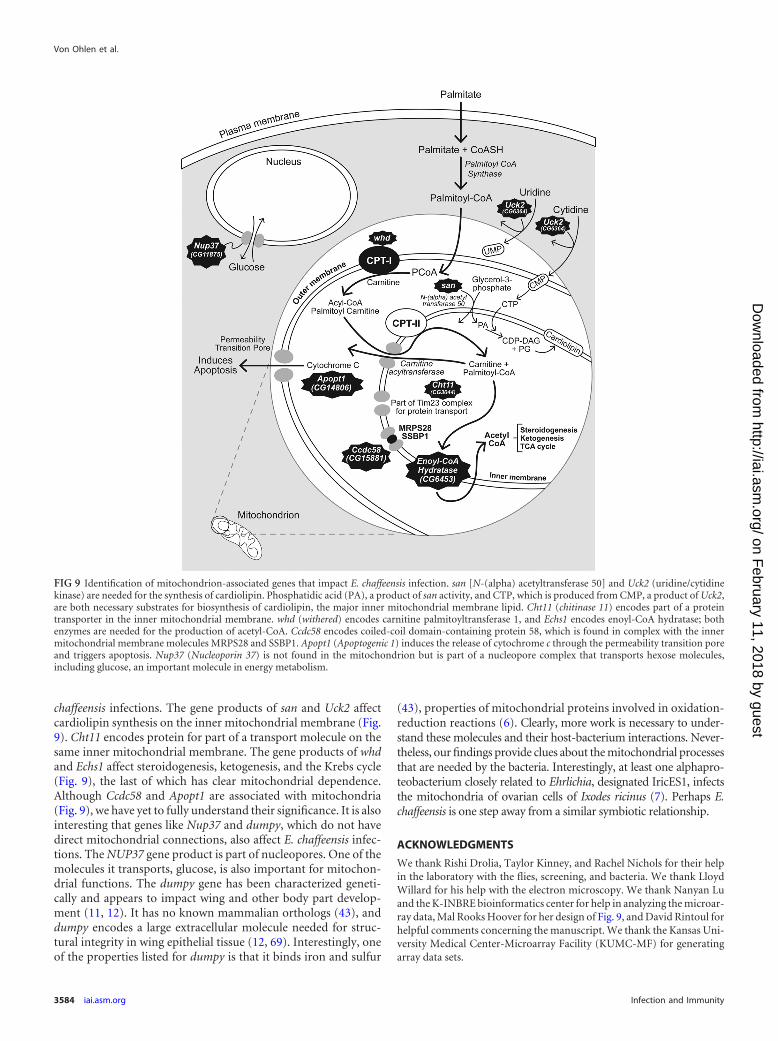
chaffeensis infections. The gene products of san and Uck2 affectcardiolipin synthesis on the inner mitochondrial membrane (Fig.9). Cht11 encodes protein for part of a transport molecule on thesame inner mitochondrial membrane. The gene products of whdand Echs1 affect steroidogenesis, ketogenesis, and the Krebs cycle(Fig. 9), the last of which has clear mitochondrial dependence.Although Ccdc58 and Apopt1 are associated with mitochondria(Fig. 9), we have yet to fully understand their significance. It is alsointeresting that genes like Nup37 and dumpy, which do not havedirect mitochondrial connections, also affect E. chaffeensis infec-tions. The NUP37 gene product is part of nucleopores. One of themolecules it transports, glucose, is also important for mitochon-drial functions. The dumpy gene has been characterized geneti-cally and appears to impact wing and other body part develop-ment (11, 12). It has no known mammalian orthologs (43), anddumpy encodes a large extracellular molecule needed for struc-tural integrity in wing epithelial tissue (12, 69). Interestingly, oneof the properties listed for dumpy is that it binds iron and sulfur
(43), properties of mitochondrial proteins involved in oxidation-reduction reactions (6). Clearly, more work is necessary to under-stand these molecules and their host-bacterium interactions. Never-theless, our findings provide clues about the mitochondrial processesthat are needed by the bacteria. Interestingly, at least one alphapro-teobacterium closely related to Ehrlichia, designated IricES1, infectsthe mitochondria of ovarian cells of Ixodes ricinus (7). Perhaps E.chaffeensis is one step away from a similar symbiotic relationship.
ACKNOWLEDGMENTS
We thank Rishi Drolia, Taylor Kinney, and Rachel Nichols for their helpin the laboratory with the flies, screening, and bacteria. We thank LloydWillard for his help with the electron microscopy. We thank Nanyan Luand the K-INBRE bioinformatics center for help in analyzing the microar-ray data, Mal Rooks Hoover for her design of Fig. 9, and David Rintoul forhelpful comments concerning the manuscript. We thank the Kansas Uni-versity Medical Center-Microarray Facility (KUMC-MF) for generatingarray data sets.
FIG 9 Identification of mitochondrion-associated genes that impact E. chaffeensis infection. san [N-(alpha) acetyltransferase 50] and Uck2 (uridine/cytidinekinase) are needed for the synthesis of cardiolipin. Phosphatidic acid (PA), a product of san activity, and CTP, which is produced from CMP, a product of Uck2,are both necessary substrates for biosynthesis of cardiolipin, the major inner mitochondrial membrane lipid. Cht11 (chitinase 11) encodes part of a proteintransporter in the inner mitochondrial membrane. whd (withered) encodes carnitine palmitoyltransferase 1, and Echs1 encodes enoyl-CoA hydratase; bothenzymes are needed for the production of acetyl-CoA. Ccdc58 encodes coiled-coil domain-containing protein 58, which is found in complex with the innermitochondrial membrane molecules MRPS28 and SSBP1. Apopt1 (Apoptogenic 1) induces the release of cytochrome c through the permeability transition poreand triggers apoptosis. Nup37 (Nucleoporin 37) is not found in the mitochondrion but is part of a nucleopore complex that transports hexose molecules,including glucose, an important molecule in energy metabolism.
Von Ohlen et al.
3584 iai.asm.org Infection and Immunity
on February 11, 2018 by guest
http://iai.asm.org/
Dow
nloaded from
The Microarray Facility is supported by the Kansas University School ofMedicine, the KUMC Biotechnology Support Facility, the Smith Intellectualand Developmental Disabilities Research Center (HD02528), and the KansasIDeA Network of Biomedical Research Excellence (RR016475). This projectwas supported by NIH grants AI088070, AI55052, AI052206, AI070908,RR16475, and RR17686; Kansas Agriculture Experiment Station AnimalHealth Project grant 481848; the Kansas Space Grant Consortium; NASAgrants NAG2-1274 and NNX08BA91G; American Heart Association grant0950036G; the Terry C. Johnson Center for Basic Cancer Research; and theKansas Agriculture Experiment Station.
REFERENCES1. Agaisse H, et al. 2005. Genome-wide RNAi screen for host factors re-
quired for intracellular bacterial infection. Science 309:1248 –1251.2. Alvarez-Dominguez C, Barbieri AM, Beron W, Wandinger-Ness A,
Stahl PD. 1996. Phagocytosed live Listeria monocytogenes influencesRab5-regulated in vitro phagosome-endosome fusion. J. Biol. Chem. 271:13834 –13843.
3. Arakane Y, Muthukrishnan S. 2010. Insect chitinase and chitinase-likeproteins. Cell. Mol. Life Sci. 67:201–216.
4. Azad A, Beard C. 1998. Rickettsial pathogens and their arthropod vectors.Emerg. Infect. Dis. 4:179 –186.
5. Basu Ball W, et al. 2011. Uncoupling protein 2 negatively regulatesmitochondrial reactive oxygen species generation and induces phospha-tase-mediated anti-inflammatory response in experimental visceral leish-maniasis. J. Immunol. 187:1322–1332.
6. Beinert H, Holm RH, Münck E. 1997. Iron-sulfur clusters: nature’smodular, multipurpose structures. Science 277:653– 659.
7. Beninati T, et al. 2004. A novel alpha-proteobacterium resides in themitochondria of ovarian cells of the tick Ixodes ricinus. Appl. Environ.Microbiol. 70:2596 –2602.
8. Brandt SM, Jaramillo-Gutierrez G, Kumar S, Barillas-Mury C, Schnei-der DS. 2008. Use of a Drosophila model to identify genes regulatingPlasmodium growth in the mosquito. Genetics 180:1671–1678.
9. Brouqui P, Lecam C, Olson J, Raoult D. 1994. Serologic diagnosis ofhuman monocytic ehrlichiosis by immunoblot analysis. Clin. Diagn. Lab.Immunol. 1:645– 649.
10. Calanni Rindina F, Baracca A, Solaini G, Rabbi A, Parenti Castelli G.1986. Effects of cholesterol on the kinetics of mitochondrial ATPase. FEBSLett. 198:353–356.
11. Carmon A, Guertin MJ, Grushko O, Marshall B, MacIntyre R. 2010. Amolecular analysis of mutations at the complex dumpy locus in Drosoph-ila melanogaster. PLoS One 5:e12319. doi:10.1371/journal.pone.0012319.
12. Carmon A, Topbas F, Baron M, MacIntyre RJ. 2010. Dumpy interactswith a large number of genes in the developing wing of Drosophila mela-nogaster. Fly 4:117–127.
13. Castorena K, Stapleford K, Miller D. 2010. Complementary transcrip-tomic, lipidomic, and targeted functional genetic analyses in culturedDrosophila cells highlight the role of glycerophospholipid metabolism inFlock House virus RNA replication. BMC Genomics 11:183.
14. Centers for Disease Control and Prevention. 2012. Notifiable diseasesand mortality tables. MMWR Morb. Mortal. Wkly. Rep. 60:1729 –1776.
15. Chacinska A, Koehler CM, Milenkovic D, Lithgow T, Pfanner N. 2009.Importing mitochondrial proteins: machineries and mechanisms. Cell138:628 – 644.
16. Cinco M, et al. 2004. Seroprevalence of tick-borne infections in forestryrangers from northeastern Italy. Clin. Microbiol. Infect. 10:1056 –1061.
17. Dedonder SE, Cheng C, Willard LH, Boyle DL, Ganta RR. 2012.Transmission electron microscopy reveals distinct macrophage- and tickcell-specific morphological stages of Ehrlichia chaffeensis. PLoS One7:e36749. doi:10.1371/journal.pone.0036749.
18. Dionne MS, Ghori N, Schneider DS. 2003. Drosophila melanogaster is agenetically tractable model host for Mycobacterium marinum. Infect. Im-mun. 71:3540 –3550.
19. Echegoyen S, et al. 1993. Cholesterol increase in mitochondria: its effecton inner-membrane functions, submitochondrial localization and ultra-structural morphology. Biochem. J. 289:703–708.
20. Elwell C, Engel JN. 2005. Drosophila melanogaster S2 cells: a modelsystem to study Chlamydia interaction with host cells. Cell. Microbiol.7:725–739.
21. Ganta RR, Cheng C, Wilkerson MJ, Chapes SK. 2004. Delayed clearance
of Ehrlichia chaffeensis infection in CD4� T-cell knockout mice. Infect.Immun. 72:159 –167.
22. Giot L, et al. 2003. A protein interaction map of Drosophila melano-gaster. Science 302:1727–1736.
23. Golitsina NL, Danehy FT, Jr, Fellows R, Cretton-Scott E, Standring DN.2010. Evaluation of the role of three candidate human kinases in the con-version of the hepatitis C virus inhibitor 2=-C-methyl-cytidine to its 5=-monophosphate metabolite. Antiviral Res. 85:470 – 481.
24. Gomez LA, Heath SH, Hagen TM. 2012. Acetyl-L-carnitine supplemen-tation reverses the age-related decline in carnitine palmitoyltransferase 1(CPT1) activity in interfibrillar mitochondria without changing the L-car-nitine content in the rat heart. Mech. Ageing Dev. 133:99 –106.
25. Hall-Baker PA, et al. 2007. Summary of notifiable diseases—UnitedStates, 2007. MMWR Morb. Mortal. Wkly. Rep. 56:1–94.
26. Hendrickson SL, et al. 2010. Genetic variants in nuclear-encoded mito-chondrial genes influence AIDS progression. PLoS One 5:e12862. doi:10.1371/journal.pone.0012862.
27. Janssen U, Davis EM, Le Beau MM, Stoffel W. 1997. Human mitochon-drial enoyl-CoA hydratase gene (ECHS1): structural organization and as-signment to chromosome 10q26.2-q26.3. Genomics 40:470 – 475.
28. Kang GJ, et al. 1989. Cyclopentenylcytosine triphosphate. Formation andinhibition of CTP synthetase. J. Biol. Chem. 264:713–718.
29. Koide S. 1998. Chitin-chitosan: properties, benefits and risks. Nutr. Res.18:1091–1101.
30. Koo IC, et al. 2008. Role for lysosomal enzyme beta-hexosaminidase inthe control of mycobacteria infection. Proc. Natl. Acad. Sci. U. S. A. 105:710 –715.
31. Kopka J, Ludewig M, Muller-Rober B. 1997. Complementary DNAsencoding eukaryotic-type cytidine-5=-diphosphate-diacylglycerol syn-thases of two plant species. Plant Physiol. 113:997–1002.
32. Korte D, Haverkort W, van Gennip A, Roos D. 1985. Nucleotide profilesof normal human blood cells determined by high-performance liquidchromatography. Anal. Biochem. 147:197–209.
33. Liekens A, et al. 2011. BioGraph: unsupervised biomedical knowledgediscovery via automated hypothesis generation. Genome Biol. 12:R57.doi:10.1186/gb-2011-12-6-r57.
34. Lin M, Rikihisa Y. 2003. Ehrlichia chaffeensis and Anaplasma phagocy-tophilum lack genes for lipid A biosynthesis and incorporate cholesterolfor their survival. Infect. Immun. 71:5324 –5331.
35. Lindsley D, Zimm G. 1992. The genome of Drosophila melanogaster.Academic Press, Inc., San Diego, CA.
36. Liu H, Bao W, Lin M, Niu H, Rikihisa Y. 2012. Ehrlichia type IVsecretion effector ECH0825 is translocated to mitochondria and curbsROS and apoptosis by upregulating host MnSOD. Cell. Microbiol. 14:1037–1050.
37. Liu Y, et al. 2011. Obligate intracellular bacterium Ehrlichia inhibitingmitochondrial activity. Microbes Infect. 13:232–238.
38. Loffler M, Fairbanks LD, Zameitat E, Marinaki AM, Simmonds HA.2005. Pyrimidine pathways in health and disease. Trends Mol. Med. 11:430 – 437.
39. Luce-Fedrow A, Von Ohlen T, Boyle D, Ganta RR, Chapes SK. 2008.Drosophila S2 cells as a model for studying Ehrlichia chaffeensis infections.Appl. Environ. Microbiol. 74:1886 –1891.
40. Luce-Fedrow A, Von Ohlen T, Chapes SK. 2009. Ehrlichia chaffeensisinfections in Drosophila melanogaster. Infect. Immun. 77:4815– 4826.
41. Lupas AN, Gruber M. 2005. The structure of �-helical coiled coils. Adv.Protein Chem. 70:37–38.
42. Machado RZ, Duartes JM, Dagnone AS, Szabo MP. 2006. Detection ofEhrlichia chaffeensis in Brazilain marsh deer (Blastocerus dichotomus).Vet. Parasitol. 139:262–266.
43. McQuilton P, St Pierre SE, Thurmond J, FlyBase Consortium. 2012.FlyBase 101—the basics of navigating FlyBase. Nucleic Acids Res. 40:D706 –D714.
44. Morgan T. 1929. Contributions to the genetics of Drosophila simulans andDrosophila melanogaster. Pub. Carnegie Inst. 399:169 –199.
45. Moyer J, Malinowski N, Treanor S, Marquez V. 1986. Antitumor activityand biochemical effects of cyclopentenyl cytosine in mice. Cancer Res.46:3325–3329.
46. Osman C, Haag M, Wieland FT, Brugger B, Langer T. 2010. A mito-chondrial phosphatase required for cardiolipin biosynthesis: the PGPphosphatase Gep4. EMBO J. 29:1976 –1987.
47. Pfaffl MW. 2001. A new mathematical model for relative quantification inreal-time RT-PCR. Nucleic Acids Res. 29:e45. doi:10.1093/nar/29.9.e45.
Host Factors in E. chaffeensis Growth
October 2012 Volume 80 Number 10 iai.asm.org 3585
on February 11, 2018 by guest
http://iai.asm.org/
Dow
nloaded from
48. Philips JA, Rubin EJ, Perrimon N. 2005. Drosophila RNAi screen revealsCD36 family member required for mycobacterial infection. Science 309:1251–1253.
49. Popov VL, Chen S-M, Feng H-M, Walker OH. 1995. Ultrastructuralvariation of cultured Ehrlichia chaffeensis. J. Med. Microbiol. 43:411– 421.
50. Saisongkorh W, et al. 2012. Tryptose phosphate broth improves Rickett-sia felis replication in mammalian cells. FEMS Immunol. Med. Microbiol.64:111–114.
51. Santic M, et al. 2009. Intracellular fate of Francisella tularensis withinarthropod-derived cells. Environ. Microbiol. 11:1473–1481.
52. Schimmel KJ, Gelderblom H, Guchelaar HJ. 2007. Cyclopentenyl cyto-sine (CPEC): an overview of its in vitro and in vivo activity. Curr. CancerDrug Targets 7:504 –509.
53. Schneider D, Shahabuddin M. 2000. Malaria parasite development in aDrosophila model. Science 288:2376 –2379.
54. Shimamoto Y, et al. 2002. Sensitivity of human cancer cells to the newanticancer ribo-nucleoside TAS-106 is correlated with expression of uri-dine-cytidine kinase 2. Jpn. J. Cancer Res. 93:825– 833.
55. Sirigireddy KR, Ganta RR. 2005. Multiplex detection of Ehrlichia andAnaplasma species pathogens in peripheral blood by real-time reversetranscriptase-polymerase chain reaction. J. Mol. Diagn. 7:308 –316.
56. Stroman P. 1974. Pyrimidine-sensitive drosophila wing mutants: with-ered (whd), tilt (tt) and dumpy (dp). Hereditas 78:157–168.
57. Stroschein-Stevenson SL, Foley E, O’Farrell PH, Johnson AD. 2006.Identification of Drosophila gene products required for phagocytosis ofCandida albicans. PLoS Biol. 4:e4. doi:10.1371/journal.pbio.0040004.
58. Strub BR, et al. 2008. Mutations of the withered (whd) gene in Drosoph-ila melanogaster confer hypersensitivity to oxidative stress and are lesionsof the carnitine palmitoyltransferase I (CPT I) gene. Genome 51:409 – 420.
59. Sun X, et al. 2008. Akt activation prevents Apop-1-induced death of cells.Biochem. Biophys. Res. Commun. 377:1097–1101.
60. Szklarczyk D, et al. 2011. The STRING database in 2011: functionalinteraction networks of proteins, globally integrated and scored. NucleicAcids Res. 39:D561–D568.
61. Van Rompay AR, Norda A, Linden K, Johansson M, Karlsson A. 2001.Phosphorylation of uridine and cytidine nucleoside analogs by two hu-man uridine-cytidine kinases. Mol. Pharmacol. 59:1181–1186.
62. Verschurr AC. 2007. Cytidine triphosphate synthetase (CTP synthetase)as a druggable target in cancer. Drugs Future 32:1071–1080.
63. Verschuur AC, et al. 2000. In vitro inhibition of cytidine triphosphatesynthetase activity by cyclopentenyl cytosine in paediatric acute lympho-cytic leukaemia. Br. J. Haematol. 110:161–169.
64. Vonkavaara M, Telepnev MV, Ryden P, Sjostedt A, Stoven S. 2008.Drosophila melanogaster as a model for elucidating the pathogenicity ofFrancisella tularensis. Cell Microbiol. 10:1327–1338.
65. Waddington C. 1940. The genetic control of wing development in Dro-sophila. J. Genet. 41:75–139.
66. Waterhouse RM, Zdobnov EM, Tegenfeldt F, Li J, Kriventseva EV.2011. OrthoDB: the hierarchical catalog of eukaryotic orthologs in 2011.Nucleic Acids Res. 39:D283–D288.
67. Wen B, Cao W, Pan H. 2003. Ehrlichiae and ehrlichial diseases in China.Ann. N. Y. Acad. Sci. 990:45–53.
68. West AP, et al. 2011. TLR signalling augments macrophage bactericidalactivity through mitochondrial ROS. Nature 472:476 – 480.
69. Wilkin MB, et al. 2000. Drosophila dumpy is a gigantic extracellularprotein required to maintain tension at epidermal-cuticle attachmentsites. Curr. Biol. 10:559 –567.
70. Xie J, Marusich MF, Souda P, Whitelegge J, Capaldi RA. 2007. Themitochondrial inner membrane protein Mitofilin exists as a complex withSAM50, metaxins 1 and 2, coiled-coil-helix coiled-coil-helix domain-containing protein 3 and 6 and DnaJC11. FEBS Lett. 581:3545–3549.
71. Xiong Q, Bao W, Ge Y, Rikihisa Y. 2008. Ehrlichia ewingii infectiondelays spontaneous neutrophil apoptosis through stabilization of mito-chondria. J. Infect. Dis. 197:1110 –1118.
72. Xiong Q, Wang X, Rikihisa Y. 2007. High-cholesterol diet facilitatesAnaplasma phagocytophilum infection and up-regulates macrophage in-flammatory protein-2 and CXCR2 expression in apolipoprotein E-defi-cient mice. J. Infect. Dis. 195:1497–1503.
73. Yasuda O, et al. 2006. Apop-1, a novel protein inducing cyclophilinD-dependent but Bax/Bak-related channel-independent apoptosis. J.Biol. Chem. 281:23899 –23907.
74. Zacour A, Silva M, Cecon P, Bambirra E, Vieira E. 1992. Effect of dietarychitin on cholesterol absorption and metabolism in rats. J. Nutr. Sci. Vi-taminol. 32:609 – 613.
75. Zhang JZ, Sinha M, Luxon BA, Yu XJ. 2004. Survival strategy of obli-gately intracellular Ehrlichia chaffeensis: novel modulation of immuneresponse and host cell cycles. Infect. Immun. 72:498 –507.
Von Ohlen et al.
3586 iai.asm.org Infection and Immunity
on February 11, 2018 by guest
http://iai.asm.org/
Dow
nloaded from