Embed Size (px)
Citation preview

RESEARCH ARTICLE
Identification of characteristic compounds of
moderate volatility in breast cancer cell lines
Mitsuru Tanaka1,2, Chung Hsuan1, Masataka Oeki1, Weilin Shen1, Asuka Goda1,
Yusuke Tahara2, Takeshi Onodera2,3, Keisuke Sanematsu2,4, Tomotsugu Rikitake2,
Eiji OkiID2,5, Yuzo Ninomiya2, Rintaro Kurebayashi6, Hideto Sonoda2,5,7,
Yoshihiko Maehara2, Kiyoshi Toko2,8, Toshiro MatsuiID1,2*
1 Department of Bioscience and Biotechnology, Faculty of Agriculture, Kyushu University, Fukuoka, Japan,
2 Research and Development Center for Five-Sense Devices, Kyushu University, Fukuoka, Japan, 3 Faculty
of Information Science and Electrical Engineering, Kyushu University, Fukuoka, Japan, 4 Section of Oral
Neuroscience, Graduate School of Dental Science, Kyushu University, Fukuoka, Japan, 5 Department of
Surgery and Science, Graduate School of Medical Sciences, Kyushu University, Fukuoka, Japan, 6 JOHNAN
Co., Kyoto, Japan, 7 Department of General Surgery, Imari-Arita Kyoritsu Hospital, Saga, Japan, 8 Institute
for Advanced Study, Kyushu University, Fukuoka, Japan
Abstract
In this study, we were challenging to identify characteristic compounds in breast cancer cell
lines. GC analysis of extracts from the culture media of breast cancer cell lines (MCF-7, SK-
BR-3, and YMB-1) using a solid-phase Porapak Q extraction revealed that two compounds
of moderate volatility, 1-hexadecanol and 5-(Z)-dodecenoic acid, were detected with
markedly higher amount than those in the medium of fibroblast cell line (KMST-6). Further-
more, LC-TOF/MS analysis of the extracts clarified that in addition to the above two fatty
acids, the amounts of five unsaturated fatty acids [decenoic acid (C10:1), decadienoic acid
(C10:2), 5-(Z)-dodecenoic acid (C12:1), 5-(Z)-tetradecenoic acid (C14:1), and tetradecadie-
noic acid (C14:2)] in MCF-7 medium were higher than those in medium of KMST-6. Interest-
ingly, H2O2-oxidation of 5-(Z)-dodecenoic acid and 5-(Z)-tetradecenoic acid produced
volatile aldehydes that were reported as specific volatiles in breath from various cancer
patients, such as heptanal, octanal, nonanal, decanal, 2-(E)-nonenal, and 2-(E)-octenal.
Thus, we concluded that these identified compounds over-produced in breast cancer cells
in this study could serve as potential precursors producing reported cancer-specific
volatiles.
Introduction
The national epidemiological profiles of cancer burden in the Global Burden of Disease study
estimated that there will be 18.1 million new cancer cases and 9.6 million cancer deaths world-
wide in 2018; lung cancer is the most commonly diagnosed cancer (11.6% of the total cases),
closely followed by breast cancer (11.6%), prostate cancer (7.1%), and colorectal cancer (6.1%)
for incidence. [1] The detection and diagnosis of cancer at earlier stages apparently determines
further treatments, and periodic health screening using various techniques, such as X-rays, [2]
PLOS ONE
PLOS ONE | https://doi.org/10.1371/journal.pone.0235442 June 29, 2020 1 / 14
a1111111111
a1111111111
a1111111111
a1111111111
a1111111111
OPEN ACCESS
Citation: Tanaka M, Hsuan C, Oeki M, Shen W,
Goda A, Tahara Y, et al. (2020) Identification of
characteristic compounds of moderate volatility in
breast cancer cell lines. PLoS ONE 15(6):
e0235442. https://doi.org/10.1371/journal.
pone.0235442
Editor: Aneta Agnieszka Koronowicz, University of
Agriculture in Krakow, POLAND
Received: February 16, 2020
Accepted: June 15, 2020
Published: June 29, 2020
Copyright: © 2020 Tanaka et al. This is an open
access article distributed under the terms of the
Creative Commons Attribution License, which
permits unrestricted use, distribution, and
reproduction in any medium, provided the original
author and source are credited.
Data Availability Statement: All relevant data are
within the manuscript and its Supporting
Information files.
Funding: This study was funded by: a grant-in-aid
from the Ministry of Education, Science, Sports,
and Culture of Japan (No. 15H01804) to KT.
JOHNAN Co. also provided support for this study
in the form of salary for RK. The specific roles of
these authors are articulated in the ‘author
contributions’ section. No additional external
funding was received for this study. The funders

blood tests, [3] mammography, [4] ultrasonography, [4] computed tomography, [5] positron
emission tomography, [6] and magnetic resonance imaging, [4] is highly recommended.
Regarding definitive diagnosis, an invasive tumour tissue biopsy followed by immunohis-
tochemistry (IHC) of cancer-specific markers, such as oestrogen receptor (ER), progesterone
receptor (PR), human epidermal growth factor receptor 2 (HER2), and Ki-67 (a nuclear pro-
tein associated with cellular proliferation for breast cancer) is a common technique. [7] How-
ever, considering physical stress of patients and the cost for cancer diagnosis, simpler, easier,
and faster diagnostic methodologies are still required.
Among the reported cancer-diagnostic methodologies, non-invasive techniques using
breath, [8] urine, [9] and hair [10] are approaches that must be of great benefit for initial can-
cer diagnosis owing to their convenience and rapidity. Our previous report showed that dog
could distinguish colorectal cancer patients from non-cancer individuals by smelling their
exhaled breath. [11] This strongly suggests the diagnostic benefit of breath as non-invasive
technique and presence of cancer-related volatiles in breath, which could be useful for the
breath-related research fields. Thus far, a number of volatiles have been identified in exhaled
breath or headspace of cell culture media by gas chromatography-mass spectrometry
(GC-MS). [12,13] However, it still remains unclear whether volatiles present in breath are spe-
cifically derived from cancer disorders, since distinguishing cancer patients’ breath from that
of the ones without cancer was achieved by pattern analysis (metabolomics) of volatile com-
pounds, but not by analysis of individual compounds. [14,15] Moreover, it is challenging to
reproducibly and precisely determine the volatile levels in exhaled breath due to their low lev-
els (in some cases, below the limit of detection by GC) and environmental contaminations
such as ingested food and smoking. [16]
Most of the experiments on volatiles in breath from cancer patients so far have been con-
ducted using the solid-phase micro-extraction (SPME) GC-MS method, [12,13] targeting the
compounds with high volatility (boiling point: 50 ˚C–250 ˚C) [17] in breath or “headspace” of
fluids. It is likely that fatty acids and lipids may be precursor candidates responsible for the
generation of endogenous volatiles. [18] These findings led us to hypothesis that cancer-spe-
cific precursors with low volatility (boiling point: 250 ˚C–400 ˚C) may still occur in biological
fluids. Thus, since breast cancer is one of the serious problems to be addressed, the aim of this
study was to identify such compounds of moderate volatility by targeting breast cancer cells. A
Porapak Q (ethylvinylbenzene-divinylbenzene copolymer) resin that is commonly used as GC
absorbent was used for the solid-phase extraction of volatiles in the culture media, since the
resin displayed an extensive absorption of diverse volatiles with middle to high boiling points.
[19,20] GC or GC-MS analysis, in combination with liquid chromatography-time-of-flight
(LC-TOF)/MS, were performed to identify compounds that were specifically present in culture
media of several breast cancer cell lines (Fig 1A).
Materials and methods
Materials and chemicals
5-(Z)-Dodecenoic acid (Product number: 445029, Lot number: 09512CR) and 5-(Z)-tetradece-
noic acid (Product number: T291600, Lot number: 3-WEN-16-3) were purchased from
Sigma-Aldrich (St. Louis, MO, USA) and Toronto Research Chemicals (Toronto, Canada),
respectively. Linoleic acid (Product number: 2054142, Lot number: M1P5948) was purchased
from Nacalai Tesque, Inc. (Kyoto, Japan). 1-Hexadecanol (Product number: H0071, Lot num-
ber: X45PJ-MM) was obtained from Tokyo Chemical Industry Co. Ltd. (Tokyo, Japan). All
other chemicals were of analytical reagent grade and were used without further purification.
PLOS ONE Characteristic compounds in breast cancer cell lines
PLOS ONE | https://doi.org/10.1371/journal.pone.0235442 June 29, 2020 2 / 14
had no role in study design, data collection and
analysis, decision to publish, or preparation of the
manuscript.
Competing interests: The authors have read the
journal’s policy and the authors of this manuscript
have the following competing interests: RK is a
paid employee of JOHNAN Co. (https://www.
johnan.com/en/). There are no patents, products in
development or marketed products associated with
this research to declare. This does not alter our
adherence to PLOS ONE policies on sharing data
and materials.

Cell culture
Human breast cancer cell lines MCF-7 (JCRB0134) and YMB-1 (JCRB0823), and fibroblast
cell line KMST-6 (JCRB0433) were obtained from Japanese Collection of Research Biore-
sources (JCRB Cell Bank, Osaka, Japan). Another human breast cancer cell line SK-BR-3 (No.
30–2007) was obtained from American Type Culture Collection (ATCC, Summit Pharmaceu-
ticals International, Tokyo, Japan). Both JCRB and ATCC provide these cell lines with the con-
firmation of a negative result of micomycoplasma testing and STR-profiling to characterise the
cell lines. Although International Cell Line Authentication Committee (ICLAC) Database of
Cross-contaminated or Misidentified Cell Lines reports a risk that YMB-1 is a substrain of
breast cancer cell line ZR-75-1, the aim of this study regarding characteristic compounds com-
monly present in breast cancer cell lines would overcome the issue. MEM media (Wako,
Osaka, Japan) for MCF-7 and KMST-6, D-MEM media (Thermo Fisher Scientific, Waltham,
MA, USA) for MCF-7, SK-BR-3, and KMST-6, and RPMI 1640 media (Thermo Fisher Scien-
tific) for YMB-1 and KMST-6 were used with supplementation of 10% fetal bovine serum
(Sigma). Cells were maintained at 37 ˚C in humidified atmosphere containing 5% CO2. Each
cell line was individually seeded and grown in a culture flask (175 cm2, IWAKI, Shizuoka,
Fig 1. Schematic diagram of analytical protocols for compounds in culture media of breast cancer cell lines by solid-phase Porapak Q extraction
(A) and volatiles from H2O2-oxidation of identified fatty acids by solid-phase micro-extraction (SPME) (B).
https://doi.org/10.1371/journal.pone.0235442.g001
PLOS ONE Characteristic compounds in breast cancer cell lines
PLOS ONE | https://doi.org/10.1371/journal.pone.0235442 June 29, 2020 3 / 14

Japan) up to a sub-confluence of 70%–80%. After this growing period, the cells were trans-
ferred to a new culture flask (Multi-Flasks 5-layer 875 cm2, Corning, NY, USA) at 5.0 × 10⁶
cells/flask. The culture medium of 250 mL/bottle was refreshed every two days and collected
into 5L-polypropylene bottle (Steritainer, Sekisui, Osaka, Japan) for 4 days as the medium col-
lecting period (total volume: 500 mL) for solid-phase Porapak Q extraction. Collected culture
medium was stored at -40 ˚C until extraction.
Extraction of volatile compounds in culture media with solid-phase
Porapak Q resin
Volatile organic compounds in culture media of cell lines were extracted using a solid-phase
Porapak Q resin according to the procedures described by Fukamachi et al. [21] Medium col-
lected from the cell line culture (500 mL) was injected to a column packed with Porapak Q
resin (50–80 mesh, Waters Co., Milford, MA, USA) with a bead volume of 20 mL (50 mm
length × 20 mm I.D.). After washing the column with 100 mL of deionized water, diethyl ether
(100 mL)-extraction was performed. Contaminated water in the ether eluate was removed by
the addition of excess anhydrous sodium sulphate (Nacalai Tesque), followed by the evapora-
tion of ether at 42 ˚C in a water bath. The obtained ether-concentrate (500 μL) was stored at
-30 ˚C prior to GC or LC analysis.
GC-FID and GC-MS analyses
Ether concentrate was analysed using a Shimadzu GC-18A equipped with an FID detector
(Shimadzu Co., Ltd., Kyoto, Japan) or a Shimadzu GC-MS QP2010 Plus on a DB-FFAP capil-
lary column (30 m × 0.32 mm I.D. with 0.25 μm film thickness, Agilent Technologies, Santa
Clara, CA, USA). An aliquot (5 μL) of the concentrate was injected into GC system in splitless
mode at a constant carrier gas (He) pressure of 70 kPa with a linear velocity of 40 cm/s. GC
separation on the abovementioned capillary column was performed at temperatures pro-
grammed at 40 ˚C for 5 min to 240 ˚C at 5 ˚C/min. MS conditions were as follows: electron
ionization (EI), positive at 70 eV; mass range, m/z 40–500; ion source temperature, 200 ˚C;
interface temperature, 220 ˚C, and scan event time, 0.3 s. GC-MS identification of target was
done by a matching (>90% of similarity) mass spectra with the NIST 14 library database, as
well as matching of retention index (RI) [22] of target with that of the standard.
LC-TOF/MS analysis
Ether concentrate was 10-fold diluted with methanol prior to LC-TOF/MS analysis. LC separa-
tion was performed using an Agilent 1200 series HPLC (Agilent Technologies) on a Cosmosil
5C18-MS-II column (2.0 mm × 150 mm, Nacalai Tesque) under a linear gradient of 0.1% for-
mic acid (FA) to 100% methanol containing 0.1% FA over 30 min at 0.2 mL/min and 40 ˚C.
MS analysis in a single negative electrospray-ionization (ESI) mode was performed using a
micrOTOF-II mass spectrometer (Bruker Daltonics, Bremen, Germany). ESI-MS conditions
were as follows: drying N2 gas flow rate, 8.0 L/min; drying temperature, 200 ˚C; nebulizer gas
pressure, 1.6 bar; capillary voltage, 3800 V. At the beginning of each run, m/z was calibrated by
10 mM sodium formate. Identification of targets was achieved by matching of mass unit and
retention time on LC column with standards. Data acquisition and analysis were carried out
using a Bruker Data Analysis 3.2 software.
PLOS ONE Characteristic compounds in breast cancer cell lines
PLOS ONE | https://doi.org/10.1371/journal.pone.0235442 June 29, 2020 4 / 14

H2O2-oxidation of unsaturated fatty acids and identification of volatiles by
SPME-GC-MS
H2O2 oxidation of 5-(Z)-dodecenoic acid and 5-(Z)-tetradecenoic acid was performed as fol-
lows (Fig 1B); each unsaturated fatty acid (10 ppm) was added to 10 mL of 0.2% H2O2 solution
in a teflon-capped 50 mL-vial and the vial stood for 6 days at 37 ˚C. A blank experiment in
0.2% H2O2 solution without fatty acid was also performed at the above-mentioned incubation
conditions. Extraction of volatile compounds in the headspace gas of the teflon-capped 50 mL-
vial was performed with SPME fibre coated with 50/30 μm divinylbenzene/carboxen/polydi-
methylsiloxane (DVB/CAR/PDMS, SUPELCO Co., Bellefonte, PA, USA). The fibre was condi-
tioned at 270 ˚C for 1 h. After the vial was placed in a water bath at 37 ˚C for 20 min, the
SPME fibre was exposed to the headspace of the vial for 45 min. The fibre was, then, trans-
ferred to an injection port of GC-MS at 250 ˚C for 10 min for desorption of SPME-absorbed
compounds. GC-MS was conducted with a Shimadzu GC-MS QP2010 Plus on a DB-WAX
capillary column (30 m × 0.32 mm I.D. with 0.5 μm film thickness, Agilent Technologies).
Chromatographic and MS conditions were the same as aforementioned. GC-MS identification
of targets was done by matching (>90% of similarity) of mass spectra with the NIST 14 library
database, as well as matching of RI of targets with that of the standards.
Statistical analyses. The statistical significance between two groups was analysed using
unpaired two-tailed Student’s t-test. All analyses were conducted with a GraphPad Prism 5
software (GraphPad Software, La Jolla, CA, USA).
Results
Identification of characteristic compounds present in culture media of
breast cancer cells by GC and GC-MS
For GC and GC-MS analyses of volatiles in the culture media, three breast cancer cell lines,
MCF-7, SK-BR-3, and YMB-1, were cultured in MEM, D-MEM, and RPMI 1640 media,
respectively. The culture media were collected at every 2 days for 4 days (total volume of 500
mL) and were subjected to solid-phase extraction using a Porapak Q resin. A fibroblast cell
line, KMST-6, as a control cell was cultured in the aforementioned three media respectively
and each was subjected to solid-phase extraction. Concentrated ether-extract (500 μL) from
solid-phase Porapak Q resin was applied to the gas chromatograph with a flame-ionization
detector (GC-FID) to identify the peaks specific for breast cancer cell lines, since FID can
widely detect organic compounds without any consideration of detection efficiency (as in the
case of MS ionization) between compounds. On comparing GC chromatograms of KMST-6
media with those of breast cancer cell media, we observed enhanced GC peaks that were com-
mon in all three different cell lines. However, enhanced peaks that did not commonly appear
in the three different media (marked with asterisk in Fig 2) were excluded from the possibility
of being breast cancer-specific volatile candidates; e.g., peaks at 10.8 min, 14.1 min, 33.8 min,
37.2 min, 38.0 min, 40.1 min, 48.6 min, and 54.1 min for MCF-7 (Fig 2a), peaks at 21.2 min,
35.8 min, and 38.9 min for SK-BR-3 (Fig 2b), and peaks at 10.8 min, 14.2 min, 17.2 min, 35.7
min, and 48.6 min for YMB-1 (Fig 2c). According to the report by Hanai et al., [23] two peaks
(denoted as P1 and P2 in Fig 2) showing significantly and commonly high intensity in the
three different breast cancer cell media were selected (Fig 2a–2c); GC peak intensities of P1
and P2 in cell cultures of MCF-7 were> 6-times higher than those in KMST-6 (n = 3,
P< 0.05) (S1 Fig). In addition, P1 and P2 observed in MEM medium for MCF-7 (Fig 2a) were
also detected when MCF-7 was cultured in D-MEM (S2 Fig). This indicated that the two peaks
were generated from cancer cell growth, but not medium composition.
PLOS ONE Characteristic compounds in breast cancer cell lines
PLOS ONE | https://doi.org/10.1371/journal.pone.0235442 June 29, 2020 5 / 14

GC-FID samples from 500 mL of culture media (MEM for MCF-7, D-MEM for SK-BR-3,
RPMI 1640 for YMB-1) were obtained through solid-phase Porapak Q extraction. KMST-6
cells were also present in each culture medium for comparison of their GC chromatograms as
controls, with those of cancer cell media. Analytical conditions for solid-phase Porapak Q
extraction and GC-FID analysis on DB-FFAP capillary column (30 m × 0.32 mm I.D.) were
described in the Methods section. GC chromatograms obtained from the extracts of breast
cancer cells and KMST-6 cells were in orange and black colour respectively. Asterisk (�) indi-
cates the predominant peaks for cancer cells within the same media of cancer and control
KMST-6 cells. Peaks denoted as P1 and P2 are predominant peaks commonly observed in all
cancer cell extracts regardless of the media.
Identification of P1 and P2 was performed using GC-MS with the aid of a similarity search
using NIST library 14 software. As shown in Fig 3, P1 and P2 were identified as 1-hexadecanol
(NIST similarity score of 93) and 5-(Z)-dodecenoic acid (NIST similarity score of 95), respec-
tively. The two compounds of moderate volatility were also confirmed by their standards
[1-hexadecanol: RI on FFAP column, 2379; 5-(Z)-dodecenoic acid: RI on FFAP column, 2520]
(S3 Fig). To the best of our knowledge, this is the first finding showing that the two com-
pounds obtained through GC analysis of the culture media were possible candidates for breast
cancer cell lines.
Fig 2. Comparison between GC-FID chromatograms of culture media for breast cancer cell lines [MCF-7 (a), SK-BR-3 (b), and YMB-1(c)] and
those for fibroblast cell (KMST-6).
https://doi.org/10.1371/journal.pone.0235442.g002
PLOS ONE Characteristic compounds in breast cancer cell lines
PLOS ONE | https://doi.org/10.1371/journal.pone.0235442 June 29, 2020 6 / 14

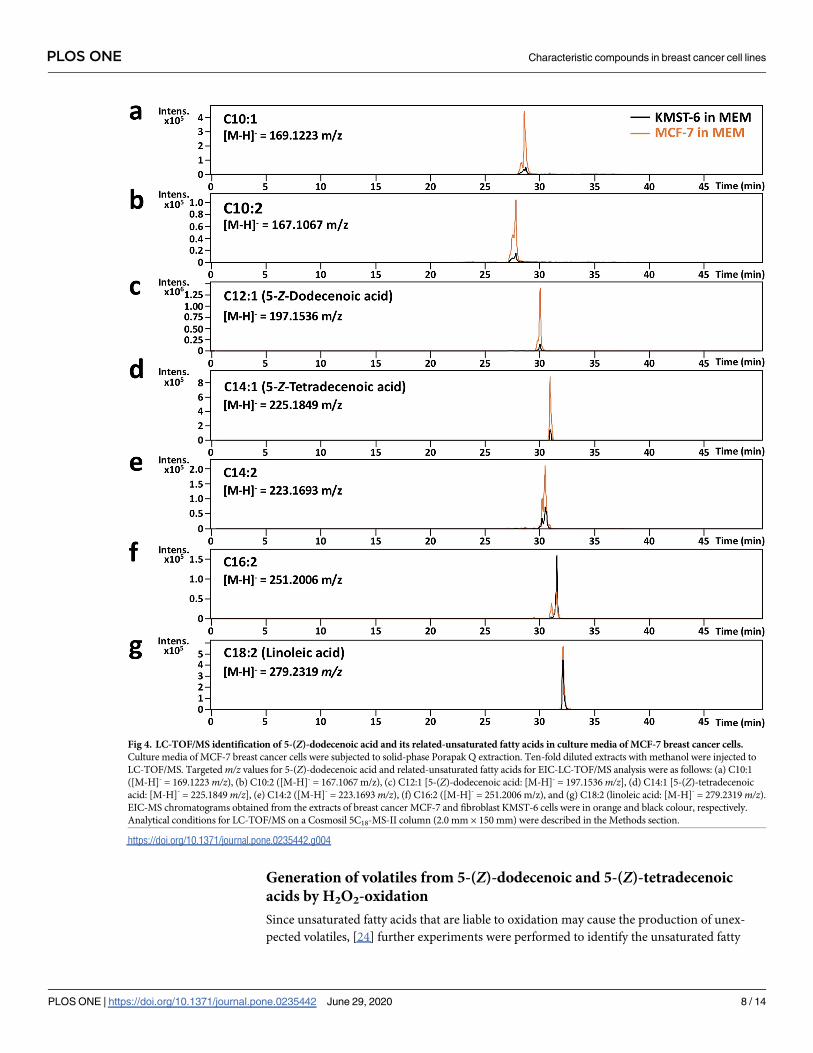
LC-TOF/MS analysis of 5-(Z)-dodecenoic acid and the related compounds
specific for MCF-7 breast cancer cell culture medium
Considering that 5-(Z)-dodecenoic acid could be a possible biomarker for breast cancer, fur-
ther experiments were performed to investigate whether 5-(Z)-dodecenoic acid-related unsat-
urated fatty acids were also abundant in MCF-7 cell culture media compared to those in
KMST-6. LC-TOF/MS (but not GC-MS) analysis of MCF-7 cultured MEM medium was per-
formed owing to aqueous properties of the targeted fatty acids in medium. As shown in Fig
4a–4g, seven peaks on extracted-ion chromatograms (EIC) corresponding to m/z values of
unsaturated fatty acids (C10:1, C10:2, C12:1, C14:1, C14:2, C16:2, and C18:2) were detected,
and five peaks (C10:1, C10:2, C12:1, C14:1, and C14:2) showed much higher intensity in MCF-
7 compared to those in KMST-6 (Fig 4a–4e). With the aid of commercially available standards,
C12:1 and C14:1 detected in MCF-7 were successfully identified as 5-(Z)-dodecenoic acid and
5-(Z)-tetradecenoic acid, respectively, whereas the position of the unsaturated bond for other
unsaturated fatty acids (except for C18:2 as linoleic acid) could not be defined due to unavail-
ability of standards.
Fig 3. GC-MS identification of 1-hexadecanol and 5-(Z)-dodecenoic acid in culture media of MCF-7 breast cancer cells. Culture media (MEM) of
MCF-7 breast cancer cells were subjected to solid-phase Porapak Q extraction. The extracts were injected to GC-MS. Targeted peaks selected by
GC-FID analyses (i.e., P1 and P2) on GC-MS total ion chromatogram (a) were determined according to RI values of 2379 and 2520, respectively. MS
spectra of P1 (b) and P2 (c) were applied to similarity search with NIST 14 library database, and a similarity score of 93% for 1-hexadecanol (d) and 95%
for 5-(Z)-dodecenoic acid (e) was obtained, respectively. Analytical conditions for GC-MS on DB-FFAP capillary column (30 m × 0.32 mm I.D. with
0.25 μm film thickness) were described in the Methods section.
https://doi.org/10.1371/journal.pone.0235442.g003
PLOS ONE Characteristic compounds in breast cancer cell lines
PLOS ONE | https://doi.org/10.1371/journal.pone.0235442 June 29, 2020 7 / 14
Generation of volatiles from 5-(Z)-dodecenoic and 5-(Z)-tetradecenoic
acids by H2O2-oxidation
Since unsaturated fatty acids that are liable to oxidation may cause the production of unex-
pected volatiles, [24] further experiments were performed to identify the unsaturated fatty
Fig 4. LC-TOF/MS identification of 5-(Z)-dodecenoic acid and its related-unsaturated fatty acids in culture media of MCF-7 breast cancer cells.
Culture media of MCF-7 breast cancer cells were subjected to solid-phase Porapak Q extraction. Ten-fold diluted extracts with methanol were injected to
LC-TOF/MS. Targeted m/z values for 5-(Z)-dodecenoic acid and related-unsaturated fatty acids for EIC-LC-TOF/MS analysis were as follows: (a) C10:1
([M-H]- = 169.1223 m/z), (b) C10:2 ([M-H]- = 167.1067 m/z), (c) C12:1 [5-(Z)-dodecenoic acid: [M-H]- = 197.1536 m/z], (d) C14:1 [5-(Z)-tetradecenoic
acid: [M-H]- = 225.1849 m/z], (e) C14:2 ([M-H]- = 223.1693 m/z), (f) C16:2 ([M-H]- = 251.2006 m/z), and (g) C18:2 (linoleic acid: [M-H]- = 279.2319 m/z).EIC-MS chromatograms obtained from the extracts of breast cancer MCF-7 and fibroblast KMST-6 cells were in orange and black colour, respectively.
Analytical conditions for LC-TOF/MS on a Cosmosil 5C18-MS-II column (2.0 mm × 150 mm) were described in the Methods section.
https://doi.org/10.1371/journal.pone.0235442.g004
PLOS ONE Characteristic compounds in breast cancer cell lines
PLOS ONE | https://doi.org/10.1371/journal.pone.0235442 June 29, 2020 8 / 14

acid-derived volatiles by 0.2% H2O2-accelerating oxidation of each acid in water (10 ppm) in a
50 mL-sealed vial for 6 days at 37 ˚C. As shown in Fig 5, GC-FID analysis of the 6-day-incu-
bates of 5-(Z)-dodecenoic acid and 5-(Z)-tetradecenoic acid revealed that four peaks from 5-
(Z)-dodecenoic acid and three peaks from 5-(Z)-tetradecenoic acid were newly and markedly
obtained compared to GC chromatograms of the blank (without both acids) sample. GC-MS
analysis in combination with RI matching [22] on DB-WAX column showed that all the iden-
tified compounds except T2 were volatile aldehydes (Tables 1 and 2). In addition, these identi-
fied aldehydes (heptanal, octanal, nonanal, decanal, 2-(E)-nonenal, and 2-(E)-octenal) were
volatiles identified in the breath or blood from patients of various cancers as reported previ-
ously, [8,25–29] e.g., heptanal in breath as a lung cancer-specific volatile. [8,25,26] This finding
allowed us to speculate that the unsaturated fatty acids identified in this study may be one of
the possible precursors of the reported cancer-specific volatiles.
Discussion
So far, research interests have been focused on the elucidation of “volatiles” in the exhaled
breath of cancer patients, since the evidential report that dog could distinguish colorectal can-
cer patients by smelling their exhaled breath. [11] Thus, research on volatiles in breath from
cancer patients [8,12] have been conducted using SPME extraction method, which is suitable
for the recovery of highly volatile compounds from gas phase. In contrast, in the present study,
Fig 5. GC-MS analyses of volatiles generated from 5-(Z)-dodecenoic acid (a) and 5-(Z)-tetradecenoic acid (b) by H2O2-oxidation. H2O2 (0.2%)
oxidation of 5-(Z)-dodecenoic acid and 5-(Z)-tetradecenoic acid (each 10 ppm in deionized water) was performed respectively in a 50-mL teflon-sealed
vial at 37 ˚C for 6 days. A blank experiment in 0.2% H2O2 solution without fatty acid was also performed at the above-mentioned incubation
conditions. Volatiles in the headspace of the vial were analysed by SPME-aided GC-MS analysis on a DB-WAX capillary column (30 m × 0.32 mm I.
D.). Analytical conditions for SPME extraction and for GC-MS analysis on DB-FFAP capillary column (30 m × 0.32 mm I.D.) were described in the
Methods section. Newly observed peaks with high MS intensity were denoted as D1 to D4 and T1 to T3 for 5-(Z)-dodecenoic acid (a) and 5-(Z)-
tetradecenoic acid (b), respectively.
https://doi.org/10.1371/journal.pone.0235442.g005
PLOS ONE Characteristic compounds in breast cancer cell lines
PLOS ONE | https://doi.org/10.1371/journal.pone.0235442 June 29, 2020 9 / 14

we used a solid-phase Porapak Q resin for the recovery of a variety of volatiles, since com-
pounds of moderate volatility such as fatty acids or lipids were targeted as precursors of these
cancer specific volatiles. For the first time, we identified two compounds, 1-hexadecanol and
5-(Z)-dodecenoic acid, by GC-FID analysis in the culture media of MCF-7, a breast cancer cell
line (Fig 2a). Both newly identified compounds were observed in breast cancer cell media
regardless of the species of the breast cancer cell line (MCF-7, SK-BR-3, and YMB-1, Fig 2a–
2c) and medium (MEM, D-MEM, and RPMI 1640, S2 Fig).
Considering the boiling points of 344 ˚C and 311 ˚C for 1-hexadecanol and 5-(Z)-dodece-
noic acid, respectively, they could evaporate into gas phase, since compounds with a high boil-
ing point of> 300 ˚C, e.g., β-naphthyl methylketone with boiling point of 301 ˚C, can behave
as a synthetic strawberry-like flavour compound. [24] Thus, a direct analysis of the headspace
of culture media of breast cancer cells is needed for further experiments, which is now in prog-
ress to confirm their vaporization into gas phase. Abaffy et al. reported that 1-hexadecanol was
identified as one of the malignant melanoma-specific volatiles in melanoma biopsy. [31] Shi-
geyama et al. also revealed that 1-hexadecanol was newly produced in the saliva of oral squa-
mous cell carcinoma patients, together with other alcohols and ketones as volatile metabolites.
[32] Therefore, it seems likely that 1-hexadecanol was a candidate cancer-specific biomarker.
It has been revealed that BRCA1 germline mutation, and HER2 and ER expressions were
candidate tumour biomarkers for the risk of breast or ovarian cancer, and for therapeutic
assessment of breast and gastric cancers, respectively. [33] Serum biomarkers such as carcino-
embryonic antigen (CEA), cancer antigen 19–9 (CA19-9), cancer antigen 125 (CA125), cancer
antigen 15–3 (CA15-3), and tissue polypeptide-specific antigen (TPS) have also been used as
diagnostic metabolites for metastatic breast cancer. [3] In contrast, challenging studies on
Table 1. Identified volatiles from 5-(Z)-dodecenoic acid by H2O2-oxidation.
Peak Rt (min) RI on DB-WAX Compound Reported identification
D1 8.63 1192 heptanal breath for lung cancer [8,25]
breath for breast cancer [27]
plasma for bone cancer [29]
plasma for Burkitt’s lymphoma [29]
plasma for large cell lymphoma [29]
D2 11.35 1296 octanal breath for lung cancer [8,26]
D3 15.08 1437 2-(E)-octenal plasma for bone cancer [29]
plasma for Burkitt’s lymphoma [29]
plasma for large cell lymphoma [29]
D4 17.78 1544 2-(E)-nonenal plasma for acute myelogenous leukemia [29]
https://doi.org/10.1371/journal.pone.0235442.t001
Table 2. Identified volatiles from 5-(Z)-tetradecenoic acid by H2O2-oxidation.
Peak Rt (min) RI on DB-WAX Compound Reported identification
T1 14.14 1400 nonanal breath for lung cancer [8,26]
breath for ovarian cancer [28]
breath for colorectal cancer [30]
T2 14.97 1433 N.I.a -
T3 16.83 1505 decanal breath for ovarian cancer [28]
breath for colorectal cancer [30]
aN.I., not identified.
https://doi.org/10.1371/journal.pone.0235442.t002
PLOS ONE Characteristic compounds in breast cancer cell lines
PLOS ONE | https://doi.org/10.1371/journal.pone.0235442 June 29, 2020 10 / 14

non-invasive cancer diagnosis have been reported using breath, [8] urine, [9] or hair. [10]
Although the non-invasive approach appears to be of great value owing to convenient and
rapid sampling, the reliability of diagnostic results remains unascertained. For example,
although many candidate volatiles in exhaled breath of breast cancer subjects have been
reported so far, [12] no crucial volatiles that can characterise the onset of breast cancer have
been elucidated yet. In this study, we successfully identified two unsaturated fatty acids, 5-(Z)-
dodecenoic acid and 5-(Z)-tetradecenoic acid, in the culture media of breast cancer cells. It is
well known in food sciences that unsaturated fatty acids such as oleic, linoleic, and linolenic
acids are liable to oxidation, causing the generation of volatile aldehydes and ketones responsi-
ble for “fatty and green” odour quality. [24] In this study, autoxidation treatment of 5-(Z)-
dodecenoic acid and 5-(Z)-tetradecenoic acid by H2O2 caused the production of volatile
aldehydes including heptanal, octanal, nonanal, decanal, 2-(E)-nonenal, and 2-(E)-octenal
(Fig 5a and 5b, and Tables 1 and 2) in agreement with the reported cancer-specific volatiles.
[8,25–29] Although the production behaviour of volatiles from the two fatty acids was
obtained in the present limited H2O2-oxidation experiments, the identified unsaturated fatty
acids in the culture media of breast cancer cells may behave as a precursor for the generation
of cancer-specific volatiles seen in exhaled breath of lung, [8,25,26] breast, [27] ovarian, [28]
and colorectal cancer patients, [30] as well as plasma from cancer patients who are children.
[29]
Cancer can alter fatty acid metabolism in response to hypoxia inducible factor-1 (HIF-1)-
mediated attenuation of medium chain acyl-CoA dehydrogenase (MCAD) and long chain
acyl-CoA dehydrogenase (LCAD). [34] It has been reported that MCAD or LCAD deficiency
caused the increase in medium chain fatty acids in human plasma, such as C8:0, C10:1, and
C10:2 for MCAD deficiency, and C12:1 [5-(Z)-dodecenoic acid], C14:1 [5-(Z)-tetradecenoic
acid], C14:2 and C16:2 for LCAD deficiency. [35] In the MEM culture medium of MCF-7, 5
unsaturated fatty acids (C10:1, C10:2, C12:1 [5-(Z)-dodecenocic acid], C14:1 [5-(Z)-tetradece-
noic acid], and C14:2) were successfully detected with a> 2.5-fold higher intensity than those
in KMST-6 (Fig 4a–4e). Thus, according to the present observations, the increase of medium
chain unsaturated fatty acid identified in this study in plasma or other biological fluids would
be a detectable symptom for breast cancer onset. We are now investigating diagnostic potential
of target fatty acids as biomarkers for breast cancer using non-invasive biological fluids.
In conclusion, in this study, we firstly demonstrated that 1-hexadecanol and 5-(Z)-dodece-
noic acid (and its related unsaturated fatty acids including 5-(Z)-tetradecenoic acid) were
important candidates in breast cancer cells but not fibroblast cells by GC-MS and/or LC-MS
analyses of cell culture media. We also found that unsaturated fatty acids could generate the
reported volatiles specific for breath from various cancer patients such as heptanal, octanal,
nonanal, decanal, 2-(E)-nonenal, and 2-(E)-octenal by H2O2 oxidation. Thus, the diagnostic
targets, medium chain unsaturated fatty acids of moderate volatility, would overcome the
issues on poor reproducibility and precision of volatile detection in exhaled breath. [16] In
summary, the present findings clearly indicate that unsaturated fatty acids of moderate volatil-
ity may be over-produced in breast cancer cells, serving as potential precursors for the produc-
tion of cancer-specific volatiles reported so far.
Supporting information
S1 Fig. GC-FID intensities of P1 (1-hexadecanol) and P2 (5-(Z)-dodecenoic acid) in cell
culture media of KMST-6 and MCF-7. GC-FID samples from 500 mL of MCF-7 and KMST-
6 culture media (MEM) were obtained through solid-phase Porapak Q extraction. Analytical
conditions for solid-phase Porapak Q extraction and for GC-FID analysis on a DB-FFAP
PLOS ONE Characteristic compounds in breast cancer cell lines
PLOS ONE | https://doi.org/10.1371/journal.pone.0235442 June 29, 2020 11 / 14

capillary column (30 m × 0.32 mm I.D.) were described in the Methods section. GC peak
intensities from individual triplicate cell cultures are shown as mean ± S.E.M. Significant dif-
ferences between groups were analysed by unpaired two-tailed Student’s t-test. Significantly
high GC intensities of P1 and P2 were obtained in cell culture of MCF-7 compared to KMST-6
(n = 3, P< 0.05).
(TIFF)
S2 Fig. Comparison between GC-FID chromatograms of D-MEM culture medium for
breast cancer MCF-7 and those for fibroblast cell (KMST-6). GC-FID samples from 500 mL
of MCF-7 culture media (D-MEM) were obtained through solid-phase Porapak Q extraction.
KMST-6 cells were also present in the same culture medium for comparison of their GC chro-
matograms as controls, with those of cancer cell media. Analytical conditions for Porapak Q
column extraction and for GC-FID analysis on a DB-FFAP capillary column (30 m × 0.32 mm
I.D.) were described in the Methods section. GC chromatograms obtained from the extracts of
MCF-7 and KMST-6 were in orange and black colour, respectively. Peaks denoted as P1 and
P2 are predominant commonly observed in all cancer cell extracts regardless of different
media (as shown in Fig 2).
(TIFF)
S3 Fig. GC-FID chromatograms of RPMI 1640 culture medium for breast cancer YMB-1
spiked with 1-hexadecanol (a) or 5-(Z)-dodecenoic acid (b). Standard 1-hexadecanol (a) or
5-(Z)-dodecenoic acid (b) (1 ppm for each standard) was spiked into Porapak Q-column
extract of RPMI 1640 culture medium for YMB-1 to be subjected for GC-FID analysis. Analyt-
ical conditions for Porapak Q column extraction and for GC-FID analysis on a DB-FFAP
capillary column (30 m × 0.32 mm I.D.) were described in the Methods section. GC-FID chro-
matograms obtained from standard spiked-media and those not from standard spiked-media
were in orange and black colour, respectively.
(TIFF)
Acknowledgments
The authors are grateful to Prof. Mitsuya Shimoda and Prof. Noriyuki Igura at Kyushu Univer-
sity for professional advices on GC-MS. The authors also thank Ms Kaori Miyazaki and Ms
Tomoko Hidaka at Kyushu University for technical assistance.
Author Contributions
Conceptualization: Yuzo Ninomiya, Hideto Sonoda, Yoshihiko Maehara, Kiyoshi Toko,
Toshiro Matsui.
Data curation: Mitsuru Tanaka, Chung Hsuan, Masataka Oeki, Weilin Shen, Asuka Goda,
Yusuke Tahara, Takeshi Onodera, Keisuke Sanematsu, Tomotsugu Rikitake, Eiji Oki, Yuzo
Ninomiya, Rintaro Kurebayashi, Hideto Sonoda, Yoshihiko Maehara, Kiyoshi Toko,
Toshiro Matsui.
Formal analysis: Mitsuru Tanaka, Masataka Oeki, Weilin Shen, Asuka Goda, Yusuke Tahara,
Takeshi Onodera, Keisuke Sanematsu, Tomotsugu Rikitake, Eiji Oki, Yuzo Ninomiya,
Hideto Sonoda, Yoshihiko Maehara, Kiyoshi Toko, Toshiro Matsui.
Funding acquisition: Kiyoshi Toko.
Investigation: Mitsuru Tanaka, Chung Hsuan, Masataka Oeki, Weilin Shen, Asuka Goda,
Hideto Sonoda, Kiyoshi Toko, Toshiro Matsui.
PLOS ONE Characteristic compounds in breast cancer cell lines
PLOS ONE | https://doi.org/10.1371/journal.pone.0235442 June 29, 2020 12 / 14

Methodology: Mitsuru Tanaka, Chung Hsuan, Weilin Shen, Keisuke Sanematsu, Hideto
Sonoda, Toshiro Matsui.
Project administration: Kiyoshi Toko, Toshiro Matsui.
Supervision: Toshiro Matsui.
Validation: Hideto Sonoda, Kiyoshi Toko, Toshiro Matsui.
Writing – original draft: Mitsuru Tanaka, Toshiro Matsui.
Writing – review & editing: Toshiro Matsui.
References
1. Collaboration GB of DC (2019) Global, regional, and national cancer incidence, mortality, years of life
lost, years lived with disability, and disability-adjusted life-years for 29 cancer groups, 1990 to 2017: a
systematic analysis for the global burden of disease study. JAMA Oncol 5: 1749–1768. https://doi.org/
10.1001/jamaoncol.2019.2996 PMID: 31560378
2. Todd J, McGrath EE (2010) Chest X-ray mass in a patient with lung cancer! QJM An Int J Med 104:
903–904. https://doi.org/10.1093/qjmed/hcq159 PMID: 20829191
3. Wang W, Xu X, Tian B, Wang Y, Du L, et al. (2017) The diagnostic value of serum tumor markers CEA,
CA19-9, CA125, CA15-3, and TPS in metastatic breast cancer. Clin Chim Acta 470: 51–55. https://doi.
org/10.1016/j.cca.2017.04.023 PMID: 28457854
4. Kim HR, Jung HK, Ko KH, Kim SJ, Lee KS (2016) Mammography, US, and MRI for preoperative predic-
tion of extensive intraductal component of invasive breast cancer: interobserver variability and perfor-
mances. Clin Breast Cancer 16: 305–311. https://doi.org/10.1016/j.clbc.2016.02.005 PMID: 27025667
5. Brandman S, Ko JP (2011) Pulmonary nodule detection, characterization, and management with multi-
detector computed tomography. J Thorac Imaging 26: 90–105. https://doi.org/10.1097/RTI.
0b013e31821639a9 PMID: 21508732
6. Truong MT, Viswanathan C, Erasmus JJ (2011) Positron emission tomography/computed tomography
in lung cancer staging, prognosis, and assessment of therapeutic response. J Thorac Imaging 26:
132–146. https://doi.org/10.1097/RTI.0b013e3182128704 PMID: 21508735
7. Goldhirsch A, Winer EP, Coates AS, Gelber RD, Piccart-Gebhart M, et al. (2013) Personalizing the
treatment of women with early breast cancer: highlights of the St Gallen International Expert Consensus
on the Primary Therapy of Early Breast Cancer 2013. Ann Oncol 24: 2206–2223. https://doi.org/10.
1093/annonc/mdt303 PMID: 23917950
8. Poli D, Goldoni M, Corradi M, Acampa O, Carbognani P, et al. (2010) Determination of aldehydes in
exhaled breath of patients with lung cancer by means of on-fiber-derivatisation SPME–GC/MS. J Chro-
matogr B 878: 2643–2651. https://doi.org/10.1016/j.jchromb.2010.01.022 PMID: 20149763
9. Matsumura K, Opiekun M, Oka H, Vachani A, Albelda SM, et al. (2010) Urinary volatile compounds as
biomarkers for lung cancer: a proof of principle study using odor signatures in mouse models of lung
cancer. PLoS One 5: e8819. https://doi.org/10.1371/journal.pone.0008819 PMID: 20111698
10. Chikawa J, Mouri Y, Shima H, Yamada K, Yamamoto H, et al. (2014) A correlation of breast cancer and
calcium levels in hair analyzed by X-ray fluorescence. J Xray Sci Technol 22: 587–603. https://doi.org/
10.3233/XST-140447 PMID: 25265920
11. Sonoda H, Kohnoe S, Yamazato T, Satoh Y, Morizono G, et al. (2011) Colorectal cancer screening with
odour material by canine scent detection. Gut 60: 814–819. https://doi.org/10.1136/gut.2010.218305
PMID: 21282130
12. Sun X, Shao K, Wang T (2016) Detection of volatile organic compounds (VOCs) from exhaled breath as
noninvasive methods for cancer diagnosis. Anal Bioanal Chem 408: 2759–2780. https://doi.org/10.
1007/s00216-015-9200-6 PMID: 26677028
13. Silva CL, Perestrelo R, Silva P, Tomas H, Camara JS (2017) Volatile metabolomic signature of human
breast cancer cell lines. Sci Rep 7: 43969. https://doi.org/10.1038/srep43969 PMID: 28256598
14. Shuster G, Gallimidi Z, Reiss AH, Dovgolevsky E, Billan S, et al. (2011) Classification of breast cancer
precursors through exhaled breath. Breast Cancer Res Treat 126: 791–796. https://doi.org/10.1007/
s10549-010-1317-x PMID: 21190078
15. Di Natale C, Macagnano A, Martinelli E, Paolesse R, D’Arcangelo G, et al. (2003) Lung cancer identifi-
cation by the analysis of breath by means of an array of non-selective gas sensors. Biosens Bioelectron
18: 1209–1218. https://doi.org/10.1016/s0956-5663(03)00086-1 PMID: 12835038
PLOS ONE Characteristic compounds in breast cancer cell lines
PLOS ONE | https://doi.org/10.1371/journal.pone.0235442 June 29, 2020 13 / 14
16. Shirasu M, Touhara K (2011) The scent of disease: volatile organic compounds of the human body
related to disease and disorder. J Biochem 150: 257–266. https://doi.org/10.1093/jb/mvr090 PMID:
21771869
17. Jones AP (1999) Indoor air quality and health. Atmos Environ 33: 4535–4564. https://doi.org/10.1016/
S1352-2310(99)00272-1
18. Hakim M, Broza YY, Barash O, Peled N, Phillips M, et al. (2012) Volatile organic compounds of lung
cancer and possible biochemical pathways. Chem Rev 112: 5949–5966. https://doi.org/10.1021/
cr300174a PMID: 22991938
19. Maneerat C, Hayata Y, Kozuka H, Sakamoto K, Osajima Y (2002) Application of the porapak Q column
extraction method for tomato flavor volatile analysis. J Agric Food Chem 50: 3401–3404. https://doi.
org/10.1021/jf011626r PMID: 12033802
20. Fukamachi M, Matsui T, Shimoda M, Nakashima M, Osajima Y (1993) Sorption depression of flavors
into modified EVA film by lowering film-flavor affinity. J Agric Food Chem 41: 929–932. https://doi.org/
10.1021/jf00030a018
21. Fukamachi M, Matsui T, Hwang Y-H, Shimoda M, Osajima Y (1996) Sorption behavior of flavor com-
pounds into packaging films from ethanol solution. J Agric Food Chem 44: 2810–2813. https://doi.org/
10.1021/jf950707i
22. van Den Dool H, Kratz P Dec. (1963) A generalization of the retention index system including linear tem-
perature programmed gas—liquid partition chromatography. J Chromatogr A 11: 463–471. https://doi.
org/10.1016/S0021-9673(01)80947-X
23. Hanai Y, Shimono K, Oka H, Baba Y, Yamazaki K, et al. (2012) Analysis of volatile organic compounds
released from human lung cancer cells and from the urine of tumor-bearing mice. Cancer Cell Int 12: 7.
https://doi.org/10.1186/1475-2867-12-7 PMID: 22364569
24. Belitz H-D, Grosch W, Schieberle P (2009) Food Chemistry. Berlin, Heidelberg: Springer Berlin Heidel-
berg. 158–247 p. https://doi.org/10.1007/978-3-540-69934-7_4
25. Phillips M, Gleeson K, Hughes JMB, Greenberg J, Cataneo RN, et al. (1999) Volatile organic com-
pounds in breath as markers of lung cancer: a cross-sectional study. Lancet 353: 1930–1933. https://
doi.org/10.1016/S0140-6736(98)07552-7 PMID: 10371572
26. Fuchs P, Loeseken C, Schubert JK, Miekisch W (2010) Breath gas aldehydes as biomarkers of lung
cancer. Int J Cancer 126: 2663–2670. https://doi.org/10.1002/ijc.24970 PMID: 19839051
27. Phillips M, Cataneo RN, Ditkoff BA, Fisher P, Greenberg J, et al. (2006) Prediction of breast cancer
using volatile biomarkers in the breath. Breast Cancer Res Treat 99: 19–21. https://doi.org/10.1007/
s10549-006-9176-1 PMID: 16502014
28. Amal H, Shi D-Y, Ionescu R, Zhang W, Hua Q-L, et al. (2014) Assessment of ovarian cancer conditions
from exhaled breath. Int J Cancer 136: E614–E622. https://doi.org/10.1002/ijc.29166 PMID: 25159530
29. Yazdanpanah M, Luo X, Lau R, Greenberg M, Fisher LJ, et al. (1997) Cytotoxic aldehydes as possible
markers for childhood cancer. Free Radic Biol Med 23: 870–878. https://doi.org/10.1016/s0891-5849
(97)00070-1 PMID: 9378366
30. Altomare DF, Di Lena M, Porcelli F, Trizio L, Travaglio E, et al. (2012) Exhaled volatile organic com-
pounds identify patients with colorectal cancer. Br J Surg 100: 144–150. https://doi.org/10.1002/bjs.
8942 PMID: 23212621
31. Abaffy T, Moller M, Riemer DD, Milikowski C, Defazio RA (2011) A case report—Volatile metabolomic
signature of malignant melanoma using matching skin as a control. J Cancer Sci Ther 3: 140–144.
https://doi.org/10.4172/1948-5956.1000076 PMID: 22229073
32. Shigeyama H, Wang T, Ichinose M, Ansai T, Lee S-W (2019) Identification of volatile metabolites in
human saliva from patients with oral squamous cell carcinoma via zeolite-based thin-film microextrac-
tion coupled with GC–MS. J Chromatogr B 1104: 49–58. https://doi.org/10.1016/j.jchromb.2018.11.
002 PMID: 30445287
33. Henry NL, Hayes DF (2012) Cancer biomarkers. Mol Oncol 6: 140–146. https://doi.org/10.1016/j.
molonc.2012.01.010 PMID: 22356776
34. Huang D, Li T, Li X, Zhang L, Sun L, et al. (2014) HIF-1-mediated suppression of acyl-CoA dehydroge-
nases and fatty acid oxidation is critical for cancer progression. Cell Rep 8: 1930–1942. https://doi.org/
10.1016/j.celrep.2014.08.028 PMID: 25242319
35. Onkenhout W, Venizelos V, van der Poel PF, van den Heuvel MP, Poorthuis BJ (1995) Identification
and quantification of intermediates of unsaturated fatty acid metabolism in plasma of patients with fatty
acid oxidation disorders. Clin Chem 41: 1467 LP–1474. https://doi.org/10.1093/clinchem/41.10.1467
PLOS ONE Characteristic compounds in breast cancer cell lines
PLOS ONE | https://doi.org/10.1371/journal.pone.0235442 June 29, 2020 14 / 14


![ANTI-ACETYL CHOLINESTERASE COMPOUNDS ISOLATED FROM …sdiarticle4.com/prh/doc/Ms_EJMP_49262.pdf · to moderate disorder [7,8,9];51 overall benefit, nevertheless, may be minor [9],](https://img.pdfslide.us/doc/110x75/5faba1638759331ac94ed20e/anti-acetyl-cholinesterase-compounds-isolated-from-to-moderate-disorder-78951.jpg)


![Biologically active compounds from bryophytesmedia.iupac.org/publications/pac/2007/pdf/7904x0557.pdf · Table 2 Characteristic odor of liverworts [7,11a]. Species Odor Asterellaspecies](https://img.pdfslide.us/doc/110x75/606d59c193119417f12a39f2/biologically-active-compounds-from-table-2-characteristic-odor-of-liverworts-711a.jpg)