Embed Size (px)
Citation preview

IDENTIFICATION OF ACYLOXYACYL HYDROLASE, A LIPOPOLYSACCHARIDE-
DETOXIFYING ENZYME, IN THE MURINE URINARY TRACT
APPROVED BY SUPERVISORY COMMITTEE
Robert S. Munford, M.D. __________________________
Leon Eidels, Ph.D. __________________________
Christopher Lu, M.D. __________________________
Kevin S. McIver, Ph.D. __________________________
Nicolai van Oers, Ph.D. __________________________

DEDICATION
Dedicated to my parents, Gene and Candace Feulner, my brother, Kenneth Feulner and Joseph Baur for their support, encouragement and love.
IDENTIFICATION OF ACYLOXYACYL HYDROLASE, A LIPOPOLYSACCHARIDE-
DETOXIFYING ENZYME, IN THE MURINE URINARY TRACT
by
J. Amelia Feulner
Presented to the Faculty of the Graduate School of Biomedical Sciences
The University of Texas Southwestern Medical Center at Dallas
In Partial Fulfillment of the Requirements
For the Degree of
DOCTOR OF PHILOSOPHY
The University of Texas Southwestern Medical Center at Dallas
Dallas, Texas
August, 2003
Copyright
by
J. Amelia Feulner 2003
All Rights Reserved

IDENTIFICATION OF ACYLOXYACYL HYDROLASE, A LIPOPOLYSACCHARIDE-DETOXIFYING ENZYME, IN THE MURINE URINARY TRACT
Publication No. ______________
J. Amelia Feulner, Ph.D.
The University of Texas Southwestern Medical Center at Dallas, 2003
Supervising Professor: Robert S. Munford, M.D.
Acyloxyacyl hydrolase (AOAH) is a lipase that removes the secondary fatty acyl chains
that are substituted to the hydroxyl groups of glucosamine-linked 3-hydroxyacyl residues in lipid
A, the bioactive center of Gram-negative bacterial lipopolysaccharides (LPS). Such limited
deacylation has been shown to attenuate cytokine and chemokine responses to LPS, suggesting a
role for AOAH in modulating (downregulating) inflammatory responses to invading Gram-
negative bacteria. Prior to the experiments described in this report, AOAH had only been found
in myeloid lineage cells (monocyte-macrophages, neutrophils and dendritic cells). In the work
presented here, AOAH was found in murine renal proximal tubule cells and in human renal
v
cortex. Proximal tubule cells are known targets for invading Gram-negative uropathogens and
we hypothesize that possessing AOAH may help them degrade the LPS contained within these
bacteria. I further found that AOAH is secreted from proximal tubules in vitro and that it can be
detected in murine urine, where it is able to deacylate purified LPS. AOAH may also associate
with downstream bladder epithelial cells (which do not express AOAH) and be processed by
them to its more enzymatically active, mature form. Bladder cells that have taken up AOAH in
vitro are able to deacylate LPS.
To determine the in vivo role of AOAH, I induced ascending urinary tract infections
(UTIs) in wild type and AOAH null mice. To my surprise, AOAH null mice were able to clear
bacteria from their urine faster than did wild type mice. An analysis of the immune response by
histological analysis of bladder tissue and enumeration of neutrophils in the urine did not show a
significant difference between wild type and AOAH null mice at any of the time points
examined. Although I do not yet understand the mechanism for such increased clearance in
AOAH null animals, we hypothesize that, due to their inability to deacylate LPS, they might
have a more effective immune response to invading Gram-negative bacteria. A more detailed
analysis of such responses to invading Gram-negative uropathogens will be important for
understanding the in vivo role of AOAH in the urinary tract.
vi

PRIOR PUBLICATIONS
J. A. Feulner, M. Lu, J. Shelton, J. Richardson, R.S. Munford, Identification of acyloxyacyl hydrolase, a lipopolysaccharide-detoxifying enzyme, in the murine urinary tract. Manuscript in submission, July 2003. A.C. Walsh, J. A. Feulner, A. Reilly. Evidence for functionally significant polymorphism of human glutamate cysteine ligase catalytic subunit: association with glutathione levels and drug resistance in the National Cancer Institute tumor cell line panel. Toxicol Sci. 2001 Jun;61(2):218-23. T.J. Sellati, D. A. Bouis, M.J. Caimano, J. A. Feulner, C.Ayers, E. Lien, and J. D. Radolf. Activation of Human Monocytic Cells by Borrelia burgdorferi and Treponema pallidum is Facilitated by CD14 and Correlates with Surface Exposure of Spirochetal Lipoproteins. The Journal of Immunology, 1999, 163: 2049-2056.
vii

Table of Contents CHAPTER ONE
LITERATURE REVIEW
LIPOPOLYSACCHARIDE (LPS) ........................................................................................................ 1
Lipopolysaccharide History ................................................................................................... 1
Basic Structure of LPS .......................................................................................................... 3
O-antigen (O specific chain).................................................................................................. 4
Core Oligosaccharide............................................................................................................. 5
Lipid A .................................................................................................................................... 6
Structure-Function Relationships of Lipid A ....................................................................... 7
Chemically Modified LPS and Synthetic Lipid A Derivatives.............................................. 9
ACYLOXYACYL HYDROLASE (AOAH) ...................................................................................... 12
Deacylation of Diverse Lipopolysaccharides by AOAH ..................................................... 14
Purification of Acyloxyacyl Hydrolase (AOAH)................................................................. 15
Basic Structure of AOAH .................................................................................................... 15
Large Subunit....................................................................................................................... 17
Small Subunit ....................................................................................................................... 19
Precursor vs. Mature AOAH ............................................................................................... 19
Mannose 6 Phosphate Residues and Receptors.................................................................. 20
Other Known Activities of AOAH ....................................................................................... 21
Localization of AOAH ......................................................................................................... 21
KIDNEY ARCHITECTURE AND RENAL PROXIMAL TUBULE CELLS .............................................. 22
URINARY TRACT INFECTIONS .................................................................................................... 24
Etiology of Urinary Tract Infections................................................................................... 25
VIRULENCE FACTORS ASSOCIATED WITH UROPATHOGENIC E. COLI.......................................... 25
Adhesins ............................................................................................................................... 25
Type I fimbriae ..................................................................................................................... 26
P pili...................................................................................................................................... 27
Lipopolysaccharide .............................................................................................................. 28
viii
Toxins ................................................................................................................................... 28
Other virulence factors of UPEC ........................................................................................ 29
Known Host Defenses to Invading Uropathogens.............................................................. 30
ASCENDING URINARY TRACT INFECTIONS (UTIS)..................................................................... 30
The role of toll-like receptor 4 (TLR4) in UTI.................................................................... 31
The role of lipid A in experimental ascending UTI ............................................................ 32
CHAPTER TWO
IDENTIFICATION OF ACYLOXYACYL HYDROLASE, A LIPOPOLYSACCHARIDE-
DETOXIFYING ENZYME, IN THE MURINE URINARY TRACT
INTRODUCTION....................................................................................................................... 34
RESULTS .................................................................................................................................... 35
AOAH IS PRODUCED IN THE KIDNEY....................................................................................... 35
AOAH IS PRESENT IN HUMAN KIDNEY .................................................................................... 42
PROXIMAL TUBULE CELLS SECRETE PRO-AOAH ................................................................... 43
AOAH IS FOUND IN VOIDED URINE .......................................................................................... 46
BLADDER CELLS TAKE UP PRO-AOAH.................................................................................... 50
BLADDER CELLS DO NOT RE-SECRETE MATURE AOAH ......................................................... 53
BLADDER CELLS THAT HAVE TAKEN UP AOAH CAN DEACYLATE LPS. ................................ 55
METHODS .................................................................................................................................. 57
Chemicals ............................................................................................................................. 57
Mouse strains ....................................................................................................................... 57
Northern Analysis ................................................................................................................ 57
AOAH Activity Assays.......................................................................................................... 57
Thin-layer chromatography................................................................................................. 58
Generation of AOAH null mice........................................................................................... 59
In situ hybridization and riboprobes ................................................................................... 59
Real-time PCR...................................................................................................................... 60
Cell culture ........................................................................................................................... 61
ix

Antibodies ............................................................................................................................. 61
Immunoprecipitation of biosynthetically radiolabeled AOAH........................................... 62
Immunoprecipitation and Western blot of murine urine ................................................... 63
Uptake and deacylation of LPS by bladder cells................................................................. 64
AOAH Elisa.......................................................................................................................... 64
DISCUSSION .............................................................................................................................. 65
CHAPTER THREE
THE IN VIVO ROLE OF ACYLOXYACYL HYDROLASE (AOAH)
INTRODUCTION....................................................................................................................... 69
RESULTS .................................................................................................................................... 71
WILD TYPE AND AOAH NULL MICE DIFFER IN RATE OF BACTERIAL CLEARANCE. ............... 71
AOAH NULL AND WILD TYPE MICE HAVE SIMILAR NUMBERS OF BACTERIA IN THEIR
BLADDERS AT 72 HOURS ........................................................................................................... 75
THE IMMUNE RESPONSE TO INVADING UPEC ......................................................................... 77
HISTOLOGICAL ANALYSIS OF C57BL/6 BLADDER AND KIDNEY SAMPLES .............................. 79
AN ANALYSIS OF INFLAMMATION IN THE BLADDERS OF C57BL/6 MICE AT 24 HOURS POST-
INFECTION................................................................................................................................. 81
C3H/HEN MICE ARE ALSO SUSCEPTIBLE TO ASCENDING UTI ............................................... 82
MICE DO NOT HAVE DETECTABLE LEVELS OF ANTI-F11 LPS IN THEIR URINE....................... 87
METHODS .................................................................................................................................. 89
Chemicals ............................................................................................................................. 89
Mouse strains ....................................................................................................................... 89
Bacterial Strains................................................................................................................... 89
AOAH Activity Assays.......................................................................................................... 90
Catheters............................................................................................................................... 90
Urine Collection ................................................................................................................... 91
Experimental Ascending Urinary Tract Infections ............................................................ 91
x

Colony-forming Unit (CFU) Determination ....................................................................... 92
Neutrophil determination .................................................................................................... 93
Bladder and Kidney CFU and Histology ............................................................................ 93
Histological Scoring Index .................................................................................................. 94
Preparation of LPS .............................................................................................................. 95
Immunization of mice with heat-killed f11 CNF 1+ and serum collection. ...................... 95
f11-LPS ELISA. ................................................................................................................... 96
DISCUSSION .............................................................................................................................. 98
CHAPTER FOUR
AOAH-FLUORESCENT FUSION PROTEIN
INTRODUCTION..................................................................................................................... 102
RESULTS .................................................................................................................................. 104
THE CREATION OF AOAH-PTIMER 1 FUSION PROTEIN ........................................................ 104
TRANSFECTION OF PAF988 INTO CHO-CD14 CELLS .......................................................... 104
MANNOSE-6-PHOSPHATE MIGHT BLOCK THE RE-UPTAKE OF AOAH BY TRANSFECTED CHO-
CD14 CELLS............................................................................................................................ 106
THE REMOVAL OF DSRED1-E5 AND REPLACEMENT WITH DSRED2 .................................... 107
THE REMOVAL OF DSRED2 AND REPLACEMENT WITH EGFP .............................................. 108
CHO K1 AND CHO-CD14 CELLS PHAGOCYTOSE BODIPY-LABELED E.COLI ...................... 110
IMMUNOHISTOCHEMISTRY OF AOAH IN HUMAN AND MURINE KIDNEY. ............................. 112
MATERIALS AND METHODS ............................................................................................. 113
Chemicals ........................................................................................................................... 113
Cell Culture ........................................................................................................................ 113
Mini-prep protocol ............................................................................................................. 114
Generation of Plasmids...................................................................................................... 114
P-timer.CMVpromoter (pAF987) ................................................................................... 114
PCR of human AOAH and creation of clone RM 1012 .................................................. 116
xi
AOAH-DsRed1-E5 fusion protein (pAF988) .................................................................. 117
AOAH-DsRed2 fusion protein (pAF1127 and pAF1128) ............................................... 118
AOAH-eGFP fusion protein (pAF1224) ......................................................................... 121
Transfections ...................................................................................................................... 122
Phagocytosis Experiments ................................................................................................. 123
Immunohistochemistry ...................................................................................................... 124
DISCUSSION ............................................................................................................................ 126
CHAPTER FIVE
DISCUSSION
AOAH EXPRESSION IN RENAL PROXIMAL TUBULE CELLS...................................................... 130
THE SECRETION OF AOAH BY PROXIMAL TUBULE CELLS IN VITRO AND IN VIVO ................... 134
ENZYME SHARING IN THE URINARY TRACT............................................................................. 137
THE ROLE OF AOAH IN ASCENDING URINARY TRACT INFECTION.......................................... 140
MALAKOPLAKIA AND INTERSTITIAL CYSTITIS ......................................................................... 144
CONCLUSIONS.......................................................................................................................... 146
REFERENCE LIST.................................................................................................................. 148
ACKNOWLEDGEMENTS ..................................................................................................... 167
VITAE........................................................................................................................................ 169
xii
List of Figures
FIGURE 1.1 – THE COMPOSITION OF A GRAM-NEGATIVE BACTERIAL MEMBRANE ........................... 3
FIGURE 1.2 GENERAL STRUCTURE OF SMOOTH LPS ....................................................................... 4
FIGURE 1.3 STRUCTURE OF S. TYPHIMURIUM/E. COLI LIPID A. ...................................................... 7
FIGURE 1.4 DIAGRAM OF AOAH BIOSYNTHESIS .......................................................................... 16
FIGURE 1.5 -- ALIGNMENTS OF SEVERAL STRETCHES OF AMINO ACIDS FROM THE LARGE SUBUNIT
OF AOAH .............................................................................................................................. 18
FIGURE 1.6 – A DIAGRAM OF THE HUMAN URINARY TRACT.......................................................... 23
FIGURE 1.7 – PANEL A: A DIAGRAM OF THE KIDNEY. PANEL B: A SCHEMATIC OF THE
GLOMERULUS AND PROXIMAL TUBULE................................................................................... 23
FIGURE 2.0. A AND B –AOAH EXPRESSION IN MURINE TISSUES BY NORTHERN BLOT .................. 36
FIGURE 2.1 - AOAH ACTIVITY IN LYSATES OF FRESHLY HARVESTED C57BL/6 MOUSE TISSUES ... 37
FIGURE 2.2 – TLC ANALYSIS OF THE DEACYLATION OF LPS BY VARIOUS AOAH SOURCES ......... 38
FIGURE 2.3 - LOCALIZATION OF AOAH MRNA IN MURINE KIDNEY BY IN SITU HYBRIDIZATION... 39
FIGURE 2.4 – LOCALIZATION OF AOAH MRNA IN WILD TYPE AND AOAH NULL MURINE KIDNEYS
BY IN SITU HYBRIDIZATION .................................................................................................... 40
FIGURE 2.5 - AOAH ACTIVITY IN LYSATES OF RENAL CORTEX, MEDULLA, AND WASHED BLADDER
............................................................................................................................................... 41
FIGURE 2.6 - REAL-TIME PCR ANALYSIS OF AOAH AND GAPDH MRNA IN RENAL CORTEX,
MEDULLA, AND BLADDER....................................................................................................... 41
FIGURE 2.7 – LPS DEACYLATION BY HUMAN KIDNEY .................................................................. 42
FIGURE 2.8 – AOAH MRNA EXPRESSION IN HUMAN TISSUES....................................................... 43
FIGURE 2.9.A – DIAGRAM OF AOAH BIOSYNTHESIS ................................................................... 45
FIGURE 2.9. B – PRODUCTION OF 35S-AOAH BY PORCINE PROXIMAL TUBULE CELLS IN VITRO.... 45
FIGURE 2.9.C – WESTERN BLOT ANALYSIS OF AOAH PRODUCTION BY LLC-PK1 CELLS IN VITRO
............................................................................................................................................... 46
FIGURE 2.10 – LPS DEACYLATION BY URINE FROM AOAH +/+ AND -/- MICE............................... 47
xiii
FIGURE 2.11- LPS DEACYLATION BY MURINE URINE IN THE ABSENCE OF DETERGENT.................. 48
FIGURE 2.12—WESTERN BLOT OF AOAH IN MURINE URINE. ....................................................... 49
FIGURE 2.13 – UPTAKE OF PRO-AOAH BY T24 BLADDER CELLS . ............................................... 52
FIGURE 2.14– UPTAKE OF LLC-PK1 AOAH BY 5637 HUMAN BLADDER CELLS ........................... 53
FIGURE 2.15 – BLADDER CELLS DO NOT SECRETE MATURE AOAH INTO THEIR MEDIUM IN VITRO.54
FIGURE 2.16 – AOAH CONFERS LPS-DEACYLATING ACTIVITY TO BLADDER CELLS ..................... 56
FIGURE 3.0 – C57BL/6 AOAH NULL MICE CLEAR BACTERIA FROM THEIR URINE FASTER THAN DO
WILD TYPE MICE..................................................................................................................... 73
FIGURE 3.1 – C57BL/6 COLONY FORMING UNITS PER ML URINE ................................................... 74
FIGURE 3.2 – COLONY FORMING UNITS PER MG BLADDER TISSUE (72 HOURS POST-INFECTION) ... 76
FIGURE 3.3—COLONY FORMING UNITS PER MG BLADDER TISSUE (24 HOURS POST-INFECTION)... 77
FIGURE 3.4 –THE NEUTROPHIL RESPONSE TO ASCENDING UTI IN C57BL/6 MICE ......................... 78
FIGURE 3.5—THE MEAN HISTOLOGICAL SCORE OF C57BL/6 BLADDERS AT 72 HOURS POST-
INFECTION.............................................................................................................................. 80
FIGURE 3.6 – THE MEAN HISTOLOGICAL SCORE OF C57BL/6 BLADDERS AT 24 HOURS POST-
INFECTION.............................................................................................................................. 82
FIGURE 3.7 – C3H/HEN COLONY FORMING UNITS PER ML URINE OVER TIME................................ 85
FIGURE 3.8 – NUMBER OF NEUTROPHILS IN THE URINE OF +/+ AND -/- C3H/HEN MICE AFTER
INFECTION WITH F11 UPEC ................................................................................................... 85
FIGURE 3.9 – COLONY-FORMING UNITS PER MG BLADDER OR KIDNEY TISSUE IN C3H/HEN MICE AT
24 AND 10 DAYS POST-INFECTION .......................................................................................... 86
FIGURE 3.10 – ANTI-F11 LPS ANTIBODY TITERS IN SERUM FROM C57BL/6 MICE. ........................ 88
FIGURE 4.0 – CHO K1 AND CHO-CD14 CELLS ARE ABLE TO INTERNALIZE BODIPY-LABELED E.
COLI. .................................................................................................................................... 111
FIGURE 4.1 – IMMUNO-LOCALIZATION OF AOAH IN THE MURINE KIDNEY ................................. 113
FIGURE 4.2– A. VECTOR MAP OF PTIMER-1. B. THE MULTIPLE CLONING SITE OF VECTOR PTIMER-
1........................................................................................................................................... 115
xiv
FIGURE 4.3 – THE CLONING STRATEGY OF PLACING HUMAN AOAH INTO PDRIVE CLONING VECTOR
............................................................................................................................................. 117
FIGURE 4.4 – PAF988, THE FINAL HAOAH-DSRED1-E5 FUSION PROTEIN.................................. 118
FIGURE 4.5 – CLONING ON PAF 1127.......................................................................................... 120
FIGURE 4.6 – PLASMID PAF1224 ................................................................................................ 122
xv
List of Tables
TABLE 1.0 – SYNTHETIC LIPID A AND DISACCHARIDE-TYPE LIPID A PRECURSORS .......................... 8
TABLE 1.1– A COMPARISON OF THE BIOACTIVITIES OF SEVERAL SYNTHETIC LIPID AS .................. 10
TABLE 1.2 – IMMUNOSTIMULATORY PROPERTIES OF VARIOUS CHEMICALLY SYNTHESIZED LIPID AS
............................................................................................................................................... 11
TABLE 3.0 – DILUTION GUIDELINES FOR ASCENDING URINARY TRACT INFECTION......................... 93
TABLE 4.0 – A LIST OF THE VARIOUS AOAH FUSION PROTEINS AND CONSTRUCTS USED TO DESIGN
THEM.................................................................................................................................... 109
xvi
Chapter One
Literature Review
Lipopolysaccharide (LPS) Lipopolysaccharide History
Fever was one of the first recorded physical findings in medicine. Early investigators
hypothesized that the inducer(s) of fever were physical entities and named them pyrogens,
stemming from the Greek root pyr, meaning fire. Debates then arose as to whether fever was a
manifestation of disease or a host defense against developing illness. Albrecht von Haller, a
pioneer in the field of lipopolysachharide (endotoxin), showed that putrid (decomposing) tissue
could induce fever in animals when re-injected intravenously 1. In 1892, Richard Pfeiffer, a
prized student of Koch, published that Vibrio cholerae had a toxin “closely attached to, and
probably an integral part of, the bacterial body” 1. This came at a time when most scientists
believed pyrogens to be secreted proteins like the other known bacterial toxins. Pfeiffer is
credited with coining the term endotoxin (although he never published it), which is still used
today 2.
Endotoxin was first purified (crudely) around 1932 by Andre Boivin and Lydia
Mesrobeanu using a trichloroacetic acid (TCA)-based method. Soon after, Walter T. J. Morgan
and Walther F. Goebel used organic solvents and water to purify endotoxin. Both groups found
1

2
endotoxin to be composed of lipid and polysaccharide with very little if any associated
protein 1. While this crude preparation was a huge advance in our understanding of LPS, it was
believed that alternative methods of purification would lead to a more highly purified product
and a better understanding of the molecule. It was Otto Westphal and Otto Luderitz who
succeeded in the landmark purification of endotoxin. Using a hot phenol-water extraction, they
were able to obtain highly purified, biologically active endotoxin from a variety of Gram-
negative bacteria. Their product lacked protein and was composed of just carbohydrate, fatty
acids, and phosphorus 3. It was they who first used the term lipopolysaccharide (LPS) to
describe endotoxin, although the term was not immediately accepted by the scientific community
of their day. Further studies by Westphal and Luderitz demonstrated that LPS was present on
both pathogenic and non-pathogenic Gram-negative bacteria. It is now known that LPS is
embedded in the outer leaflet of the outer membrane of Gram-negative bacteria (Figure 1.1) and
that mutants with defects in the early stages of LPS biosynthesis are non-viable 4.

3
Figure 1.1 – The composition of a Gram-negative bacterial membrane 4. The inner or cytoplasmic membrane surrounds the bacterial cell. The periplasm, which contains peptidoglycan, is surrounded by the outer membrane. Lipopolysaccharide (LPS) is embedded in the outer leaflet of the outer membrane and is composed of three distinct components; lipid A, oligosaccharide core, and O-antigen. The oligosaccharide core contains a unique sugar, 2-keto-2-deoxyoctonate (KDO).
Basic Structure of LPS The availability of purified LPS made studies of the individual components of LPS
possible 1. The first bacterial LPS to be chemically characterized were those of the
Enterobacteriaceae family (pathogenic and non-pathogenic bacteria located in the gut), such as
Salmonella and Escherichia. When grown on solid agar, the colony morphology of all wild type
strains of enteric bacteria look similar; it is complete and smooth (as opposed to some mutants,
which have rough, irregular colony edges). After studying hundreds of enteric LPSs, Luderitz
4
proposed that all endotoxins are composed of three general components; the O-specific side
chain, the oligosaccharide core, and lipid A (Figure 1.1 and 1.2) 1;5.
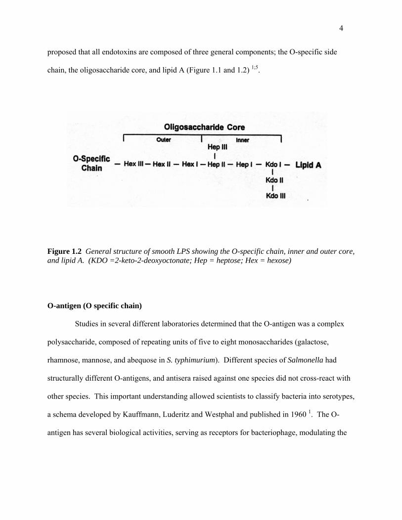
Figure 1.2 General structure of smooth LPS showing the O-specific chain, inner and outer core, and lipid A. (KDO =2-keto-2-deoxyoctonate; Hep = heptose; Hex = hexose)
O-antigen (O specific chain) Studies in several different laboratories determined that the O-antigen was a complex
polysaccharide, composed of repeating units of five to eight monosaccharides (galactose,
rhamnose, mannose, and abequose in S. typhimurium). Different species of Salmonella had
structurally different O-antigens, and antisera raised against one species did not cross-react with
other species. This important understanding allowed scientists to classify bacteria into serotypes,
a schema developed by Kauffmann, Luderitz and Westphal and published in 1960 1. The O-
antigen has several biological activities, serving as receptors for bacteriophage, modulating the
5
activation of the alternative complement pathway, and inhibiting the attachment of the
membrane attack complex to the bacterial outer membrane 4.
Core Oligosaccharide It was later discovered that not all Gram-negative bacteria possess an O-antigen. Such
bacteria are termed rough (R) because they form rigid, incomplete colonies on solid agar and
autoagglutinate in saline. Studies of the core oligosaccharide were facilitated by the
characterization of Salmonella minnesota and Salmonella typhimurium mutants. Several rough
(R)-mutants were shown to have truncated polysaccharide cores due to defects in genes that code
for glycosyl or phosphoryl transferases. Rough mutants synthesizing the entire core (but lacking
O-antigen) were termed Ra. Those lacking the terminal sugar were named Rb, and the mutant
possessing the shortest core was named Re 6. Luderitz, Westphal, and colleagues proposed the
structure of the S. minnesota core in 1967. Although core regions differ among bacterial species,
all core regions contain an unusual sugar, 2-keto-3-deoxyoctonate (KDO). Other known residues
are heptose, glucose, galactose, and N-acetylglucosamine [reviewed in 7]. The minimum
requirement for cell viability is a single molecule of KDO, as is present in Re Salmonella and E.
coli LPS 8. Based on the most severe mutant (Re), one can deduce that KDO must be directly
attached to lipid A, the toxic moiety of LPS. Not much is known about the biological activities
of the outer core region of LPS, but it is believed that both the outer and inner core carry epitopes
for antibodies 4.

6
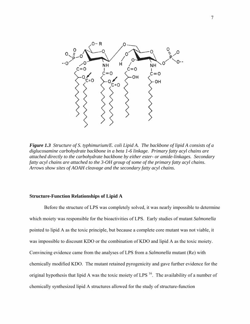
Lipid A Westphal and Luderitz are credited with coining the term lipid A. As described in a 1954
review, they isolated a chloroform- and pyridine- soluble lipid structure from intact LPS by a
thirty-minute treatment with HCl at elevated temperature 1;9. Determining the structure of the
extracted lipid was considerably more difficult than structuring either the core or O-antigen and
it wasn’t until 1983 that Takayama and colleagues published the complete and correct structure
of lipid A 10. It is now known that lipid A consists of a unique diglucosamine backbone (D-
GlcN) that is β(1’-6) interlinked. Two phosphate groups are attached to the backbone at the 1
and 4’ positions. These phosphates are sometimes modified with polar groups such as 4-amino-
4-deoxy-L-arabinose (Ara4N) and/or ethanolamine, both of which are removed by the mild acid
treatment used to purify and study lipid A. The fatty acid composition of lipid A was first
described in E. coli. A total of six fatty acids chains are attached to the lipid A backbone, two
via amide linkages and four via ester linkages. The amino-linked fatty acids were exclusively 3-
hydroxymyristate, while the ester-linked fatty acids were of varying length (myristate, laurate, or
palmitate). It was later discovered that two of the four ester-linked fatty acids were not directly
attached to the lipid A backbone, but were actually bound to the 3-hydroxyl groups of one
amide- and one ester-linked fatty acid 11. Munford et al. termed these piggybacked, acyloxyacyl
linked, fatty acids secondary, while those attached directly to the lipid A backbone were termed
“primary” 10-12. The ester- or amide-linked primary fatty acids are all 3-hydroxymyristates
[14:0(3-OH)]. In independent studies, the structure of S. typhimurium lipid A was determined to
be essentially identical to that of E. coli. Around 1985 investigators chemically synthesized E.
coli lipid A 13. This compound, called LA-15-PP (506), was later shown to be indistinguishable
from lipid A purified from natural bacterial sources in a variety of biological assays 4;14;15.
7
Figure 1.3 Structure of S. typhimurium/E. coli Lipid A. The backbone of lipid A consists of a diglucosamine carbohydrate backbone in a beta 1-6 linkage. Primary fatty acyl chains are attached directly to the carbohydrate backbone by either ester- or amide-linkages. Secondary fatty acyl chains are attached to the 3-OH group of some of the primary fatty acyl chains. Arrows show sites of AOAH cleavage and the secondary fatty acyl chains.
Structure-Function Relationships of Lipid A Before the structure of LPS was completely solved, it was nearly impossible to determine
which moiety was responsible for the bioactivities of LPS. Early studies of mutant Salmonella
pointed to lipid A as the toxic principle, but because a complete core mutant was not viable, it
was impossible to discount KDO or the combination of KDO and lipid A as the toxic moiety.
Convincing evidence came from the analyses of LPS from a Salmonella mutant (Re) with
chemically modified KDO. The mutant retained pyrogenicity and gave further evidence for the
original hypothesis that lipid A was the toxic moiety of LPS 16. The availability of a number of
chemically synthesized lipid A structures allowed for the study of structure-function
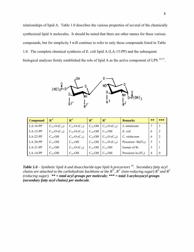
8
relationships of lipid A. Table 1.0 describes the various properties of several of the chemically
synthesized lipid A molecules. It should be noted that there are other names for these various
compounds, but for simplicity I will continue to refer to only those compounds listed in Table
1.0. The complete chemical synthesis of E. coli lipid A (LA-15-PP) and the subsequent
biological analyses firmly established the role of lipid A as the active component of LPS 15;17.
Compound R3’ R2’ R3 R2 Remarks ** *** LA-16-PP C14-O-(C14) C14-O-(C12) C14-OH C14-O-(C16) S. minnesota 7 3
LA-15-PP C14-O-(C14) C14-O-(C12) C14-OH C14-OH E. coli 6 2
LA-22-PP C14-OH C14-O-(C14) C14-OH C14-O-(C14) C. violaceum 6 2
LA-20-PP C14-OH C14-OH C14-OH C14-O-(C16) Precursor- Ib(IVb) 5 1
LA-21-PP C14-OH C14-O-(C16) C14-OH C14-OH Isomer of Ib 5 1
LA-14-PP C14-OH C14-OH C14-OH C14-OH Precursor Ia (IVa) 4 0
Table 1.0 – Synthetic lipid A and disaccharide-type lipid A precursors 16. Secondary fatty acyl chains are attached to the carbohydrate backbone at the R3’, R2’ (non-reducing sugar) R3 and R2 (reducing sugar). ** = total acyl groups per molecule; *** = total 3-acyloxyacyl groups (secondary fatty acyl chains) per molecule.
9
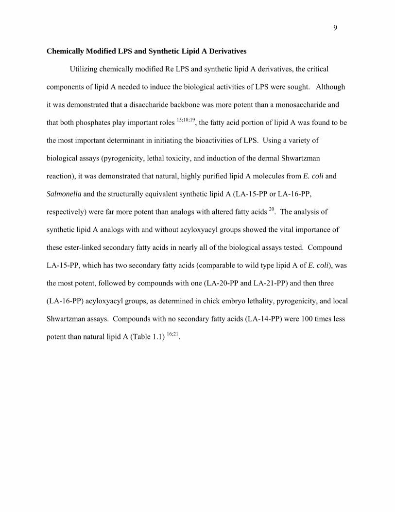
Chemically Modified LPS and Synthetic Lipid A Derivatives
Utilizing chemically modified Re LPS and synthetic lipid A derivatives, the critical
components of lipid A needed to induce the biological activities of LPS were sought. Although
it was demonstrated that a disaccharide backbone was more potent than a monosaccharide and
that both phosphates play important roles 15;18;19, the fatty acid portion of lipid A was found to be
the most important determinant in initiating the bioactivities of LPS. Using a variety of
biological assays (pyrogenicity, lethal toxicity, and induction of the dermal Shwartzman
reaction), it was demonstrated that natural, highly purified lipid A molecules from E. coli and
Salmonella and the structurally equivalent synthetic lipid A (LA-15-PP or LA-16-PP,
respectively) were far more potent than analogs with altered fatty acids 20. The analysis of
synthetic lipid A analogs with and without acyloxyacyl groups showed the vital importance of
these ester-linked secondary fatty acids in nearly all of the biological assays tested. Compound
LA-15-PP, which has two secondary fatty acids (comparable to wild type lipid A of E. coli), was
the most potent, followed by compounds with one (LA-20-PP and LA-21-PP) and then three
(LA-16-PP) acyloxyacyl groups, as determined in chick embryo lethality, pyrogenicity, and local
Shwartzman assays. Compounds with no secondary fatty acids (LA-14-PP) were 100 times less
potent than natural lipid A (Table 1.1) 16;21.

10
Table 1.1– A comparison of the bioactivities of several synthetic lipid A and disaccharide-type lipid A precursors 16. ND, not determined.
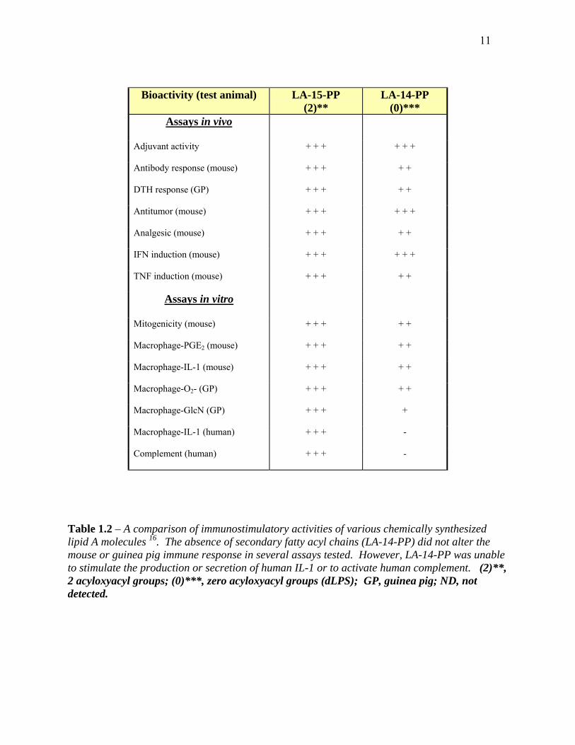
In addition to the bioactivities of LPS mentioned above (pyrogenicity, tissue toxicity, and
the dermal Shwartzman reaction), LPS is known to modulate many immune functions. The use
of various chemically synthesized lipid A molecules (listed in Table 1.0) in a variety of in vivo
and in vitro immunoassays yielded some interesting results. Compound LA-14-PP, which lacks
secondary (acyloxyacyl) groups, retained nearly all of its in vivo immunostimulatory ability
(Table 1.2) 16. For example, compound LA-14-PP was only deficient in activating the human
complement pathway and stimulating the production and secretion of human IL-1 16;21.
11
Bioactivity (test animal) LA-15-PP (2)**
LA-14-PP (0)***
Assays in vivo
Adjuvant activity + + + + + +
Antibody response (mouse) + + + + +
DTH response (GP) + + + + +
Antitumor (mouse) + + + + + +
Analgesic (mouse) + + + + +
IFN induction (mouse) + + + + + +
TNF induction (mouse) + + + + +
Assays in vitro
Mitogenicity (mouse) + + + + +
Macrophage-PGE2 (mouse) + + + + +
Macrophage-IL-1 (mouse) + + + + +
Macrophage-O2- (GP) + + + + +
Macrophage-GlcN (GP) + + + +
Macrophage-IL-1 (human) + + + -
Complement (human) + + + -
Table 1.2 – A comparison of immunostimulatory activities of various chemically synthesized lipid A molecules 16. The absence of secondary fatty acyl chains (LA-14-PP) did not alter the mouse or guinea pig immune response in several assays tested. However, LA-14-PP was unable to stimulate the production or secretion of human IL-1 or to activate human complement. (2)**, 2 acyloxyacyl groups; (0)***, zero acyloxyacyl groups (dLPS); GP, guinea pig; ND, not detected.

12
It has been suggested that, upon ingestion by phagocytes, Gram-negative bacteria are
rapidly killed. LPS, on the other hand, can be detected for days to months later 22. Munford
hypothesized that LPS might be catabolized within the cell and he began searching for enzymes
(lipases) that might remove fatty acyl chains from lipid A. At this time, the correct structure of
lipid A had not yet been elucidated and secondary fatty acyl chains were not known to exist. In
order to begin his studies, he obtained a Salmonella typhimurium mutant, PR122 (from Paul Rick
at USUHS). This strain lacks glucosamine deaminase and is therefore unable to metabolize
glucosamine to glucose, so it incorporates radiolabeled glucosamine (N-Ac-14C-glucosamine)
into the carbohydrate backbone of LPS. Other bacterial components might also be labeled in this
process, but are removed during LPS purification. In addition to labeling the carbohydrate
backbone of lipid A, the fatty acyl chains were biosynthetically labeled with 3H by adding 3H-
acetate to the bacterial growth medium. The resulting purified LPS (double-labeled LPS
substrate) has a 14C-labeled carbohydrate backbone and tritiated fatty acyl chains. It was only
after the demonstration that neutrophils released the non-hydroxylated fatty acyl chains from the
double-labeled LPS substrate that Wollenweber et al. reported the existence of acyloxyacyl
linkages in lipid A 23.
Acyloxyacyl Hydrolase (AOAH)
In 1983, Hall and Munford identified an enzyme from neutrophils that partially
deacylated Salmonella typhimurium LPS 12. This enzyme, acyloxyacyl hydrolase (AOAH), was
present in the granule fraction of human neutrophils and could remove the secondary
13
(nonhydroxylated laurate, myristate, and palmitate) fatty acids from the lipid A backbone of
LPS. The primary fatty acids (3-OH-14:0) remained linked to the diglucosamine backbone 12.
In subsequent studies, partially purified acyloxyacyl hydrolase isolated from HL-60 cells
(human promyelocytes) was used to deacylate LPS. The partially deacylated LPS product is
termed deacylated LPS (dLPS); its lipid A moiety structurally resembles compound LA-14-PP
(also called 406, precursor 1b, and lipid IVA). The dermal Shwartzman reaction, an assay for
tissue toxicity, was utilized to test the potency of normal and AOAH-deacylated LPS (dLPS). In
this assay, New Zealand White rabbits were injected intradermally with either enzyme-treated or
control LPS, followed by an intravenous dose of LPS 20-24 hours later. Rabbits that were given
dLPS had no hemorrhagic necrosis at the site of the intradermal injection, while the sites that
were injected with intact LPS had lesions of 3 mm or greater. A dose response analysis revealed
that dLPS was more than 100- fold less toxic than intact LPS 24. These results were in keeping
with data previously generated from chemically synthesized tetracyl lipid As 16;21.
As mentioned earlier, chemically synthesized lipid A molecules lacking acyloxyacyl-
linked fatty acyl chains (compound LA-14-PP) retain their immunostimulatory ability, including
the ability to stimulate mouse splenocyte mitogenesis (Table 1.2). In order to verify these results
using AOAH-treated LPS (dLPS), Hall and Munford tested the ability of LPS and dLPS to
stimulate murine splenocyte division. The results were similar to those obtained with compound
LA-14-PP; when splenocytes were incubated with dLPS, their rate of division was reduced by a
factor of 6 to 20 as compared to splenocytes incubated with intact LPS 24. While there was thus
an effect on mitogenesis, it was not as significant as the dramatic effect seen between LPS and
dLPS in tissue toxicity assays (dermal Shwartzman reaction). Combined, these experiments
gave validation to the studies of chemically synthesized lipid A molecules, for the enzymatically

14
deacylated lipid A was still attached to the core and O-antigen, which was not the case with the
chemical derivatives. This work showed the importance of the secondary acyl chains in the
bioactivity of LPSs, thus proving that the polysaccharide chain clearly plays a secondary role in
the toxicity of LPS.
Since the elucidation of the correct lipid A structure, it has been shown that tetraacyl lipid
A analogs, including AOAH-treated LPS (dLPS) and compound LA-14-PP, are able to
antagonize LPS in human cells 25. Deacylated LPS has been shown to inhibit neutrophil
adherence to LPS stimulated endothelial cells 26, and also to inhibit prostaglandin E2 production
by neutrophils in vitro 27. These compounds have also been shown to inhibit TNFα release from
LPS-stimulated whole blood ex vivo 28 and abrogate the ability of LPS to stimulate endothelial
cells in vitro 29. Combined, these data clearly show the importance of acyloxyacyl groups in the
bioactivities of LPS.
Deacylation of Diverse Lipopolysaccharides by AOAH Although the general structure of lipid A is highly conserved, LPSs isolated from
different bacteria can differ in many ways. The extent of phosphorylation, the polar group
modifications of these phosphate groups, and the extent and type of acyloxyacyl groups attached
to the lipid A backbone can vary in bacterial species. To study the specificity of AOAH for
secondary (non-hydroxylated) fatty acids, the LPSs of Escherichia coli, Haemophilus influenzae,
Neisseria meningitidis, Neisseria gonorrhoeae, and Pseudomonas aeruginosa were treated with
AOAH. Despite the structural differences in lipid A structure (namely the location and nature of
the acyloxyacyl groups), AOAH was able to deacylate all LPSs to the same degree (~30% of the
15
total, or all of the secondary fatty acyl chains were removed). AOAH removed only the
secondary fatty acyl chains, regardless of fatty acid chain length or the placement of the
secondary fatty acyl chains on the reducing or non-reducing glucosamine. In each case, the
primary fatty acyl chains (hydroxylated) remained attached to the lipid A backbone 30.
Purification of Acyloxyacyl Hydrolase (AOAH) In 1989, Hall and Munford described the purification of acyloxyacyl hydrolase (AOAH).
AOAH, when purified from HL60 cells, had the same specificity for secondary fatty acyl chains,
a similar pH optimum (4.5), and the same Km (~0.55 µM) as did the original enzyme(s) isolated
from the granule fraction of neutrophils 12;31. The purified enzyme had an apparent size of 52 to
60 kDa and was composed of two disulfide-linked, glycosylated subunits. The AOAH cDNA
was cloned in 1991 and the recombinant protein stably expressed in BHK570 cells 32.
Recombinant AOAH retained all of the characteristics of purified AOAH and is used in many of
our studies 32.
Basic Structure of AOAH Studies of recombinant AOAH protein produced a better understanding of the structure of
AOAH and some of its supposed functions. Purified AOAH runs as a single band of
approximately 60 kDa on a non-reducing SDS-polyacrylamide gel. Reduction of AOAH results
in a separation of two subunits, of 50 kDa (large subunit) and ~10-14 kDa (small subunit). The
sequence of AOAH reveals five potential (N-glycosylation) sites, one in the small subunit and
four in the large subunit 32. While the roles of such N-glycosylation sites are unknown, it has
since been shown that the small subunit glycosylation site is not essential for either enzymatic
16
deacylation or intracellular localization 33. The roles of the large subunit glycosylation sites are,
at this time, unknown. In an attempt to reduce the complexity of the enzymes structure so that
AOAH could be crystallized, several N-glycosylation mutants were constructed. While some
glycosylation site mutants retained activity, others did not. Due to the lack of an appropriate
antibody for detection by Western blot, it was never determined if the non-functional proteins
were actually produced, since removing glycosylation sites may make proteins more susceptible
to degradation.
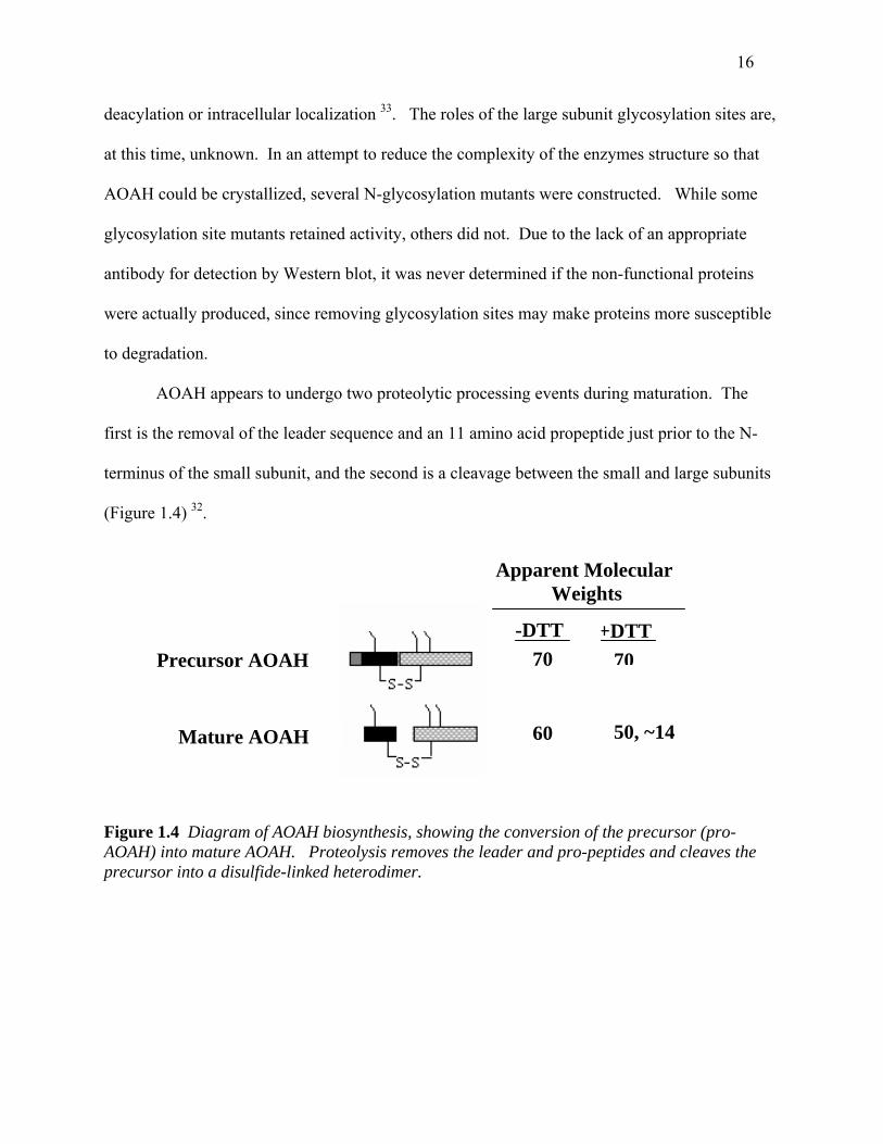
AOAH appears to undergo two proteolytic processing events during maturation. The
first is the removal of the leader sequence and an 11 amino acid propeptide just prior to the N-
terminus of the small subunit, and the second is a cleavage between the small and large subunits
(Figure 1.4) 32.
+DTT -DTT 70 70
60 50, ~14 Mature AOAH
Precursor AOAH
Apparent Molecular Weights
Figure 1.4 Diagram of AOAH biosynthesis, showing the conversion of the precursor (pro-AOAH) into mature AOAH. Proteolysis removes the leader and pro-peptides and cleaves the precursor into a disulfide-linked heterodimer.

17
Large Subunit
The large subunit of AOAH contains the sequence Gly-X-Ser-X-Gly, which is found at
the active sites of many lipases. In fact, AOAH closely resembles members of the GDSL family
of lipases, which have a conserved Ser-Asp-His catalytic triad 34 that is thought to be the active
site for their various activities. Replacement of AOAH’s serine (Ser263) with Leu reduced its
activity toward LPS by about 99% 33. As mentioned earlier, the large subunit of AOAH is
glycosylated although the importance of these sites is currently unknown. The large subunit of
AOAH is highly conserved as shown (in part) below (Figure 1. 5 A-C).

18
Human AOAHMouse AOAHRabbit AOAH
Dictyostelium discoideumInositol Deacylase Trypanosome Protein
Consensus
Human AOAHMouse AOAHRabbit AOAH
Dictyostelium discoideumInositol Deacylase Trypanosome Protein
Consensus
Contains the active site serine.
Contains the active site aspartate.
B
A
C
Human AOAHMouse AOAHRabbit AOAH
Dictyostelium discoideumInositol Deacylase Trypanosome Protein
Consensus
Contains the active site histidine.
Figure 1.5 -- Alignments of several stretches of amino acids from the large subunit of AOAH, derived from cDNAs from human, rabbit, mouse, Dictyostelium discoideum, and Trypanosome cDNA. The ovals represent areas of conservation including the serine (A), aspartate (B), and histidine (C) motifs present in all GDSL lipase family members 34.
19
Small Subunit The small subunit of AOAH shares sequence identity with a family of proteins called
saposins. All saposin-like proteins (SAPLIPs) have six common cysteine residues and a N-
linked glycosylation site that have been hypothesized to form a secondary structure of four
disulfide-linked amphipathic helical bundles 35. All members of the family interact with lipids,
but seem to have diverse functions in vivo. The small subunit of AOAH is essential for the
intracellular location and stability of AOAH and may also be involved in substrate recognition.
The evidence for this comes from experiments where only the large subunit of AOAH was
expressed in BHK 570 cells. In these cells, AOAH was less stable, less enzymatically active,
and did not localize to the same intracellular compartment as did the full-length protein. In
addition, a deletion of 32 amino acids within the small subunit of AOAH (including two of the
six cysteines) resulted in an unstable protein that had approximately 40% of its native activity
33;35. As is the case with other SAPLIPs, the function of the N-linked glycosylation is unclear.
Deletion of the small subunit glycosylation site by site-directed mutagenesis did not alter the
protein’s stability, intracellular location, or its secretion. It did, however, increase its activity
toward LPS by about 3-fold 33.
Precursor vs. Mature AOAH Studies such as those described above were extremely helpful in understanding the
structure-function relationships of AOAH, but they were not complete. When stably expressed
in BHK 570 cells, recombinant AOAH is secreted into the culture supernatant as a
approximately 70 kDa propeptide or precursor protein. The cell lysate fraction contains both the
precursor and the previously described mature form of AOAH (~60 kDa). It was hypothesized
20
that, during maturation, the propeptide underwent intracellular proteolytic cleavage. In order to
test this hypothesis, partially purified recombinant precursor was treated with trypsin or
chymotrypsin, followed by analysis by a reducing or non-reducing SDS-PAGE. Both trypsin
and chymotrypsin mimicked natural proteolytic cleavage, causing AOAH to separate into its
large and small subunits on reducing SDS-PAGE. Maturation of AOAH (either naturally or via
chymotrypsin treatment) increases its ability to deacylate LPS by 10- to 20- fold. Although
AOAH is able to remove fatty acids from glycerophosphatidylcholine (GPC) in vitro 36, its
activity toward this substrate is not altered by maturation 37.
Mannose 6 Phosphate Residues and Receptors
Newly synthesized proteins that contain an Asn-X-Ser/Thr motifs are covalently
modified in the trans-Golgi network (TGN) by the addition of Asn-linked sugar chains that often
contain mannose-6-phosphate (M6P) residues. Such proteins are recognized by mannose-6-
phosphate receptors in the TGN and are either targeted to endosomes/lysosomes or are secreted
from the cell 38. Two mannose-6-phosphate receptors have been described. The first, an
integral membrane glycoprotein with an apparent molecular weight of 215,000, binds M6P
containing proteins independent of divalent cations. This receptor also binds insulin-like growth
factor II (IGFII) and thus the receptor has been termed the CI-M6P/IGFII receptor. The second
M6P receptor is also an integral membrane glycoprotein with an apparent molecular weight of
46,000. Because of its enhanced ligand binding affinity in the presence of divalent cations it has
been termed the cation dependent (CD)-MPR 39;40. As shown in Chapter Two, the secreted form
of AOAH (pro-AOAH) uses M6P receptors on the plasma membrane to gain entry into cells.
21
Other Known Activities of AOAH
In addition to its role in deacylating LPS, the enzyme has been shown to have several
other activities in vitro. AOAH preferentially cleaves saturated fatty acids from
glycerophospholipids, lysophospholipids, and diacylglycerol with little to no preference for
position (sn-1 vs sn-2, the names given to the two fatty acyl chains present in the above
compounds). These studies were done by incubating either native or recombinant AOAH with
glycerophospholipid substrates that had either saturated or unsaturated fatty acid chains in the sn-
2 position and a saturated fatty acid at sn-1. When the sn-2 fatty acid was unsaturated, AOAH
released only the saturated fatty acid from sn-1. When both sn-1 and sn-2 were occupied by
saturated fatty acids, AOAH was able to release both saturated fatty acids 36. These results
indicated that fatty acid structure, and not position on the carbohydrate backbone, determined the
enzyme’s specificity. It was also shown that AOAH was able to transfer acyl chains to several
lipid acceptors, and that the presence of free fatty acids in the reaction mixture did not inhibit
such transfer. This suggests that AOAH is transferring fatty acyl chains from donor to acceptor
rather than non-specifically associating with any available, free, fatty acyl chains 36. These
findings raise the possibility that AOAH may have a function(s) other than deacylating
(detoxifying) LPS in vivo.
Localization of AOAH As described earlier, AOAH was first detected in the granule fraction of human
neutrophils and later purified from the HL-60 human promyelocyte cell line. Since its
purification, AOAH has been detected in human and mouse myeloid lineage cells such as
monocytes, macrophages, and, more recently, dendritic cells. My dissertation work will describe
22
the detection of AOAH in renal proximal tubule cells. This is the first description of AOAH in a
non-myeloid cell. Such a finding encouraged me to study the role of AOAH in the urinary tract
and the following paragraphs will discuss the current literature on such infections and some
general properties of renal proximal tubule cells.
Kidney Architecture and Renal Proximal Tubule Cells
Our entire blood volume is filtered through the kidneys about 65 times each day. In
doing so, the kidney(s) regulate our water and electrolyte balance and eliminate many metabolic
waste products. The functional unit of the kidney, the nephron, is responsible for such functions
and is composed of many cell types. Renal proximal tubule cells function within the kidney to
regulate the water, ion, and small molecule concentrations in the blood. They are the first cell
type in the nephron to actively reabsorb and secrete such molecules, which are filtered from the
blood in the glomerulus. In doing so, they help to maintain the osmotic pressure and ionic
composition of the fluids of the body 41.
23
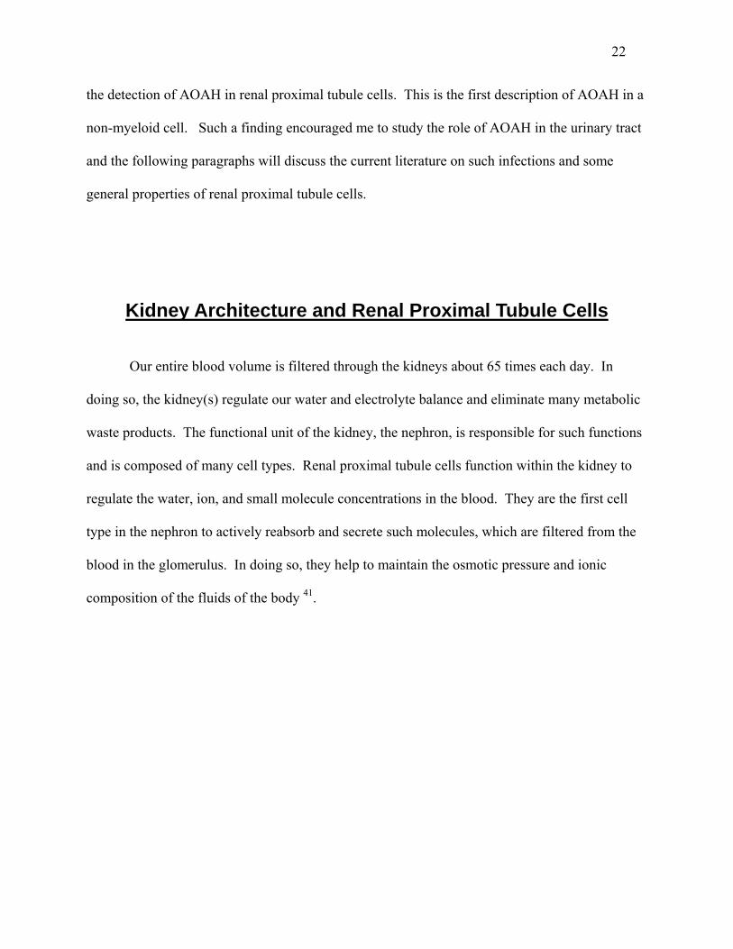
Figure 1.6 – A diagram of the human urinary tract. Bacteria normally enter through the urethra and may ascend into the bladder, ureter(s), and kidneys(s). This figure was obtained from the website: mcdb.colorado.edu/courses/ 3280/class08.html.
A
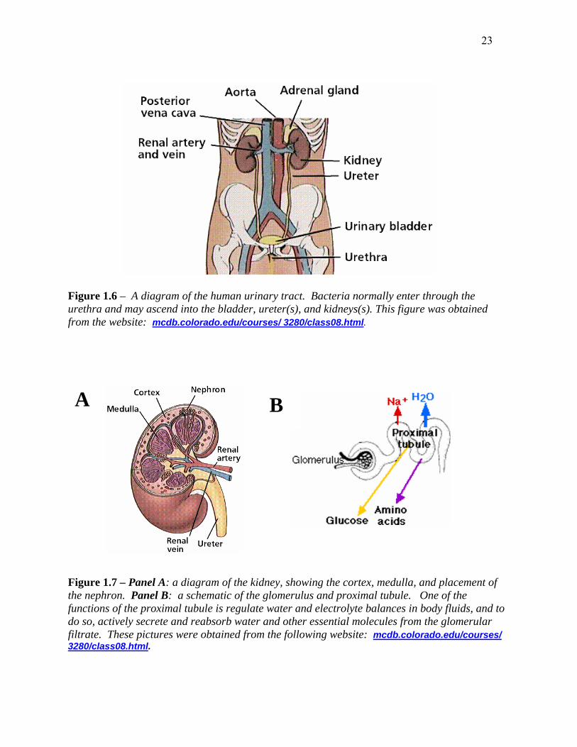
B
Figure 1.7 – Panel A: a diagram of the kidney, showing the cortex, medulla, and placement of the nephron. Panel B: a schematic of the glomerulus and proximal tubule. One of the functions of the proximal tubule is regulate water and electrolyte balances in body fluids, and to do so, actively secrete and reabsorb water and other essential molecules from the glomerular filtrate. These pictures were obtained from the following website: mcdb.colorado.edu/courses/ 3280/class08.html.
24
Urinary Tract Infections
Urinary tract infections (UTIs) are a significant cause of morbidity in the developed
world and are one of the most common reasons for clinical visits to primary care, hospital, and
extended-care facilities 42. Cystitis, or bladder infection, is the most common manifestation of
urinary tract infection. Symptoms include frequent or urgent voiding and suprapubic pain.
Pyelonephritis, or infection of the kidney, is a more serious complication of urinary tract
infection because of the destruction of kidney cells and the potential of the bacteria to enter the
bloodstream. Symptoms of pyelonephritis include all of those described for cystitis plus flank
pain, nausea, vomiting, fever, sweats, and malaise 43. Pyelonephritis sometimes leads to
bacteraemia 44. UTIs affect women more frequently than men, probably due to the anatomy of
the female urinary system as compared to that of the male (the female urethra is shorter and in
closer proximity to areas of bacterial colonization such as the colon). It is estimated that one-
third of American women will have a UTI before the age of 65 and that, of those women, 25 to
30% will have one or more recurrences within 3 to 6 months of their initial infection 45.
Uncomplicated urinary tract infections (which account for the majority of infections in
adult women) are defined as those that occur in otherwise healthy individuals with normal
immune status, respond well to antibiotic treatment, and in which recurrences are due to re-
infections with strains other than the initial pathogen 44. Complicated UTIs normally occur in
individuals with urinary tract abnormalities and/or immune system functions such as diabetes,
AIDS, and liver insufficiency. Complicated UTIs do not respond well to antibiotic therapy and
recurrences are often due to relapse with the same pathogen 44;46. Complicated UTIs will not be
discussed in greater detail in this dissertation.
25
Etiology of Urinary Tract Infections Approximately 80% of uncomplicated, community-acquired urinary tract infections are
caused by uropathogenic E. coli (UPEC), which are facultative anaerobic Gram-negative rods.
Other Gram-negative bacteria such as Proteus mirabilis, Klebsiella pneumoniae, and
Pseudomonas aeruginosa are also known to cause UTIs, but mostly in individuals with
abnormalities in their urinary system or underlying immune dysfunction (complicated UTI).
Staphylococcus saprophyticus, a facultative anaerobic, Gram-positive coccus, accounts for
approximately 10 to 15% of UTIs 44;47. Because of the prevalence of UPEC as the causative
agent of UTIs and the existence of well-established murine models of infection, we chose to
focus our studies on UPEC-induced urinary tract infections in the mouse.
The majority of bacteria that enter the urinary system do so via an ascending route; very
few reach the kidneys via the bloodstream 44. They must first gain access to the urethra and, if
they survive the battery of host defenses that aim to eliminate them, they may travel to the
bladder, ureters, and kidneys (Figure 1.6 and 1.7 A).
Virulence Factors Associated with Uropathogenic E. Coli
Adhesins (pili, fimbriae) E. coli, like many enteric bacteria, is a heterogeneous species with members that differ
widely in their ability to cause disease. With that said, strains that are able to colonize the
bladders and/or kidneys during urinary tract infections typically have several common features.
Arguably the most important virulence factors are the adhesins (also called pili or fimbriae),

26
which mediate bacterial binding and entry into bladder or kidney epithelium and result in the
initiation of cellular inflammatory responses. Without these proteinaceous bacterial appendages,
bacteria would be unable to gain a foothold on the host epithelium and would likely not cause
disease. Martinez et. al. have demonstrated that type I pili are necessary to mediate not only
adherence, but also invasion of bacteria into bladder epithelial cells 48. Several adhesins have
been described in the literature, these include type I, P, S, F1C, and Dr fimbriae, Afimbrial
adhesin I (AFA I) and III (AFA III), Non-fimbrial adhesin 1 (Nfa-1), M and G-adhesin, and
Curli 44. Here I will describe only the type I and P fimbriae, the two most relevant to my
experimental system.
Type I fimbriae Because of their common occurrence on UPEC, type I fimbriae have been well studied.
Although different pili bind specific cellular targets, their structures are strikingly similar.
Therefore, the following description applies to both type I and P pili (as well as other adhesins).
Genes that encode the structural and non-structural components of pili are located on large
operons that usually consist of 9-12 genes. These genes include a structural subunit (Fim A; Pap
A), accessory proteins (Fim I, C, D, F, and G; Pap H, C, D, E, and F), regulatory proteins (Fim B
and E; Pap I and B), and the adhesin (Fim H; Pap G). The biogenesis of the pilus will not be
discussed here, but like that of other adhesins, it involves a chaperone-usher pathway.
Adherence and invasion by type I piliated bacteria is mediated by Fim H binding to
mannosylated glycoproteins such as CD48, collagens, laminin, and fibronectin which are found a
variety of host tissues 43;49. It has recently been shown that type I pili bind UP1a, an integral
membrane glycoprotein located on the lumenal surface of the bladder. In so doing, type I pili
27
induce exfoliation of the bladder epithelium via an apoptosis-like mechanism 50. While binding
to the bladder seems beneficial for the uropathogen, exfoliation is thought to be an effective
innate host defense mechanism, clearing many bacteria from the urinary tract.
P pili While type I pili can often be isolated from both pathogenic and non-pathogenic bacteria,
p pili are rarely isolated from non-uropathogens and are the most commonly isolated fimbriae
type from UPEC 51;52. The adhesin, Pap G, mediates binding to glycolipid receptors (alpha-Gal-
beta-(1-4)-Gal moieties), which are found on uroepithelial cells, renal proximal tubules, and
renal vascular endothelium 44. P blood group antigens, which are found on erythrocytes and
uroepithelial cells, have also been shown to bind p pili. In fact, women with p-positive
erythrocytes are more likely to get UTIs than are women who do not express such antigens. It is
believed that p pili bind to the p blood group antigens expressed on uroepithelial cells 44. Like
type I pili, p pili also utilize a chaperone-usher pathway for pilus biogenesis. Studies in
cynomogus monkeys have shown the vital importance of the p pilus in colonizing the kidneys.
In these studies, p pilus negative strains of bacteria were able to colonize the bladder, but were
unable to adhere to or cause pyelonephritis 53. Likewise, studies in human volunteers have
shown that p pili enhance the ability of bacteria to colonize the urinary tract 54. In contrast, it
has been shown that both p piliated and non p piliated strains of bacteria were able to bind to and
invade proximal tubule cell in vitro 55;56.
28
Lipopolysaccharide Particular O-antigens of LPS are often associated with uropathogenicity. The most
common UTI- associated O-groups are O1, 2, 4, 6, 7, 8, 16, 18, 25, 50, and 75. In comparison to
fecal isolates, UTI isolates are less diverse in their O serotypes (ie. similar O-groups predominate
in UTI urine cultures). Women with vaginal colonization of serotypes 2, 4, 6, and 75 often
develop UTI with these same serotypes. In contrast, women with other vaginal serotypes do not
normally experience UTIs 49. It is currently unknown what is unique about such serotypes. In
addition to the O-antigen, the lipid A moiety of LPS is known to play a role in the virulence of
type I piliated UPEC. Strains of bacteria that lack functional lipid A moieties are unable to
stimulate appropriate inflammatory responses in bladder and kidney epithelial cells in vitro 57.
Mice that are unable to recognize LPS due to a mutation in TLR4 do not recruit neutrophils to
the urine or bladder tissue and subsequently fail to clear UTIs 42;58-61. In contrast, despite having
a dysfunctional lipid A, bacteria that express p pili are still able to stimulate appropriate cytokine
and chemokine responses in vitro and in murine models of UTI 57;62. These data will be
discussed in greater detail in Chapter 3 and in the discussion.
Toxins Most UPEC produce toxins such as alpha-haemolysin (~50%) and cytotoxic necrotizing
factor 1 (cnf1) 49. Alpha-haemolysin is a heat-labile exotoxin that is encoded by genes hly A, B,
C, D, and tolC, which are located on chromosomal pathogenicity islands or on transmissible
plasmids 63. Alpha-haemolysin is a pore-forming cytolysin that lyses erythrocytes by disrupting
transmembrane ion gradients, raising intracellular osmotic pressure, and eventually lysing the
cell. In addition to lysing red blood cells, alpha-hemolysin is thought to be able to lyse other
29
mammalian cells. In vitro data suggest that it may be able to lyse monocytes and granulocytes,
but that it has little activity against lymphocytes 49;64. Cnf 1, also a common toxin of UPEC, is
associated with the O4 and O6 serotypes 65;66. Cnf 1 affects the host cell cytoskeleton by post-
translationally modifying the Rho GTP-binding protein responsible for formation of the cellular
microfilament network 67. In addition, in vitro work has suggested that Cnf 1 may increase the
phagocytic behavior of epithelial cells, allowing the bacteria more efficient entry into cells 44.
Other virulence factors of UPEC Uropathogens, like all E. coli, require iron for survival and thus encode the siderophores
aerobactin and enterobactin, which help them sequester iron from their host. The mechanism by
which aerobactin, which is predominately found on enteric pathogens, sequesters iron is well
studied. Once secreted, the small siderophore is able to extract Fe3+ from host iron-binding
proteins and channel it into the bacteria via an outer membrane receptor complex 68.
Enterobactin is expressed by both pathogenic and non-pathogenic bacteria and will not be
discussed here 44. In addition to iron acquisition systems, uropathogens also utilize
polysaccharide capsules in their quest for host colonization. UPEC are known to produce type
K1, 2, 5, 6, 12, 13, 29, and 51 capsules and to use these polysaccharides to evade host immune
recognition and to inhibit opsonization 69. Small percentages of UPEC, but a high percentage of
Proteus mirabilis, produce urease. Urease is associated with an increased susceptibility to stone
formation and pyelonephritis and acts by hydrolysing urea to ammonia and carbamate. Urease is
further able to hydrolyse carbamate to ammonia and carbonic acid, thus increasing the pH of the
urine and precipitating previously soluble polyvalent ions 44.
30
Known Host Defenses to Invading Uropathogens Although they have been less well studied, several host responses to invading
uropathogens are worth noting. UPEC are able to grow in urine despite the low pH, the force of
flow during urination, and the high osmolarity. The host produces several inhibitors that are
secreted into the urine. Tamm-Horsfall protein (THP), one such inhibitor, is a glycoprotein that
binds to S fimbriae leading to the elimination of S-fimbriated strains from the urinary tract 70.
Other constitutive secretory components of normal urine are defensins, secretory IgA,
uromucoid, and urea. As previously noted, binding of type I pili to bladder epithelial cells
initiates exfoliation of the superficial cells that line the bladder, which is considered an innate
host defense mechanism 71. One of the most important host defenses toward invading
uropathogens are neutrophils and macrophages which flux to site(s) of bacterial colonization and
contribute greatly to bacterial clearance 43.
Ascending Urinary Tract Infections (UTIs)
In order to study the in vivo role of AOAH, I chose to induce unobstructed, ascending
urinary tract infections in mice. Mice were the most suitable animal due to existing experimental
protocols, the availability of AOAH null animals, and low cost. The method of Hagberg et. al.
was chosen and will be described in detail in the methods section of Chapter Three. Briefly, six
to ten week old female mice are anesthetized and given a 50 µl injection of UPEC (suspended in
PBS) via a soft polyethylene catheter into the bladder 72. This is by far the most commonly

31
utilized model of UTI and while other models exist, they may not accurately mimic natural
ascending UTI.
The role of toll-like receptor 4 (TLR4) in UTI Even before the discovery of TLRs, it was known that, unlike C3H/HeN mice, C3H/HeJ
mice were hyporesponsive to LPS. It is now known that C3H/HeJ mice have a point mutation in
the toll-like receptor 4 (TLR4) gene which renders them unresponsive to LPS stimulation 73;73;74.
When subjected to ascending experimental UTI with UPEC, C3H/HeJ mice are unable to mount
appropriate inflammatory responses (ie. neutrophils in urine and bladder and IL-6 in the urine)
and fail to clear bacteria as efficiently as do C3H/HeN controls 42;58;59;61. Recent data have
shown that type I piliated bacteria not only invade bladder epithelium but are able to replicate
and persist within bladder cells for months 75. It has been suggested that the failure to recognize
LPS contributes to the prolonged bladder colonization seen in infected C3H/HeJ mice 61.
Although bacteria have been detected in bladder cells up to six weeks after infection, I have not
seen any data to suggest a difference between C3H/HeN and HeJ mice during such prolonged
infection. Therefore, the ability to recognize LPS might mediate early or immediate immune
responses to invading uropathogens but play a smaller role in more chronic infections. More
recent data have suggested that mice that are transgenic for a mutant form of TLR4 in either
bladder epithelial or hematopoietic stem cells are unable to mount appropriate inflammatory
responses to UPEC and do not clear bacteria from their urinary tracts as efficiently as wild type
controls 76. In this study, TLR4+ hematopoietic cells alone were not sufficient to activate
appropriate immune responses or clear the bacteria 76. Combined, these results suggest that
TLR4 expression in the urinary tract and on immune cells plays a vital role in the recognition
32
and clearance of Gram-negative uropathogens. Without LPS recognition, mice are unable to
mount appropriate innate immune responses that are necessary for bacterial clearance.
The role of lipid A in experimental ascending UTI Although little in vivo work has addressed the role of lipid A in the establishment or
persistence of UTI, several investigators have addressed its role in vitro. Polymyxin B (an
antibiotic that inhibits the biological activities of LPS), bactericidal permeability-increasing
protein (BPI)(a protein known to bind to and inhibit the bioactivities of LPS), and detoxified LPS
(derived from a msbB E. coli mutant) were all able to reduce the IL-6 and IL-8 response of A498
kidney and 5637 bladder epithelial cells to type I piliated UPEC infection 57;75. In contrast, the
presence or absence of stimulatory LPS made no difference in infections with p piliated strains
of bacteria 62. Mutational inactivation of the msbB gene, which encodes an acyltransferase
responsible for adding myristate (a secondary fatty acid) onto lipid A precursors, renders LPS
non-toxic. Compared to wild type UPEC, msbB mutants were unable to elicit characteristic IL-6
or IL-8 inflammatory responses when they were used to activate epithelial cells. As was
previously shown with polymyxin B, BPI, and detoxified LPS, the phenotype was seen only in
type I piliated bacteria, not with p piliated strains 57;62;71. This work suggests that type I pili and
LPS work in concert to stimulate the epithelial cell inflammatory response to UPEC, but that p
piliated strains function in an LPS-independent manner.
The in vivo role of LPS in UTI was examined by Frendeus et al. in C3H/HeJ and
C3H/HeN mice. In this study, it was determined that a msbB mutation had no effect on the
ability of p piliated UPEC to stimulate neutrophil recruitment into the urine after experimental
ascending infection in C3H/HeN mice. C3H/HeJ mice were unable to recruit neutrophils to sites
33
of infection, regardless of the bacterial msbB genotype 62. The authors did not use a type I
piliated strain of bacteria in their studies. They concluded that p pili function in vivo in an LPS-
independent manner to stimulate the characteristic inflammatory response to UPEC and that,
surprisingly, TLR4 is essential for this response.
It is obvious that LPS plays a vital role in the modulation of Gram-negative UTI, since a
failure to recognize LPS places mice at an increased risk of prolonged bacterial colonization.
AOAH’s previously described roles in modulating the bioactivities of LPS and its expression in
the urinary tract make UTIs an interesting model to study. Throughout this dissertation I
describe my efforts to understand the role of AOAH in the murine urinary tract.
CHAPTER TWO
Identification of Acyloxyacyl Hydrolase, a Lipopolysaccharide-detoxifying Enzyme, in the Murine
Urinary Tract
Introduction
Gram-negative bacterial lipopolysaccharide (LPS) is a potent inducer of local and
systemic inflammatory responses. Within the urinary tract, members of the receptor complex
that initiates inflammatory responses to LPS (CD14 and TLR4) have been detected in
uroepithelial cells in the bladder both in vivo 77 and in the T24, J82, and 5637 human bladder cell
lines 71;77;78. Murine renal proximal tubule cells, which possess several toll like receptors (TLR1,
2, 3, 4, and 6), CD14 and MD-2, are also invaded by uropathogenic bacteria during ascending
urinary tract infections 53;79. In addition to possessing such pattern recognition receptors, both
bladder and proximal tubule epithelial cells are known to secrete IL-6 and IL-8 in response to
bacterial and/or purified LPS stimulation 71. Therefore, epithelial cells in the urinary tract are
able to recognize and initiate innate immune responses to invading Gram-negative bacteria.
Acyloxyacyl hydrolase (AOAH) is a lipase that removes secondary fatty acyl chains
(lauroyl, myristoyl, palmitoyl) that are substituted to the hydroxyl groups of glucosamine-linked
3-hydroxyacyl residues in lipid A, the bioactive center of LPSs 12. As discussed in Chapter one,
such limited deacylation has been shown to attenuate cytokine and chemokine responses to LPS,
in keeping with the important role that acyloxyacyl linkages play in lipid A bioactivity 26;29;80 and
34

35
in the ability of Gram-negative bacteria to stimulate inflammation 29;81. Prior to the
experiments described in this report, AOAH had been found in myeloid-lineage cells (ie.
neutrophils, monocyte-macrophages and dendritic cells) which can deacylate both purified LPSs
and the LPS contained in intact Gram-negative bacteria 82-84. I report here the unexpected
finding that AOAH is also produced by renal cortical tubule epithelial cells (probably proximal
tubules), which secrete it into the urine, where it can act on LPS. I also present evidence that the
proximal tubule-derived enzyme can be taken up by downstream cells within the urinary tract
and used by them to deacylate LPS. These observations raise the possibility that LPS
deacylation plays a role in limiting inflammatory reactions to Gram-negative bacteria that enter
the urinary tract.
Results
AOAH is produced in the kidney
I first determined the abundance of AOAH mRNA in different tissues by using a 1 kb,
32P-labeled fragment of AOAH cDNA to probe a commercially-prepared membrane (BD
Biosciences Clontech) that contained polyadenylated RNAs extracted from 8 tissues. We found
intense hybridization to kidney mRNA (Figure 2.0a). An identical result was obtained using a
different (non-overlapping) 350 bp 32P-labeled fragment of AOAH cDNA as a probe (Figure
2.0b). In a survey of tissue lysates, several wild type murine tissues were homogenized in 0.2%
Triton X-100 and assayed for AOAH activity. My results indicate that AOAH activity was also
greatest in kidney (Figure 2.1). An analysis of the ethanol-soluble 3H-lipids (from the tissue
activity experiment) using thin-layer chromatography confirmed that only secondary acyl chains

36
were released from radiolabeled LPS, consistent with the known specificity of AOAH 12 (Figure
2.2).
A
2.4 kb
2.4 kb
B
Figure 2.0. A –AOAH expression in murine tissues. Northern analysis of multiple tissue RNAs (Clontech) using a 1 kb 32P-labeled cDNA probe from the 5’ coding region of AOAH (top). The blot was stripped and re-probed to detect murine β-actin (bottom). B – Northern analysis of multiple tissue RNAs (Clontech) using a labeled 350 bp cDNA probe from the 3’ non-coding region of mouse AOAH.
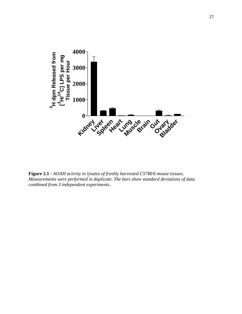
37
Kidney
Liver
SpleenHea
rtLung
MuscleBrai
nGut
Ovary
Bladder
0
1000
2000
3000
40003 H
dpm
Rel
ease
d fr
om[3 H
/14C
] LPS
per
mg
Tiss
ue p
er H
our
Figure 2.1 - AOAH activity in lysates of freshly harvested C57Bl/6 mouse tissues. Measurements were performed in duplicate. The bars show standard deviations of data combined from 3 independent experiments.
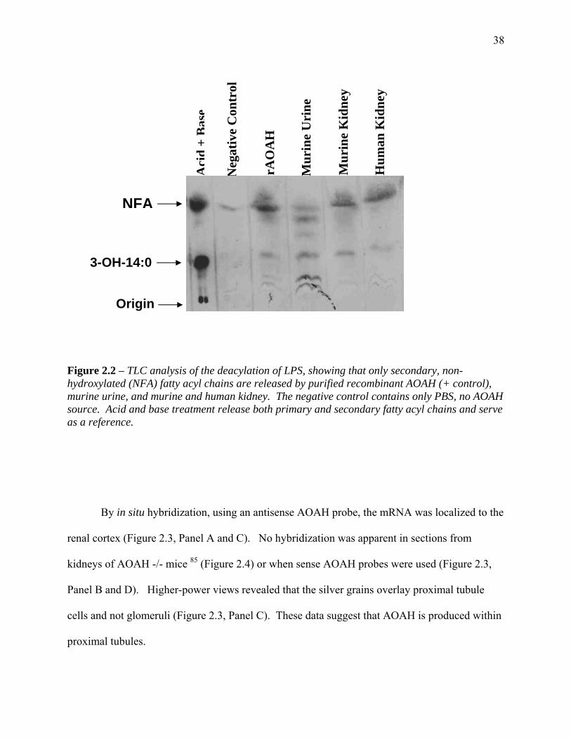
38
Mur
ine
Uri
ne
Aci
d+
Bas
e
Neg
ativ
e C
ontr
ol
rAO
AH
Mur
ine
Kid
ney
NFA
3-OH-14:0
Origin
Hum
an K
idne
y
Figure 2.2 – TLC analysis of the deacylation of LPS, showing that only secondary, non-hydroxylated (NFA) fatty acyl chains are released by purified recombinant AOAH (+ control), murine urine, and murine and human kidney. The negative control contains only PBS, no AOAH source. Acid and base treatment release both primary and secondary fatty acyl chains and serve as a reference.
By in situ hybridization, using an antisense AOAH probe, the mRNA was localized to the
renal cortex (Figure 2.3, Panel A and C). No hybridization was apparent in sections from
kidneys of AOAH -/- mice 85 (Figure 2.4) or when sense AOAH probes were used (Figure 2.3,
Panel B and D). Higher-power views revealed that the silver grains overlay proximal tubule
cells and not glomeruli (Figure 2.3, Panel C). These data suggest that AOAH is produced within
proximal tubules.

39
Figure 2.3 - Localization of AOAH mRNA in murine kidney by in situ hybridization. A and C, antisense probe. B and D, sense probe. The antisense probe hybridized to the renal cortex (panel A) and was found over cortical tubules (panel C). The bars indicate 500 µm (A and B) or 20 µm (C and D). This experiment was repeated 3 times; each experiment used kidney from a different mouse.
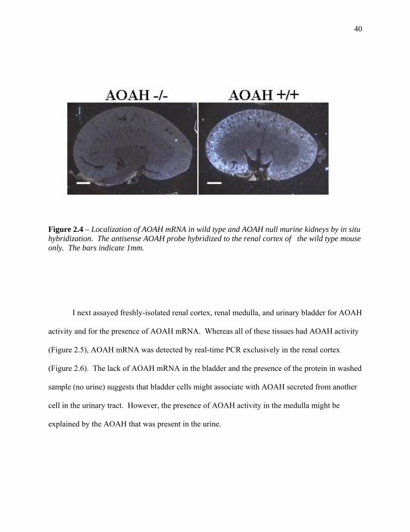
40
Figure 2.4 – Localization of AOAH mRNA in wild type and AOAH null murine kidneys by in situ hybridization. The antisense AOAH probe hybridized to the renal cortex of the wild type mouse only. The bars indicate 1mm.
I next assayed freshly-isolated renal cortex, renal medulla, and urinary bladder for AOAH
activity and for the presence of AOAH mRNA. Whereas all of these tissues had AOAH activity
(Figure 2.5), AOAH mRNA was detected by real-time PCR exclusively in the renal cortex
(Figure 2.6). The lack of AOAH mRNA in the bladder and the presence of the protein in washed
sample (no urine) suggests that bladder cells might associate with AOAH secreted from another
cell in the urinary tract. However, the presence of AOAH activity in the medulla might be
explained by the AOAH that was present in the urine.

41
Cortex Medulla Bladder0
25
50
75
100R
elat
ive
AOAH
Act
ivity
(per
mg
tota
l pro
tein
)
**
* Figure 2.5 - AOAH activity in lysates of renal cortex, medulla, and washed bladder. Each bar shows the mean and SE of 3 or more measurements. The mean activity per mg protein in 7 bladder samples was 2.5% of the activity in total kidney lysates. Statistics were performed with GraphPad software using a paired, two-tailed, t test. * p=0.159, ** p=0.0002.
Cortex Medulla Bladder0
25
50
75
100
mR
NA,
AO
AH/G
APD
H(A
rbitr
ary
units
) Figure 2.6 - Real-time PCR analysis of AOAH and GAPDH mRNA in renal cortex, medulla, and bladder. GAPDH mRNA was used as the reference control. The experiment was performed in duplicate and repeated twice, using tissues from different mice, with similar results.
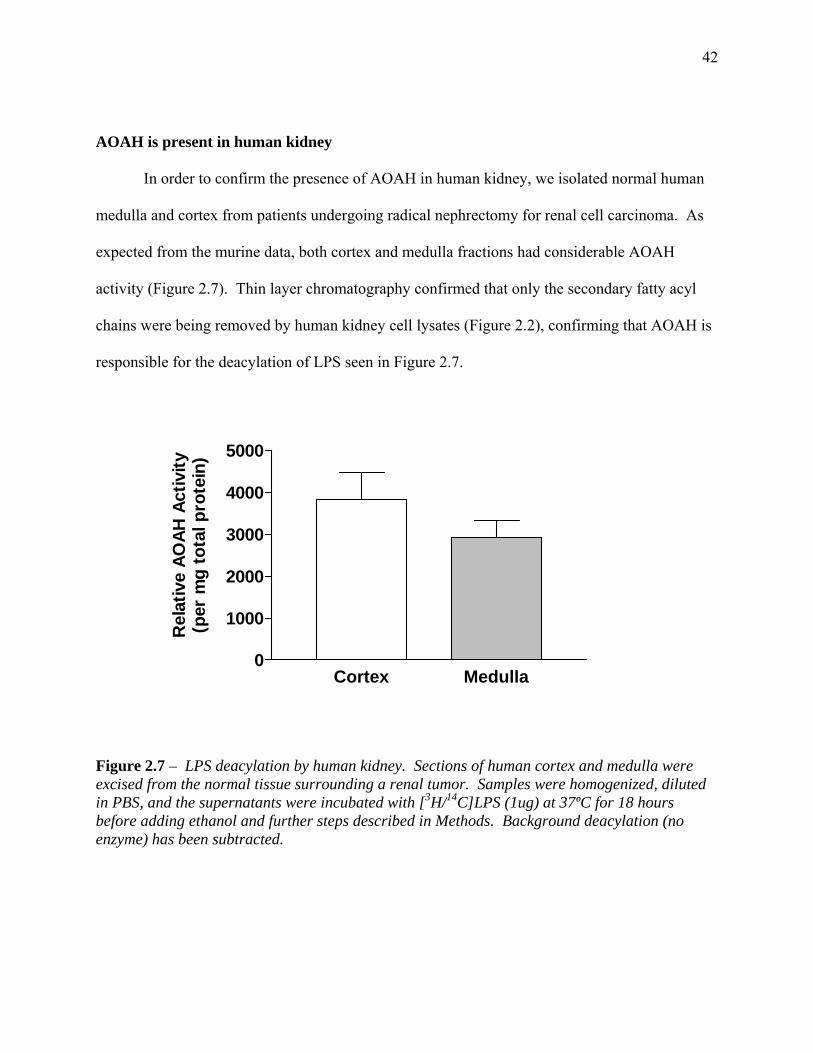
42
AOAH is present in human kidney In order to confirm the presence of AOAH in human kidney, we isolated normal human
medulla and cortex from patients undergoing radical nephrectomy for renal cell carcinoma. As
expected from the murine data, both cortex and medulla fractions had considerable AOAH
activity (Figure 2.7). Thin layer chromatography confirmed that only the secondary fatty acyl
chains were being removed by human kidney cell lysates (Figure 2.2), confirming that AOAH is
responsible for the deacylation of LPS seen in Figure 2.7.
Cortex Medulla0
1000
2000
3000
4000
5000
Rel
ativ
e AO
AH A
ctiv
ity(p
er m
g to
tal p
rote
in)
Figure 2.7 – LPS deacylation by human kidney. Sections of human cortex and medulla were excised from the normal tissue surrounding a renal tumor. Samples were homogenized, diluted in PBS, and the supernatants were incubated with [3H/14C]LPS (1ug) at 37ºC for 18 hours before adding ethanol and further steps described in Methods. Background deacylation (no enzyme) has been subtracted.
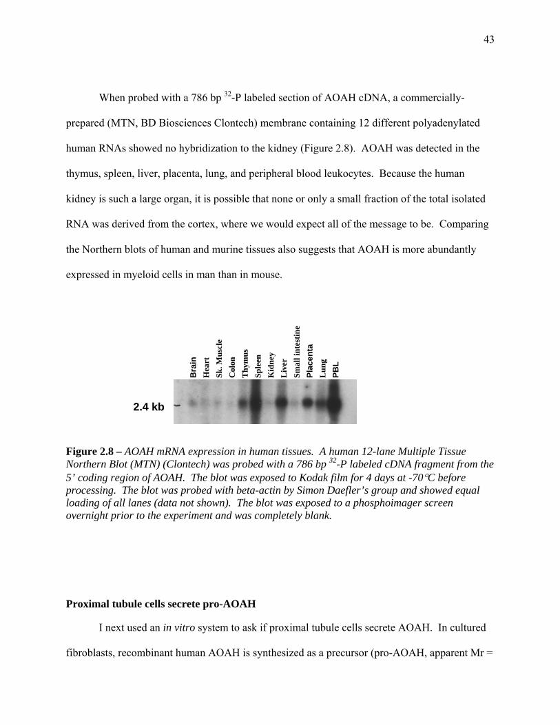
43
When probed with a 786 bp 32-P labeled section of AOAH cDNA, a commercially-
prepared (MTN, BD Biosciences Clontech) membrane containing 12 different polyadenylated
human RNAs showed no hybridization to the kidney (Figure 2.8). AOAH was detected in the
thymus, spleen, liver, placenta, lung, and peripheral blood leukocytes. Because the human
kidney is such a large organ, it is possible that none or only a small fraction of the total isolated
RNA was derived from the cortex, where we would expect all of the message to be. Comparing
the Northern blots of human and murine tissues also suggests that AOAH is more abundantly
expressed in myeloid cells in man than in mouse.
2.4 kb
Bra
in
Hea
rt
Sk. M
uscl
e C
olon
T
hym
us
Sple
en
Kid
ney
Liv
er
Smal
l int
estin
e Pl
acen
ta
Lun
g PB
L
Figure 2.8 – AOAH mRNA expression in human tissues. A human 12-lane Multiple Tissue Northern Blot (MTN) (Clontech) was probed with a 786 bp 32-P labeled cDNA fragment from the 5’ coding region of AOAH. The blot was exposed to Kodak film for 4 days at -70°C before processing. The blot was probed with beta-actin by Simon Daefler’s group and showed equal loading of all lanes (data not shown). The blot was exposed to a phosphoimager screen overnight prior to the experiment and was completely blank. Proximal tubule cells secrete pro-AOAH
I next used an in vitro system to ask if proximal tubule cells secrete AOAH. In cultured
fibroblasts, recombinant human AOAH is synthesized as a precursor (pro-AOAH, apparent Mr =
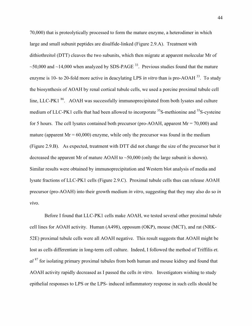
44
70,000) that is proteolytically processed to form the mature enzyme, a heterodimer in which
large and small subunit peptides are disulfide-linked (Figure 2.9.A). Treatment with
dithiothreitol (DTT) cleaves the two subunits, which then migrate at apparent molecular Mr of
~50,000 and ~14,000 when analyzed by SDS-PAGE 33. Previous studies found that the mature
enzyme is 10- to 20-fold more active in deacylating LPS in vitro than is pro-AOAH 33. To study
the biosynthesis of AOAH by renal cortical tubule cells, we used a porcine proximal tubule cell
line, LLC-PK1 86. AOAH was successfully immunoprecipitated from both lysates and culture
medium of LLC-PK1 cells that had been allowed to incorporate 35S-methionine and 35S-cysteine
for 5 hours. The cell lysates contained both precursor (pro-AOAH, apparent Mr = 70,000) and
mature (apparent Mr = 60,000) enzyme, while only the precursor was found in the medium
(Figure 2.9.B). As expected, treatment with DTT did not change the size of the precursor but it
decreased the apparent Mr of mature AOAH to ~50,000 (only the large subunit is shown).
Similar results were obtained by immunoprecipitation and Western blot analysis of media and
lysate fractions of LLC-PK1 cells (Figure 2.9.C). Proximal tubule cells thus can release AOAH
precursor (pro-AOAH) into their growth medium in vitro, suggesting that they may also do so in
vivo.
Before I found that LLC-PK1 cells make AOAH, we tested several other proximal tubule
cell lines for AOAH activity. Human (A498), opposum (OKP), mouse (MCT), and rat (NRK-
52E) proximal tubule cells were all AOAH negative. This result suggests that AOAH might be
lost as cells differentiate in long-term cell culture. Indeed, I followed the method of Triffilis et.
al 87 for isolating primary proximal tubules from both human and mouse kidney and found that
AOAH activity rapidly decreased as I passed the cells in vitro. Investigators wishing to study
epithelial responses to LPS or the LPS- induced inflammatory response in such cells should be

45
aware of the presence (or likely absence) of AOAH, a LPS-deacylating enzyme, in their cell
lines.
Precursor
Apparent Molecular Weights (in KDa)
A Mature Figure 2.9.A – Diagram of AOAH biosynthesis, showing the conversion of the precursor (pro-AOAH) into mature AOAH. Proteolysis removes the leader and pro-peptides and cleaves the precursor into a disulfide-linked heterodimer.
B
Figure 2.9. B – Production of 35S-AOAH by porcine proximal tubule cells in vitro. Labeled AOAH was immunoprecipitated with either anti-murine AOAH monoclonal antibody 2F3-2A4 (+), or control IgG (-) as described in Methods and studied by SDS-PAGE (+/- DTT) and autoradiography. M = media, L = lysate.
46
Figure 2.9.C – Western blot analysis of AOAH production by LLC-PK1 cells in vitro. Media and lysate fractions were immunopreciptated with antibody 2F3-2A4, run on SDS-PAGE in the presence of DTT, and analyzed by Western blot as described in Methods. The ~55 kDa band seen in the lysate and beads alone lane is the heavy chain of the IgG used to immunoprecipitate AOAH. The LLC-PK1 Media lane is underexposed and the cross-reaction (antibody) is not detected.
AOAH is found in voided urine.
Consistent with secretion of AOAH by cortical tubule cells in vivo, AOAH activity was
present in freshly voided murine urine (Figure 2.10, 2.11). Urine from AOAH null mice was
not active (Figure 2.10), indicating that the deacylating activity detected in wild-type urine is due
to AOAH. Thin layer chromatographic analysis of the above reaction revealed that only the
secondary, non-hydroxylated fatty acids were removed by wild type urine, in keeping with
AOAH being responsible for the deacylation of LPS (Figure 2.2). The reaction mixture used in

47
AOAH activity assays contains Triton-X, a detergent that is not present in the urinary system. In
order to simulate in vivo conditions better, I assayed freshly voided murine urine for its ability to
deacylate 3H-[LPS] substrate in the absence of detergent. My results indicate that urine is able to
deacylate radiolabeled LPS substrate under these conditions (Figure 2.11), suggesting that
soluble AOAH may act on extracellular LPS within the urine.
+/+ -/-
0
500
1000
1500
2000
3 H dp
m R
elea
sed
from
[3 H/14
C] L
PS b
y10
ul U
rine
***
Figure 2.10 – LPS deacylation by urine from AOAH +/+ and -/- mice. Urine (10 µl) was added to 490 µl AOAH reaction mixture containing 1 µg [3H/14C]LPS and incubated at 37°C for 18 hrs before adding ethanol and further steps as described in Methods. Statistics were performed with GraphPad software using a two-tailed, paired t test (*** p<0.001).

48
37°C 4°C
0250050007500
1000012500
3 H dp
m R
elea
sed
from
[3 H/14
C] L
PS b
y10
ul U
rine
**
Figure 2.11- LPS deacylation by murine urine. Fresh urine (10µl) was incubated with [3H/14C]LPS (0.5µg) at 4°C (control) or 37°C for 18 hrs. AOAH reaction mixture was then added to provide protein for co-precipitation of intact LPS, followed by ethanol. The remaining steps are described in Methods. Statistics were performed with GraphPad software using a two-tailed, paired t test (*** p= 0.0017).
To determine if the AOAH in the urine is the precursor or the mature form, I collected
urine from both wild type and AOAH null mice, precipitated AOAH with an anti-mAOAH
monoclonal antibody, and performed a Western blot as described in Methods. Urine AOAH
was found to be in the mature (Mr~60,000, reduced 50,000) form (Figure 2.12). Although the
low pH of the urine may create an environment that favors proteolytic cleavage of pro-AOAH as
it descends through the urinary tract, I was unable to show that urine from AOAH -/- mice can
cleave pro-AOAH in vitro (data not shown). Alternatively, epithelial cells might process the
precursor and return the mature enzyme to the urine, or mature AOAH may be secreted by
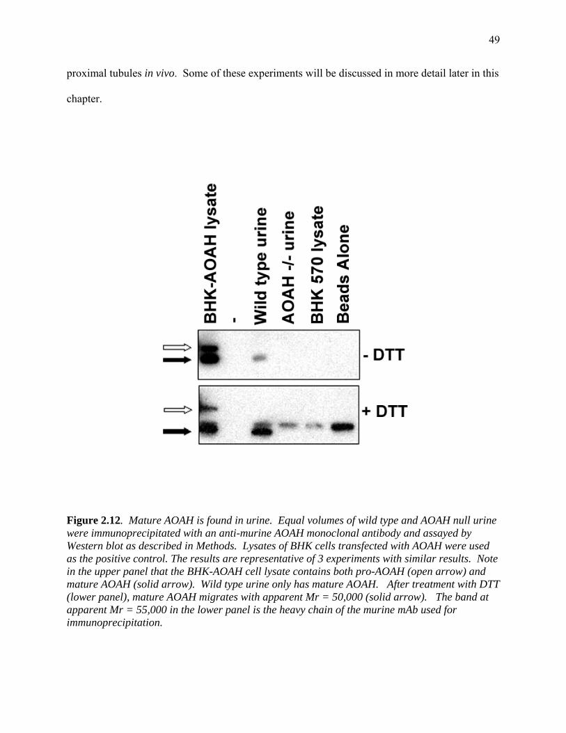
49
proximal tubules in vivo. Some of these experiments will be discussed in more detail later in this
chapter.
Figure 2.12. Mature AOAH is found in urine. Equal volumes of wild type and AOAH null urine were immunoprecipitated with an anti-murine AOAH monoclonal antibody and assayed by Western blot as described in Methods. Lysates of BHK cells transfected with AOAH were used as the positive control. The results are representative of 3 experiments with similar results. Note in the upper panel that the BHK-AOAH cell lysate contains both pro-AOAH (open arrow) and mature AOAH (solid arrow). Wild type urine only has mature AOAH. After treatment with DTT (lower panel), mature AOAH migrates with apparent Mr = 50,000 (solid arrow). The band at apparent Mr = 55,000 in the lower panel is the heavy chain of the murine mAb used for immunoprecipitation.

50
Bladder cells take up pro-AOAH
In order to test the hypothesis that secreted AOAH could be used by non-expressing
bladder cells, I added medium containing pro-AOAH (from confluent AOAH-transfected BHK
570 cells) to cultures of T24 human bladder cells. Binding of AOAH to T24 bladder cells was
readily detected, and it could be blocked by adding mannose-6-phosphate (M6P) or ammonium
chloride but not glucose-6-phosphate (G6P) or mannose (Figure 2.13. Panels A and B). These
results strongly suggest that the uptake of AOAH, a heavily N-glycosylated protein 32;33, is
mannose-6-phosphate receptor-dependent. Since the specific activity of the enzyme (3H-fatty
acids released from [3H/14C]LPS per µg AOAH protein) was ~50-fold higher in the T24 cell
lysates than in the BHK medium (Figure 2.13, panel C), it is likely that pro-AOAH is processed
to mature AOAH by the T24 cells 33.
Previous work by Staab and colleagues found that the maturation of AOAH is dependent
upon low pH 33. Her data, obtained by analyzing the cell lysate fractions of AOAH-transfected
BHK 570 cells before and after treatment with 10 mM ammonium chloride (NH4Cl) (an agent
known to raise the intracellular pH), showed that ammonium chloride treatment blocked the
expression of mature AOAH. In apparent disagreement with these data, I found that ammonium
chloride was able to block the association of AOAH with T24 cells, but had no effect on the
maturation of the enzyme (specific activity, Figures 2.13. Panel C). Since AOAH uptake is
mediated by the M6P receptor and expression of the M6P receptor on the cell surface may be
disrupted by ammonium chloride 88;89 ammonium chloride probably prevents the internalization
of AOAH, but does not interfere with maturation by proteolytic leavage. This interpretation
suggests that AOAH maturation may not be acid-dependent.

51
We also found that other cell lines (IMCD3 mouse collecting duct, 5637 human bladder,
and CHO-CD14 chinese hamster ovary cells) are able to take up pro-AOAH (from either LLC-
PK1 or AOAH transfected BHK 570 cells) in a M6P-dependent fashion (Figure 2.14).

52
B
Ficomacthexthreacus
A
****ng
Cel
l-ass
ocia
ted
AO
AH
Pro
tein
**
Rel
ativ
e A
OA
H A
ctiv
ity
**C
Rel
ativ
e Sp
ecifi
c A
ctiv
ity
gure 2.13 – A. Uptake of pro-AOAH by T24 bladder cells after incubation with AOAH-ntaining medium for 5 hrs in the presence or absence of 10 mM M6P, G6P, NH4Cl or annose. Washed cells were lysed and AOAH protein was assayed by ELISA. B. AOAH tivity in T24 lysates, expressed relative to the activity observed in cells that took up AOAH in e absence of inhibitor. Each bar shows the mean and SE of data from 4 independent periments. C. AOAH specific activity (activity/ng protein) in T24 cell lysates. Compared with e AOAH added in the medium, cell-associated AOAH had much greater specific activity, flecting its activation by the T24 cells. M6P inhibited AOAH binding (A) but did not prevent tivation of the cell-associated AOAH (C). Statistics were performed with GraphPad software ing a two-tailed, paired t test (*p< 0.1, **p< 0.001, ***p< 0.0001).
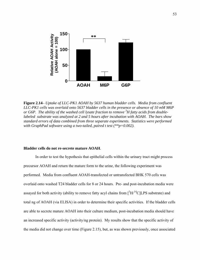
53
AOAH M6P G6P0
50
100
150
Rel
ativ
e AO
AH A
ctiv
ity(A
OAH
alo
ne =
100
) ** Figure 2.14– Uptake of LLC-PK1 AOAH by 5637 human bladder cells. Media from confluent LLC-PK1 cells was overlaid onto 5637 bladder cells in the presence or absence of 10 mM M6P or G6P. The ability of the washed cell lysate fraction to remove 3H fatty acids from double-labeled substrate was analyzed at 2 and 5 hours after incubation with AOAH. The bars show standard errors of data combined from three separate experiments. Statistics were performed with GraphPad software using a two-tailed, paired t test (**p=0.002). Bladder cells do not re-secrete mature AOAH.
In order to test the hypothesis that epithelial cells within the urinary tract might process
precursor AOAH and return the mature form to the urine, the following experiment was
performed. Media from confluent AOAH-transfected or untransfected BHK 570 cells was
overlaid onto washed T24 bladder cells for 8 or 24 hours. Pre- and post-incubation media were
assayed for both activity (ability to remove fatty acyl chains from [3H/14C]LPS substrate) and
total ng of AOAH (via ELISA) in order to determine their specific activities. If the bladder cells
are able to secrete mature AOAH into their culture medium, post-incubation media should have
an increased specific activity (activity/ng protein). My results show that the specific activity of
the media did not change over time (Figure 2.15), but, as was shown previously, once associated

54
with the T24 cells, AOAH had a great increase in specific activity. These data suggest that while
bladder cells are able to mature AOAH, they do not return it to their medium (or that the culture
conditions do not allow such release). These data do not rule out the possibility that another cell
type in the urinary tract is responsible for maturing and re-secreting AOAH or that bladder cells
may re-secrete AOAH in vivo.
Pre-incu
bation m
edia
Post-incu
bation m
edia
T24 ce
ll-ass
ociated
T24 ce
lls al
one0
25000
50000
75000
100000
3 H fa
tty
acid
s re
leas
edfr
om [3 H
/14C
]LPS
per
ng
AOAH
pro
tein
Figure 2.15 – Bladder cells do not secrete mature AOAH into their medium in vitro. T24 bladder cells were washed with PBS and incubated with AOAH-transfected or untransfected BHK 570 cell media for 8 or 24 hours at 37ºC. Media and lysates were assayed for AOAH activity and total AOAH protein (ELISA) as described in Methods. The experiment was done in duplicate and repeated twice with similar results.
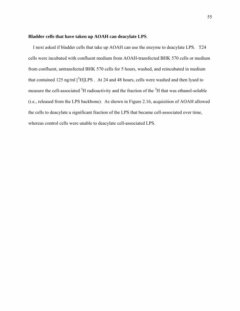
55
Bladder cells that have taken up AOAH can deacylate LPS. I next asked if bladder cells that take up AOAH can use the enzyme to deacylate LPS. T24
cells were incubated with confluent medium from AOAH-transfected BHK 570 cells or medium
from confluent, untransfected BHK 570 cells for 5 hours, washed, and reincubated in medium
that contained 125 ng/ml [3H]LPS . At 24 and 48 hours, cells were washed and then lysed to
measure the cell-associated 3H radioactivity and the fraction of the 3H that was ethanol-soluble
(i.e., released from the LPS backbone). As shown in Figure 2.16, acquisition of AOAH allowed
the cells to deacylate a significant fraction of the LPS that became cell-associated over time,
whereas control cells were unable to deacylate cell-associated LPS.
56
A
24 480
10
20
30
40
Hours
Cel
l-as
soci
ated
[3 H] L
PS,
dpm
per
µg
Cel
l Pro
tein
B
0 12 24 36 48
-100
0
100
200
300
400
Time (hours)
3 H d
pm R
elea
sed
perµ
gC
ell P
rote
in
*
***
Figure 2.16 – AOAH confers LPS-deacylating activity to bladder cells. T24 cells were allowed to take up AOAH for 5hrs, washed, and then incubated with 3H-LPS (125 ng/ml) for the times indicated. Control cells were incubated with medium that did not contain AOAH. Whereas control and AOAH-containing cells took up similar amounts of 3H-LPS (A), only the AOAH-containing cells removed 3H-fatty acids from the LPS backbone (B). The data represent combined results of three separate experiments; the error bars represent 1 SEM. Solid squares and bars, T24 cells with AOAH. Open circles and bars, control T24 cells. Statistics were performed with GraphPad software using a two-tailed, paired t test (***p = 0.0009, *p = 0.0147).

57
METHODS
Chemicals. Unless otherwise indicated, chemicals were purchased from Sigma-Aldrich
Chemical Co, St. Louis, MO.
Mouse strains. Specific pathogen-free mice were housed in the UT Southwestern Animal
Resource Center and fed a standard diet. ICR (Harlan, Indianapolis, IN), C57Bl/6 (Harlan) or
129S6/SvEvTac (Taconic, Germantown, NY) mice were used. AOAH null 129 and C57Bl/6
mice were produced as described by Lu et al 85.
Northern Analysis. A Mouse Multiple Tissue Northern Blot (BD Biosciences Clontech) was
probed with a 32P-radiolabeled 1 kb (Asp718 to HindIII) cDNA fragment of the 5’ coding region
of mouse AOAH cDNA (Genebank # AF018172). The blot was stripped and re-probed with a
350 bp cDNA probe from the 3’ (AhdI to XbaI) non-coding region of mouse AOAH as above.
The blot was then stripped and reprobed with a 500 bp EcoRI to EcoRI fragment of the murine β-
actin cDNA (a kind gift from I. Shimomura) 90. A Human Multiple Tissue Northern Blot (BD
Biosciences Clontech) was probed with a 786 bp 32P- labeled cDNA fragment from the 5’ coding
region of human AOAH.
AOAH Activity Assays. Three month-old female C57Bl/6 mice were anesthetized (ketamine-
acepromazine), sacrificed by cervical dislocation, then selected tissues were rinsed with ice-cold
saline and weighed. Each tissue was transferred to a microfuge tube that contained 500 µl lysis
buffer (0.2% triton X-100 in PBS, pH 7.2, with 0.5 µg/ml aprotinin, 0.5 µg/ml leupeptin, and 2.5

58
mM EDTA) on ice, and sonicated (Branson Sonifier 450, VWR, West Chester, PA) to complete
disruption (approximately 30 sec). The lysates were diluted 1:10 in sterile PBS and 10 µl was
assayed for AOAH activity at 37°C as previously described 91, using a double-labeled [3H-acyl
chains/14C-glucosamine backbone] S. typhimurium LPS as substrate. Mouse urine was assayed
using the same reaction mixture 91 or by adding 0.5 µg double-labeled S. typhimurium LPS
substrate and sodium acetate, pH 5 (final concentration = 3 mM) to 10 µl urine (final volume =
15µl) and incubating at either 4°C or 37°C for 18 hrs.
Thin-layer chromatography. Twenty µls murine urine and 100 µls human or murine kidney
lysate (sonicated briefly in 0.1% Triton X-100 in PBS) was incubated in 400 µls AOAH reaction
mix plus approximately 1µg purified [3 H]LCD25-O9 LPS and incubated overnight at 37°C. A
chloroform/methanol extraction was performed to isolate the soluble fatty acyl chains. Briefly,
450 µl of the overnight reaction was placed in a glass tube to which 3.75x volume
chloroform:methanol 1:2 (v:v) was added. Samples were vortexed, incubated at RT for 10 min.,
and extracted in 1.25 volume chloroform, 1 volume water, and 1/100th volume glacial acetic
acid. The chloroform layer was removed, washed three times and dried under argon. Samples
were resuspended in 50 µl chloroform:methanol 1:1 (v:v) and loaded onto a silica gel G TLC
plate. Primary and secondary fatty acids (for reference) were obtained by hydrolyzing LPS
sequentially with 4 M HCl and 4 M NaOH. The hydrolyzed fatty acids were extracted into
chloroform, dried under argon, resuspended in 50µl chloroform:methanol 1:1 (v:v) and loaded
onto a silica G plate. The TLC was run in petroleum ether:diethyl ether:acetic acid; 70:30:1.
The plate was sprayed with 3H-enhance, incubated at -70°C for one week and visualized by
autoradiography using Kodak film.

59
Generation of AOAH null mice. C3H/HeN and C57Bl/6 AOAH knockout mice were
generated as follows. The murine AOAH gene was disruption in 129/SvEvTac ES cells by
inserting a neomycin resistance gene into the first exon of AOAH, eliminating a 705 bp region
that encodes untranslated mRNA, the translation start site, the leader and pro-peptide sequences,
and 41 amino acids of the small subunit of AOAH. Mouse DNA was screened by Southern blot,
using an EcoRI-BamHI probe derived from the 5’ genomic sequence upstream of the long arm of
the targeting vector. 129/SvEvTac heterozygous mice were crossbred to the F2 generation at
which time they were crossbred with wild type C57Bl/6 and C3H/HeN mice. Heterozygous
mice from each backcross were bred through 8 generations, so that only 0.39% of the genes were
of 129/SvEvTac origin. Progeny of F8 homozygote knockouts and wild type littermates were
used in all experiments presented in this thesis.
In situ hybridization and riboprobes. A 1 kb fragment of the 5’ coding region of AOAH
cDNA (5’-Asp718 to 3’-HindIII) was inserted into pBlueScript KS+ (Stratagene, La Jolla, CA) at
the Asp718 and HindIII sites. The plasmid was linearized with BglII and a 650 bp antisense
riboprobe was generated by in vitro transcripton from the T7 promoter using the Ambion
Maxiscript kit (Ambion, Austin, TX). A 517 bp sense riboprobe was similarly generated using
the T3 promoter according to the manufacturer’s instructions. Briefly, 300 ng of template DNA
was transcribed with T7 (antisense) or T3 (sense) polymerase in the presence of 200 µCi of [α -
35S]-UTP (800 Ci/mmol) (Amersham, Piscataway, NJ) for 1 hr at 37ºC. DNAse I was added for
15 min at 37ºC and all enzymatic activity was then terminated with 1 µl of 0.5M EDTA. The
volume was brought to 75 µl with DEPC water and 50 µl was run through a Rnase-free G-50
60
column (Roche, Basel Switzerland). The probes were stored at -80ºC and used within 2 days of
preparation.
Female ICR mice (Harlan) were anesthetized (ketamine-acepromazine) and tissues were
isolated following transcardial perfusion with cold heparinized DEPC-saline and then with
chilled 4% formaldehyde/DEPC-PBS, pH 7.4 (freshly prepared from paraformaldehyde).
Samples were incubated in 4% formaldehyde for 16 hrs and then transferred to sterile DEPC-
saline. Kidneys were dehydrated and paraffin-embedded, and 4 µm sections were placed onto
microscope slides treated with Vectabond (Vector Laboratories, Burlingame, CA). Slides were
stored desiccated at 4ºC until use. In situ hybridization was preformed as previously described
92, using the riboprobes described above. The in situ hybridization using wild type and AOAH
null mice were performed as above using 129 female mice.
Real-time PCR. Total RNA was isolated from bladders and from pooled renal cortex and
medulla fractions obtained from wild type mice (RNAqueous Kit, Ambion, Austin, TX). A
region of the AOAH cDNA was amplified using primers Seq_mAOAH-ex12F
(CCAACTCTCTGGTGTAACTGGATTT) and Seq_mAOAH-ex12R
(TCTCAAACGATGGTAAATGGATTTT). TaqMan® MGB probe (FAMTM dye-labeled)
ACGAGTGGAATTGAAG and primers were designed and synthesized by Applied Biosystems
(Foster City, CA). Plasmid pMF612, which contains murine AOAH cDNA, was used as a
reference molecule for the standard curve calculation. TaqMan® Rodent GAPDH Control
Reagents were used to measure GAPDH gene expression. All real-time PCR reactions were
performed by M. Lu in a 25 µl mixture with Taqman one step RT-PCR Master Mix Reagents Kit
on the ABI PRISM® 7700 Sequence Detection System.
61
Cell culture. LLC-PK1 porcine proximal tubule cells (ATCC, CL-101) were cultured in low-
glucose DMEM (Invitrogen, Carlsbad, CA), 5% heat-inactivated FCS (Hyclone, Logan, UT),
penicillin, streptomycin, glutamine (PSG, Invitrogen) at 37ºC in an atmosphere of 5% CO2. T24
human bladder cells (ATCC, HTB-4) were cultured in Vitacell McCoy's 5a Medium (ATCC)
supplemented with 10% FCS and 2% PSG at 37ºC, 5%CO2. AOAH-transfected BHK 570 cells
33 and untransfected BHK cells were cultured in DMEM with glutamine and 4.5 g/L glucose
(Fisher Scientific), 10% FCS, and 2% PSG as above. 5637 human bladder cells (HTB-9) and
CHO K1 (CCL-61) were obtained from ATCC and IMCD3 cells were a kind gift of Dr.
Christopher Lu (UT Southwestern).
Antibodies. Rabbit anti-human AOAH polyclonal antibodies (E553) bind porcine, but not
murine, AOAH. The available murine monoclonal antibodies to human AOAH also do not bind
murine AOAH. After numerous unsuccessful attempts at producing antibodies to murine
AOAH in rabbits, we (W. Lai, Dept. Pathology) immunized AOAH -/- mice thrice, at monthly
intervals, with 100 µg of a plasmid that expressed the murine AOAH cDNA from a CMV
promoter. We then administered approximately 109 plaque-forming units of an adenoviral vector
that produces murine AOAH, prepared as previously described 93. When the mice still did not
have high anti-AOAH antibody titers, we (Mei Zhang) expressed the large subunit of murine
AOAH in E. coli using the pET-30 LIC vector (Novagen); inclusion bodies were subjected to
preparative SDS-PAGE and bands containing recombinant AOAH were cut out and injected,
emulsified in complete or incomplete Freund adjuvant, at monthly intervals (3 injections).
Although the antibody titers remained low, we fused spleens from immunized mice with SP 2/0-
62
IL-6 myeloma cells and screened the resulting hybridomas to isolate anti-murine AOAH IgG
antibodies. Approximately one liter of hybridoma supernatant was purified on a protein A
Sepharose column and concentrated using an Amicon Centricon YM-100 (Millipore, Bedford,
MA). The purified antibody, 2F3-2A4, is a mouse anti-mouse AOAH monoclonal antibody of
the IgG2a, kappa isotype.
Immunoprecipitation of biosynthetically radiolabeled AOAH. One million LLC-PK1 cells
were seeded into Costar brand 75 cm2 tissue culture-treated flasks (Corning, Acton, MA). Two
days later, the cells were washed twice with pre-warmed, sterile PBS, pH 7.2, and incubated with
methionine- and cysteine-free DMEM (Sigma) at 37ºC, 5% CO2 for 20 to 30 min. 1 mCi 35S-
labeled methionine and cysteine (1175 Ci/mmol) (ICN, Costa Mesa, CA) was then added to 5 ml
pre-warmed, methionine- and cysteine-free DMEM with 50 µl dialyzed heat-inactivated FCS
(Hyclone, Logan, UT) and overlaid onto washed LLC-PK1 cells. Incubation was continued at
37°C in 5% CO2 for 5 hrs. The flasks were then placed on ice, the medium was removed and
saved at 4°C, and the monolayers were washed thrice with PBS, pH 7.2. I then added 0.6 ml
RIPA buffer (150 mM NaCl, 1% NP-40, 0.5% sodium deoxycholate, 0.1% SDS, 50 mM Tris-Cl
pH 8.0, 10 mM PMSF and 2.5 mM EDTA) on ice, rocking occasionally for 20 min. The cells
were pelleted by centrifugation (5 min, at 4°C) and the lysate supernatants were transferred to
fresh microfuge tubes. The media and lysates were pre-cleared with 50 µl of a 50% slurry of
Pansorbin (CalBiochem, La Jolla, CA) that had been allowed to bind non-immune rabbit IgG.
Specific immunoprecipitation was then performed using rabbit anti-human AOAH IgG bound to
protein A Sepharose CL-4B (Amersham Biosciences, Uppsla, Sweden).

63
Immunoprecipitation and western blot of murine urine. C57Bl/6 mouse urine was stored at
4°C overnight after adding protease inhibitors (1 mM PMSF and 2 µg/ml leupeptin). Protein A
Sepharose beads were washed 5 times with IP wash buffer (0.1% Triton X-100, 50 mM Tris-Cl
pH 7.4, 300 mM NaCl, 5 mM EDTA, 0.02% sodium azide, and 1 mM PMSF, 2 µg/ml
leupeptin), and resuspended in 1 ml wash buffer on ice. The pH of the urine was adjusted to 7.6
with 1 M Tris, pH 8.0, and brought to a final concentration of 0.1% SDS, 0.1% Triton X-100,
and 1 mM PMSF and 10 µM leupeptin. Three ml urine was pre-cleared at 4°C for 1 h by adding
150 µl of a 50% slurry of protein A Sepharose in IP wash buffer and gentle rotation. The
samples were spun and the supernatants were poured into fresh 15 ml conical tubes to which 300
µl of protein A Sepharose (pre-bound with 20 µg mouse anti-murine AOAH mA2F3-2A4 in a
50% slurry) and 30 µg BSA were added. After gentle rotation for 1 hr at 4°C, the beads were
washed 4 times with IP wash buffer and once with PBS. Twenty µl of sample buffer (100 mM
Tris-Cl, pH 6.8, 4% SDS, 0.2% bromophenol blue, and 20% glycerol) was added and the
samples were immediately heated (100°C, 5 min) and loaded onto a 4-20% gradient gel (BioRad,
Hercules, CA). The gel was run at 100V for approximately 2 hours and then transferred to a
ImmobilonTM-P membrane (Millipore, Bedford, MA) at 4°C overnight. The blot was blocked in
4% dry milk/TTBS (2 mM Tris-HCl, pH 7.5, 15 mM NaCl, 0.05% Tween 20) for one hr at room
temperature and probed with primary antibody 2F3-2A4 (4.5 µg/ml) for one hour at RT.
Following 5 washes in TTBS, the blot was incubated with peroxidase-conjugated sheep anti-
mouse IgG antibody (Amersham) for 1 hr at RT. The membrane was washed five times as
above, treated with SuperSignal West Pico ECL reagent (Pierce, Rockford, IL), and exposed to
Kodak film to visualize the bands.
64
Uptake and deacylation of LPS by bladder cells. T24 cells were seeded into 6 well culture
dishes (2 x 10⁵ cells per well). Two days later, media from confluent AOAH- transfected BHK
cells and untransfected BHK 570 cells were cleared by centrifugation and added to washed T24
cells in the presence or absence of 10 mM mannose-6-phosphate (M6P), 10 mM glucose-6-
phosphate (G6P), 10 mM D(+) mannose, or 10mM ammonium chloride. After 5 hr at 37°C, the
cells were washed 3 times with PBS and re-incubated with 0.8 ml of T24 cell media that
contained 100 ng E. coli LCD25 [3H]LPS, specific activity = 4200 dpm/ng 94. The cells in two
wells were immediately lifted and placed at 4°C. The remaining cells were incubated at 37°C,
5% CO2 for 1 hr, washed twice with PBS, and re-incubated with fresh T24 cell media for 24 and
48 hrs. Pelleted cells were washed once with PBS and lysed in 0.6 ml PBS, 0.1% Triton X-100.
0.25 ml was removed to a fresh microfuge tube and the insoluble [3H]LPS was removed by
centrifugation after adding 0.625% Triton X-100, 1.25 mg/ml BSA, and 1 ml 100% ethanol and
incubation at –20°C for 20 min. The radioactivity in the ethanol-soluble fraction and the original
lysate was counted (Packard Tri-carb 2100TR, PerkinElmer, Boston, MA) and analyzed to
obtain the percentage of the 3H dpm that was released from the [3H]LPS over time. Protein
assays were done on cell lysates by using the Bradford method (BioRad).
AOAH ELISA. Ninety-six well, square-bottom, NUNC brand Maxisorb ELISA plates (Fisher
Scientific) were coated with 0.5 µg/ml 3C5 (8F8) monoclonal mouse anti-human AOAH
antibody in 0.1M NaHCO3 overnight at 4°C. Sample wells were washed 3 times in PBST
(0.005% Tween 20 in PBS, pH 7.2) and blocked with 1% milk/PBST for 1 hr at 37°C. Samples
and standards were diluted in 1% milk/PBST and incubated at 4°C overnight or at 37°C for 2
hrs. After washing, secondary antibody (E553, purified polyclonal rabbit anti-human AOAH,

65
final concentration 10 µg/ml in 1% milk/PBST) was added for one hour at 37°C. After washing
5 times, the tertiary antibody (biotin goat anti-rabbit (Zymed, San Francisco, CA) was then
added (0.075 µg/ml) for 1 hr at 37°C. 2 µg/ml alkaline phosphatase-conjugated streptavidin
(Jackson Laboratories) in 1% milk/PBST was added to each well and incubated for 30 min at
37°C. The plate was washed as above and developed with alkaline phosphatase substrate (5 mM
Sigma 104 and 0.1M Sigma Alkaline buffer 221 in water). Optical densities were read at 405
nm and analyzed on an Elisa Plate Reader (MRX Revelation, Dynex Technologies, Chantilly,
Virginia).
DISCUSSION
Prior to these studies, AOAH had been found only in the myeloid cells (neutrophils,
monocyte-macrophages, and dendritic cells) that comprise the front-line innate defense against
invading bacteria and fungi. In the work presented here, AOAH mRNA was localized to renal
cortical tubule cells of the murine kidney. It was further shown that AOAH is secreted from
proximal tubule cells in vitro and present in freshly voided murine urine. The presence of
AOAH protein in the washed urinary bladder, combined with the absence of mRNA in this
tissue, suggests that the enzyme may be produced and secreted by cortical tubule cells in vivo
and “shared” with downstream cells in the urinary tract. In support of this hypothesis, the
secreted form of AOAH (pro-AOAH) was able to associate with bladder epithelial cells in vitro
and was used by these cells to deacylate cell-associated LPS. I believe this to be the first
description of enzyme sharing in the urinary tract. Although the urinary bladder has AOAH

66
activity, I did not attempt to immunolocalize AOAH within this tissue due to my inability to
localize AOAH in the kidney (where I expect AOAH to be the most concentrated). As discussed
in Chapter 4, once appropriate antibodies and/or immunhistochemical techniques are optimized,
immunolocalization experiments should be performed on the urinary bladder.
As expected, only the precursor form of AOAH was detected in the culture medium of
LLC-PK1, porcine proximal tubule cells. I therefore hypothesized that freshly voided murine
urine would contain the precursor form of AOAH. To my surprise, I found that voided murine
urine contains the mature, highly active form of AOAH. It is possible that the low pH of the
urine may create an environment that favors proteolytic cleavage of pro-AOAH as it descends
through the urinary tract. Alternatively, proximal tubules may secrete the mature form in vivo or
epithelial cells that line the urinary tract might process the precursor and return the mature
enzyme to the urine. Western blot analysis of pro-AOAH before and after incubation with
AOAH null urine suggested that urine alone is unable to mature AOAH, for pro-AOAH did not
mature in the presence of urine. T24 human bladder cells were able to mature pro-AOAH, but
only that which was cell-associated, for the culture medium did not accumulate any mature
AOAH over time. While this suggests that bladder cells are unable to release mature AOAH in
vitro, it does not rule out the possibility that bladder or other cells in the urinary tract mature
AOAH in vivo. I believe more experiments are necessary to determine the mechanism by which
mature AOAH is produced in urine.
In the original descriptions of AOAH, Staab et. al. reported a dependence on pH for the
maturation of AOAH. In her studies, the addition of 10 mM ammonium chloride to AOAH
transfected BHK570 cells completely ablated the presence of mature AOAH in cell lysate
fractions (mature AOAH has never been seen in the cell supernatant) 33. In apparent opposition,
67
my studies have shown that ammonium chloride treatment (either pre-treatment or given
concurrent with the AOAH source) does not prevent the maturation of the AOAH that has
associated with T24 bladder cells, although it does block association. The addition of
ammonium chloride disrupts the intracellular location of M6P receptors, sequestering them in
distinct endosomal compartments where they are not usually present. Not only does this
effectively remove M6P receptors from the cell surface but it also affects the intracellular
trafficking of newly synthesized proteins with M6P moieties 95. The data presented by Staab et.
al might now be interpreted differently. Newly synthesized and precursor AOAH that had been
secreted from the cell would be unable to traffic to their appropriate intracellular locations,
possibly inhibiting their maturation. These data do not address whether AOAH must first be
secreted from the cell in order to then re-enter and become mature, or whether some pro-AOAH
might be targeted to endosomes for maturation before secretion. It should also be noted that my
system for detecting maturation (activity assays and ELISA) is much more sensitive (based on
my experiments with IP/Western blot vs. ELISA) than the Staab et. al method (pulse chase of
radiolabeled [35S]methionine and [35S]cysteine) and therefore it is possible that there is some
maturation of AOAH present in the Staab et. al method that has gone undetected. Removing
M6P receptors from the cell surface would be expected to inhibit association of pro-AOAH with
T24 bladder cells, but should have no effect on the maturation of any AOAH that did become
cell-associated.
The data presented here describe a potential mechanism of enzyme sharing in the urinary
tract. AOAH, an enzyme well described for its role in detoxifying LPS, is produced and
secreted by renal proximal tubule cells and detected in voided murine urine as a mature, highly
active protein. The mere presence of AOAH in the urine, combined with its ability to deacylate
68
LPS in the absence of detergent (Figure 2.13), suggests a possible role in defense against
invading uropathogens. In addition to its potential role in the detoxification of LPS on bacteria
that gain entry into the fluid-filled urinary system, AOAH may also play a role in modulating the
host response to cell-associated bacteria in the bladder and kidney. In vitro, AOAH is able to
associate with bladder cells and once associated, is used by these cells to deacylate LPS. I
believe that this may be beneficial to the bladder cells, for the inability to deacylate the LPS on
resident, replicating bacteria might lead to an overstimulation of the immune response and
possible immune damage. The same is true of the proximal tubule cells themselves. It is well
documented that p piliated (pyelonephritic) bacteria have the ability to gain entry into the kidney
and once there, bind to renal proximal tubule cells. The presence of AOAH in these cells may
help downregulate prolonged LPS induced inflammatory responses in the kidney. Based on the
observations presented here, future studies will focus on the role, if any, of AOAH in Gram-
negative urinary tract infections.
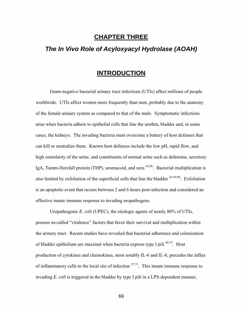
CHAPTER THREE
The In Vivo Role of Acyloxyacyl Hydrolase (AOAH)
INTRODUCTION
Gram-negative bacterial urinary tract infections (UTIs) affect millions of people
worldwide. UTIs affect women more frequently than men, probably due to the anatomy
of the female urinary system as compared to that of the male. Symptomatic infections
arise when bacteria adhere to epithelial cells that line the urethra, bladder and, in some
cases, the kidneys. The invading bacteria must overcome a battery of host defenses that
can kill or neutralize them. Known host defenses include the low pH, rapid flow, and
high osmolarity of the urine, and constituents of normal urine such as defensins, secretory
IgA, Tamm-Horsfall protein (THP), uromucoid, and urea 43;96. Bacterial multiplication is
also limited by exfoliation of the superficial cells that line the bladder 43;50;96. Exfoliation
is an apoptotic event that occurs between 2 and 6 hours post-infection and considered an
effective innate immune response to invading uropathogens.
Uropathogenic E. coli (UPEC), the etiologic agents of nearly 80% of UTIs,
possess so-called “virulence” factors that favor their survival and multiplication within
the urinary tract. Recent studies have revealed that bacterial adherence and colonization
of bladder epithelium are maximal when bacteria express type I pili 48;75. Host
production of cytokines and chemokines, most notably IL-6 and IL-8, precedes the influx
of inflammatory cells to the local site of infection 57;71. This innate immune response to
invading E. coli is triggered in the bladder by type I pili in a LPS-dependent manner,
69
70
since polymyxin B or detoxified LPS inhibit IL-6 production in cultured bladder
epithelial cells that have been invaded by Gram-negative uropathogens 71. Moreover,
the cytokine response of cultured epithelial cells was abrogated after infection with a
msbB strain (lacking secondary fatty acyl chains) 57.
As mentioned previously, AOAH is a host lipase that cleaves the non-
hydroxylated (secondary) fatty acyl chains from the lipid A portion of LPS. In human
cells, this event leads to the detoxification of LPS, as reflected by its diminished ability to
induce cellular inflammatory responses. In vitro data have suggested that deacylated LPS
may function differently in mouse cells. In one experiment, the difference in the ability
of dLPS and fully acylated LPS to stimulate murine splenocyte mitogenesis was much
smaller (~6-20 fold reduction with dLPS) than would be expected for human splenocytes
(~100 fold reduction with dLPS) 97. It is now appreciated that there are subtle differences
in the structures of murine and human TLR4s that might account for this species-specific
difference 98;99. For example, murine TLR4 might recognize a LPS structure independent
of secondary fatty acyl chains, such that deacylating LPS (with AOAH) does not
completely inhibit TLR4’s ability to signal. On the other hand, human TLR4 probably
recognizes LPS in a way that is dependent on the secondary fatty acyl chains. Removing
them (dLPS) abrogates signaling through TLR4. These data suggest that dLPS may not
be as strong an antagonist in murine cells, but it probably does not exclude a role for
AOAH in the detoxification of LPS in the mouse. As described in Chapter two of this
thesis, AOAH is highly expressed in renal proximal tubules of both mice and humans. It
is secreted from these cells and is easily detected in the urine and urinary bladder of wild
type mice. In vitro data suggest that secreted AOAH may associate with bladder cells

71
and be used by them to deacylate associated LPS. This chapter will describe my efforts
to determine the in vivo role of AOAH in the urinary tract during Gram-negative urinary
tract infection.
Results Wild type and AOAH null mice differ in rate of bacterial clearance. In order to examine the in vivo role of AOAH, we challenged AOAH null and
wild type C57Bl/6 mice with the uropathogenic E. coli strain f11 CNF 1+, originally
isolated from a patient with cystitis 100 and provided to us by C. Virginia Lockatell (V.A.
Medical Center, Baltimore Maryland). We chose this strain for several reasons. Firstly,
it has been well characterized by Warren et. al 100 and shown to cause urinary tract
infection when injected into the urethra of mice. Secondly, cystitis strains have been
shown to cause a more severe (duration and intensity) bladder infection in mice as
compared to pyelonephritic strains. As mentioned in the literature review, p piliated
(pyelonephritic) bacteria are the most common UPEC isolated from human patients with
kidney infection and therefore one might assume that p pili are necessary for kidney
infection. This is not the case, for Warren et. al have shown that of the several cystitis
and pyelonephritic strains tested, cystitis strains (including f11) caused a more severe
kidney infection 100. Thirdly, strain f11 CNF 1+ expresses type I pili that allows for its
identification by a simple agglutination test (mannose inhibited agglutination of 1%
yeast, as described in Methods). A fourth consideration is the fact that type I pili

72
stimulate the host immune system via an LPS-dependent mechanism, whereas p pili
initiate responses independently of LPS.
In my experiments, six to ten week-old female wild type and AOAH null C57Bl/6
mice were injected intra-urethrally with (1 x 10⁹ CFU/ml [5 x 10⁷ total]) bacteria in PBS.
Urine was collected at 6, 12, 24, 48, and 72 hours post-infection and assayed for bacterial
growth (colony counts on MacConkey agar) and neutrophils (enumeration using a
hemocytometer). Our results indicate that, at early time points, both wild type and
AOAH null mice have similar bacterial loads in their urine, but that by 48 and 72 hours
post-infection, the AOAH null mice have significantly fewer bacteria in their urine than
do wild type mice (Figure 3.0 and 3.1 A-E).

73
0 24 48 72 96
102
103
104
105
106
107
108
Hours
CFU
/ml U
rine
wtkocontrol
* ** Figure 3.0 – C57BL/6 AOAH null mice clear bacteria from their urine faster than do wild type mice. The data represent combined results of 5 independent experiments, (~30 mice per group per time point); the error bars represent 1 SD. * p=0.0034, ** p=0.018
74
72 Hours
24 Hours
C D
48 HoursE
Figure 3.1 – C57BL/6 colony forming units per ml urine. A. Six-hour time point. B. Twelve-hour time point. C. Twenty-four hour time point. D. Forty-eight hour time point. E. Seventy-two hour time point. The data represent combined results from five separate experiments; each data point represents an individual mouse; bars are medians. Statistics were performed using GraphPad software (Mann-Whitney, 2-tailed, unpaired t test). +/+ -/- ctr
102
103
104
105
106
107
108
109
CFU
/ml U
rine
6 Hours 12 Hours
A B
+/+ -/- ctr
102
103
104
105
106
107
108
109C
FU/m
l urin
e
+/+ -/- ctr
102
103
104
105
106
107
108
109
CFU
/ml U
rine
+/+ -/- ctr
102
103
104
105
106
107
108
109
CFU
/ml U
rine
+/+ -/- ctr
102
103
104
105
106
107
108
109
CFU
/ml U
rine
*

75
AOAH null and wild type mice have similar numbers of bacteria in their bladders at 72 hours. Previously considered extracellular “pathogens”, UPEC are now being re-
examined in light of recent evidence that type I piliated bacteria are able to bind to,
invade, and persist within bladder epithelial cells for weeks to months 75. In vitro, 5637
bladder cells respond to piliated bacteria with an initial IL-6 response, peaking between
two and twenty-four hours, and falling to baseline levels despite the persistence of
intracellular bacteria 71. In order to assess the number of bacteria present within the
bladder tissue following experimental ascending UTI, C57BL/6 mice were sacrificed at
72 hours post-infection. This time point was chosen because it allowed me to follow the
course of infection to its endpoint (an early collection time requires the mouse to be
sacrificed before we gain essential data concerning the outcome of infection). To our
surprise, both wild type and AOAH null mice had bacteria present in their bladders (at
levels similar to reports from other investigators), but there was no significant difference
in the bacterial loads between wild type and AOAH null mice at 72 hours (Figure 3.2).
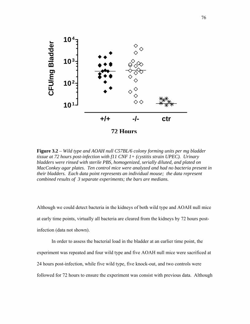
76
+/+ -/- ctr
101
102
103
104
CFU
/mg
Bla
dder
s Figure 3.2 – Wild type and AOAH nutissue at 72 hours post-infection withbladders were rinsed with sterile PBMacConkey agar plates. Ten controtheir bladders. Each data point reprcombined results of 3 separate expe
Although we could detect bacteria in
at early time points, virtually all bact
infection (data not shown).
In order to assess the bacteria
experiment was repeated and four wi
24 hours post-infection, while five w
followed for 72 hours to ensure the e
72 Hour
ll C57BL/6 colony forming units per mg bladder f11 CNF 1+ (cystitis strain UPEC). Urinary S, homogenized, serially diluted, and plated on l mice were analyzed and had no bacteria present in esents an individual mouse; the data represent riments; the bars are medians.
the kidneys of both wild type and AOAH null mice
eria are cleared from the kidneys by 72 hours post-
l load in the bladder at an earlier time point, the
ld type and five AOAH null mice were sacrificed at
ild type, five knock-out, and two controls were
xperiment was consist with previous data. Although
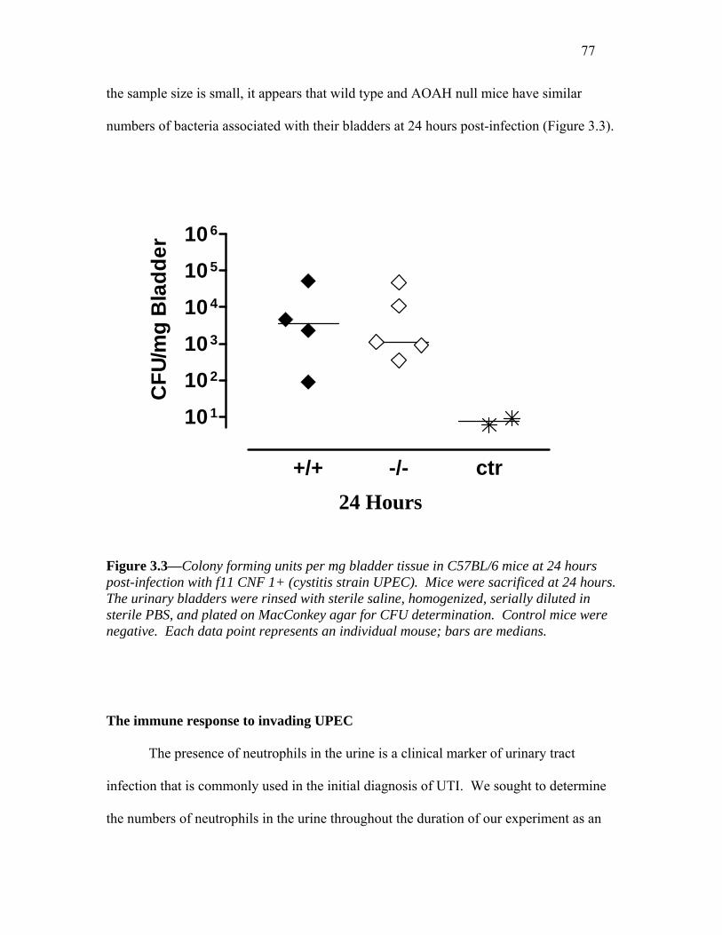
77
the sample size is small, it appears that wild type and AOAH null mice have similar
numbers of bacteria associated with their bladders at 24 hours post-infection (Figure 3.3).
+/+ -/- ctr
101
102
103
104
105
106
CFU
/mg
Bla
dder
24 Hours Figure 3.3—Colony forming units per mg bladder tissue in C57BL/6 mice at 24 hours post-infection with f11 CNF 1+ (cystitis strain UPEC). Mice were sacrificed at 24 hours. The urinary bladders were rinsed with sterile saline, homogenized, serially diluted in sterile PBS, and plated on MacConkey agar for CFU determination. Control mice were negative. Each data point represents an individual mouse; bars are medians. The immune response to invading UPEC The presence of neutrophils in the urine is a clinical marker of urinary tract
infection that is commonly used in the initial diagnosis of UTI. We sought to determine
the numbers of neutrophils in the urine throughout the duration of our experiment as an
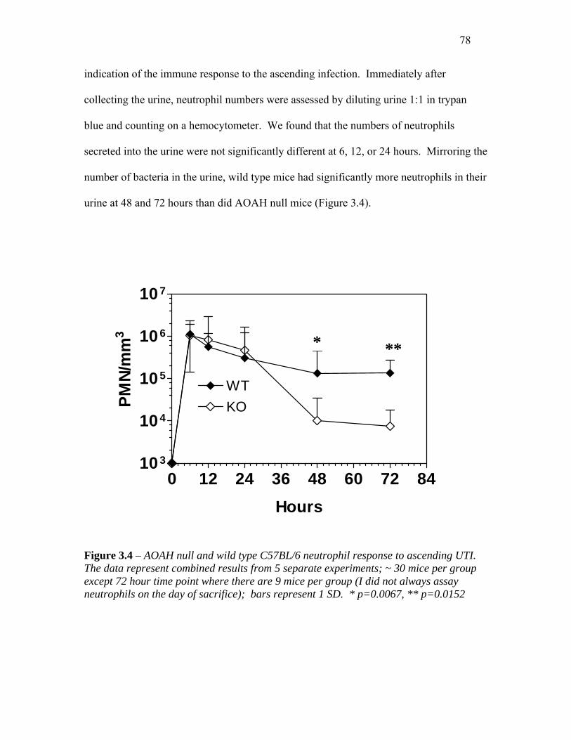
78
indication of the immune response to the ascending infection. Immediately after
collecting the urine, neutrophil numbers were assessed by diluting urine 1:1 in trypan
blue and counting on a hemocytometer. We found that the numbers of neutrophils
secreted into the urine were not significantly different at 6, 12, or 24 hours. Mirroring the
number of bacteria in the urine, wild type mice had significantly more neutrophils in their
urine at 48 and 72 hours than did AOAH null mice (Figure 3.4).
0 12 24 36 48 60103
104
105
106
107
WTKO
* *
Hours
PMN/
mm
3 *
Figure 3.4 – AOAH null and wild type C57BL/6 neutrophil reThe data represent combined results from 5 separate experimexcept 72 hour time point where there are 9 mice per group (neutrophils on the day of sacrifice); bars represent 1 SD. * p
**
72 84
sponse to ascending UTI. ents; ~ 30 mice per group I did not always assay =0.0067, ** p=0.0152

79
Histological analysis of C57Bl/6 bladder and kidney samples.
In order to assess the local inflammatory response to ascending UTI, bladders
were collected at the time of sacrifice (72h), bisected, and fixed in 4% formaldehyde as
described in Methods. After two days in fixative, the bladders were paraffin-embedded
and 4 µm sections were cut and stained with hematoxylin-and-eosin (H&E). Dr. Zhou, a
renal pathologist, kindly scored each specimen. He was blinded to the experimental
grouping of the mice and scored on a scale of 0-3 as described in Methods. Briefly, each
section was scored for the extent of inflammatory cell influx into the tissue and interstitial
edema. Our results indicate that, at 72 hours, there is no significant difference between
wild type and AOAH null mice in their mean histological scores. Both wild type and
AOAH null mice had a significant amount of inflammation as compared to the PBS
injected control mice (Figure 3.5).
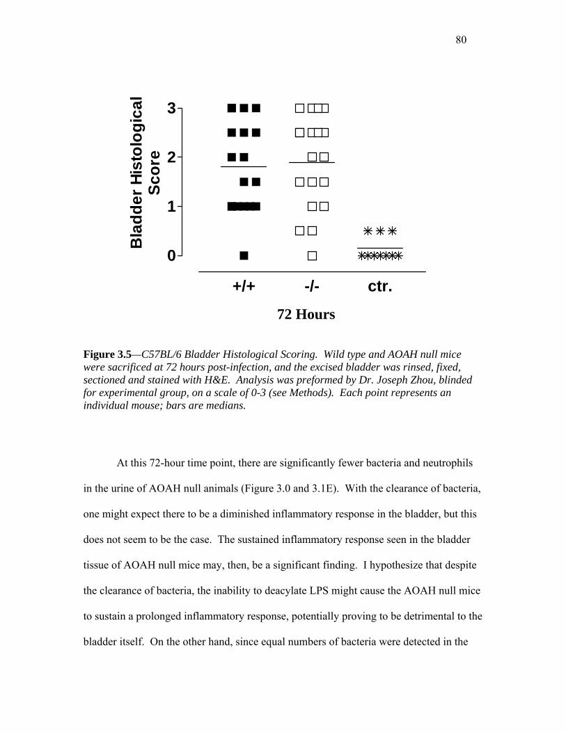
80
+/+ -/- ctr.
0
1
2
3
Bla
dder
His
tolo
gica
lSc
ore
Figure 3.5—C57BL/6 Bladder Histologicawere sacrificed at 72 hours post-infection,sectioned and stained with H&E. Analysisfor experimental group, on a scale of 0-3 (individual mouse; bars are medians.
At this 72-hour time point, there ar
in the urine of AOAH null animals (Figure
one might expect there to be a diminished
does not seem to be the case. The sustaine
tissue of AOAH null mice may, then, be a
the clearance of bacteria, the inability to de
to sustain a prolonged inflammatory respo
bladder itself. On the other hand, since eq
72 Hours
l Scoring. Wild type and AOAH null mice and the excised bladder was rinsed, fixed, was preformed by Dr. Joseph Zhou, blinded see Methods). Each point represents an
e significantly fewer bacteria and neutrophils
3.0 and 3.1E). With the clearance of bacteria,
inflammatory response in the bladder, but this
d inflammatory response seen in the bladder
significant finding. I hypothesize that despite
acylate LPS might cause the AOAH null mice
nse, potentially proving to be detrimental to the
ual numbers of bacteria were detected in the

81
bladders at the time of sacrifice, one might expect to find a similar level of tissue
inflammation.
In an attempt to localize bacteria within the bladder epithelium, unstained sections
of bladder from infected and control wild type and AOAH null mice were Gram-stained
and analyzed by Dr. Zhou, blinded to the experimental grouping of the mice.
Unfortunately, no bacteria were detected in any of four wild type, four AOAH null, or
two control animals. Hultgren and colleagues have shown by electron microscopy that
internalized UPEC are not evenly dispersed throughout the bladder tissue; rather, they
form focal lesions within the epithelium. In fact, Mulvey et. al have called such lesions
“bacterial factories” 75. A recent publication by Hultgren and colleagues has suggested
that E. coli form biofilms within the bladder epithelial cells and has termed such bacterial
focuses “biofilm-like pods”. It is possible that the 4 µm sections examined in my studies
were not representative of the entire bladder. Sections taken from other planes of the
bladder might have contained bacteria (as would be expected from the CFU/mg bladder
determinations). Alternatively, 72 hours may be too late to detect bacteria in the bladder
tissue. An earlier time point, such as the 6 and 24 hour time point used by Mulvey and
colleagues, may allow for better visualization due to the increased numbers of bacteria
present.
An analysis of inflammation in the bladders of C57Bl/6 mice at 24 hours post-infection I next wondered if the inflammatory response within the bladders of wild type and
AOAH null mice might be different at 24 hours, just prior to the clearance of bacteria
from AOAH null urine. To assess this, C57BL/6 mice were sacrificed at 24 hours post-
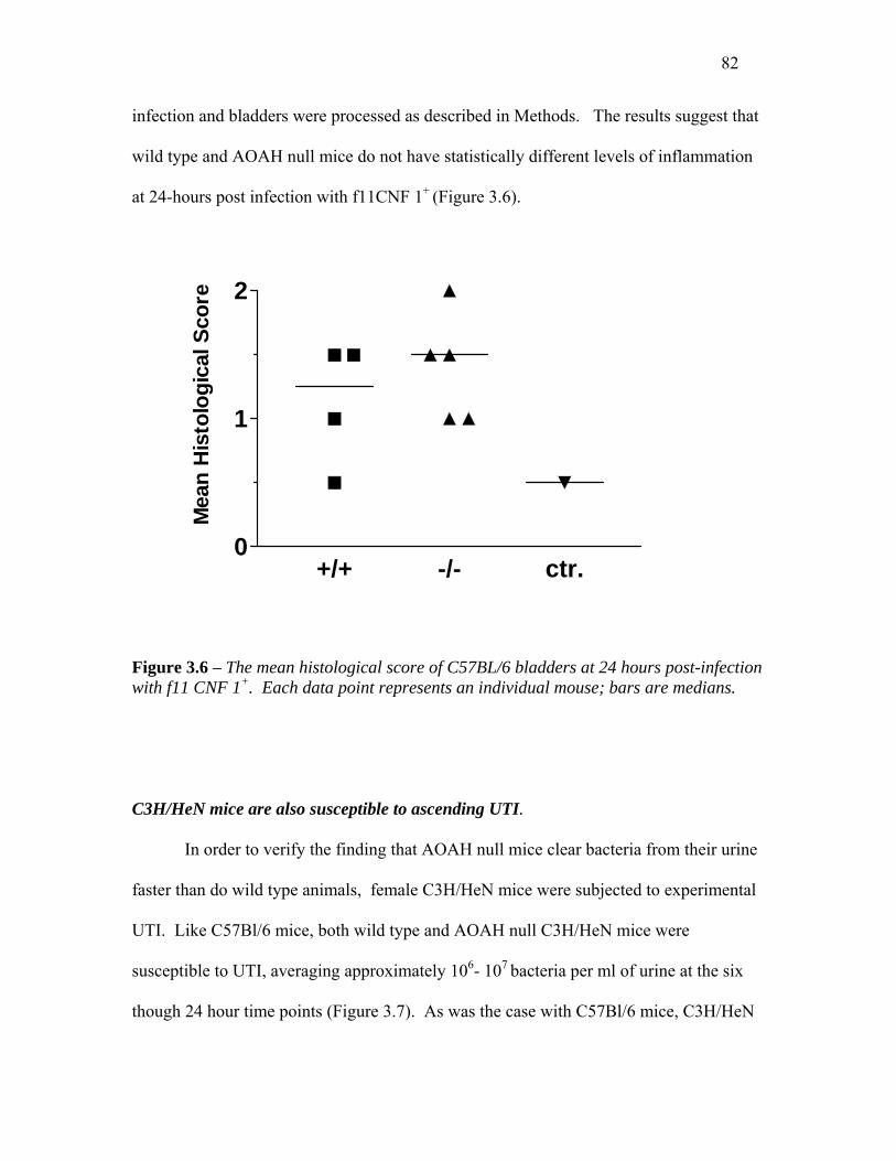
82
infection and bladders were processed as described in Methods. The results suggest that
wild type and AOAH null mice do not have statistically different levels of inflammation
at 24-hours post infection with f11CNF 1+ (Figure 3.6).
+/+ -/- ctr.0
1
2
Mea
n H
isto
logi
cal S
core
Figure 3.6 – The mean histological score of C57BL/6 bladders at 24 hours post-infection with f11 CNF 1+. Each data point represents an individual mouse; bars are medians. C3H/HeN mice are also susceptible to ascending UTI. In order to verify the finding that AOAH null mice clear bacteria from their urine
faster than do wild type animals, female C3H/HeN mice were subjected to experimental
UTI. Like C57Bl/6 mice, both wild type and AOAH null C3H/HeN mice were
susceptible to UTI, averaging approximately 106- 107 bacteria per ml of urine at the six
though 24 hour time points (Figure 3.7). As was the case with C57Bl/6 mice, C3H/HeN
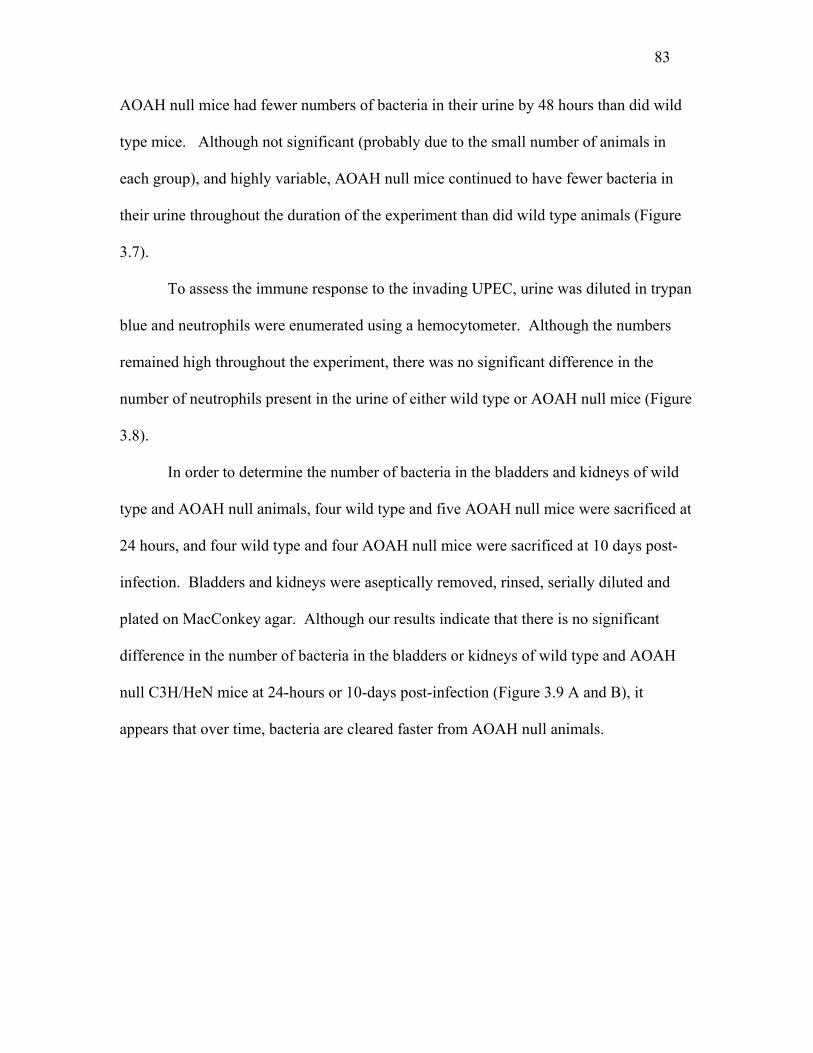
83
AOAH null mice had fewer numbers of bacteria in their urine by 48 hours than did wild
type mice. Although not significant (probably due to the small number of animals in
each group), and highly variable, AOAH null mice continued to have fewer bacteria in
their urine throughout the duration of the experiment than did wild type animals (Figure
3.7).
To assess the immune response to the invading UPEC, urine was diluted in trypan
blue and neutrophils were enumerated using a hemocytometer. Although the numbers
remained high throughout the experiment, there was no significant difference in the
number of neutrophils present in the urine of either wild type or AOAH null mice (Figure
3.8).
In order to determine the number of bacteria in the bladders and kidneys of wild
type and AOAH null animals, four wild type and five AOAH null mice were sacrificed at
24 hours, and four wild type and four AOAH null mice were sacrificed at 10 days post-
infection. Bladders and kidneys were aseptically removed, rinsed, serially diluted and
plated on MacConkey agar. Although our results indicate that there is no significant
difference in the number of bacteria in the bladders or kidneys of wild type and AOAH
null C3H/HeN mice at 24-hours or 10-days post-infection (Figure 3.9 A and B), it
appears that over time, bacteria are cleared faster from AOAH null animals.
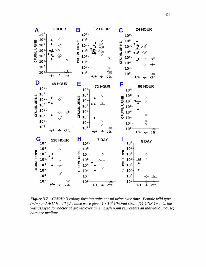
84
G H I
12 HOUR
+/+ -/- ctr.102
103
104
105
106
107
108
109
CFU
/ML
UR
INE
6 HOUR
+/+ -/- ctr102
103
104
105
106
107
108
109C
FU/M
L U
RIN
E
48 HOUR
+/+ -/- ctr.102
103
104
105
106
107
108
109
CFU
/ML
UR
INE
72 HOUR
+/+ -/- ctr. 102
103
104
105
106
107
108
109
CFU
/ML
UR
INE
96 HOUR
+/+ -/- ctr.102
103
104
105
106
107
108
109
CFU
/ML
UR
INE
+/+ -/- ctr102
103
104
105
106
107
108
109
CFU
/ML
UR
INE
24 HOUR
120 HOUR
+/+ -/- ctr.102
103
104
105
106
107
108
109
CFU
/ML
UR
INE
7 DAY
+/+ -/- ctr.102
103
104
105
106
107
108
109
CFU
/ML
UR
INE
8 DAY
+/+ -/- ctr.102
103
104
105
106
107
108
109
CFU
/ML
UR
INE
FE D
A B C
Figure 3.7 – C3H/HeN colony forming units per ml urine over time. Female wild type (+/+) and AOAH null (-/-) mice were given 1 x 10⁹ CFU/ml strain f11 CNF 1+ . Urine was assayed for bacterial growth over time. Each point represents an individual mouse; bars are medians.

85
A B
C D
E F
6 HOURS
+/+ -/-103
104
105
106
107
108
PMN
/MM
3
12 HOURS
+/+ -/-10 3
10 4
10 5
10 6
10 7
10 8
PMN
/MM
324 HOURS
+/+ -/-103
104
105
106
107
108
PMN
/MM
3
48 HOURS
+/+ -/-103
104
105
106
107
108PM
N/M
M3
72 HOURS
+/+ -/-103
104
105
106
107
108
PMN
/MM
3
96 HOURS
+/+ -/-103
104
105
106
107
108
109
1010
PMN
/MM
3
Figure 3.8 – Number of neutrophils in the urine of +/+ and -/- C3H/HeN mice after infection with f11 UPEC. Urine was collected as described in methods, diluted 1:1 in 1% trypan blue, and placed under a hemocytometer for enumeration. Each point represents an individual mouse; bars are medians.
86
FigureAOAH negativ
A
24 HOURSBladder+/+ -/-10 0
10 1
10 2
10 3
10 4
10 5
10 6C
FU/M
G B
LAD
DER
10 DAYBladder
+/+ -/-100
101
102
103
104
CFU
/MG
BLA
DD
ER
C
3.9 – Colony-forming units per mg blanull C3H/HeN mice at 24-hour and 10e at all time points and in all tissues.
B
24 HOURSKidney
+/+ -/-10 0
10 1
10 2
10 3
10 4
CFU
/MG
KID
NEY
10 DAYKidney
+/+ -/-100
101
102
103
104
CFU
/MG
KID
NEY
D
dder or kidney tissue in wild type and -day post-infection. Control mice were

87
Mice do not have detectable levels of anti-f11 LPS in their urine.
I next wondered if AOAH null mice might have an abundance of anti-LPS
antibodies in their urine that could explain their ability to clear uropathogens faster than
wild type mice. Although it is unlikely that the mice have seen the particular strain of
bacteria (f11 CNF 1+) used to give them UTIs, it’s possible that they possess anti-LPS
antibodies that might cross-react with the LPS present in the f11 strain. I used an ELISA
to detect anti-f11 LPS in the urine of both wild type and AOAH null C57Bl/6 and
C3H/HeN mice. To begin these studies, pre-immune serum was collected from five
AOAH null and four wild type C57Bl/6 mice that had previously been given UTIs with
the f11 strain. Mice were immunized by injecting a sub-cutaneous dose of heat-killed f11
CNF 1+ bacteria as described in the Methods. Two weeks later, serum was collected and
assayed for anti-f11 LPS IgG, IgM, and IgA antibodies. AOAH null mice had
significantly more anti-f11 IgG, IgM, and IgA f11 LPS antibodies in their post-infection
serum than did wild type mice (Figure 3.10 A-C). Wild type and AOAH null mice had
similar levels of anti-f11 LPS IgG, IgM, and IgA antibodies in their pre-immune serum
(Figure 3.10 B-E) Neither wild type or AOAH null (C57Bl/6 or C3H/HeN) mice had any
anti-f11 LPS IgG, IgM, or IgA in their urine (data not shown).

88
Post-immunization Pre-immunization
A D
Figure 3.10 – Anti-f11 LPS antibody titers in serum from C57Bl/6 mice. Mice were immunized with heat-killed f11 CNF 1+ as described in Methods. A. Post-immune serum, anti-f11 LPS IgM B. Post-immune serum, anti-f11 LPS IgG. C. Post-immune serum, anti-f11 LPS IgA. D. Pre-immune serum, anti-f11 LPS IgM. E. Pre-immune serum, anti-f11 LPS IgG. F. Pre-immune serum, anti-f11 LPS IgA. (A-C): Each point represents four wild type and five AOAH null mice, assayed in duplicate. (D-F): Each point represents three wild type and three AOAH null mice, assayed in duplicate. The error bars represent SEM.
F
E0 100 200 300 400 500
0.0
0.1
0.2
0.3
0.4
0.5
KOWT
Dilution
OD
0 100 200 300 400 5000.0
0.1
0.2
0.3
KOWT
Dilution
OD
C
B 0 100 200 300 400 500
0
1
2
Dilution
OD
0 100 200 300 400 5000
1
2
Dilution
OD
IgM
IgG
0 100 200 300 400 5000.00
0.05
0.10
0.15
KOWT
Dilution
OD
0 100 200 300 400 5000.0
0.1
0.2
0.3
Dilution
IgA
OD

89
Methods Chemicals. Unless otherwise indicated, chemicals were purchased from Sigma-Aldrich
Chemical Co, St. Louis, MO.
Mouse strains. Specific pathogen-free mice were housed in the UT Southwestern
Animal Resource Center and fed a standard diet. ICR (Harlan, Indianapolis, IN),
C57Bl/6, or C3H/HeN, wild type and AOAH null 85 mice were used.
Bacterial Strains. E. coli strain f11 CNF 1+ (f11) was obtained from C. Virginia
Lockatell, V.A. Medical Center, Baltimore Maryland, and is described in 100. Strain f11
expresses a type I pilus which can be verified by a simple agglutination test.
Agglutination was assessed before and during every experiment by adding 50-100µls
bacterial suspension (or one colony) to 50-100 µls of 1% yeast (Fleischmann’s® active
dry yeast, bought at the grocery store) +/- an equal volume of 2.5 mM D-mannose on a
sterile petri dish and gently mixing. Agglutination is evident within one minute. E. coli
strain FN414 is a previously described non-pathogenic strain originally isolated from the
stool of a healthy individual. It was also obtained from C. Virginia Lockatell, V.A.
Medical Center, Baltimore Maryland, and is referenced in 100. It does not express a type I
pilus and therefore does not agglutinate a 1% yeast solution. Approximately one week
before each experiment, bacteria were streaked onto Trypticase Soy Agar (TSA) plates,
incubated at 37°C overnight, and stored at 4°C for a maximum of one week before use.
Three days prior to the experiment, one to five colonies were inoculated into 5 mls LB
medium and grown overnight at 37°C with shaking. The five ml culture was transferred
to 50 mls LB medium and allowed to grow at 37°C with shaking for 2 days. On the
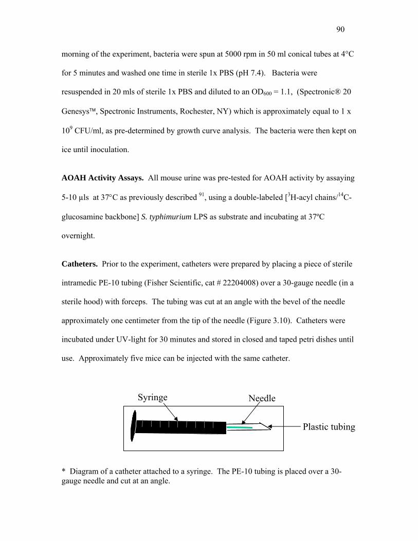
90
morning of the experiment, bacteria were spun at 5000 rpm in 50 ml conical tubes at 4°C
for 5 minutes and washed one time in sterile 1x PBS (pH 7.4). Bacteria were
resuspended in 20 mls of sterile 1x PBS and diluted to an OD600 = 1.1, (Spectronic® 20
Genesys™, Spectronic Instruments, Rochester, NY) which is approximately equal to 1 x
109 CFU/ml, as pre-determined by growth curve analysis. The bacteria were then kept on
ice until inoculation.
AOAH Activity Assays. All mouse urine was pre-tested for AOAH activity by assaying
5-10 µls at 37°C as previously described 91, using a double-labeled [3H-acyl chains/14C-
glucosamine backbone] S. typhimurium LPS as substrate and incubating at 37ºC
overnight.
Catheters. Prior to the experiment, catheters were prepared by placing a piece of sterile
intramedic PE-10 tubing (Fisher Scientific, cat # 22204008) over a 30-gauge needle (in a
sterile hood) with forceps. The tubing was cut at an angle with the bevel of the needle
approximately one centimeter from the tip of the needle (Figure 3.10). Catheters were
incubated under UV-light for 30 minutes and stored in closed and taped petri dishes until
use. Approximately five mice can be injected with the same catheter.
Needle Syringe Plastic tubing * Diagram of a catheter attached to a syringe. The PE-10 tubing is placed over a 30-gauge needle and cut at an angle.

91
Urine Collection. The perineal skin of all mice was sprayed with 70% EtOH and the
mice were returned to their own clean cage and allowed to dry. Urine was collected onto
sterile petri dishes (Fisher Scientific) after picking up the mice and sometimes gently
pushing on their bladders. Urine that came into contact with feces or other areas of the
perineum was discarded and not analyzed.
Experimental Ascending Urinary Tract Infections. Just prior to an experiment, time
zero urine was collected from all mice and stored on ice. Six to ten week-old wild type
and AOAH null, female, C57Bl/6 or C3H/HeN mice were anesthetized IP with 0.05 mls
ketamine-acepromazine (2.5 mls ketamine (Ketaset, Fort Dodge Animal Health, Burns
Veterinary Supply, Rockville Centre, NY), 0.5 mls acepromazine (Aceproject, Vetus
Animal Health, Burns Veterinary Supply, Rockville Centre, NY), and 7.0 mls sterile
water) and placed in sterile, clean cages. Once asleep, the mice were secured, lying
supine, onto a piece of Styrofoam with two large rubber bands over each hind leg.
Bacteria (1 x 10⁹ CFU/ml) were drawn into a 1 ml tuberculin syringe and a pre-made and
sterilized catheter was attached. The catheter was gently inserted into the urethra of the
mouse, first entering perpendicular to the mouse and then slowly angling to become more
parallel. The catheter was never forced; when it is in the correct place, it will glide into
the bladder and one will know it is in the correct place. Once in place, 0.05 mls of
bacteria were gently and slowly expulsed into the bladder. The catheter was slowly
removed and no more manipulations were made. The mice were placed in clean cages,

92
which had been placed on an electric heating-pad (high setting)(Walgreen’s) and allowed
to awaken (~20-30 minutes). Control mice (at least two per group) were injected as
above with 0.05 mls sterile PBS (pH 7.2). The mice remained in the lab throughout the
duration of the experiment so as not to infect the mouse colony and for ease of urine
collection. All cages were placed on heating pads (low setting) and were covered at night
to ensure complete darkness.
Colony-forming Unit (CFU) Determination. MacConkey agar (Voigt Global
Distribution LLC) plates were prepared weeks in advance of the experiment by
autoclaving media and pouring 15 mls per plate into sterile petri dishes. Plates were
cooled overnight at room temperature, dried overnight at 37ºC, and stored, sealed in
original packaging at 4ºC until use. Four (or more) hours before use, plates were
removed from their packaging and placed at 37ºC to warm-up. Urine, bladder or kidney
lysates were serially diluted (Table 3.0) into sterile PBS (pH 7.2) and 100 µls were
plated. Bacteria were spread by either the traditional glass rod method or by placing 3 to
5 glass beads (3 mM in diameter) (Fisher Scientific) onto each plate and shaking for
approximately 30 seconds. The glass bead method is much faster and works better than
the traditional glass rod method. Plates were incubated at 37ºC overnight and stored at
4ºC until colonies could be counted. If possible, two dilutions were counted (between 50
and 500 colonies) and the average was recorded. The presence of type I pili was
confirmed by mannose-sensitive agglutination of 1% yeast at least twice during the
experiment.
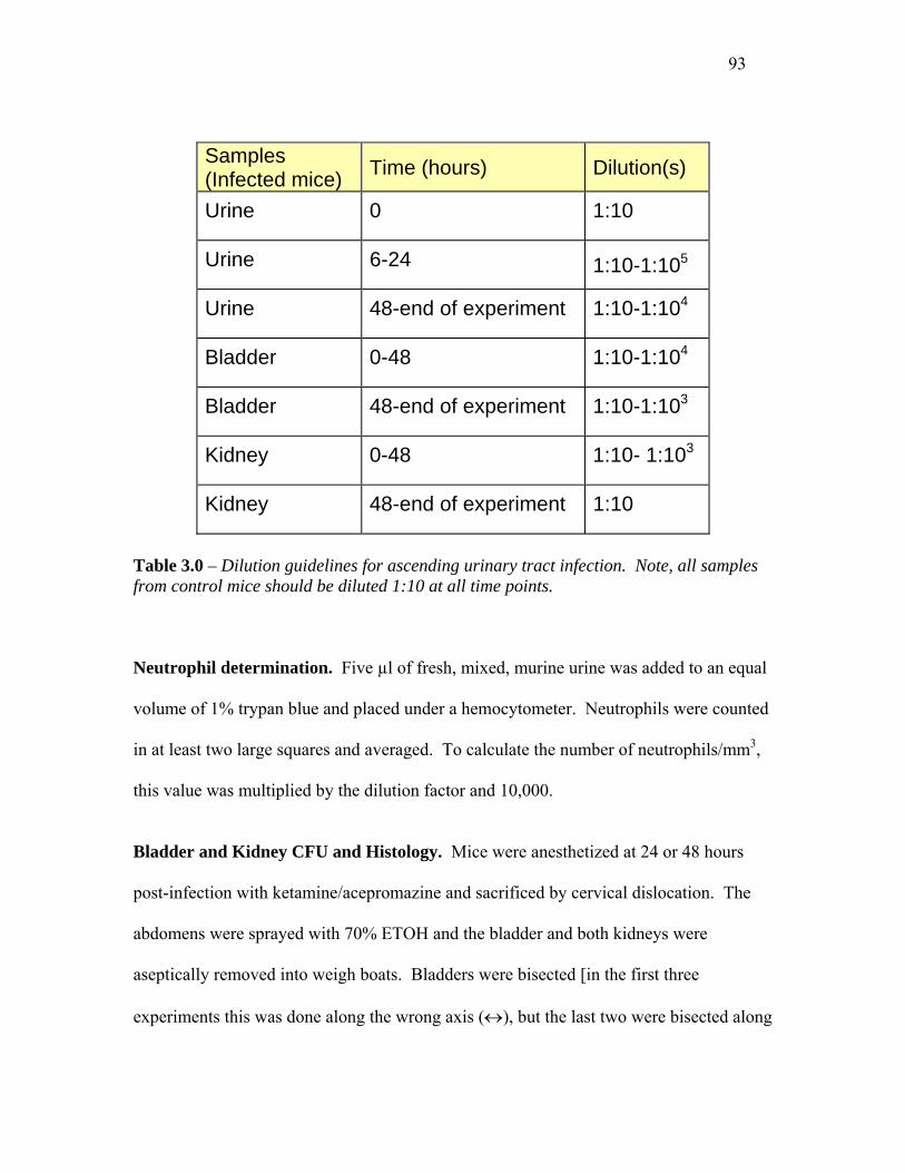
93
Samples (Infected mice) Time (hours) Dilution(s)
Urine 0 1:10
Urine 6-24 1:10-1:105
Urine 48-end of experiment 1:10-1:104
Bladder 0-48 1:10-1:104
Bladder 48-end of experiment 1:10-1:103
Kidney 0-48 1:10- 1:103
Kidney 48-end of experiment 1:10
Table 3.0 – Dilution guidelines for ascending urinary tract infection. Note, all samples from control mice should be diluted 1:10 at all time points.
Neutrophil determination. Five µl of fresh, mixed, murine urine was added to an equal
volume of 1% trypan blue and placed under a hemocytometer. Neutrophils were counted
in at least two large squares and averaged. To calculate the number of neutrophils/mm3,
this value was multiplied by the dilution factor and 10,000.
Bladder and Kidney CFU and Histology. Mice were anesthetized at 24 or 48 hours
post-infection with ketamine/acepromazine and sacrificed by cervical dislocation. The
abdomens were sprayed with 70% ETOH and the bladder and both kidneys were
aseptically removed into weigh boats. Bladders were bisected [in the first three
experiments this was done along the wrong axis (↔), but the last two were bisected along

94
the correct, sagittal axis ( ↑ )] and rinsed with sterile saline to remove any residual urine.
Half of the bladder and one kidney was weighed, placed into a 5 ml polystyrene FACs
tube (Falcon #2054), pre-filled with 0.025% Triton X-100 in PBS on ice, for future
colony-forming unit determination. One half of the bladder and the other kidney were
immersed in 4% formaldehyde (freshly prepared from paraformaldehyde in 1x PBS) and
stored on ice. Tissues in formaldehyde were rocked for two days at 4ºC at which time the
formaldehyde was replaced with PBS. Tissues were dehydrated, paraffin-embedded, and
4 µm sections were placed onto microscope slides and stained for hematoxylin-and-eosin
(H&E) 92.
Tissues in 0.025% Triton X-100 were homogenized to complete disruption with a
clean (washed in 70% ETOH, and two sterile water washes) hand-held Tissue Tearor
(Biospec Products, Inc. Model 985-370) for approximately 30 seconds. Disrupted
tissues were serially diluted in sterile PBS and 100 µl were plated onto MacConkey agar
plates as per Table 3.0.
Histological Scoring Index. Bladders were scored, blinded to experimental group, on a
scale of 0-3 as previously described 100. In brief, a score of zero (no inflammation); one,
mild (infiltration of low numbers of neutrophils in the lamina propria, little or no
interstitial edema, and the absence of regenerative hyperplasia in the luminal epithelium);
two, moderate (moderate numbers of neutrophils in the lamina propria, moderate
interstitial edema, and moderate generalized hyperplasia of the lumina epithelium); and
three, severe (diffuse infiltration of large numbers of neutrophils in the lamina propria,
severe interstitial edema, and severe generalized hyperplasia of the luminal epithelium).

95
Preparation of LPS. Strain f11 CNF 1+ (RM 945) was streaked onto a Trypticase Soy
Agar (TSA) plate from a glycerol stock and grown overnight at 37ºC. Several colonies
were transferred to 500 mls of LB and grown for 48 hours at 37ºC with shaking. Bacteria
were spun at 1500 rpm (Beckman Coulter, Fullerton, CA) for 10 minutes at 4ºC, washed
once in 20 mls of sterile water, and resuspended in 21 mls of water. Twenty mls of
phenol (27g of phenol resuspended in 3 mls water) was added to the 20 ml bacterial
suspension in a 68ºC water bath with continuous mixing. The mixture was kept at 68ºC
(with mixing) for approximately 20 minutes (or until one phase is clearly present). In
order to separate into two phases, the mixture was placed in an ice/water bath for 20
minutes. The sample was spun at 3500 rpm (Beckman Coulter, Fullerton, CA) for 15
minutes and the top layer (LPS and water) was removed to a fresh tube. Twenty mls of
water was added to the LPS/phenol preparation and re-extracted as above. The final
sample was dialyzed in a 10-14,000 MW cut-off dialysis tube against 3, 1L changes of
water over two days and one change into 20 mM Tris, pH 8.0. The LPS was removed
from the tubing, treated with 1 mg/ml DNaseI, 10 mg/ml RNaseI for several hours at
37ºC, and then 5 µg/ml Proteinase K for 3 hours at 37ºC. The sample was split in half
and spun at 35,000 rpm in the ultracentrifuge (Sorval Ultracentrifuge, OTT65B) with
rotor T 865 overnight at 4ºC. The supernatant was poured off and the LPS resuspended
in 0.5 ml water. The LPS preparation was run on a 15% SDS-polyacrylamide gel and
silver-stained to test its purity. This process was repeated to obtain more LPS.
Immunization of mice with heat-killed f11 CNF 1+ and serum collection. Strain f11
CNF 1+ (RM 945) was streaked onto a Trypticase Soy Agar (TSA) plate from a glycerol

96
stock and grown overnight at 37ºC. Several colonies were transferred to 500 mls of LB
and grown for 48 hours at 37ºC with shaking. Bacteria were washed and resuspended to
a concentration of 1 x 10⁹ CFU/ml (OD = 1.1) in PBS. An aliquot was boiled for 30
minutes at 85 -100°C and then placed at 4°C until use. Blood was drawn from the tail
vein of four wild type and five AOAH null mice that had previously been given UTIs
with strain f11 CNF 1+. Immediately after the blood draw, mice were given a sub-
cutaneous injection of 100 µls heat-killed bacteria into their back. Post-immunization
serum was drawn 2 weeks post-immunization. Blood was stored at 4°C overnight, spun
at 50% max speed at 4°C, and serum collected to a fresh tube.
f11-LPS ELISA. Ninety-six well, square-bottom, NUNC brand Maxisorb ELISA plates
(Fisher Scientific) were coated in a 1:100 dilution of f11-LPS (diluted in 0.1M NaHCO3)
overnight at 4°C. Sample wells were washed 3 times in PBST (0.005% Tween 20 in
PBS, pH 7.2) and blocked with 10% HI-FCS/PBS (block buffer) for 1 hr at 37°C.
Samples (1:5 dilutions of urine) and standards (1:25, 1:100, or 1:400 dilutions of post-
immune serum) were diluted in block buffer and incubated at 4°C overnight or at 37°C
for 2 hrs. After washing, secondary antibody (goat anti-mouse IgM-HRP, goat anti-
mouse IgG-HRP, or goat anti-mouse IgA-biotin) was added at a dilution of 1:1000 in
block buffer for one hour at 37°C. After washing 5 times, the IgM and IgG plates were
developed with TMB substrate reagent (PharMingen)(equal volumes of A and B mixed
immediately before use). The IgA plates were incubated with 2 µg/ml alkaline
phosphatase-conjugated streptavidin (Jackson Laboratories) in block buffer for 30 min at
37°C. The IgA plate was washed as above and developed with alkaline phosphatase

97
substrate (5 mM Sigma 104 and 0.1M Sigma Alkaline buffer 221 in water). Optical
densities were read at 405 nm (IgA) and 450 nm test, 570 nm reference (IgG and IgM)
and analyzed on an ELISA Plate Reader (MRX Revelation, Dynex Technologies,
Chantilly, Virginia).
98
Discussion
Urinary tract infections are a significant cause of morbidity and account for
thousands of medical visits each year in the developed world. In addition to the need for
a better understanding of UTIs and their sequelae (ie. pyelonephritis, renal scarring,
sepsis) for treatment purposes, the urinary system is also a useful model for studies of the
innate epithelial cell responses to Gram-negative infection. While many scientists focus
such studies on myeloid lineage cells (monocytes, macrophages, and dendritic cells),
fewer have investigated the role that epithelial cells play in host defense. As discussed
earlier, AOAH is highly expressed in the kidney and in vitro is able to associate with
bladder cells, providing them with the ability to deacylate LPS. The enzyme is also
present in the urine, where it is able to deacylate LPS. I aimed to assess the role of
AOAH in vivo, and to do so, induced ascending UTIs in wild type and AOAH null mice.
My results have shown that C57Bl/6 mice are susceptible to Gram-negative UTI. At the
0, 6, 12, and 24-hour time points, one can detect similar numbers of bacteria in the urine
of both wild type and AOAH null mice. However, AOAH null mice have significantly
fewer bacteria in their urine at 48 and 72 hours post-infection than do wild type animals.
One hypothesis to explain these data might be that due to their inability to
deacylate LPS, AOAH null mice mount a more vigorous inflammatory response to LPS.
Such a heightened response might in turn help the AOAH null mice to clear the infection
faster than wild type mice. In apparent opposition to this hypothesis is the finding that
C57BL/6 wild type and AOAH null mice have similar numbers of neutrophils in their
99
urine at all early time points. In fact, it is the wild type animals that eventually (48 and
72 hours post-infection) have significantly more neutrophils present in their urine.
Analysis of bladder tissue at 24- and 72-hours post-infection also suggests a similar level
of inflammation, since there is no difference in the mean histological score at either of
these time points. To investigate further the immune response to invading uropathogens,
urine was assayed for IL-6 and KC (the mouse IL-8 homolog) over the time course of
infection. Unfortunately, any signal was below the level of detection. We are currently
testing C3H/HeN urine for both IL-6 and KC, hoping this strain will have higher levels of
cytokines. It is possible that such a response exists and I have been unable to detect it.
A second hypothesis is that AOAH null mice might produce more natural, LPS
antibodies (that cross-react with the LPS present on uropathogens). Unlike an acquired
immune response, natural antibody acts rather quickly and if present might explain the
faster clearance of uropathogens by AOAH null mice. This hypothesis is based on
preliminary results by Mingfang Lu, who has shown that AOAH null mice produce more
polyclonal antibody in response to LPS or LOS immunization. Unfortunately, I was
unable to detect any anti-f11 LPS in the urine of wild type or AOAH null C57Bl/6 or
C3H/HeN mice by ELISA.
Recently, Martinez et. al have shown that type I piliated bacteria are able to
invade and persist within epithelial cells of the murine bladder for months, avoiding
recognition by the immune system 50. It is speculated that these “bacterial factories” may
be present in humans and account for recurrent UTIs, since many times the second
infection is caused by the same bacterial strain that caused the first. How UPEC avoid
clearance and remain quiescent is currently unknown. Importantly, the IL-6 response of
100
cultured bladder cells infected with UPEC is transient, peaking at two hours and returning
to baseline thereafter. It is possible that AOAH plays a role in dampening the immune
response to the LPS present on these intracellular bacteria, thereby allowing them to
persist within the bladder epithelium for some time. In this way, the bacteria could be
thought of as using the host machinery to their advantage, allowing them to escape
immune recognition or clearance. However, my experiments do support this hypothesis.
I see equal numbers of bacteria in the bladder tissue of both wild type and AOAH null
mice at 72 hours post-infection (Figure 3.2). If the absence of AOAH allows bacteria to
persist intracellularly, one might expect to fine more bacteria in the bladders of AOAH
null mice at later time points. It might be interesting to follow infected mice for an
extended period of time to see if wild type mice eventually clear intracellular bacteria
faster than AOAH null.
If AOAH functions to dampen the innate immune response to LPS on intracellular
uropathogens, the absence of AOAH might lead to a sustained LPS-induced
inflammatory response within the bladder. It is possible that such a sustained response
would benefit the host, helping to rid it of bacteria. On the other hand, one could imagine
a sustained immune response as potentially harmful to the host, possibly causing
inflammation induce damage to bladder or kidney tissue. Histological analysis of the
bladder tissue from C3H/HeN mice infected with uropathogens might help us address this
question. Because the mice are infected for such a long period of time (8 or more days),
a potential immune mediated tissue injury might be detected, for C57Bl/6 mice clear
pathogens in 2-3 days. Alternatively, one could induce a series of UTIs in the same mice

101
over a period of months, and assess the bladder and kidney histology after such repeated
stress.
If the phenotype we are seeing is the result of AOAH deficiency, infecting both
wild type and AOAH null mice with a strain of bacteria that has a lipid A structure
similar to AOAH-treated LPS (dLPS), should result in a similar level of infection and
clearance. Although a strain exists, the msbB mutant, it lacks the appropriate pili and
virulence factors that enable it to cause significant infection in mice. Much work would
be needed to render the bacterium infectious in an in vivo model.
Non-pathogenic strain FN414 was originally isolated from the stool of a healthy
individual and does not express a type I pilus. For this reason, it is difficult to readily
distinguish the infecting (inoculum) strain from normal flora or possible contaminants. I
attempted to select for naladixic acid resistant (NalR) mutants by growing the bacteria on
progressively more concentrated LB agar plates supplemented with naladixic acid (15
µg/ml 100 µg/ml). I was unable to obtain viable, healthy NalR E. coli at any of the
concentrations. The colonies were much smaller and of a different texture (more solid or
stiff) than wild type FN414. When Gram-stained (after growing overnight on 15 µg/ml
naladixic acid), they had an unusual elongated morphology and the bacteria seemed to
form chains. In contrast, I obtained NalR colonies of strain f11 CNF 1+ with relative
ease, and although less dramatic, they too had an elongated morphology when Gram-
stained. Because strain f11 possess type I pili, they are easily distinguished from other,
contaminating bacteria. Future studies where one would like to use a non-pathogenic
strain of bacteria should address this issue and maybe try making a mutant resistant to a
different antibiotic.

Chapter Four
AOAH-Fluorescent Fusion Protein
Introduction
While the role of AOAH in deacylating LPS is clear, where this reaction occurs is
less clear. One of the most common questions regarding AOAH is, “where is it doing its
job?” Little work has investigated the intracellular location of AOAH and the reason for
this is several-fold. First, the abundance of AOAH in myeloid lineage cells is extremely
low and the protein is nearly impossible to detect by immunoflourescence. Secondly,
antibodies to murine AOAH proved very difficult to obtain, and have only recently been
produced. Janet Staab conducted the only published study of AOAH’s intracellular
location. She showed diffuse, punctate, and cytoplasmic staining of AOAH-transfected
BHK 570 cells (fibroblasts) by indirect immunofluorescence using a polyclonal rabbit
anti-human antibody 33. It was also demonstrated that, in vitro, the addition of 10mM
ammonium chloride, which raises intracellular pH, blocks the processing of AOAH to its
mature form. Ammonium chloride did not alter the localization of recombinant AOAH
in the transfected fibroblasts. Deleting the entire small subunit or a critical portion of it
(the first two cysteines) led to a more diffuse cytoplasmic staining pattern 33. This,
combined with in vitro data showing the ability of ammonium chloride to block
102

103
processing of AOAH to its mature form, led to the hypothesis that AOAH is localized in
lysosome-like structures. Bacteria are also targeted for destruction within lysosomes. It
is reasonable to think that AOAH may be found in lysosomes and act on bacteria there. It
should be noted that all of these data were obtained from recombinant AOAH in
fibroblasts, and may not apply to AOAH in its natural environment.
Macrophages are important innate immune cells that respond quickly and
efficiently to invading bacteria. They express most of the pattern recognition molecules
necessary for efficient recognition, internalization (phagocytosis), destruction of bacteria
(within phagolysosomes), and recruitment of inflammatory mediators (ie. neutrophils) to
the site of infection. AOAH protein is detected in human and murine macrophages 85, but
in low abundance. Localization of AOAH within these important immune cells would
help us to better understand AOAH’s role in deacylating LPS.
Although uropathogenic E. coli were traditionally considered extracellular
pathogens, it is now believed that both bladder and proximal tubule epithelial cells
internalize them. Gram-negative bacteria have been detected in vivo and in vitro in both
types of cells both by gentamicin protection assays and by electron microscopy 48;50;55;101-
104. Although the intracellular location of UPEC is currently unknown, it is believed that
they evade phagosomes and lysosomes, and therefore escape death by the numerous anti-
microbial factors located within these cellular compartments. Mulvey and colleagues
have demonstrated the ability of UPEC to survive for extended periods of time within the
bladder epithelium 50;75.
Today there are many tools for studying the localization of proteins within cells.
We have purified a mouse anti-mouse AOAH monoclonal antibody that has been very
104
successful in Western blot analysis and in immunoprecipitating human, murine, and
porcine AOAH. Unfortunately, this antibody has not been reliable in
immunohistochemistry experiments. I decided to investigate the intracellular location of
AOAH using a fluorescent-labeled AOAH fusion protein. My long-term goal was to co-
localize AOAH with E. coli or purified LPS within cells. In addition, several attempts
were made to localize AOAH within murine kidney sections using various anti-AOAH
antibodies.
Results
The creation of AOAH-pTimer 1 fusion protein. There are many fluorescent vectors to choose from when designing a fusion
protein for localization studies, and even more have become available since I finished
these experiments. BD Biosciences Clontech, which has a large variety of plasmids for
these purposes, recently made available a plasmid called pTimer-1. This plasmid
encodes DsRed1-E5, a mutant of the red fluorescent protein DsRed1, which has two
amino acid substitutions that both increase its fluorescence and allow the protein to
change color as it ages (from green to red approximately three hours after protein
expression). I created a C-terminal (the 3’ end of human AOAH fused to DsRed-1-E5)
protein by placing the coding sequence of human AOAH upstream and in frame with the
DsRed-1-E5 gene. The final product, pAF988, has the human CMV promoter driving the
expression of human AOAH fused to DsRed1-E5 (Figure 4.3).
Transfection of pAF988 into CHO-CD14 cells.

105
I chose to express the AOAH fusion protein in CHO-CD14 cells because of their
known ease of transfection and the presence of CD14, the receptor for LPS. In chapter
two of this dissertation I demonstrated that exogenous AOAH (from LLC-PK1 or AOAH
transfected BHK 570 cells) is taken up by CHO-CD14 cells in a mannose-6-phosphate
dependent manner. For these reasons, CHO-CD14 cells seemed to be a good model for
studying the effect of LPS on AOAH’s intracellular location, whether AOAH and LPS
co-localize, and the localization of AOAH after secretion and re-uptake.
To determine the localization of AOAH within CHO-CD14 cells, I transfected
pAF988 (hAOAH-DsRed1-E5 driven by the CMV promoter) and pAF987 (DsRed1-E5
driven by CMV promoter) with Superfect transfection reagent (Qiagen). After 24 hours,
~20% of cells transfected with the AOAH fusion protein were stained red (cytoplasmic),
and few cells were green. Cells transfected with the control plasmid (DsRed1-E5 alone)
were stained both green and red in both the cytoplasm and nucleus. I repeated this
experiment, this time plating CHO-CD14 cells onto cover slips and studying under a
better microscope at 24 hours post-transfection. Twenty to thirty percent of the cells
transfected with the control plasmid (pAF987) were colored (green and red staining of
nuclei and cytoplasm). Cells transfected with the AOAH-fusion protein (pAF988) were
about 20% transfected, the cytoplasm was mostly red, with very little green staining.
The experiment was repeated two times and each time, although the transfection
efficiency was low, ~20%, the results were similar. In an attempt to increase transfection
efficiency, I tested Lipofectamine 2000 (Invitrogen) transfection reagent and compared it
to Superfect (Qiagen). Lipofectamine 2000 seemed to work better (~50% transfection

106
efficiency), did not require a medium change post-transfection, and was used in all
subsequent experiments.
Mannose-6-Phosphate might block the re-uptake of AOAH by transfected CHO-CD14 cells. In order to assess the effect of M6P on the localization of AOAH, CHO-CD14
cells were transfected with pAF987 (control, DsRed1-E5) and pAF988 (hAOAH-
DsRed1-E5). Two hours post-tranfection, 10 mM M6P was added to both control and
test cells in duplicate and the cells were incubated at 37°C, 5% CO2 for an additional 24
hours. CHO-CD14 cells transfected with pAF987 alone had ~20% green (strong) and
20% red (strong) cytoplasmic and nuclear staining. Cells transfected with pAF988 were
~10% green and 5% red with both colors localized to the cytoplasm. Interestingly, the
addition of 10 mM M6P abolished the red fluorescence in the cells transfected with the
AOAH fusion protein, but had no effect on control cells (pAF987). The percentage of
green cells did not change (~10%). Work presented earlier in this dissertation and by
previous Munford lab members has characterized AOAH’s ability to be secreted from
cells as a precursor and to be processed by either the same cell or other cells (with M6P
receptors) to form the mature, more enzymatically active form of the protein. It is
possible that secretion and re-uptake is necessary for the maturation of AOAH. We know
that M6P blocks the uptake of AOAH in CHO-CD14 cells and finding that M6P blocks
the expression of red protein might imply that blocking the re-uptake of secreted AOAH
blocks nearly all processing of AOAH. Because DsRed1-E5 changes color from green to
red over time, seeing only green protein suggests that the cell no longer contains mature

107
(old) protein. M6P does not appear to affect the expression of proteins, for the control
transfection (DsRed1-E5) was not affected.
In an attempt to localize AOAH in cells that have taken up AOAH, media from
cells transfected with pAF988 (AOAH fusion protein) and pAF987 (control DsRed1-E5
alone) was overlaid onto confluent CHO-CD14 cells. This medium was collected 24
hours after the original transfection and after a change in medium, so that no DNA would
be present). After a five-hour incubation with the media, both control and AOAH
recipient cells were visualized under a fluorescent microscope and both sets had red cells.
These results suggest that when overlaid onto CHO-CD14 cells, the fluorescent protein
alone is able to enter cells. This experiment should be repeated, this time adding M6P to
the medium to see if the uptake is inhibited.
The removal of DsRed1-E5 and replacement with DsRed2. In order to reduce the complexity of the system and so that AOAH and LPS might
be co-localized, the DsRed1-E5 gene was replaced with DsRed2 as described in Methods
and used to transfect CHO-CD14 cells. Using the Lipofectamine 2000 transfection
reagent, pAF1127 (AOAH-DsRed2) and control pRSM 978 (DsRed2 alone) were
transfected into CHO-CD14 cells and visualized by fluorescence microscopy at 24 and 48
hours post-transfection. After numerous attempts and varying both the amount of DNA
transfected and Lipofectamine 2000 used, I was never able to obtain a good transfection.
Both the control and fusion protein killed the majority of cells. Approximately 5% of the
cells were red, but even those cells looked sick. It was determined that in my system,
DsRed2 is toxic and should not be used for future studies. One explanation for this
108
toxicity is the strong CMV promoter. Dr. Alan Varley has created several truncations of
the CMV promoter, creating promoters that express reporter genes to various, increasing
levels. It is possible that some of these constructs might be used to reduce the levels of
protein in the cells and to reduce cellular toxicity. At the time of these experiments the
truncated promoters were not well characterized and it was decided not to use these, but
rather, to remove the DsRed2 gene and replace it with eGFP.
The removal of DsRed2 and replacement with eGFP.
The enhanced green fluorescent protein, eGFP (BD Biosciences Clontech), is well
characterized, stable, and non-toxic. Plasmid pAF1224 (hCMV-hAOAH-eGFP), was
generated and transfected into CHO-CD14 cells along with a control plasmid, pRK785,
which expresses the eGFP protein under control of the human CMV promoter. The
results of this experiment were promising. Twenty-four hours after transfection, cells
transfected with both the AOAH fusion protein and control plasmid were green. The
control cells were stained in both the cytoplasm and nucleus while the AOAH fusion
protein was localized to the cytoplasm only. Although only a small percentage of the
cells were transfected, it appeared as though the staining was in a specific location (not
diffuse) near the nucleus (similar to proteins that localize to the golgi).

109
Construct Strain ID Fluorescence AOAH
Activity? Location of fluorescence in CHO-CD14 cells
pTimer 1 RM 986 DsRed1-E5 No N/A
hCMV-pTimer 1 RM 987 pTimer 1 (green red) No
Green and red staining in the cytoplasm and nucleus.
pDrive + hAOAH RM 1012 None N/A N/A
hCMV-hAOAH-pTimer1 RM 988 pTimer1
(green red)
Yes (Lysates only)
The cells were red in the cytoplasm only. Cells were green when M6P was added.
hCMV-hAOAH-DsRed2
RM 1127 DsRed2 (Red) No Toxic to cells
hCMV-hAOAH-eGFP RM 1224 eGFP (green) No* The cells were green in
the cytoplasm only.
hCMV-.eGFP RK785 eGFP (green) No The cells were green in the cytoplasm and nucleus.
Table 4.0 – A list of the various AOAH fusion proteins and constructs I used to design them. N/A = not assayed; * The transfection efficiency was very low in this experiment and might explain the lack of AOAH activity.
110
CHO K1 and CHO-CD14 cells phagocytose Bodipy-labeled E.coli.
Once we have localized AOAH within cells, we hope to co-localize it with whole
bacteria or purified LPS. For these experiments to work, CHO-CD14 cells must be able
to phagocytose bacteria. To test their ability to internalize Gram-negative bacteria, cells
were plated onto 6-well dishes and infected with Bodipy-labeled E. coli. Bodipy is a
green fluorophore, like FITC or GFP, that has a similar emission and excitation spectra,
although it is brighter and more stable than either of these alternatives. Bacteria were
spun onto the cells and incubated for one hour in the absence and presence of
cytochalasin D, an inhibitor of phagocytosis. To our surprise, both CHO K1 and CHO-
CD14 cells were able to internalize the K12 strain of bacteria (Bodipy-labeled). In both
cases the internalization was inhibited by cytochalasin D (Figure 4.0). These results
suggest that once transfected with AOAH-eGFP, CHO-CD14 cells can be infected with
red labeled bacteria, allowing the localization studies to be performed.

111
Cou
nts
BA
FL1-Height
Cou
nts
FL1-Height
Cou
nts
Cou
nts
C D
FL1-Height FL1-Height
Figure 4.0 – CHO K1 and CHO-CD14 cells are able to internalize Bodipy labeled E. coli. Panel A and C. CHO K1 (A) or CHO-CD14 (C) cells stained with murine anti-human CD14 antibody 63D3 (green) or control IgG2a (red). Cells alone are in purple. Only the CHO-CD14 cells express CD14. Panel B and D. CHO K1 (B) or CHO-CD14 (D) cells were incubated with Bodipy-labeled E. coli in the presence (red) or absence (green) of cytochalasin D, an inhibitor of phagocytosis. After a one hour incubation with bacteria, the cells were rinsed with PBS, lifted in 2 mM EDTA, and analyzed +/- 0.1% trypan blue by FACscan. Only the (+) trypan blue results are shown. Both CHO K1 and CHO-CD14 cells were able to internalize E.coli.

112
Immunohistochemistry of AOAH in human and murine kidney. In an attempt to localize AOAH within human or mouse kidney by
immunohistochemistry, a collaboration with Dr. James Richardson (UT-Southwestern)
was arranged. With his lab’s assistance, sections of normal human kidney (medulla and
cortex), excised from patients with renal tumors, were fixed, paraffin embedded, and cut
(4µm slices). Slices were processed, using two murine anti-human AOAH monoclonal
antibodies [3C5 (8F8) and 3G4 (3F3)], as described in Methods. To our delight, positive
signal was detected in the proximal tubule cells of tissues that were incubated with a
1:200 dilution of primary antibody 3G4 (3F3). Slides that were incubated with a PBS,
IgG control or the 3C5 (8F8) primary antibody were all negative. This experiment was
repeated two times with similar results. After consulting with Dr. Richardson, it was
determined that the staining was not strong enough to be convincing. Future experiments
were never able to increase the level of staining in human kidney samples. When
monoclonal antibody to murine AOAH became available, murine kidneys were tested as
above. Antibody 2F3-2A4, a purified mouse anti-mouse monoclonal antibody, was
moderately successful in these assays, and in one experiment stained the lumenal side of
proximal tubule cells (Figure 4.1) . Unfortunately, these experiments were not very
reproducible. I think that these experiments might be repeated, this time altering the way
in which the tissue is processed. Because AOAH is a secreted protein, it is possible that
the perfusion of the mouse flushed some of the protein out of the tubular lumens,
effectively reducing the amount of AOAH available for detection.
Immunohistochemistry on frozen sample or tissue fixed without prior perfusion might be
more successful.
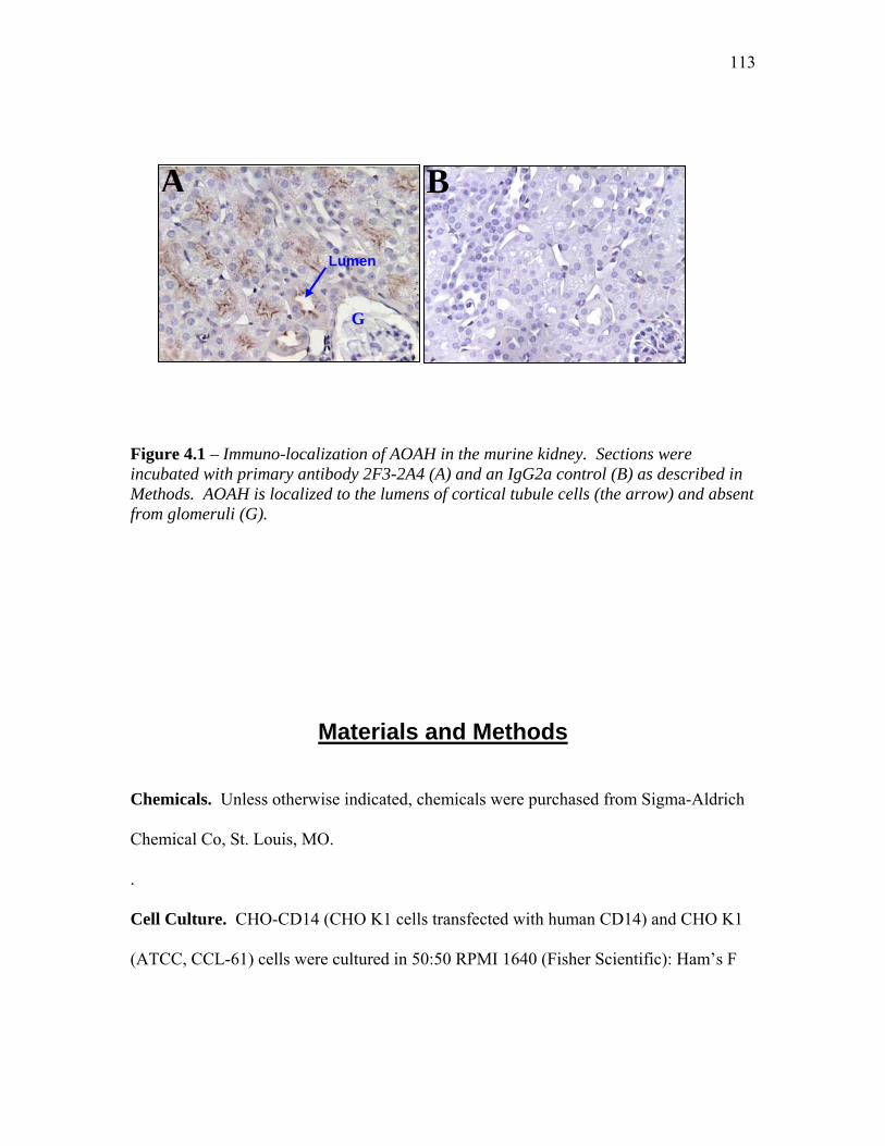
113
G
A B
Lumen
Figure 4.1 – Immuno-localization of AOAH in the murine kidney. Sections were incubated with primary antibody 2F3-2A4 (A) and an IgG2a control (B) as described in Methods. AOAH is localized to the lumens of cortical tubule cells (the arrow) and absent from glomeruli (G).
Materials and Methods
Chemicals. Unless otherwise indicated, chemicals were purchased from Sigma-Aldrich
Chemical Co, St. Louis, MO.
.
Cell Culture. CHO-CD14 (CHO K1 cells transfected with human CD14) and CHO K1
(ATCC, CCL-61) cells were cultured in 50:50 RPMI 1640 (Fisher Scientific): Ham’s F
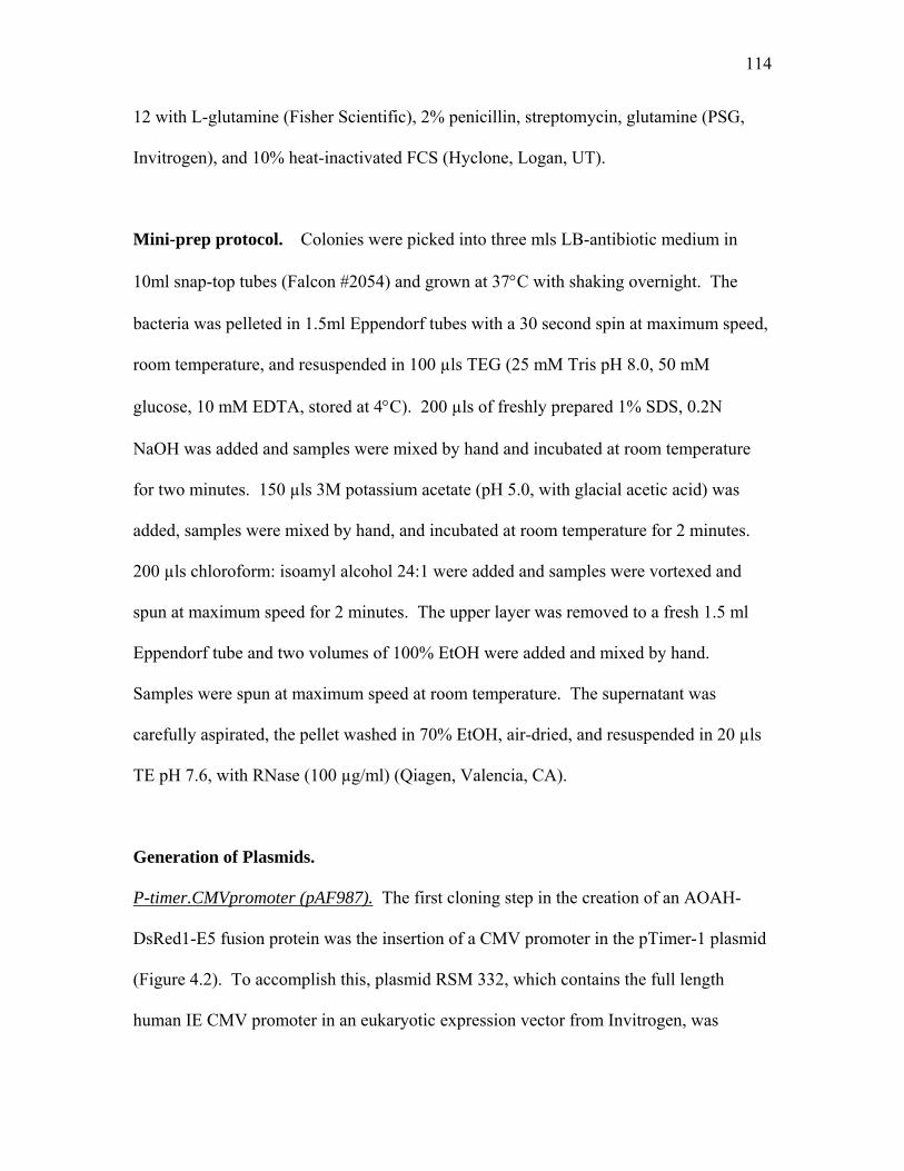
114
12 with L-glutamine (Fisher Scientific), 2% penicillin, streptomycin, glutamine (PSG,
Invitrogen), and 10% heat-inactivated FCS (Hyclone, Logan, UT).
Mini-prep protocol. Colonies were picked into three mls LB-antibiotic medium in
10ml snap-top tubes (Falcon #2054) and grown at 37°C with shaking overnight. The
bacteria was pelleted in 1.5ml Eppendorf tubes with a 30 second spin at maximum speed,
room temperature, and resuspended in 100 µls TEG (25 mM Tris pH 8.0, 50 mM
glucose, 10 mM EDTA, stored at 4°C). 200 µls of freshly prepared 1% SDS, 0.2N
NaOH was added and samples were mixed by hand and incubated at room temperature
for two minutes. 150 µls 3M potassium acetate (pH 5.0, with glacial acetic acid) was
added, samples were mixed by hand, and incubated at room temperature for 2 minutes.
200 µls chloroform: isoamyl alcohol 24:1 were added and samples were vortexed and
spun at maximum speed for 2 minutes. The upper layer was removed to a fresh 1.5 ml
Eppendorf tube and two volumes of 100% EtOH were added and mixed by hand.
Samples were spun at maximum speed at room temperature. The supernatant was
carefully aspirated, the pellet washed in 70% EtOH, air-dried, and resuspended in 20 µls
TE pH 7.6, with RNase (100 µg/ml) (Qiagen, Valencia, CA).
Generation of Plasmids.
P-timer.CMVpromoter (pAF987). The first cloning step in the creation of an AOAH-
DsRed1-E5 fusion protein was the insertion of a CMV promoter in the pTimer-1 plasmid
(Figure 4.2). To accomplish this, plasmid RSM 332, which contains the full length
human IE CMV promoter in an eukaryotic expression vector from Invitrogen, was
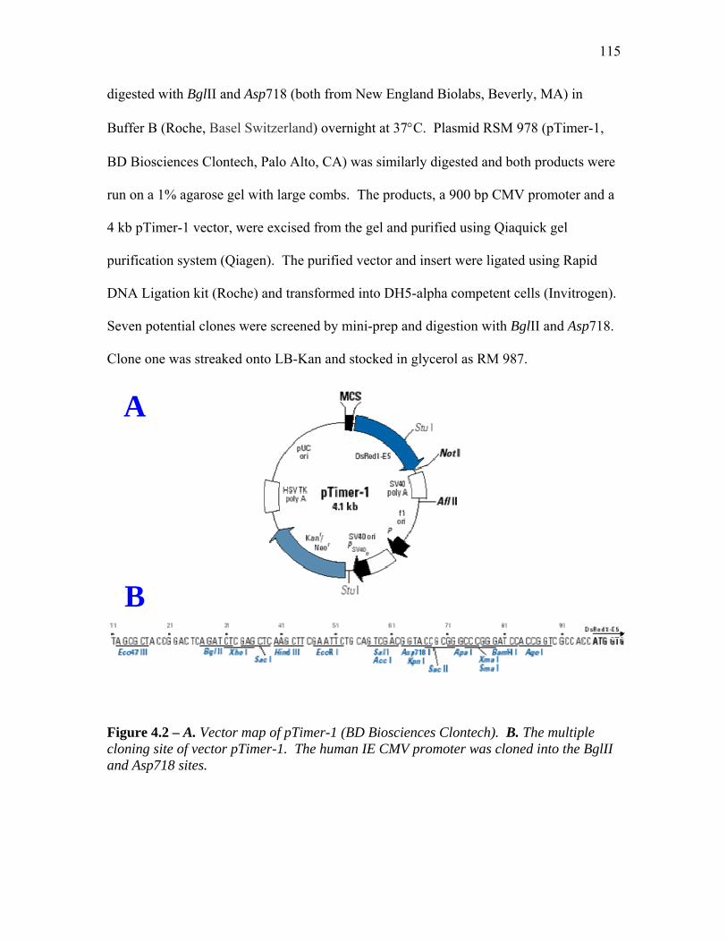
115
digested with BglII and Asp718 (both from New England Biolabs, Beverly, MA) in
Buffer B (Roche, Basel Switzerland) overnight at 37°C. Plasmid RSM 978 (pTimer-1,
BD Biosciences Clontech, Palo Alto, CA) was similarly digested and both products were
run on a 1% agarose gel with large combs. The products, a 900 bp CMV promoter and a
4 kb pTimer-1 vector, were excised from the gel and purified using Qiaquick gel
purification system (Qiagen). The purified vector and insert were ligated using Rapid
DNA Ligation kit (Roche) and transformed into DH5-alpha competent cells (Invitrogen).
Seven potential clones were screened by mini-prep and digestion with BglII and Asp718.
Clone one was streaked onto LB-Kan and stocked in glycerol as RM 987.
A
B
Figure 4.2 – A. Vector map of pTimer-1 (BD Biosciences Clontech). B. The multiple cloning site of vector pTimer-1. The human IE CMV promoter was cloned into the BglII and Asp718 sites.

116
PCR of human AOAH and creation of clone RM 1012. The coding region of human
AOAH was amplified from plasmid pAF956 (pAF956 is the new name for pJSK2301,
Janet Staab’s wt hAOAH with 3 amino acid corrections in pSelect-1 downstream of T7
promoter. I re-stocked and gave it an accession number) using primers:
AF3’hAOAH.AgeI.23 (5’- ACC GGT TTG TGC CCG CCT TGG TC- 3’) and
AF5’hAOAH.AgeI.23 (5’- ACC GGT ATG CAG TCC CCC TGG AA-3’) ordered from
(Integrated DNA Technologies, Inc., Coralville, IA). The PCR was performed with
High-Fidelity Taq polymerase (Roche), an annealing temperature of 65°C, and a 1.5-
minute extension at 72°C, and 30 cycles. The final product was gel purified from a 1%
agarose gel using Qiaquick Gel Purification System (Qiagen) and TA cloned into pDrive
(Qiagen). Several clones were positive and three (1, 3, and 15) were streaked onto LB-
Kan plates, glycerol stocked, and stored at -80°C in freezer position 2, 3, 2, 10 for future
use. Clone number one was sequenced using the following primers: hAOAH.seq#1.af
(5’- GGGTAGAGATGACACAATGG-3’); hAOAH.seq#2.af (5’-
GCGCAGATATGAATGCTG-3’); hAOAH.seq#3.af (5’-CTGTAATGGCATTTGGGG-
3’); hAOAH.seq#4.af (5’- GGTGTTGGACTATCCCG-3’); and hAOAHseq#5.af (5’-
CTTCAGAGAGAGCAGAGC-3’) (Integrated DNA Technologies, Inc., Coralville, IA).
Clone one had 2 mutations, the first, an “A” to “G” at position 1206 which corresponded
to a Thr Ala amino acid change, and a second at position 1504, that did not change
the amino acid. Both of these mutations were determined not to affect the activity of
AOAH. This clone was given the accession number RM 1012 (plasmid = pAF1012).
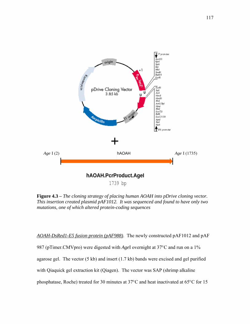
117
hAOAH.P
Age I (2)
Figure 4.3 – The cloning strategy ofThis insertion created plasmid pAF1mutations, one of which altered prot
AOAH-DsRed1-E5 fusion protein (p
987 (pTimer.CMVpro) were digested
agarose gel. The vector (5 kb) and in
with Qiaquick gel extraction kit (Qia
phosphatase, Roche) treated for 30 m
+
crProduct.AgeI1739 bp
hAOAH Age I (1735)
placing human AOAH into pDrive cloning vector. 012. It was sequenced and found to have only two ein-coding sequences
AF988). The newly constructed pAF1012 and pAF
with AgeI overnight at 37°C and run on a 1%
sert (1.7 kb) bands were excised and gel purified
gen). The vector was SAP (shrimp alkaline
inutes at 37°C and heat inactivated at 65°C for 15
118
minutes. The treated vector and insert were ligated with Rapid DNA Ligation kit
(Roche) and transformed into DH5-alpha competent cells (Invitrogen). Positive clones
number two and five (screened with AgeI and AgeI + BglII restriction enzymes) were
streaked onto LB-Kan plates and glycerol stocked. They were given the accession
number AF 988 clone 2 and AF 988 clone 2 (pCMV.p-Timer.hAOAH fusion).
pAF988(pTimer-1.hAOAH.CMVpro)6691 bp
DsRed1-E5 gene
Kanamycin/neomycin resistance gene
hAOAH
SV40 early mRNA polyadenylation signal
SV40 Polyadenylation signal
HSV thymidine kinase (TK) polyadenylation signal
HSV thymidine kinase (TK) polyadenylation signal
DsRed1-C Sequencing Primer
Ampicillin resistance (B-Lactamase) promoter
CMVpro
T7pro
Kosak
SV40 origin of replication
pUC plasmid replication origin
Asp 718 (912)
Age I (933)
Age I (2666)
Bgl II (28)
Bgl II (1438)
Figure 4.4 – pAF988, the final hAOAH-DsRed1-E5 fusion protein. AOAH was digested with AgeI from pAF1012 and inserted into the AgeI site in the multiple cloning region of pAF987. Clones number two and five were glycerol stocked and stored at -80°C. Maxi-preps were also performed and used in future transfection experiments.
AOAH-DsRed2 fusion protein (pAF1127 and pAF1128). To create this plasmid, the
previously described pAF988 was partially digested with AgeI (New England Biolabs)
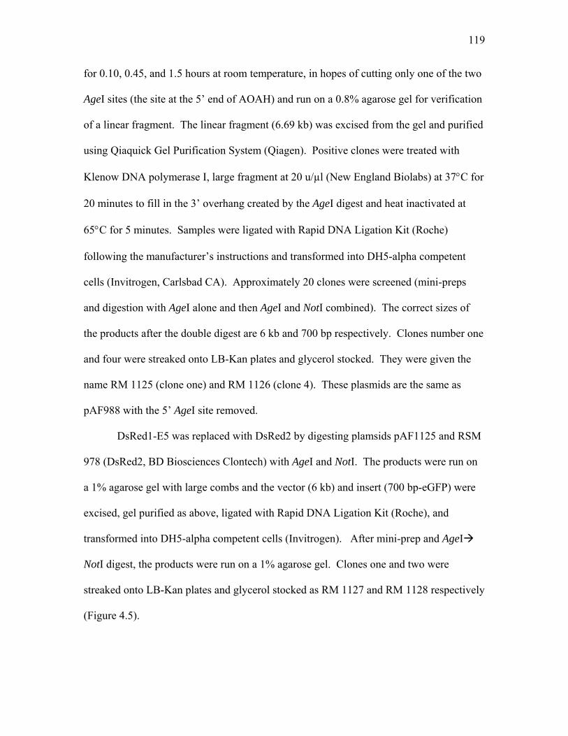
119
for 0.10, 0.45, and 1.5 hours at room temperature, in hopes of cutting only one of the two
AgeI sites (the site at the 5’ end of AOAH) and run on a 0.8% agarose gel for verification
of a linear fragment. The linear fragment (6.69 kb) was excised from the gel and purified
using Qiaquick Gel Purification System (Qiagen). Positive clones were treated with
Klenow DNA polymerase I, large fragment at 20 u/µl (New England Biolabs) at 37°C for
20 minutes to fill in the 3’ overhang created by the AgeI digest and heat inactivated at
65°C for 5 minutes. Samples were ligated with Rapid DNA Ligation Kit (Roche)
following the manufacturer’s instructions and transformed into DH5-alpha competent
cells (Invitrogen, Carlsbad CA). Approximately 20 clones were screened (mini-preps
and digestion with AgeI alone and then AgeI and NotI combined). The correct sizes of
the products after the double digest are 6 kb and 700 bp respectively. Clones number one
and four were streaked onto LB-Kan plates and glycerol stocked. They were given the
name RM 1125 (clone one) and RM 1126 (clone 4). These plasmids are the same as
pAF988 with the 5’ AgeI site removed.
DsRed1-E5 was replaced with DsRed2 by digesting plamsids pAF1125 and RSM
978 (DsRed2, BD Biosciences Clontech) with AgeI and NotI. The products were run on
a 1% agarose gel with large combs and the vector (6 kb) and insert (700 bp-eGFP) were
excised, gel purified as above, ligated with Rapid DNA Ligation Kit (Roche), and
transformed into DH5-alpha competent cells (Invitrogen). After mini-prep and AgeI
NotI digest, the products were run on a 1% agarose gel. Clones one and two were
streaked onto LB-Kan plates and glycerol stocked as RM 1127 and RM 1128 respectively
(Figure 4.5).
120
Figure 4.5 – Cloning on pAF by partial digest and re-ligatigene by double digestion with
This site was removed.
pAF988(pTimer-1.hAOAH.CMVpro)6691 bp
DsRed1-E5 gene
hAOAH
adenylation signal
adenylation signal
DsRed1-C Sequencing Primer
CMVpro
T7pro
Kosak
on origin
Asp 718 (912)
Age I (933)
Age I (2666)
Bgl II (28)
Bgl II (1438)
Not I
1o
127. The 5’ Age I site from plasmid pAF988 was removed n. The DsRed1-E5 gene was replaced with the DsRed2 AgeI and Not I.
DsRed1-E5 was replaced with DsRed2.
121
AOAH-eGFP fusion protein (pAF1224). Faith Sharp, a rotation student, under my
supervision performed the following cloning steps. Plasmid AF1127 (DsRed2-AOAH
fusion protein) and pRK785 (plasmid DsRed1-N1 from BD Biosciences Clontech with
the DsRed1 gene replaced with the enhanced green fluorescence gene, eGFP) were
digested with AgeI and NotI overnight at 37°C. Products were run on a 1% agarose gel
and the 6 kb vector (pAF1127 minus the DsRed2 gene), and 717 bp insert (eGFP), were
excised and purified with Qiaquick gel extraction kit (Qiagen). Vector and insert were
ligated using Rapid DNA Ligation kit (Roche) and transformed into DH10B electro
competent cells (Invitrogen). Colonies were screened with several restriction enzyme
digests: AgeI, NotI, and KpnI to linearize the plasmid at 6.7 kb, NotI + AgeI to give 6.0
kb and 735 bp fragments, and finally with KpnI + AgeI to give 5.0 kb and 1.7 kb
fragments. Positive clone #2 was streaked onto LB-Kan plates, grown overnight at 37°C,
and glycerol stocked. It was given the accession number RM 1224 and stored in the lab’s
bacterial strain stocks at -80°C. Two plasmid preps were made and one stored in the
labs plasmid stocks at -80°C and the other in Y9.330, -20°C, AF Fusion Protein box.

122
pAF12246736 bp
Figure 4.6 – Plasmid pAF1224 (hAOAH-eGFP). Plasmid pAF1224 was created by replacing the DsRed2 gene from pAF1127 with eGFP from pRK785.
Transfections. CHO-CD14 cells were plated at ~ 2 x 10⁵ cells per well in 12-well plates
on 18 mm circle, glass cover slips at #1 thickness (Fisher Scientific cat # 12-545-100),
that had been flamed prior to putting into wells. Transfection was conducted when the
cells reached ~80% confluency (usually in 2 days) using either Superfect (Qiagen) or
Lipofectamine 2000 (Invitrogen) transfection reagents. For Superfect transfections, one
µg of DNA was diluted in 100 µl serum and antibiotic-free CHO medium; four µl of
Superfect reagent was then added slowly and incubated at room temperature for ten
Kanamycin/neomycin resistance gene
hAOAH
eGFP
CMVpro
T7pro
pUC plasmid replication origin
BglII (28)
BglII (1442)
AgeI (2670)
NotI (3405)
123
minutes. During the incubation, the CHO-CD14 cells were washed twice with PBS pH
7.2, and 0.6 mls of fresh serum and antibiotic-free CHO medium was added to the cell
monolayer. After the ten-minute incubation, 100 µl of diluted DNA/Superfect was
added to each well. Cells were incubated for two hours at which time the medium was
changed and the cells were re-incubated for 24 or 48-hours.
Transfections using the Lipofectamine 2000 Reagent (Invitrogen) were performed
as follows. One µg of DNA was diluted into 50 µl serum and antibiotic-free CHO
medium. Two µl of Lipofectamine 2000 reagent was diluted in 50 µl of the serum and
antibiotic-free medium and incubated at room temperature for five minutes. During the
incubation, CHO-CD14 cells were washed two times with PBS and 0.5 ml of fresh serum
and antibiotic-free medium was added to each well. After the five-minute incubation, the
diluted DNA and Lipofectamine were combined and incubated at room temperature for
20 minutes. One hundred µl of this mixture was added to each well of CHO-CD14 cells.
It was determined that changing the media at 5 hours post-transfection did not increase
the efficiency of transfection. The cells can be cultured in serum free media for about
two days before significant cell death is seen. This may actually increase the transfection
efficiency.
Phagocytosis Experiments. Two days prior to the experiment, CHO-CD14 cells were
passed into 6-well plates at 2 x 10⁵ cells per well. XS52 cells were used as a positive
control and were plated at 1 x 10⁶ cells per well two days prior to the experiment. On the
day of the experiment, duplicate negative control cells were treated with 10 µM
cytochalasin D (1:200 from 2M stock) and all cells were given fresh medium and

124
incubated at 37°C, 5% CO2 for 30 minutes to one hour. During this incubation, Bodipy-
labeled E. coli (Molecular Probes, Eugene, OR, cat # E-2864) were thawed on ice,
sonicated for 30 seconds at 6-watts in a water-bath sonicator (Braunsonic 1510, B. Braun,
Melsungen, AG), and analyzed under a fluorescent microscope to ensure there were no
large clumps of bacteria. 100 µl of Bodipy were added to each well and spun at 800 rpm
(~55 x g) at room temperature for 5 minutes on the Sorvall tabletop centrifuge
(RT6000B, Kendro LabProducts, Newton, CT). Cells were re-incubated at 37°C, 5%
CO2 for 1.5 hours. The media was removed, cells washed once in 1x PBS, and lifted into
3 ml FACS tubes (Falcon) with 2 mM EDTA diluted in PBS. Cells were run through a
Fluorescence activated cell sorter (FACS) +/- trypan blue and data were analyzed by Cell
Quest software (Becton Dickinson).
Immunohistochemistry. Sections of normal human kidney (medulla and cortex) were
excised from renal tissue after their removal from patients undergoing radical
nephrectomy. Sections about the size and thickness of a quarter were immediately placed
in 4% formaldehyde/DEPC-PBS (freshly prepared from paraformaldehyde) and
incubated at 4°C for 16 hours. The formaldehyde was replaced with sterile PBS, pH 7.2
and incubated at 4°C for an additional 16 hours. Kidneys were dehydrated and paraffin-
embedded, and 4 µm sections were placed onto microscope slides treated with Vectabond
(Vector Laboratories, Burlingame, CA). Slides were stored desiccated at 4ºC until use.
Murine kidneys were obtained from 129 mice (Harlan) that were anesthetized (ketamine-
acepromazine) and transcardially perfused with cold heparinized DEPC-saline. Tissues
were deparaffinized twice in xylene for 5 minutes and then 3 times in 100% EtOH 2
125
minutes. Slides were dipped in water and then incubated in PBS for 5 minutes. Tissues
were permeabilized in 0.3% Triton X-100/ PBS for 2 incubations, each 5 minutes,
followed by 2 PBS washes. PBS was used as the buffer in all subsequent steps. Non-
specific secondary interactions were blocked with 3% normal horse serum (Vector
Laboratories) for 30 minutes at room temperature. Excess blocking serum was decanted
off the slide by tapping on a paper towel and the areas around the tissue were wiped with
a Kim-wipe. Dilutions of primary antibody (see below) were placed over the tissue and
slides were incubated overnight at 4ºC in a covered dish. After washing in PBS, a
biotinylated horse anti-mouse antibody (Vector Laboratories) was diluted 1:200 in PBS
and applied to the tissue for 30 minutes at room temperature. After a PBS wash,
horseradish peroxidase streptavidin (Vector Laboratories) was diluted 1:500 and
incubated for 30 minutes at room temperature. During this incubation, fresh DAB-
chromagen (Dako, Caprinteria, CA) was prepared by placing 1 tablet into 10 mls of
sterile 0.5 M Tris-Cl, pH 7.6 and allowing it to dissolve. Just prior to use, hydrogen
peroxide (3%, Sigma-Aldrich) was added to a final concentration of 0.3%. After a PBS
wash, DAB-chromagen was added in 2, five-minute incubations. The slides were placed
under running water for approximately 5 minutes and then counter-stained with filtered
hematoxylin for one second. Sections were washed under running water for 5 minutes,
dehydrated 3 times in 100% EtOH for 3 minutes each, and equilibrated 3 times in xylene
for 3 minutes each. Slides were mounted with Permount (Fisher Scientific) and cover-
slips were applied. Human kidney sections were stained with murine monoclonal
antibodies 3C5 (8F8) and 3G4 (3F3). Various concentrations of primary antibody were
tested (1:10 1:10,000). A 1:200 dilution of 3G4 (3F3) was successful in detecting
126
human AOAH in kidney samples. Murine kidneys were incubated with primary antibody
4E5-B3 and B6 (ascites fluid antibody) and purified monoclonal antibody 2F3-2A4.
Only a 1:100 dilution of 2F3-2A4 monoclonal antibody was successful in the detection of
AOAH in the murine kidney.
Discussion In this chapter, I have discussed the generation of three separate AOAH-fusion
proteins. The first, a C-terminal fusion of human AOAH to DsRed1-E5 (pTimer-1), was
an apparent success. Upon transfection, both green and red cytoplasmic staining could be
visualized in transfected CHO-CD14 cells. The addition of 10mM M6P abolished all red
fluorescence in cells transfected with the AOAH-fusion protein, but had no effect on cells
transfected with pTimer-1 alone. This potentially interesting result, if repeated, might
give further evidence to our hypothesis that AOAH must be secreted and subsequently re-
enter the cell in order to mature to the more enzymatically active form of AOAH.
Because DsRed1-E5 changes color from green to red as it matures, finding only green
protein within the cell implies that “old” protein must either be secreted from the cell or
destroyed. Differentiating between these two events should be relatively easy. One
could collect lysates and supernatants from transfected cells and assay them for AOAH
activity as well as for fluorescence on a flourometer. If AOAH is indeed secreted and
taken back up again, the AOAH obtained from the supernatants might initially be green,
and if removed from the cells it would change fluorescence to red over time. I have

127
never detected mature AOAH in the supernatants of LLC-PK1 or AOAH-transfected
BHK 570 cells. If AOAH is destroyed within the cell, one should not be able to detect
AOAH activity in the supernatant. One would assume that red fluorescence would not be
detectable at any early time points, but only after the protein ages.
The second fusion protein, a C-terminal fusion of human AOAH to DsRed2, did
not work. This fusion was created to reduce the complexity of the previous pTimer-1
system. If successful, all red fluorescence would be derived from the AOAH-fusion
protein. Co-staining transfected cells with various organelle-specific markers would
allow us to determine better AOAH’s intracellular location. The single color fusion
protein should also allow us to use our available sources of green fluorescent E. coli and
purified LPS. Once successfully transfected, CHO-CD14 cells could be infected with
Bodipy-labeled E. coli or GFP-labeled purified LPS and one could search for the co-
localization of green and red fluorescence. Unfortunately, this fusion-protein (pAF1127)
was toxic and killed the vast majority of transfected cells. It was nearly impossible to
find a red cell at any time after transfection and the experiments using this plasmid were
terminated.
The third fusion protein, a C-terminal fusion of human AOAH to enhanced green
fluorescent protein (eGFP), is probably a success. Preliminary experiments suggest that
transfection efficiency in CHO-CD14 cells is approximately 20-30% and that it is non-
toxic. These experiments need to be repeated. If consistent results are obtained,
experiments to determine the exact intracellular location of AOAH should be conducted.
I suggest starting with a marker for lysosomes. Molecular Probes Inc., have many of
these transfection reagents and the experiments should be relatively simple. CHO-CD14

128
cells phagocytose Bodipy-labeled E. coli and should therefore phagocytose red-
fluorescent labeled bacteria, although a preliminary experiment should be done to prove
that this is the case. Once we know that the cells phagocytose red-labeled bacteria, we
can begin co-localization studies. One could also do this experiment with fluorescent-
labeled purified LPS. At this time, we do not know if AOAH acts on intact bacteria or
LPS that has been shed from Gram-negative bacteria. I believe it will act on both, but
may do so in different sub-cellular locations. The above experiments will help us to
answer such questions.
Renal cortical tubule cells express abundant AOAH activity. While initial
attempts at immuno-localizing AOAH within these cells were discouraging, I believe it
warrants further investigation. While we only have one antibody capable of recognizing
murine AOAH, several antibodies, both human and mouse, recognize porcine (pig)
AOAH. Immunohistochemical experiments on pig kidneys may allow us to localize
AOAH within proximal tubule cells. If successful, immuno-electron microscopy may be
utilized to localize better AOAH within specific cellular organelles. Bladder cells do not
express AOAH, but in vitro they are able to acquire exogenous AOAH from the
supernatants of AOAH-producing cells. It may not be possible to detect such small
quantities of AOAH in the bladder tissue, although it would be an interesting experiment.
While no definitive results have come from the work presented in this chapter, I
believe we are well on our way to discovering the intracellular location of AOAH. The
plasmids described here should provide a starting point for others to create more useful
fusion proteins. Whoever takes over these projects should be prepared to spend
numerous hours on a good fluorescence microscope. The good microscopes are, at this
129
time, very costly and one needs to spend a lot of time to understand truly how to use them
to their fullest advantage.
Chapter Five
Discussion
Acyloxyacyl hydrolase (AOAH) is a lipopolysaccharide-deacylating enzyme that
until recently has been found only in myeloid lineage cells. By cleaving the secondary
fatty acyl chains from the bioactive center of LPS, lipid A, AOAH reduces the toxicity of
LPS and greatly reduces its ability to stimulate human endothelial cells in vitro as well as
in several models of inflammation 12;24;29. In addition to this function, AOAH may also
modulate innate immune responses to invading Gram-negative bacteria. In the work
presented here, I have described several important discoveries that contribute to a better
understanding of AOAH. There are many future directions to pursue regarding this work
and I hope to outline some of those here.
AOAH Expression in Renal Proximal Tubule Cells
One of my first discoveries in the lab was the finding that AOAH is expressed in
renal cortical tubule cells. This came as a surprise, for AOAH had never been localized
to a non-myeloid lineage cell before. Not only was AOAH present in murine tubule
cells, but its activity was also detected in lysates of human kidney and in first-passage
130

131
primary murine and human renal proximal tubule cells. AOAH expression was lost as
the primary cells differentiated in culture. One of the most commonly used human
proximal tubule cell-lines, A498, used extensively by other labs to study the LPS-induced
renal inflammatory response, does not express AOAH. Because this cell line lacks the
only enzyme capable of deacylating and detoxifying LPS, one must conclude that it is not
an appropriate model for such studies. In an attempt to find a renal proximal tubule cell
line that expresses AOAH , I tested numerous cell lines for AOAH activity. Only LLC-
PK1, porcine proximal tubule cells, had AOAH activity. LLC-PK1 cells were able to
secrete the precursor form of AOAH (Figure 2.9B and 2.9C) into their medium as was
previously demonstrated for recombinant AOAH stably expressed in BHK 570 fibroblast
cells 33. LLC-PK1 cells retain many properties of native proximal tubule cells 86;105 and
therefore are a representative cell line to use for in vitro studies. The available rabbit and
murine monoclonal anti-AOAH antibodies were able to recognize porcine AOAH, which
allowed me to perform immunoprecipitation and Western blot analyses. As described in
chapter two of this dissertation, the lack of appropriate antibodies to murine AOAH has
made immunohistochemical studies in mouse tissues difficult. An alternative might be to
use pig tissue for such studies.
Several lines of evidence, including gentamicin protection and electron
microscopic assays, suggest that Gram-negative bacteria are able to bind to, and invade,
proximal tubule cells 55;56;106-108. Studies in cynomogus monkeys have suggested that
bacteria must possess p-pili in order to bind to proximal tubule cells 53. This work,
combined with the finding that bacteria without such pili are rarely detected in the
kidneys of humans or mice with Gram-negative UTI, has led to the hypothesis that the
132
presence of uropathogens in the kidney (in particular proximal tubule cells) is dependent
upon p pili. On the other hand, both pyelonephritic (pap operon positive) and fecal (pap
operon negative) isolates were able to bind to and invade human first-passage renal
cortical tubule cells 55;56. Regardless of the necessity for p pili-mediated invasion of the
kidney, once bacteria have gained access to the kidney it is evident that proximal tubule
cells are their targets. It seems reasonable that proximal tubule cells should contain
enzymes, such as AOAH, that degrade the bacteria that they ingest.
In order to test the hypothesis that AOAH functions in renal proximal tubule cells
to deacylate LPS contained in internalized bacteria, one could utilize the porcine LLC-
PK1 cells. The first step in these studies would be to perform internalization assays
(either by the traditional gentamicin-protection assay or by allowing cells to internalize
fluorescently-labeled bacteria and analyzing uptake by FACS) using various strains of E.
coli. The experiments conducted by Donnenberg and Warren 55;56 suggest that the
presence of a p pilus is not necessary for the bacteria to gain entry into the tubule cells
and therefore it is reasonable to assume that E.coli lacking p pili might be utilized. I
tested the ability of LLC-PK1 cells to phagocytose Bodipy-labeled (K12) E. coli and
found that while XS52 dendritic cells were able to internalize these bacteria, LLC-PK1
cells were not. I next wondered if a pyelonephritic strains of E. coli (expressing p pili)
might be internalized by LLC-PK1 cells. After labeling pyelonephritic strains RSM 947
(DS17) and RSM 953 (AAEC185/pPil110-35) and their non-piliated control, RSM 948
(DS17-8) 53, and RSM 954 (AAEC185/pPil110-35, G-) with Bodipy, I assayed their
ability to be internalized by both XS52 dendritic and LLC-PK1 cells. Neither the
pyelonephritic or control strains were internalized by LLC-PK1 cells. All strains were

133
internalized by XS52 dendritic cells, but not as effiecintly as commercially prepared
Bodipy-E.coli (K12).
Murine renal proximal tubule cells possess several toll-like receptors (TLRs 1, 2,
3, 4, and 6), CD14 and MD-2 79 and therefore have the pattern recognition receptors
necessary for LPS recognition. Using our double-labeled [3H/14C]LPS substrate, I
performed an experiment to investigate the ability of LLC-PK1 cells to deacylate cell-
associated LPS. Unfortunately, I was unable to show significant deacylation of purified
LPS by LLC-PK1 cells. These experiments should be repeated, using fresh LPS
substrate and if successful, experiments that test the ability of LLC-PK1 cells to deacylate
LPS on internalized Gram-negative bacteria might then be performed. While the
experiments using purified LPS will be relatively straightforward, preparing the substrate
for the internalization of whole E. coli might prove more difficult, especially if p pili are
necessary for invasion. LCD25 E. coli, a rough Ra mutant, is unable to produce acetate
or utilize it as a carbon or energy source. When one incubates it with [14C]acetate, the
label is incorporated exclusively into the fatty acids 94. Although not tested, we have no
reason to believe that LCD25 bacteria express p pili. If need be, one can transform
LCD25 with a plasmid that contains the pap operon, test for agglutination of sheep
erythrocytes, and then label the fatty acyl chains with [14C]acetate. By using positive
(XS52 dendritic) and negative (A498 proximal tubule cells which do not express AOAH)
controls, one might show the dependence on AOAH for the deacylation of internalized
Gram-negative bacteria in proximal tubules.

134
The Secretion of AOAH by Proximal Tubule Cells In Vitro and In Vivo
As mentioned above, AOAH is secreted from renal proximal tubule cells in vitro.
As has been previously demonstrated for recombinant AOAH in transfected BHK 570
fibroblasts, the secreted form of the protein runs as a single band on SDS-PAGE at an
apparent molecular weight of 70,000. It is termed pro-AOAH. Cell lysate fractions from
LLC-PK1 and AOAH-transfected BHK 570 cells contain both pro-AOAH and the
mature, more enzymatically active form of AOAH. Mature AOAH has an apparent
molecular weight of 60,000 on a non-reducing SDS-polyacrylamide gel and 50,000 on a
reducing gel.
AOAH is also present in cortical tubule cells in vivo. This was demonstrated by
AOAH activity assays on human and murine kidney lysates (Figures 2.1, 2.6, and 2.7)
and by in situ hybridization experiments in murine kidney tissue (Figures 2.3 and 2.4).
The presence of AOAH in renal proximal tubules and their known ability, in vitro, to
secrete AOAH, led me to speculate that AOAH would also be secreted from proximal
tubule cells in vivo. A small volume (5 µl) of urine from wild type, but not AOAH -/-
mice, was able to remove 3H fatty acyl chains from the double labeled [3H/14C]LPS
substrate (Figure 2.10 and 2.11) and only secondary (acyloxyacyl-linked) chains were
removed (Figure 2.2). While these data suggest that AOAH is being released from
tubule cells in vivo, they do not indicate which form (pro- or mature AOAH) is present.
To determine this, AOAH was immunoprecipitated from both wild type and AOAH-/-
mice, run on a reducing and non-reducing polyacrylamide gel, and immunoblotted with

135
antibodies specific for murine AOAH. To our surprise, only mature AOAH was detected
in murine urine (Figure 2.12).
There are several explanations for this finding, some of which have already been
tested. It is possible, although not very likely, that proximal tubule cells secrete the
mature form of AOAH in vivo. If culture conditions for primary murine proximal tubule
cells were identified, one could argue that their supernatant might contain AOAH in its
true, in vivo, state. However, these cells have been manipulated and probably do not
represent the true in vivo state of tubule cells. In addition, it has been very difficult to
culture primary tubule cells; even in our successful attempts, cells lost their AOAH
activity relatively quickly. A second explanation for the presence of mature AOAH in
the urine is that AOAH might be converted to the mature form by some component of the
urine during its transit through the kidney and into the bladder. The low pH of the urine
may provide an environment that promotes proteolytic cleavage of AOAH into its mature
form. To address this possibility, urine from AOAH -/- mice was incubated with
concentrated pro-AOAH ex-vivo. Our attempts to show conversion of pro-AOAH to
mature AOAH by Western blot were unsuccessful. It’s possible that the conditions of
this experiment were not optimized or that Western blot is not a sensitive enough assay.
Thirdly, it is possible that AOAH is being converted to the mature form by cells in the
urinary tract as it transits into the bladder. These cells might, then, re-secrete the mature
form into the urine. This is also difficult to test, since it must be done in vitro, where
there are extensive numbers of cell types to test and conditions might not be true to the in
vivo state. Nevertheless, I attempted to address this hypothesis in cultured T24 bladder
cells. Upon incubation with pro-AOAH, T24 bladder cells were unable to secrete the
136
mature form of AOAH into their culture medium, although they were able to mature cell-
associated AOAH (Figure 2.15). It’s possible that another cell type (distal tubule,
collecting duct, or ureter cell) is responsible for such maturation or that this is not the
maturation mechanism.
Importantly, only the precursor form of AOAH (pro-AOAH) can be taken up by
bladder cells in vitro, and therefore it seems unlikely that the mature form of AOAH is
becoming cell-associated in vivo. Because we find AOAH activity in washed bladder
tissue, I am inclined to believe that we do not yet understand the mechanism of uptake
and maturation in vivo. It is possible that proximal tubule cells are indeed secreting pro-
AOAH, but that all of it is becoming cell-associated during its transit in the urinary tract,
allowing for little or none to be detected in our assays.
As discussed in Chapter Two, AOAH activity can also be detected in lysates of
human cortex and medulla (Figure 2.7). Despite repeated attempts, we were unable to
detect AOAH activity in normal or infected (cystitis) human urine (data not shown).
Attempts to concentrate the urine did not allow for the detection of AOAH activity. It is
possible that the kidneys of human and mouse differ in respect to AOAH expression or
secretion or that we were unable to detect the AOAH present in human urine. Firstly, it’s
possible that AOAH is not secreted from human renal proximal tubule cells. This is
unlikely due to the fact that we detect AOAH activity in both the human cortex and
medulla and because all cell types tested to date have secreted the precursor form of
AOAH into their culture medium in vitro or murine urine in vivo. Secondly, AOAH
might be expressed in very low abundance in the human urinary tract making it difficult
for us to detect. A third, related, explanation is that the bladder (or other downstream

137
cells) is taking up nearly all of the secreted AOAH, leaving little enzyme available for
detection in our assays.
Understanding the mechanisms by which AOAH is secreted by tubule cells and
matured within the urinary tract might prove difficult, but this information is very
important for understanding AOAHs role in deacylating the LPS in Gram-negative
uropathogens (or commensal bacteria).
Enzyme Sharing in the Urinary Tract
Another important finding presented in this dissertation is the observation that
AOAH, secreted from proximal tubule cells in vitro, can be “shared” with other cells.
As presented in Chapter two, AOAH protein (as measured by activity) and mRNA (as
measured by real-time PCR) were present in isolated murine renal cortex. However,
renal medulla and urinary bladder samples had AOAH activity but lacked AOAH mRNA
(Figures 2.5 and 2.6). Although the activity in the medulla might be explained by the
presence of urine within the tissue, the activity in the washed bladder tissue argues that
the cells are able to take up AOAH from the urine. This finding, combined with data
presented in Chapter Two (Figure 2.19 A-C), suggests that bladder cells are able to take
up, and subsequently mature, pro-AOAH in vitro. In addition to finding that pro-AOAH
is taken up by bladder cells, it was also demonstrated that 10 mM M6P and ammonium
chloride, but not G6P or mannose, could block such uptake. It is not surprising that the
uptake of AOAH, a heavily N-glycosylated protein, is mediated by the M6P receptor.
Kornfeld, in a 1989 review, suggested that the lysosome is the final destination for
138
soluble proteins internalized by endocytosis. He also noted that this transport is
dependent on recognition of M6P residues by M6P receptors 39. It makes sense then that
AOAH, known for its role in detoxifying LPS on Gram-negative bacteria, should traffic
to lysosomes, where bacteria are known to be targeted for destruction.
There are several experiments that might strengthen the evidence for the role of
M6P residues and receptors in the uptake of AOAH. Cells can be transiently depleted of
M6P receptors by an infection with an adenovirus expressing a ribozyme (a RNA
molecule that can cleave RNA in a sequence specific way) specific for M6P/IGF2R
mRNA 109. One could use such cells (or cells treated with siRNAs specific for the M6P
receptor mRNA) to test their ability to take up pro-AOAH from their culture medium.
One would expect that cells lacking M6P receptors would be unable to associate with
pro-AOAH. Another relatively simple experiment would be to use phosphatases to
remove phosphates from AOAH and subsequently test the modified protein’s ability to
become associated with cells. Again, if the association is mediated by M6P receptors,
one would expect phosphatase treatment to inhibit such associations. A third experiment,
which would require the acquisition of M6P receptor antibodies, would be to test the
ability of such antibodies to block the association of AOAH with recipient cells.
BHK cells that have been transfected with CD-M6PR secrete nearly all newly
synthesized M6P-containing proteins. When co-transfected with both the CD- and CI-
M6P/IGFII receptor, cells restore the intracellular targeting of M6P-containing proteins
to endosomes and reduce their secretions 110. These findings suggest that the CD-M6P
receptor is responsible for the secretion of nascent polypeptides with M6P moieties and
that in the absence of the CI-M6P/IGFII receptor, very few of these proteins are found

139
intracellularly. We wondered if P388D1 murine macrophage-like cells, which lack the
CI-M6P/IGFII receptor (but possess the 46 kDa CD-M6PR)(14), would express AOAH
and if so, whether or not there would be an abundance of AOAH in the culture media but
not in the cells. We hypothesized that, due to the lack of CI-M6P receptors at the plasma
membrane, any AOAH activity would be detected in the culture medium. There was
some confusion about our available P388D1 cell-line, since it had been obtained from
another lab and we could not verify that the cells were indeed P388D1 cells (ATCC
literature claims that P388D1 cells should be in suspension and our cells were not). We
obtained new P388D1 cells from ATCC (CCL-46) and assayed both the cell lysate and
medium for AOAH activity. The results were highly variable and therefore inconclusive.
The first assay, done in duplicate, suggested that both cell lysate and supernatant samples
from P388D1 cells contain a significant amount of AOAH activity. However, a second
and third assay, done 3 and 12 days later, suggested that the supernatant did indeed have
more AOAH than the cell lysate fraction. In two attempts to assay the P388D1 cells
again, months later, neither the supernatant or cell lysate expressed AOAH. This might
suggest that expression of AOAH is lost as the cells are passed in culture over time.
We believe this to be the first description of potential “enzyme sharing” in the
urinary tract. Although other proteins (such as hormones) act at locations distant from
their origin, we were unable to find another such example in the urinary system. In
addition, hormones are signaling molecules whose function is to transmit information
from one site to another. AOAH, however, is not known to be a signaling molecule;
rather, it is part of the cellular machinery. There is no (known) reason why bladder cells
shouldn’t make their own AOAH. Although this is mere speculation, the original (and
140
main) role for AOAH might have been within proximal tubule cells. Here, the enzyme
might function to deacylate internalized Gram-negative bacteria or to perform tasks
completely separate from those involved in host defense against invading uropathogens.
Bladder cells might have taken advantage of the AOAH present in urine, thus allowing
them to associate with and use AOAH to degrade the LPS on internalized Gram-negative
bacteria. By allowing bladder cells to deal with invading pathogens before they make
their way to the kidneys, AOAH might help to prevent kidney infections (pyelonephritis),
which are usually more serious diseases.
While “enzyme sharing” is certainly an interesting concept, it should be noted that
in vitro, recombinant AOAH and murine urine have been shown to deacylate purified
LPS. This suggests that in vivo, AOAH might function to deacylate LPS shed from
Gram-negative bacteria even in the absence of cells (in the urine, for example). Indeed,
an association with bladder cells might not be necessary for the deacylation and
detoxification of bacteria in the urinary tract.
The Role of AOAH in Ascending Urinary Tract Infection
The above-described observations, combined with the fact that the majority of
UTIs are the result of colonization of the urinary tract with uropathogenic E. coli 44, led
me to speculate about possible roles for AOAH in ascending urinary tract infections.
Initially, we wondered if AOAH, by degrading the LPS in invading bacteria, might
contribute to the bacteriostatic nature of murine urine. If so, one would expect infected
141
AOAH null mice to have a more prolonged infection (as assessed by positive urine,
bladder, or kidney cultures) than wild type mice. This did not turn out to be the case. In
fact, as described in Chapter three (Figures 3.0 and 3.1), wild type mice had positive
urine cultures for days longer than did AOAH null animals.
We next wondered if the inflammatory response to UTI might be more intense or
of longer duration in AOAH null mice. Because AOAH is the only known enzyme
capable of removing secondary acyl chains from lipid A, it seems reasonable that
animals without this ability (AOAH null mice) would be more likely to possess fully
acylated (highly stimulatory) LPS than would wild type animals. In fact, data from
several studies where C3H/HeN and C3H/HeJ (hyporesponsive to LPS due to a TLR4
mutation) were infected with UPEC suggest that LPS is necessary for the influx of
neutrophils into both the urine and bladder tissue. Although both strains of mice were
infected to similar levels, C3H/HeJ mice were unable to recruit neutrophils to either their
urine or bladder, had no IL-6 present in their urine 111 and failed to clear bacteria from
their bladders as efficiently as did C3H/HeN mice 42;58-61. It seems reasonable, then, that
overstimulation of the immune system by fully acylated LPS might cause a more
sustained inflammatory response and possible immune-mediated tissue injury.
In light of these data, we wondered whether, upon inducing experimental
ascending UTIs in both wild type and AOAH null mice, AOAH null mice might exhibit a
more intense (duration or intensity) inflammatory response to the infection. To assess
this, I analyzed the number of neutrophils present in the urine following ascending UTI
with a pathogenic strain of E. coli. I found that there was no significant difference in the
number of neutrophils present in the urine of wild type or AOAH null mice at any of the
142
early time points (6, 12, 24 hours post-infection) examined. As the bacteria were cleared
from wild type animals (48 and 72 hours post-infection), I also saw a drop in neutrophil
numbers in the urine (Figure 3.4).
Although enumeration of urinary neutrophils is commonly used to assess
inflammatory responses within the urinary tract, we wondered whether this was indeed an
appropriate measure of local inflammatory responses. As discussed previously, UPEC
invade both bladder epithelium and renal proximal tubule cells 55;56;106-108 and both tissues
either express or have acquired AOAH activity. We wondered if differences between
wild type and AOAH null mice might be better seen in the bladder or kidney. To test this
hypothesis, these tissues were collected at 72 hours post-infection, when both wild type
and AOAH null mice had cleared bacteria from their urine. Tissues were fixed, paraffin
embedded, cut, and stained with hematoxylin and eosin and scored by Dr. Joseph Zhou, a
renal pathologist at UT-Southwestern, for the extent of inflammation present. I chose the
72 hour time point because it allowed me to follow the entire course of infection without
sacrificing any mice. Our results, described in Chapter three and Figure 3.5, indicate that
there is no significant difference in the level of inflammation in either the bladder or
kidney at this time point. In fact, the kidney showed no signs of inflammation in either
wild type or AOAH null mice. Because we infected with a cystitis, rather than a
pyelonephritic, strain of E. coli, we did not expect to see a phenotype in the kidney. We
wondered whether this 72 hour time point might be too late -- if we had missed a critical
early time point that would explain the clearance of bacteria from AOAH null mice, since
one might expect a correlation between clearance of bacteria from the urinary tract and a
preceding inflammatory response. To address this concern, the experiment was repeated

143
and four wild type and five AOAH null mice were sacrificed at 24 hours post-infection.
The results indicated that, at 24 hours post-infection, there is no significant difference in
the inflammatory response between wild type and AOAH null mice in the bladder
(Figure 3.6). I believe there were too few mice to accurately address this question and
that this experiment should be repeated with more mice in each group.
Because these experiments require the use of a large number of animals, it is
difficult to obtain an appropriate sample number for statistical analysis. Ideally, one
would induce infection in approximately 55 mice, collecting urine from all and
sacrificing 10 test and 1 control at each 6, 12, 24, 48, and 72-hour time point. Such an
experiment would allow us to obtain important information about the inflammatory
responses in the bladder and kidney throughout the duration of the experiment without
compromising our ability to assess the final outcome of the infection. Ten mice at each
sacrifice point should be sufficient to see any statistically significant difference that might
be present. Such an experiment would require a coordinated effort on the part of several
individuals in the lab.
It should also be noted here that C57BL/6 mice respond very differently to
ascending UTI than do C3H/HeN mice. While both strains are susceptible to and show
similar immune responses to the invading bacteria, C3H/HeN mice have a more sustained
infection and inflammatory response than do C57BL/6 mice. As described in Chapter 3,
bacteria were present in the urine and bladders of C3H/HeN mice up to 8 days post-
infection, whereas C57BL/6 mice had cleared their infections by day two. While I
believe both strains are potentially useful for our studies, we might be better able to
144
detect an inflammatory phenotype in the C3H/HeN strain of mice due to the higher base-
line level of infection.
Finally, although it can be assumed that any phenotypic difference in the
responses of wild type and AOAH null mice are the result of the absence (or presence) of
AOAH, it might be interesting to test this assumption directly. Using a strain of E. coli
which lacks secondary fatty acyl chains (the msbB mutant), one could induce ascending
infections in wild type and AOAH null mice. One would expect that the level of
infection in the wild type and AOAH null mouse would be virtually identical, since
neither animal would have the “advantage” of being able to deacylate the LPS present on
invading bacteria. These experiments might be difficult because to my knowledge, such
strains do not yet exist.
Malakoplakia and Interstitial Cystitis
Malakoplakia is an uncommon inflammatory disease of unknown etiology that is
most frequently diagnosed in middle-aged women with histories of recurrent Gram-
negative urinary tract infections 112. It is believed that the disease begins, possibly after a
transient period of immunosuppression, with the ingestion of bacteria or bacterial
products by macrophages within the urinary tract (kidney > bladder). Once internalized,
the bacteria seem to be contained within the phagolysosomes, eventually persisting as
dense amorphous aggregates that become encrusted with calcium phosphate and/or
calcium hydroxyapatite. Such calcified structures are termed Michaelis-Gutmann bodies

145
(MGBs). Prolonged antibiotic therapies resolve the lesions and suggest a bacterial
etiology 112.
Xanthogranulomatous pyelonephritis (XGP) is another uncommon disease that
has been speculated to be a more severe or chronic form of malakoplakia. Patients with
XGP have characteristic “stuffed macrophages” that form nodular lesions in the kidneys
and other organs 112. Several investigators have been successful in inducing both
malakoplakia and XGP in animal models. One of the most common methods of inducing
these diseases in rats is ureteral obstruction followed by intravenous E. coli injections 113.
Interestingly, maintaining ureteral obstruction for months after the intravenous dose of
LPS seems to produce kidney lesions that resemble those in XGP patients 114. In each
case, the manifestation of disease seems to be the result of Gram-negative bacterial
infection within the urinary tract and inadequate destruction of bacterial products within
macrophages.
Because of AOAH’s presence throughout the urinary tract and its previously
described expression in macrophages, it might be possible that a deficiency in AOAH
would make one more susceptible to malakoplakia (or XGP). To test this hypothesis, one
could obstruct one ureter with a ligature (using the opposite kidney as a control) while
injecting an intravenous, sub-lethal, dose of E. coli [adapted from 113]. The ligature is
removed two days later and wild type and AOAH null mice are assessed at various time
points (3 days to a month post-infection) for difference in the size, number, or extent of
tissue involvement between the groups. Even if we do not detect the presence of MGBs,
the experiments might uncover other roles for AOAH in a more chronic inflammatory-
disease model. In fact, AOAH might have a greater role in chronic infections, where
146
regulating the responses to LPS might be extremely important so as not to induce
immune-mediated tissue damage.
Conclusions
AOAH is highly conserved throughout evolution and plays an important role in
the detoxification of LPS on Gram-negative bacteria. Although somewhat surprising, the
discovery of AOAH in renal proximal tubule cells seems to make sense. Proximal
tubule cells are equipped with several receptors (CD14, TLR4, and MD-2) that aid in the
recognition of Gram-negative bacteria and they are known targets for such uropathogens
during ascending UTI. One might expect such cells to contain enzymes, such as AOAH,
that help to defend them from overwhelming infection and inflammation. The presence
of AOAH in murine urine, combined with its ability to deacylate purified LPS in the
absence of added detergents, suggests that AOAH might also be able to deacylate free
LPS that is shed from Gram-negative bacteria in the urine. Although this suggests that
bacteria do not have to become cell-associated in order for AOAH to detoxify their LPS,
the ability of AOAH to function in bladder cells would be an added advantage to the host.
The discovery of “enzyme sharing” in vitro suggests that AOAH will be able to deacylate
the LPS on internalized uropathogens in the bladder in vivo. The findings described in
this dissertation help us understand how our immune system responds to Gram-negative

147
bacterial LPS and should give insight into the ways in which our bodies deal with
ascending urinary tract infections.
148
Reference List
1. Rietschel,E.T. and O.Westphal. 1999. Endotoxin: Historical perspectives. In
Endotoxin in Health and Disease. H.Brade, S.M.Opal, S.N.Vogel, and
D.C.Morrison, editors. Marcel Dekker, Inc., New York. 1-30.
2. Hitchcock,P.J., L.Leive, P.H.Makela, E.T.Rietschel, W.Strittmatter, and
D.C.Morrison. 1986. Lipopolysaccharide nomenclature - past, present, and
future. J Bacteriol 166:699-705.
3. Westphal,O. and K.Jann. 1965. Bacterial lipopolysaccharides: extraction
with phenol-water and further applications of the procedure. Methods
Carbohydr.Chem. 5:83-91.
4. Rietschel,E.T., L.Brade, B.Lindner, and U.Zahringer. 1992. Molecular
biochemistry of lipopolysaccharides. In Bacterial Endotoxic
Lipopolysaccharides. D.C.Morrison and J.L.Ryan, editors. CRC Press, Boca
Raton,FL. 3-42.
5. Luderitz,O., A.M.Staub, and O.Westphal. 1966. Immunochemistry of O and
R antigens of Salmonella and related Enterobacteriaceae. Bacteriol.Rev.
30:192-255.
6. Luderitz,O., C.Galanos, H.J.Risse, E.Ruschmann, S.Schlecht, G.Schmidt,
H.Schulte-Holthausen, R.Wheat, O.Westphal, and J.Schlosshardt. 1966.

149
Structural relationship of Salmonella O and R antigens. Ann.N.Y.Acad.Sci.
133:349-374.
7. Holst,O. and H.Brade. 1992. Chemical structure of the core region of
lipopolysaccharides. In Bacterial Endotoxic Lipopolysaccharides.
D.C.Morrison and J.L.Ryan, editors. CRC Press, Boca Raton, FL. 135-170.
8. Brade,H. and E.T.Rietschel. 1984. Alpha-2----4-interlinked 3-deoxy-D-
manno-octulosonic acid disaccharide. A common constituent of
enterobacterial lipopolysaccharides. Eur.J.Biochem. 145:231-236.
9. Mukerjee,P., M.Kastowsky, S.Obst, and K.Takayama. 1999.
Lipopolysaccharide preparations in aqueous media: implications for solution
vs. suspension. In Endotoxin in Health and Disease. H.Brade, S.M.Opal,
S.N.Vogel, and D.C.Morrison, editors. Marcel Dekker, Inc., New York. 221-
228.
10. Qureshi,N., K.Takayama, D.Heller, and C.Fenselau. 1983. Position of ester
groups in the lipid A backbone of lipopolysaccharides obtained from
Salmonella typhimurium. J.Biol.Chem. 258:12947-12951.
11. Wollenweber,H.-W., K.W.Broady, O.Luderitz, and E.Th.Rietschel. 1982.
The chemical structure of lipid A. Demonstration of amide-linked 3-
acyloxyacyl residues in Salmonella minnesota Re lipopolysaccharide.
Eur.J.Biochem. 124:191-198.

150
12. Hall,C.L. and R.S.Munford. 1983. Enzymatic deacylation of the lipid A
moiety of Salmonella typhimurium lipopolysaccharides by human
neutrophils. Proc.Natl.Acad.Sci.USA 80:6671-6675.
13. Imoto,M., H.Yoshimura, N.Sakaguchi, S.Kusumoto, and T.Shiba. 1985. Total
synthesis of Escherichia coli lipid A. Tetrahedron Lett. 26:1545-1548.
14. Kotani,S., H.Takada, M.Tsujimoto, T.Ogawa, I.Takahashi, T.Ikeda,
K.Otsuka, H.Shimauchi, N.Kasai, J.Mashimo, S.Nagao, A.Tanaka,
S.Tanaka, K.Harada, K.Nagaki, H.Kitamura, T.Shiba, S.Kusumoto,
M.Imoto, and H.Yoshimura. 1985. Synthetic lipid A with endotoxic and
related biological activities comparable to those of a natural lipid A from an
Escherichia coli Re-mutant. Infect.Immun. 49:225-237.
15. Galanos,C., O.Luderitz, E.T.Rietschel, O.Westphal, H.Brade, B.Brade,
M.Freudenberg, U.Schade, M.Imoto, H.Yoshimura, S.Kusumoto, and
T.Shiba. 1985. Synthetic and natural Escherichia coli free lipid A express
identical endotoxic activities. Eur.J.Biochem. 148:1-5.
16. Takada,H. and S.Kotani. 1992. Structure-function relationships of lipid A. In
Bacterial Endotoxic Lipopolysaccharides, Vol. I. D.C.Morrison and
J.L.Ryan, editors. CRC Press, Boca Raton. 107-135.
17. Imoto,M., H.Yoshimura, M.Yamamoto, T.Shimamoto, S.Kusumoto, and
T.Shiba. 1984. Chemical synthesis of phosphorylated tetra-acyl disaccharide
151
corresponding to a biosynthetic precursor of lipid A. Tetrahedron Lett.
25:2667-2670.
18. Galanos,C., V.Lehmann, O.Luderitz, E.T.Rietschel, O.Westphal, H.Brade,
L.Brade, M.A.Freudenberg, T.Hansen-Hagge, T.Luderitz, G.McKenzie,
U.Schade, W.Strittmatter, K.Tanamoto, U.Zahringer, M.Imoto,
H.Yoshimura, M.Yamamoto, T.Shimamoto, S.Kusumoto, and T.Shiba. 1984.
Endotoxic properties of chemically synthesized lipid A part structures.
Eur.J.Biochem. 140:221-227.
19. Homma,J.Y., M.Matsuura, S.Kanegasaki, Y.Kawakubo, Y.Kojima,
N.Shibukawa, Y.Kumazawa, A.Yamamoto, K.Tanamoto, T.Yasuda,
M.Imoto, H.Yoshimura, S.Kusumoto, and T.Shiba. 1985. Structural
requirements of lipid A responsible for the functions: A study with
chemically synthesized lipid A and its analogues. J Biochem 98:395-406.
20. Kanegasaki,S., K.Tanamoto, T.Yasuda, J.Y.Homma, M.Matsuura,
M.Nakatsuka, Y.Kumazawa, A.Yamamota, T.Shiba, S.Kusumoto, M.Imoto,
H.Yoshimura, and T.Shimamoto. 1986. Structure-activity relationship of
Lipid A: Comparison of biological activities of national and synthetic Lipid
A's with different fatty acid compositions. J.Biochem. 99:1203-1210.
21. Takada,H., S.Kotani, M.Tsujimoto, T.Ogawa, I.Takahashi, K.Harada,
C.Katsukawa, S.Tanaka, T.Shiba, S.Kusumoto, M.Imoto, H.Yoshimura,
M.Yamamoto, and T.Shimamoto. 1985. Immunopharmacological activities of
152
a synthetic counterpart of a biosynthetic lipid A precursor molecule and of
its analogs. Infect.Immun. 48:219-227.
22. Erwin,A.L. and R.S.Munford. 1992. Processing of LPS by phagocytes. In
Bacterial Endotoxic Lipopolysaccharides. Molecular Biochemistry and
Cellular Biology. D.C.Morrison and J.L.Ryan, editors. CRC Press, Boca
Raton, FL. 405-434.
23. Wollenweber,H.-W., U.Seydel, B.Lindner, O.Luderitz, and E.Th.Rietschel.
1984. Nature and location of amide-bound (R)-3-acyloxyacyl groups in lipid
A of lipopolysaccharides from various gram-negative bacteria.
Eur.J.Biochem. 145:265-272.
24. Munford,R.S. and C.L.Hall. 1986. Detoxification of bacterial
lipopolysaccharides (endotoxins) by a human neutrophil enzyme. Science
234:203-205.
25. Lynn,W.A. and D.T.Golenbock. 1992. Lipopolysaccharide antagonists.
Immunol.Today 13:271-276.
26. Pohlman,T.H., R.S.Munford, and J.M.Harlan. 1987. Deacylated
lipopolysaccharide inhibits neutrophil adherence to endothelium induced by
lipopolysaccharide in vitro. J.Exp.Med. 165:1393-1402.
27. Riedo,F.X., R.S.Munford, W.B.Campbell, J.S.Reisch, K.R.Chien, and
R.D.Gerard. 1990. Deacylated lipopolysaccharide inhibits plasminogen
activator inhibitor-1, prostacyclin, and prostaglandin E2 induction by
153
lipopolysaccharide but not by tumor necrosis factor-alpha. J.Immunol.
144:3506-3512.
28. Kovach,N.L., E.Yee, R.S.Munford, C.R.H.Raetz, and J.M.Harlan. 1990.
Lipid IVA inhibits synthesis and release of tumor necrosis factor induced by
LPS in human whole blood ex vivo. J.Exp.Med. 172:77-84.
29. Somerville,J.E., Jr., L.Cassiano, B.Bainbridge, M.D.Cunningham, and
R.P.Darveau. 1996. A novel Escherichia coli lipid A mutant that produces an
antiinflammatory lipopolysaccharide. J.Clin.Invest. 97:359-365.
30. Erwin,A.L. and R.S.Munford. 1990. Deacylation of structurally diverse
lipopolysaccharides by human acyloxyacyl hydrolase. J.Biol.Chem.
265:16444-16449.
31. Munford,R.S. and C.L.Hall. 1989. Purification of acyloxyacyl hydrolase, a
leukocyte enzyme that removes secondary acyl chains from bacterial
lipopolysaccharides. J.Biol.Chem. 264:15613-15619.
32. Hagen,F.S., F.J.Grant, J.L.Kuijper, C.A.Slaughter, C.R.Moomaw, K.Orth,
P.J.O'Hara, and R.S.Munford. 1991. Expression and characterization of
recombinant human acyloxyacyl hydrolase, a leukocyte enzyme that
deacylates bacterial lipopolysaccharides. Biochemistry 30:8415-8423.
33. Staab,J.F., D.L.Ginkel, G.B.Rosenberg, and R.S.Munford. 1994. A saposin-
like domain influences the intracellular localization, stability, and catalytic
activity of human acyloxyacyl hydrolase. J.Biol.Chem. 269:23736-23742.
154
34. Upton,C. and J.T.Buckley. 1995. A new family of lipolytic enzymes? T.I.B.S.
20:178-179.
35. Munford,R.S., P.O.Sheppard, and P.J.O'Hara. 1995. Saposin-like proteins
(SAPLIP) carry out diverse functions on a common backbone structure.
J.Lipid Res. 36:1653-1663.
36. Munford,R.S. and J.P.Hunter. 1992. Acyloxyacyl hydrolase, a leukocyte
enzyme that deacylates bacterial lipopolysaccharides, has phospholipase,
lysophospholipase, diacylglycerollipase, and acyltransferase activities in
vitro. J.Biol.Chem. 267:10116-10121.
37. Staab,J.F., S.Fosmire, M.Zhang, A.W.Varley, and R.S.Munford. 1999.
Distinctive structural features are shared by human, lapine, and murine
acyloxyacyl hydrolases. J.Endotoxin Res. 5:205-208.
38. von Figura,K. and A.Hasilik. 1986. Lysosomal enzymes and their receptors.
Annu.Rev.Biochem. 55:167-193.
39. Kornfeld,S. and I.Mellman. 1989. The biogenesis of lysosomes. Annu.Rev.Cell
Biol. 5:483-525.
40. Kornfeld,S. 1992. Structure and function of the mannose 6-
phosphate/insulinlike growth factor II receptors. Annu.Rev.Biochem. 61:307-
330.

155
41. Burkitt,H.G., B.Young, and J.W.Heath. 1993. Urinary System. In Wheater's
Functional Histology. Churchill Livingstone, New York. 282-303.
42. Hopkins,W.J., A.Gendron-Fitzpatrick, E.Balish, and D.T.Uehling. 1998.
Time course and host responses to Escherichia coli urinary tract infection in
genetically distinct mouse strains. Infect.Immun. 66:2798-2802.
43. Mulvey,M.A. 2002. Adhesion and entry of uropathogenic Escherichia coli.
Cell Microbiol. 4:257-271.
44. Mühldorfer,I., W.Ziebuhr, and J.Hacker. 2001. Escherichia coli in urinary
tract infections. In Molecular Medical Microbiology. M.Sussman, editor.
Academic Press, London. 1515-1540.
45. Hooton,T.M. and W.E.Stamm. 1997. Diagnosis and treatment of
uncomplicated urinary tract infection. Infect.Dis.Clin.North Am. 11:551-581.
46. Wagenlehner,F.M. and K.G.Naber. 2000. Hospital-acquired urinary tract
infections. J.Hosp.Infect. 46:171-181.
47. Ronald,A. 2002. The etiology of urinary tract infection: traditional and
emerging pathogens. Am.J.Med. 113 Suppl 1A:14S-19S.
48. Martinez,J.J., M.A.Mulvey, J.D.Schilling, J.S.Pinkner, and S.J.Hultgren.
2000. Type 1 pilus-mediated bacterial invasion of bladder epithelial cells.
EMBO J. 19:2803-2812.

156
49. Johnson,J.R. 2003. Virulence factors in Escherichia coli urinary tract
infections. Clin.Microbiol.Rev. 4:80-128.
50. Mulvey,M.A., Y.S.Lopez-Boado, C.L.Wilson, R.Roth, W.C.Parks, J.Heuser,
and S.J.Hultgren. 1998. Induction and evasion of host defenses by type 1-
piliated uropathogenic Escherichia coli. Science 282:1494-1497.
51. Johanson,I., R.Lindsted, and C.Svanborg. 1992. Roles of the pap- and prs-
encoded adhesins in Escherichia coli adherence to human uroepithelial cells.
Infect.Immun. 60:3416-3422.
52. Sampaio,E.P., E.N.Sarno, R.Galilly, Z.A.Cohn, and G.Kaplan. 1991.
Thalidomide selectively inhibits tumor necrosis factor α production by
stimulated human monocytes. J.Exp.Med. 173:699-703.
53. Roberts,J.A., B.-I.Marklund, D.Ilver, D.Haslam, M.B.Kaack, G.Baskin,
M.Louis, R.Möllby, J.Winberg, and S.Normark. 1994. The Gal(α1-4)Gal-
specific tip adhesin of Escherichia coli P-fimbriae is needed for
pyelonephritis to occur in the normal urinary tract. Proc.Natl.Acad.Sci.USA
91:11889-11893.
54. Wullt,B., G.Bergsten, H.Connell, P.Rollano, N.Gebretsadik, R.Hull, and
C.Svanborg. 2000. P fimbriae enhance the early establishment of Escherichia
coli in the human urinary tract. Mol.Microbiol. 38:456-464.
55. Donnenberg,M.S., B.Newman, S.J.Utsalo, A.L.Trifillis, J.R.Hebel, and
J.W.Warren. 1994. Internalization of Escherichia coli into human kidney
157
epithelial cells: comparison of fecal and pyelonephritis-associated strains.
J.Infect.Dis. 169:831-838.
56. Warren,J.W., H.L.T.Mobley, and A.L.Trifillis. 1988. Internalization of
Escherichia coli into human renal tubular epithelial cells. J.Infect.Dis.
158:221-223.
57. Hedlund,M., B.Frendeus, C.Wachtler, L.Hang, H.Fischer, and C.Svanborg.
2001. Type 1 fimbriae deliver an LPS- and TLR4-dependent activation signal
to CD14-negative cells. Mol.Microbiol. 39:542-552.
58. Hagberg,L., R.Hull, S.Hull, J.R.McGhee, S.M.Michalek, and E.C.Svanborg.
1984. Difference in susceptibility to gram-negative urinary tract infection
between C3H/HeJ and C3H/HeN mice. Infect.Immun. 46:839-844.
59. Haraoka,M., L.Hang, B.Frendeus, G.Godaly, M.Burdick, R.Strieter, and
C.Svanborg. 1999. Neutrophil recruitment and resistance to urinary tract
infection. J.Infect.Dis. 180:1220-1229.
60. Linder,H., I.Engberg, I.M.Baltzer, K.Jann, and C.Svanborg-Eden. 1988.
Induction of inflammation by Escherichia coli on the mucosal
level:requirement for adherence and endotoxin. Infect.Immun. 56:1309-1313.
61. Shahin,R.D., I.Engberg, L.Hagberg, and C.Svanborg-Eden. 1987. Neutrophil
recruitment and bacterial clearance correlated with LPS responsiveness in
local gram-negative infection. J.Immunol. 138:3475-3480.

158
62. Frendeus,B., C.Wachtler, M.Hedlund, H.Fischer, P.Samuelsson, M.Svensson,
and C.Svanborg. 2001. Escherichia coli P fimbriae utilize the Toll-like
receptor 4 pathway for cell activation. Mol.Microbiol. 40:37-51.
63. Gentschev,I., G.Dietrich, and W.Goebel. 2002. The E. coli alpha-hemolysin
secretion system and its use in vaccine development. Trends Microbiol. 10:39-
45.
64. Gadeberg,O.V., I.Orskov, and J.M.Rhodes. 1983. Cytotoxic effect of an
alpha-hemolytic Escherichia coli strain on human blood monocytes and
granulocytes in vitro. Infect.Immun. 41:358-364.
65. Falzano,L., C.Fiorentini, G.Donelli, E.Michel, C.Kocks, P.Cossart,
L.Cabanie, E.Oswald, and P.Boquet. 1993. Induction of phagocytic behavior
in human epithelial cells by Escherichia coli cytotoxic necrotizing factor type
1. Mol.Microbiol. 9:1247-1254.
66. Fiorentini,C., G.Donelli, P.Matarrese, A.Fabbri, S.Paradisi, and P.Boquet.
1995. Escherichia coli cytotoxic necrotizing factor 1: evidence for induction
of actin assembly by constitutive activation of the p21 Rho GTPase.
Infect.Immun. 63:3936-3944.
67. De Rycke,J., P.Mazars, J.P.Nougayrede, C.Tasca, M.Boury, F.Herault,
A.Valette, and E.Oswald. 1996. Mitotic block and delayed lethality in HeLa
epithelial cells exposed to Escherichia coli BM2-1 producing cytotoxic
necrotizing factor type 1. Infect.Immun. 64:1694-1705.
159
68. Carbonetti,N.H. and P.H.Williams. 1984. A cluster of five genes specifying
the aerobactin iron uptake system of plasmid ColV-K30. Infect.Immun. 46:7-
12.
69. Jann,B., A.S.Shashkov, D.S.Gupta, and K.Jann. 1992. The O18 antigens
(lipopolysaccharides) of Escherichia coli. Structural characterization of the
O18A, O18A1, O18B and O18B1-specific polysaccharides. Eur.J.Biochem.
210:241-248.
70. Svanborg,C., M.Hedlund, H.Connell, W.Agace, R.D.Duan, A.Nilsson, and
B.Wullt. 1996. Bacterial adherence and mucosal cytokine responses.
Receptors and transmembrane signaling. Ann.N.Y.Acad.Sci. 797:177-190.
71. Schilling,J.D., M.A.Mulvey, C.D.Vincent, R.G.Lorenz, and S.J.Hultgren.
2001. Bacterial invasion augments epithelial cytokine responses to
Escherichia coli through a lipopolysaccharide-dependent mechanism.
J.Immunol. 166:1148-1155.
72. Hagberg,L., I.Engberg, R.Freter, J.Lam, S.Olling, and C.Svanborg-Eden.
1983. Ascending, unobstructed urinary tract infection in mice caused by
pyelonephritogenic Escherichia coli of human origin. Infect.Immun. 40:273-
283.
73. Hoshino,K., O.Takeuchi, T.Kawai, H.Sanjo, T.Ogawa, Y.Takeda, K.Takeda,
and S.Akira. 1999. Cutting Edge: Toll-like receptor 4 (TLR4)-deficient mice

160
are hyporesponsive to lipopolysaccharide: Evidence for TLR4 as the Lps
gene product. J.Immunol. 162:3749-3752.
74. Poltorak,A., X.He, I.Smirnova, M.-Y.v.H.C.Liu, X.Du, D.Birdwell, E.Alejos,
M.Silva, C.Galanos, M.Freudenberg, P.Ricciardi-Castagnoli, B.Layton, and
B.Beutler. 1998. Defective LPS signaling in C3H/HeJ and C57BL/10ScCr
mice: mutations in Tlr4 gene. Science 282:2085-2088.
75. Mulvey,M.A., J.D.Schilling, and S.J.Hultgren. 2001. Establishment of a
persistent Escherichia coli reservoir during the acute phase of a bladder
infection. Infect.Immun. 69:4572-4579.
76. Schilling,J.D., S.M.Martin, C.S.Hung, R.G.Lorenz, and S.J.Hultgren. 2003.
Toll-like receptor 4 on stromal and hematopoietic cells mediates innate
resistance to uropathogenic Escherichia coli. Proc.Natl.Acad.Sci.U.S.A
100:4203-4208.
77. Bäckhed,F., M.Söderhäll, P.Ekmann, S.Normark, and A.Richter-Dahlfors.
2001. Induction of innate immune responses by Escherichia coli and purified
lipopolysaccharide correlate with organ- and cell-specific expression of Toll-
like receptors within the human urinary tract. Cellular Microbiology 3:153-
158.
78. Schilling,J.D., S.M.Martin, D.A.Hunstad, K.P.Patel, M.A.Mulvey,
S.S.Justice, R.G.Lorenz, and S.J.Hultgren. 2003. CD14- and toll-like
receptor-dependent activation of bladder epithelial cells by
161
lipopolysaccharide and type 1 piliated Escherichia coli. Infect.Immun.
71:1470-1480.
79. Tsuboi,N., Y.Yoshikai, S.Matsuo, T.Kikuchi, K.Iwami, Y.Nagai, O.Takeuchi,
S.Akira, and T.Matsuguchi. 2002. Roles of toll-like receptors in C-C
chemokine production by renal tubular epithelial cells. J.Immunol. 169:2026-
2033.
80. Kitchens,R.L., R.J.Ulevitch, and R.S.Munford. 1992. Lipopolysaccharide
(LPS) partial structures inhibit responses to LPS in a human macrophage
cell line without inhibiting LPS uptake by a CD14-mediated pathway.
J.Exp.Med. 1760:485-494.
81. Guo,L., K.B.Lim, J.S.Gunn, B.Bainbridge, R.P.Darveau, M.Hackett, and
S.I.Miller. 1997. Regulation of lipid A modifications by Salmonella
typhimurium virulence genes phoP-phoQ. Science 276:250-253.
82. Munford,R.S. and C.L.Hall. 1985. Uptake and deacylation of bacterial
lipopolysaccharides by macrophages from normal and endotoxin-
hyporesponsive mice. Infect.Immun. 48:464-473.
83. Luchi,M. and R.S.Munford. 1993. Binding, internalization, and deacylation
of bacterial lipopolysaccharides by human neutrophils. J.Immunol. 151:959-
969.
162
84. Weinrauch,Y., S.S.Katz, R.S.Munford, P.Elsbach, and J.Weiss. 1999.
Deacylation of purified LPS by cellular and extracellular components of a
sterile rabbit peritoneal inflammatory exudate. Infect.Immun. 67:3376-3382.
85. Lu,M., M.Zhang, R.L.Kitchens, S.Fosmire, A.Takashima, and R.S.Munford.
2003. Stimulus-dependent deacylation of bacterial lipopolysaccharide by
dendritic cells. J.Exp.Med. 197:1745-1754.
86. Nielsen,R., H.Birn, S.K.Moestrup, M.Nielsen, P.Verroust, and
E.I.Christensen. 1998. Characterization of a kidney proximal tubule cell line,
LLC-PK1, expressing endocytotic active megalin. J.Am.Soc.Nephrol. 9:1767-
1776.
87. Trifillis,A.L., A.L.Regec, and B.F.Trump. 1985. Isolation, culture and
characterization of human renal tubular cells. J.Urol. 133:324-329.
88. Brown,W.J., E.Constantinescu, and M.G.Farquhar. 1984. Redistribution of
mannose-6-phosphate receptors induced by tunicamycin and chloroquine.
J.Cell Biol. 99:320-326.
89. Brown,W.J., J.Goodhouse, and M.G.Farquhar. 1986. Mannose-6-phosphate
receptors for lysosomal enzymes cycle between the Golgi complex and
endosomes. J.Cell Biol. 103:1235-1247.
90. Shimomura,I., Y.Bashmakov, and J.D.Horton. 1999. Increased levels of
nuclear SREBP-1c associated with fatty livers in two mouse models of
diabetes mellitus. J.Biol.Chem. 274:30028-30032.

163
91. Munford,R.S. and A.L.Erwin. 1992. Eucaryotic lipopolysaccharide
deacylating enzyme. Meth.Enzymol. 209:485-492.
92. Shelton,J.M., M.H.Lee, J.A.Richardson, and S.B.Patel. 2000. Microsomal
triglyceride transfer protein expression during mouse development. J.Lipid
Res. 41:532-537.
93. Coulthard,M.G., J.Swindle, R.S.Munford, R.D.Gerard, and R.S.Meidell.
1996. Adenovirus-mediated transfer of a gene encoding acyloxyacyl
hydrolase (AOAH) into mice increases tissue and plasma AOAH activity.
Infect.Immun. 64:1510-1515.
94. Munford,R.S., L.C.DeVeaux, J.E.Cronan, Jr., and P.D.Rick. 1992.
Biosynthetic radiolabeling of bacterial lipopolysaccharide to high specific
activity. J.Immunol.Methods 148:115-120.
95. Baur,M., T.-O.Schmid, and B.Landauer. 1989. Role of phospholipase A in
multiorgan failure with special reference to ARDS and acute renal failure
(ARF). Klin.Wochenschr. 67:196-202.
96. Schilling,J.D., M.A.Mulvey, and S.J.Hultgren. 2001. Dynamic interactions
between host and pathogen during acute urinary tract infections. Urology
57:56-61.
97. Erwin,A.L., R.E.Mandrell, and R.S.Munford. 1991. Enzymatically
deacylated Neisseria LPS inhibits murine splenocyte mitogenesis induced by
LPS. Infect.Immun. 59:1881-1887.

164
98. Poltorak,A., P.Ricciardi-Castagnoli, S.Citterio, and B.Beutler. 2000. Physical
contact between lipopolysaccharide and Toll-like receptor 4 revealed by
genetic complementation. Proc.Natl.Acad.Sci.USA 97:2163-2167.
99. Lien,E., T.K.Means, H.Heine, A.Yoshimura, S.Kusumoto, K.Fukase,
M.J.Fenton, M.Oikawa, N.Qureshi, B.Monks, R.W.Finberg, R.R.Ingalls, and
D.T.Golenbock. 2000. Toll-like receptor 4 imparts ligand-specific recognition
of bacterial lipopolysaccharide. J.Clin.Invest. 105:497-504.
100. Johnson,D.E., C.V.Lockatell, R.G.Russell, J.R.Hebel, M.D.Island,
A.Stapleton, W.E.Stamm, and J.W.Warren. 1998. Comparison of
Escherichia coli strains recovered from human cystitis and pyelonephritis
infections in transurethrally challenged mice. Infect.Immun. 66:3059-3065.
101. Fukushi,Y., S.Orikasa, and M.Kagayama. 1979. An electron microscopic
study of the interaction between vesical epithelium and E. coli. Invest.Urol.
17:61-68.
102. McTaggart,L.A., R.C.Rigby, and T.S.Elliott. 1990. The pathogenesis of
urinary tract infections associated with Escherichia coli, Staphylococcus
saprophyticus and S. epidermidis. J.Med.Microbiol. 32:135-141.
103. Palmer,L.M., T.J.Reilly, S.J.Utsalo, and M.S.Donnenberg. 1997.
Internalization of Escherichia coli by human renal epithelial cells is
associated with tyrosine phosphorylation of specific host cell proteins.
Infect.Immun. 65:2570-2575.

165
104. Springall,T., N.S.Sheerin, K.Abe, V.M.Holers, H.Wan, and S.H.Sacks. 2001.
Epithelial secretion of C3 promotes colonization of the upper urinary tract
by Escherichia coli. Nat.Med. 7:801-806.
105. Dowland,L.K., V.A.Luyckx, A.H.Enck, B.Leclercq, and A.S.L.Yu. 2000.
Molecular cloning and characterization of an intracellular chloride channel
in the proximal tubule cell line, LLC-PK1. J.Biol.Chem. 275:37765-37773.
106. Kabore,A.F., M.Simard, and M.G.Bergeron. 1999. Local production of
inflammatory mediators in an experimental model of acute obstructive
pyelonephritis. J.Infect.Dis. 179:1162-1172.
107. Chippendale,G.R., J.W.Warren, A.L.Trifillis, and H.L.T.Mobley. 1994.
Internalization of Proteus mirabilis by human renal epithelial cells.
Infect.Immun. 62:3115-3121.
108. Cotran,R.S. 1969. The renal lesion in chronic pyelonephritis:
immunofluorescent and ultrastructural studies. J.Infect.Dis. 120:109-117.
109. Chen,Z., Y.Ge, N.Landman, and J.X.Kang. 2002. Decreased expression of
the mannose 6-phosphate/insulin-like growth factor-II receptor promotes
growth of human breast cancer cells. BMC Cancer 2:18.
110. Chao,H.H., A.Waheed, R.Pohlmann, A.Hille, and K.von Figura. 1990.
Mannose 6-phosphate receptor dependent secretion of lysosomal enzymes.
EMBO J 9:3507-3513.
166
111. de Man,P., C.van Kooten, L.Aarden, I.Engberg, H.Linder, and C.Svanborg-
Eden. 1989. Interleukin-6 induced at mucosal surfaces by gram-negative
bacterial infection. Infect.Immun. 57:3383-3388.
112. Dobyan,D.C., L.D.Truong, and G.Eknoyan. 1993. Renal malacoplakia
reappraised. Am.J.Kidney Dis. 22:243-252.
113. Tan,H.K. and R.H.Heptinstall. 1969. Experimental pyelonephritis -- a light
and electron microscopic study of the periodic acid-Schiff positive interstitial
cells. Lab Invest 20:62-69.
114. Povysil,C. and L.Konickova. 1972. Experimental xanthogranulomatous
pyelonephritis. Invest.Urol. 9:313-318.

167
ACKNOWLEDGEMENTS
I would first like to thank my mentor, Dr. Robert Munford for providing me with
an excellent scientific education and for his constant support, and advice. His attentions
to detail, persistence, ethics, and scientific know-how have taught me what a great
scientist should aspire to. I would also like to thank several members of the Munford lab
(past and present) who have been a joy to work with: Dr. Richard Kitchens, Dr. Alan
Varley, John Hardwick, Hong Li, Susan Lin, Dr. Julye Nesbitt, Nikki Reading, Faith
Sharp, and Patty Thompson. I would especially like to thank Mingfang Lu and Jami
Cowan Robinson. I could not have made it through graduate school without the two of
you and I will forever be proud to have spent such valuable time with you.
I would like to thank Dr. Nicolai van Oers and Dr. Justin Radolf for allowing me
to rotate in their labs. Thanks to Dr. Timothy Selatti, who taught me to be thorough and
honest. Thanks to the Microbiology Graduate Program and my committee members, Dr.
Leon Eidels, Dr. Christopher Lu, Dr. Kevin McIver, and Dr. Nicolai van Oers for your
advice and commitment during my training.
Thanks to Dr. Philip Perlman, Dr. Nancy Street and Nancy McKinney for giving
me such wonderful opportunities to serve on committees and advisory panels and always
treating me as a colleague and friend.
Finally, I’d like to thank my family and friends for their support and
encouragement during my time in Dallas. I especially thank my Grandma Feulner, Alice
and Grandpa Ron for all of the letters and long-distance love. Thank you to all of my
friends (I hope you all know who you are). Without you this journey would have been so

168
long! I will miss all of you dearly. To Joe, Darren, Jeremiah, Carilee, Stephanie and
Jeffrey Milush: good luck in your future endeavors, you are all such wonderful people
and I am fortunate to have met you!

VITAE J. Amelia Feulner was born in Saratoga Springs, New York on May 18, 1976 and is the
only daughter of Gene Artell Feulner and Candace Rickett Feulner. She has an older
brother, Kenneth Feulner, who lives in St. Louis, Missouri with his wife Colleen Moses
Feulner and their son, Logan Paul Feulner. After graduating from Schuylerville Central
High School she attended Russell Sage College in Troy, New York. In May 1998 she
graduated, Summa Cum Laude, highest honors, with a Bachelor of Arts degree in
Biology. In August of 1998 she entered the Graduate School of Biomedical Sciences at
The University of Texas Southwestern Medical Center at Dallas where she joined the
Molecular Microbiology program. During her time at UT-Southwestern, she has been
awarded the S. Edward Sulkin Student Scholarship in Microbiology, the Dean’s
Discretionary Award for excellence in leadership (three years in a row), a Graduate
Student Organization Travel Scholarship, and a Microbiology journal-club award.
Permanent Address: 127 Grant Avenue Extension Queensbury, NY 12804