Embed Size (px)
Citation preview
JOURNAL OF BACTERIOLOGY, Nov. 2010, p. 5887–5897 Vol. 192, No. 220021-9193/10/$12.00 doi:10.1128/JB.00745-10Copyright © 2010, American Society for Microbiology. All Rights Reserved.
Identification of YsrT and Evidence that YsrRST Constitute a UniquePhosphorelay System in Yersinia enterocolitica�
Kimberly A. Walker,1,3* Markus W. Obrist,1,3 Shirly Mildiner-Earley,3† and Virginia L. Miller1,2,3
Departments of Genetics1 and Microbiology and Immunology,2 University of North Carolina School of Medicine, Chapel Hill,North Carolina 27599, and Department of Molecular Microbiology, Washington University School of
Medicine, St. Louis, Missouri 631103
Received 25 June 2010/Accepted 10 September 2010
Two-component systems (TCS) and phosphorelay systems are mechanisms used by bacteria and fungi toquickly adapt to environmental changes to produce proteins necessary for survival in new environments.Bacterial pathogens use TCS and phosphorelay systems to regulate genes necessary to establish infectionwithin their hosts, including type III secretion systems (T3SS). The Yersinia enterocolitica ysa T3SS is activatedin response to NaCl by YsrS and YsrR, a putative hybrid sensor kinase and a response regulator, respectively.Hybrid TCS consist of a sensor kinase that typically has three well-conserved sites of phosphorylation:autophosphorylation site H1, D1 within a receiver domain, and H2 in the histidine phosphotransferase (HPt)domain. From H2, the phosphoryl group is transferred to D2 on the response regulator. A curious feature ofYsrS is that it lacks the terminal HPt domain. We report here the identification of the HPt-containing protein(YsrT) that provides this activity for the Ysr system. YsrT is an 82-residue protein predicted to be cytosolic and�-helical in nature and is encoded by a gene adjacent to ysrS. To demonstrate predicted functions of YsrRSTas a phosphorelay system, we introduced alanine substitutions at H1, D1, H2, and D2 and tested the mutantproteins for the ability to activate a ysaE-lacZ reporter. As expected, substitutions at H1, H2, and D2 resultedin a loss of activation of ysaE expression. This indicates an interruption of normal protein function, most likelyfrom loss of phosphorylation. A similar result was expected for D1; however, an intriguing “constitutive on”phenotype was observed. In addition, the unusual feature of a separate HPt domain led us to compare thesequences surrounding the ysrS-ysrT junction in several Yersinia strains. In every strain examined, ysrT is aseparate gene, leading to speculation that there is a functional advantage to YsrT being an independentprotein.
Yersinia enterocolitica is a human pathogen that causes sev-eral gastrointestinal conditions, with symptoms that includefever, vomiting, and diarrhea. In an otherwise healthy host, theinfection is usually self-limiting, lasting 1 to 2 weeks (44).However, individuals with high blood iron levels or with com-promised immune systems can develop a systemic Y. enteroco-litica infection that is often fatal (8). Pigs are a major reservoirof some biotypes of pathogenic Y. enterocolitica (28), and whileit is often isolated from the tonsils and intestinal tract, Y.enterocolitica does not cause obvious disease in swine (10). Thebacteria are shed into the environment via feces or can con-taminate pork products during processing, both of which canlead to contamination of human food and water supplies. Inaddition, Y. enterocolitica is frequently found during surveys offood-borne pathogens in milk supplies (22). The prevalence ofY. enterocolitica in contaminated meat and milk products maybe enhanced by its ability to grow at cool temperatures, even aslow as 0°C (3).
Based on biochemical properties, Y. enterocolitica strains
are subdivided into several biovars. Interestingly, there is aconsiderable range in the severity of disease caused by thedifferent biovars. Biovar 1B strains are the most highlypathogenic for humans, and 1B is the only biovar that islethal in a mouse model of yersiniosis (37, 38, and refer-ences therein). Not surprisingly, these isolates contain vir-ulence factors not found in other biovars (19, 41). Amongthese is the chromosomally encoded Ysa type III secretionsystem (T3SS). In mouse infection studies, deletion of ap-paratus genes (ysa) or individual effector genes (ysp) resultsin a modest attenuation only at 24 h postinfection (27, 45)and a 10-fold increase in the 50% lethal dose, both followingoral inoculation (17). Thus, the role of the Ysa T3SS inpathogenicity is subtle in the mouse model, leading to spec-ulation that the secreted effectors (called Ysps) are impor-tant for gastroenteritis but not for systemic infection (45). Invitro, secretion of Ysps through the Ysa T3SS apparatus isonly detected when the bacteria are cultured at 26°C and inthe presence of a high concentration of salt (e.g., NaCl) (17,50). Subsequent studies provided evidence that the genesencoding both the apparatus and effector proteins are up-regulated by growth in a high concentration of salt, indicat-ing that the salt dependence is at the level of transcription(45–47).
As Y. enterocolitica exists as both a commensal and apathogenic organism and has the capacity to grow in a widerange of temperatures, the expression of many genes must
* Corresponding author. Mailing address: Department of Microbi-ology and Immunology, University of North Carolina at Chapel Hill,804 Mary Ellen Jones Bldg., Campus Box 7290, Chapel Hill, NC27599-7290. Phone: (919) 966-9773. Fax: (919) 962-8103. E-mail:[email protected].
† Present address: Department of Pathology and Laboratory Med-icine, University of Pennsylvania, Philadelphia, PA.
� Published ahead of print on 24 September 2010.
5887
on June 19, 2018 by guesthttp://jb.asm
.org/D
ownloaded from
be tightly regulated to ensure survival under these oftenstressful conditions. One mechanism widely used by pro-karyotes to respond to changes in their environment is signaltransduction via two-component systems (TCS) (49). In thecanonical TCS, a membrane-bound sensor histidine kinase(HK) autophosphorylates at a conserved histidine (His) res-idue in response to an environmental cue. The phosphorylgroup is then transferred to a conserved aspartate (Asp)residue in the receiver domain of a response regulator (RR)protein. RR proteins also contain an effector domain, whichis often a DNA-binding moiety; phosphorylation of the re-ceiver domain activates the effector domain to bind DNAand alter gene transcription (13). A common variation of thecanonical TCS is a phosphorelay (2). Phosphorelays involvetwo or more proteins and three phosphoryl group transferevents (His3Asp3His3Asp). In one subclass of the phos-phorelay group, a hybrid sensor kinase contains not only anHK domain but also receiver (REC) and histidine phospho-transferase (HPt) domains, and the first two phosphotrans-fer events are within the sensor protein. The final phospho-
transfer event is to the RR protein. Additionally, variationson this theme have been reported where three or four pro-teins are required to provide the domains necessary for thephosphorelay (5, 12, 36, 39).
Studies conducted in our laboratory and others showedthat the putative TCS proteins YsrR and YsrS are requiredfor expression of the ysa apparatus genes and specifically ofthe ysaE promoter (Fig. 1A) (30, 45–47). Deletion of eithergene results in a lack of NaCl-induced activation of the ysagenes. Furthermore, the level of ysaE expression in a �ysrSmutant strain is the same as that in a wild-type strain grownin the absence of NaCl, thus implying that YsrS responds tothe NaCl (46). One conundrum of this model is that YsrSappears to belong to the hybrid class of sensor kinases butlacks the terminal HPt domain presumably required totransfer the phosphate to YsrR. In this study, we report theidentification of a gene encoding this HPt function anddemonstrate genetically that the Ysr proteins do indeedconduct a phosphorelay cascade that leads to activation ofthe ysaE promoter and, hence, the ysa and ysp genes.
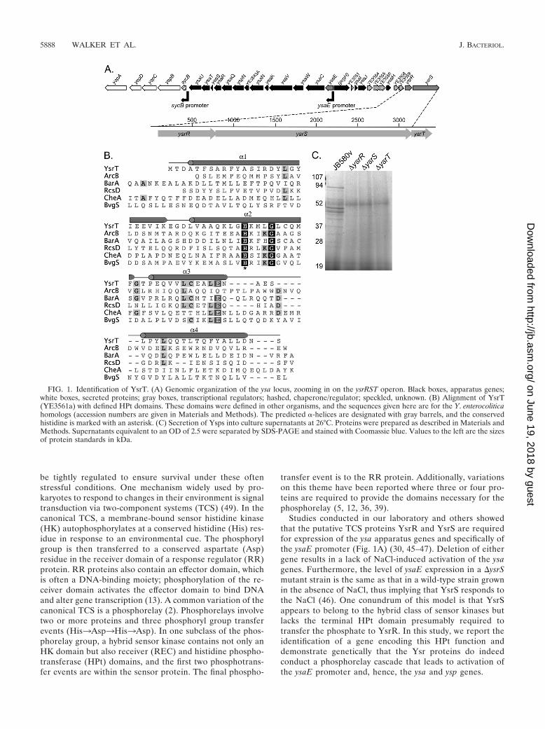
FIG. 1. Identification of YsrT. (A) Genomic organization of the ysa locus, zooming in on the ysrRST operon. Black boxes, apparatus genes;white boxes, secreted proteins; gray boxes, transcriptional regulators; hashed, chaperone/regulator; speckled, unknown. (B) Alignment of YsrT(YE3561a) with defined HPt domains. These domains were defined in other organisms, and the sequences given here are for the Y. enterocoliticahomologs (accession numbers are given in Materials and Methods). The predicted �-helices are designated with gray barrels, and the conservedhistidine is marked with an asterisk. (C) Secretion of Ysps into culture supernatants at 26°C. Proteins were prepared as described in Materials andMethods. Supernatants equivalent to an OD of 2.5 were separated by SDS-PAGE and stained with Coomassie blue. Values to the left are the sizesof protein standards in kDa.
5888 WALKER ET AL. J. BACTERIOL.
on June 19, 2018 by guesthttp://jb.asm
.org/D
ownloaded from
MATERIALS AND METHODS
Bacterial strains and growth conditions. The bacterial strains and plasmidsused in this work are listed in Table 1 and described below. All cultures ofEscherichia coli strains were grown in LB (1% tryptone, 0.5% yeast extract, 170mM NaCl; Difco) at 37°C. Cultures of Y. enterocolitica were grown at 26°C in LB,L broth (1% tryptone, 0.5% yeast extract, 0 mM NaCl), or L broth with 290 mMNaCl (referred to as LB-290). For preparation of secreted proteins, brain heartinfusion (BHI; Difco) and BHI with 490 mM NaCl were used. Antibiotics wereadded as needed at the following concentrations: kanamycin, 100 �g/ml; nalidixicacid, 20 �g/ml; chloramphenicol, 12.5 �g/ml.
Plasmid and strain construction. The plasmids and strains used in this studyare listed in Table 1, and the primers used are listed in Table 2. All of theplasmids made were confirmed by restriction digest patterns and sequencing.
In-frame deletions were constructed as described previously (46). Briefly, forysrS, fragments of approximately 500 bp upstream and downstream were inde-pendently amplified using primers ysrS-delA2/delB2 (upstream) or ysrS-delC2/delD2 (downstream). These fragments were digested with SalI and BamHI(upstream) or BamHI and NotI (downstream), ligated into pSR47S cut with SalIand NotI, and transformed into S17-1 � pir. The resulting plasmid, pKW77, wasintroduced into the desired Y. enterocolitica strains by conjugation. Followingcounterselection, confirmation of the deleted gene was determined by diagnosticPCR using at least one primer outside the region cloned in pKW77. For ysrT, thesame procedure was followed; the primer pairs used were ysrT-delA/delB andysrT-delC/D, and the resulting plasmid was pKW62.
Complementing clone for ysrT. The complementing clone for ysrT was con-structed by amplifying the gene with some additional upstream sequence toinclude the native ribosome binding site using primers KW197 and KW198. Theresulting 300-bp product was digested with SalI and BamHI and ligated intothose sites of pWKS130 to generate pYsrTWT (pKW63).
Point mutations made by overlap extension for ysrS. Point mutations wereintroduced into the ysrS gene using overlap extension PCR (18). Overlappingforward and reverse primers were designed with the desired mutation. Theseprimers were used in two separate PCRs with appropriate cognate primers. Thetwo products were gel purified and used as template DNA for PCR with theflanking forward and reverse primers to amplify the intact full-length product.The product was digested with SalI and NotI and cloned into those sites ofpWKS130 to yield pYsrSH320A (pKW79) and pYsrSD714A (pKW80). These plas-mids are identical to pYsrSWT, except that they contain the alanine substitutions.The primer pairs used were as follows. For H320A, the primers used to introducethe point mutation were H320A-R and H320A-F, used with cognate primersysrS-OE F1 and ysrS-RP3, respectively. For D714A, the primers used to intro-duce the point mutation were D714A-R and D714A-F, used with cognate prim-ers ysrS-OE F1 and ysrS-RP3, respectively.
Point mutations by megaprimer PCR for ysrT and ysrR. To generate analanine substitution for His38 in ysrT, primer KW199 (containing the mutation)was used with KW198 to amplify an �150-bp product that was gel purified andused as a reverse megaprimer in a second PCR with forward primer KW197. Theresulting 300-bp fragment was digested with SalI and BamHI and ligated intothose sites of pWKS130 to generate pYsrTH38A (pKW76). A second constructwas made for generation of the chromosomal copy of ysrT-H38A in a similarfashion. Primer KW199 was used with ysrT-delD to make a 370-bp megaprimerthat was subsequently used in a second PCR with ysrT-delA. This product wasdigested with SalI and NotI and ligated into those sites of pSR47S to generatepKW78. The D75A mutation in ysrR was similarly constructed. Primers M13Rand D75A-F were used with pYsrRWT as template DNA to generate a mega-primer that was subsequently used with ysrR-delA to amplify the full codingsequence. This product was digested with SalI and KpnI and ligated into thosesame sites of pWKS130 to generate pYsrRD75A (pKW72). To generate a chro-
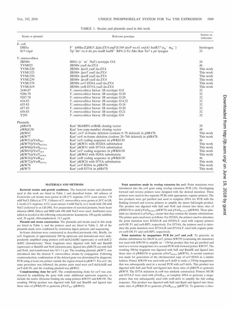
TABLE 1. Strains and plasmids used in this work
Strain or plasmid Relevant genotype Source orreference
E. coliDH5� F� �80lacZ�M15 �(lacZYA-argF)U169 deoP recA1 endA1 hsdR17 (rK
� mK�) Invitrogen
S17-1�pir Tpr Strr recA thi pro hsdR hsdM� RP4::2-Tc::Mu::Km Tn7 � pir lysogen 33
Y. enterocoliticaJB580v 8081v (r� m� Nalr) serotype O:8 24YVM925 JB580v ysaE-lacZYA 46YVM1320 JB580v �ysrS ysaE-lacZYA This workYVM1323 JB580v �ysrT ysaE-lacZYA This workYVM1250 JB580v �ysrR ysaE-lacZYA This workYVM1239 JB580v �rcsB ysaE-lacZYA This workYVM1378 JB580v ysrT-H38A ysaE-lacZYA This workYVM1419 JB580v ysrR-D75A ysaE-lacZYA This work2440-87 Y. enterocolitica biovar 1B serotype O:8 329286-78 Y. enterocolitica biovar 1B serotype O:20 329287-78 Y. enterocolitica biovar 1B serotype O:20 32634-83 Y. enterocolitica biovar 1B serotype O:4,32 32655-83 Y. enterocolitica biovar 1B serotype O:18 32657-83 Y. enterocolitica biovar 1B serotype O:20 32658-83 Y. enterocolitica biovar 1B serotype O:21 32Y295 Y. enterocolitica biovar 1B serotype O:8 32
PlasmidspSR47S Kanr MobRP4 oriR6K cloning vector 29pWKS130 Kanr low-copy-number cloning vector 48pKW62 Kanr; ysrT in-frame deletion (codons 8–78 deleted) in pSR47S This workpKW77 Kanr ysrS in-frame deletion (codons 28–768 deleted) in pSR47S This workpKW31/pYsrSWT Kanr ysrS coding sequence in pWKS130 46pKW79/pYsrSH320A Kanr pKW31 with H320A substitution This workpKW80/pYsrSD714A Kanr pKW31 with D714A substitution This workpKW63/pYsrTWT Kanr ysrT coding sequence in pWKS130 This workpKW76/pYsrTH38A Kanr pKW63 with H38A substitution This workpKW24/pYsrRWT Kanr ysrR coding sequence in pWKS130 46pKW72/pYsrRD75A Kanr pKW24 with D75A substitution This workpKW78 Kanr ysrT-H38A in pSR47S This workpKW75 Kanr ysrR-D75A in pSR47S This work
VOL. 192, 2010 UNIQUE PHOSPHORELAY SYSTEM FOR Ysa T3SS EXPRESSION 5889
on June 19, 2018 by guesthttp://jb.asm
.org/D
ownloaded from
mosomal copy of ysrR-D75A, the megaprimer was synthesized using genomicDNA for the template and primers ysrR-delA and D75A-R. The full-lengthproduct was amplified using the megaprimer and ysrR-delD. This product wasdigested with SalI and NotI and cloned into those same sites of pSR47S togenerate pKW75.
Introduction of chromosomal point mutations for ysrR and ysrT. Chromo-somal copies of genes encoding point mutations were constructed using the samemethods employed to delete genes. Plasmids pKW78 (ysrT) and pKW75 (ysrR)were conjugated into the Y. enterocolitica strain bearing a deletion of the targetedgene. Following counterselection, individual colonies were screened by PCR forthe presence of the full-length gene. The regions surrounding the engineeredpoint mutations were amplified and sequenced to verify that the desired strainhad been constructed without errors. The resulting strains are referred to asysrT-H38A (YVM1378) and ysrR-D75A (YVM1419).
�-Galactosidase assays. Saturated cultures grown overnight in L broth werediluted into fresh L broth or LB-290 to an initial optical density at 600 nm(OD600) of 0.2 and grown for 2 h at 26°C with aeration. Antibiotics were addedas necessary to retain plasmids and chromosomal integrations. Assays wereperformed as described previously (31). Individual assays were conducted with atleast three independent cultures for each strain tested, and the assays wererepeated at least three times to ensure reproducibility. Representative assays areshown.
Preparation of secreted proteins. Proteins secreted into culture supernatantswere collected essentially as described previously (46), except that the basegrowth medium was BHI or BHI with 490 mM NaCl. Briefly, overnight culturesgrown in LB at 26°C were subcultured to an OD of 0.2 into BHI or BHI–490 mMNaCl and grown for 5 to 6 h at 26°C on a roller drum. The cells were pelleted anddiscarded. Supernatants were filtered and precipitated with 10% trichloroaceticacid at 4°C overnight. Protein pellets were resuspended in sample buffer at aconcentration of 1 OD equivalent per 10 �l. Approximately 2.5 OD equivalentswas subjected to 10% sodium dodecyl sulfate-polyacrylamide gel electrophoresis(SDS-PAGE) and stained with Coomassie blue.
DNA sequencing and analysis. All DNA sequencing was performed by EtonBioscience, Inc., at their North Carolina facility. For sequencing of the ysrS-ysrTregion of the biovar 1B isolates (listed in Table 1), genomic DNA was preparedand used as templates for PCR using primers ysrS-delC and ysrT-delD. The PCRproducts were gel purified and sequenced with primer ysrS-delC2. The sequencesfor Y. mollaretii (ATCC 43969, accession no. AALD00000000) (6), Y. aldovae(ATCC 35236, accession no. ACCB00000000) (6), JB580v (Y. enterocolitica 8081,accession no. NC008800) (41), and Y. enterocolitica WA-C (WA-314, accessionno. AJ344214) (21) were obtained from the NCBI repository. Alignments were
conducted using the CLUSTAL W algorithm (40) in the Geneious Pro version 5software package (BioMatters, Ltd., New Zealand).
Secondary structural analysis and modeling. Analysis of the secondary struc-ture prediction for YsrT was performed using the Phyre server (http://www.sbg.bio.ic.ac.uk/phyre) (23). The sequences for the designated HPt-containing pro-teins in Y. enterocolitica were obtained using the following accession numbers:ArcB (YE3733), YP_001007887.1; BarA (YE0742), YP_001005086.1; RcsD(YE1398), YP_001005711.1; CheA (YE2577), YP_001006780.1; BvgS (YE2663),YP_001008383.1. The residues that make up the HPt domain were obtainedfrom the MiST database (43), and the alignment was generated using CLUSTALW as described above. The YsrS receiver domain was modeled using SWISS-MODEL (16) and generated using PyMOL (The PyMOL Molecular GraphicsSystem, version 1.2; Schrodinger, LLC). Domain sequences from YsrR, YsrS,and YsrT were identified with the simple modular architecture research tool(SMART) and used to perform a BLASTp search (1). A subset of homologousproteins whose domains had been defined either genetically or biochemicallywere aligned using the CLUSTAL W algorithm (40) in the Geneious Pro version5 software package (BioMatters, Ltd., New Zealand).
RESULTS
Identification of YsrT. We and others previously demon-strated that YsrS and YsrR are required to activate expressionof the ysaE promoter (30, 45, 46). While these proteins werepredicted to be a hybrid TCS, we noticed that YsrS lacks anHPt domain. Hybrid TCS have four sites of phosphorylationin a His(H1)3Asp(D1)3His(H2)3Asp(D2) phosphorelay.Typically, H1, D1, and H2 are contained on the sensor and D2is found on the RR but examples exist where this is not the case(49). Initial attempts to identify the protein containing the HPtdomain for the Ysr phosphorelay were unsuccessful. All of thegenes that could be identified at the time as encoding HPtproteins in Y. enterocolitica (YE2577 [cheA], YE0724 [barA],YE3733 [arcB], and YE1398 [rcsD]) were inactivated by plas-mid insertion within the gene and the resulting mutants weretested for the ability to secrete Ysps into culture supernatants.None of these proteins appeared to be the intermediate be-
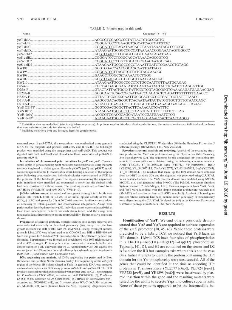
TABLE 2. Primers used in this work
Name Sequencea (533)
ysrT-delA ................................................................................ACGCGTCGACGCCCTATTACTCTGCCGCTGysrT-delB ................................................................................CGGGATCCCTGAAGGTGGCATCAGTCATGTTCysrT-delC ................................................................................CGGGATCCCTAGATAACAGCTAAATAAATAGCCCCGGCysrT-delD................................................................................ATAAGAATGCGGCCGCCATAAAAACCGGAAATAGTGGCCCysrS-delA2 ..............................................................................GCGTCGACCTCGTAGCGGGTGAAACAGATGAGysrS-delB2...............................................................................CGGGATCCCTCGGCAGCATAAACAGCCGTCGysrS-delC2...............................................................................CGGGATCCCCGGTTGCACGCGAACAATGGCAGysrS-delD2 ..............................................................................ATAAGAATGCGGCCGCCTAAATTGATCTCGAACCTGTAGGKW197 ....................................................................................GCGTCGACCAATGGCAGCAATTGATACGKW198 ....................................................................................CGGGATCCTTAGCTGTTATCTAGCAAGGCKW199 ....................................................................................GAAGCTCGGCGCTAAAATGCTGGGKW209 ....................................................................................GCGTCGACGGCGTCGGGTTAATCAAGCGCKW210 ....................................................................................ATAAGAATGCGGCCGCCTCTGGCAATTGTTAATGCAGAGD75A-R ..................................................................................CGCTACGAGGGAATATGCCAGTAATAGTACTTCAATCTCAGGGTTGCD75A-F ...................................................................................GTACTATTACTGGCATATTCCCTCGTAGCGGGTGAAACAGATGAGAACGAGH320A-R ................................................................................GCGCAATTCGGCCGCAATAACCGACAGCTCCAGATTGTTTTTTGAACCCH320A-F .................................................................................GTTATTGCGGCCGAATTGCGCACGCCGCTGATTGGTATTTTAACCD714A-R ................................................................................AAGCCGACAGGCGGTCACAATAATAGTATGGTGGTGTTGTAACCAGCD714A-F .................................................................................ATTATTGTGACCGCCTGTCGGCTTGATGAGAGCGACGGCTTTGAACYsrS OE-F1b ..........................................................................GCGTCGACGGGCTTACTTCAAACACTGATTTCYsrS RP3b...............................................................................ATAAGAATGCGGCCGCTCAGTCATGTTCTTTTTCCTTAGYsrR delAb.............................................................................ACGCGTCGACGCAGGATAATCCGATGAAATCTCGYsrR delDb.............................................................................ATAAGAATGCGGCCGCGCTTGGTAAACCACTCAATCAGCG
a Restriction sites are underlined (six- to eight-base sequences). In primers used to introduce alanine substitutions, the mutated codons are italicized and the basesthat were substituted to code for alanine are bolded.
b Published elsewhere (46) and included here for completeness.
5890 WALKER ET AL. J. BACTERIOL.
on June 19, 2018 by guesthttp://jb.asm
.org/D
ownloaded from
tween YsrS and YsrR, as Ysp secretion was unaffected by thesemutations (data not shown). Upon release of the annotated Y.enterocolitica genome, we noticed that a small open readingframe (ORF), designated YE3561a, was located adjacent toysrS (Fig. 1A). BLASTp results revealed that this hypotheticalprotein had homology to HPt domains, and this is supported byaligning YE3561a with all of the known HPt domains found inY. enterocolitica (Fig. 1B). YE3561a encodes an 82-amino-acidprotein that contains a single histidine residue (H38) locatedwithin a short stretch of somewhat conserved residues found inHPt domains (13). Protein structure prediction using the Phyreserver (23) predicted a high helical content for YE3561a, withthe conserved H located on an exposed region of an alphahelix. These findings are consistent with the notion thatYE3561a serves as an HPt protein.
To investigate if YE3561a encodes a protein important forthe function of the Ysa T3SS, we constructed an in-framedeletion of this gene and tested the resulting strain for thepresence of Ysps in culture supernatants. We found that nosecreted proteins could be detected from cultures grown underinducing conditions when YE3561a was deleted (Fig. 1C). Thisresult is the same as that observed in strains lacking ysrR andysrS (45–47). We further tested this mutant in a strain carryinga chromosomal ysaE-lacZ reporter and found that ysaE expres-sion was not activated when the bacteria were grown underinducing conditions. These data are described in detail in thefollowing sections and shown in Fig. 2. Because of the criticalrole in ysa expression and its putative function as part of aphosphorelay, we have renamed YE3561a ysrT and refer to itas such here.
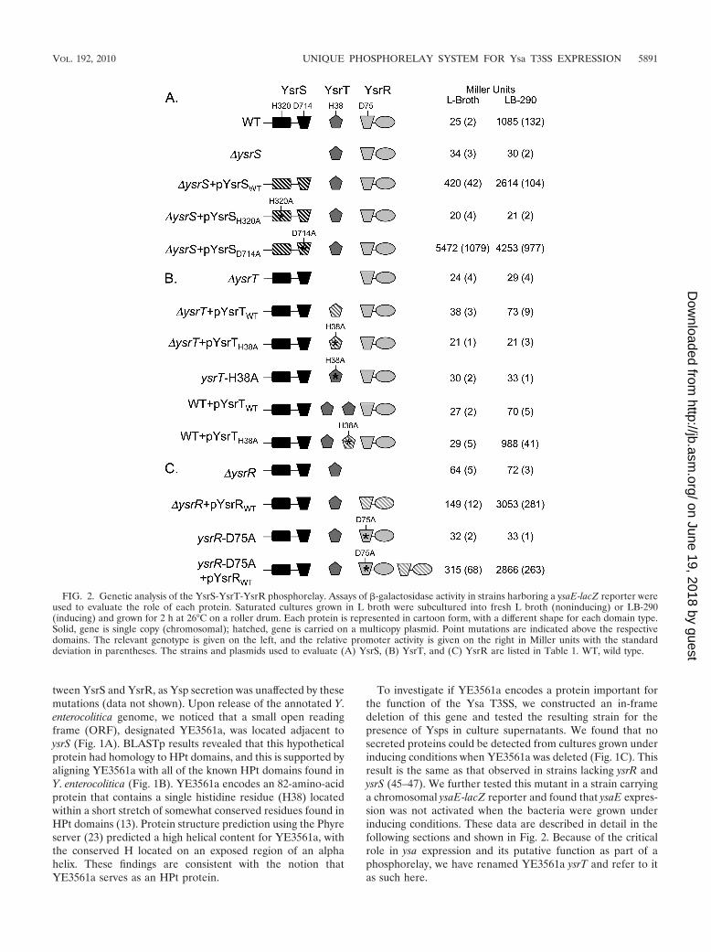
FIG. 2. Genetic analysis of the YsrS-YsrT-YsrR phosphorelay. Assays of -galactosidase activity in strains harboring a ysaE-lacZ reporter wereused to evaluate the role of each protein. Saturated cultures grown in L broth were subcultured into fresh L broth (noninducing) or LB-290(inducing) and grown for 2 h at 26°C on a roller drum. Each protein is represented in cartoon form, with a different shape for each domain type.Solid, gene is single copy (chromosomal); hatched, gene is carried on a multicopy plasmid. Point mutations are indicated above the respectivedomains. The relevant genotype is given on the left, and the relative promoter activity is given on the right in Miller units with the standarddeviation in parentheses. The strains and plasmids used to evaluate (A) YsrS, (B) YsrT, and (C) YsrR are listed in Table 1. WT, wild type.
VOL. 192, 2010 UNIQUE PHOSPHORELAY SYSTEM FOR Ysa T3SS EXPRESSION 5891
on June 19, 2018 by guesthttp://jb.asm
.org/D
ownloaded from
YsrS, YsrT, and YsrR are phosphorelay proteins. With all ofthe components of a hybrid two-component system identifiedand shown to be requisite factors in transcriptional activationof the ysaE promoter, we wanted to determine if YsrSTR wereindeed functioning as phosphorelay proteins. To this end, ala-nine substitutions were made at the histidine and aspartateresidues predicted to participate in the phosphorelay. Theseresidues were chosen based on alignments with other phos-phorelay proteins in which the key residues had been defined(Fig. 1B; see also Fig. 3 and 4) (9). Strains carrying eitherplasmid or chromosomal copies of the mutated genes wereused to evaluate their ability to activate ysaE expression.
Analysis of YsrS. In the wild-type strain (YVM925), ysaE-lacZ expression levels are very low in the absence of NaCl (Lbroth), resulting in about 25 Miller units (MU). In the pres-ence of 290 mM NaCl (LB-290), ysaE-lacZ expression levelsreach about 1,085 MU, a 43-fold activation (Fig. 2A). Consis-tent with previous observations with different ysrS mutantstrains, no activation of ysaE-lacZ is observed in LB-290 in the�ysrS mutant strain (YVM1320), indicating that YsrS is re-quired for the salt-dependent activation of ysaE expression(45–47). This phenotype can be complemented by expressingysrS on a low-copy-number plasmid (pYsrSWT), resulting in420 MU in L broth and 2,614 MU in LB-290 (Fig. 2A). Theelevated levels are likely the consequence of the multicopyplasmid. Plasmid pYsrSH320A carries ysrS with an alanine sub-stitution at H320, the predicted site of autophosphorylation(H1). When pYsrSH320A is transformed into YVM1320(�ysrS), ysaE expression is the same as in a strain with no
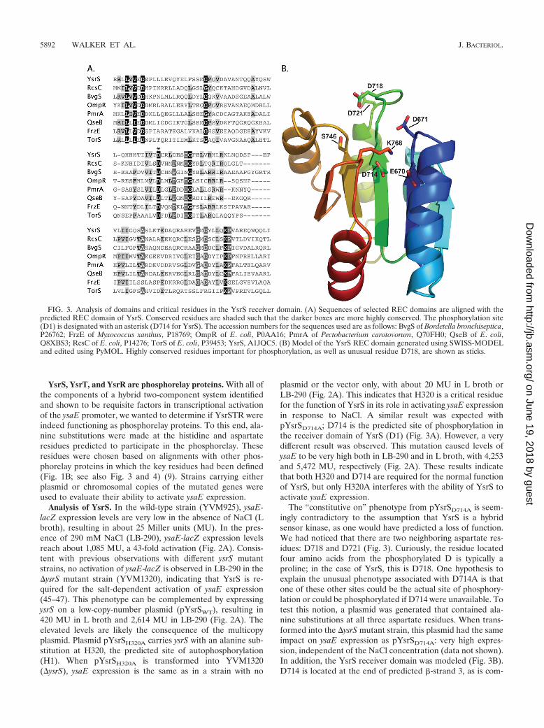
plasmid or the vector only, with about 20 MU in L broth orLB-290 (Fig. 2A). This indicates that H320 is a critical residuefor the function of YsrS in its role in activating ysaE expressionin response to NaCl. A similar result was expected withpYsrSD714A; D714 is the predicted site of phosphorylation inthe receiver domain of YsrS (D1) (Fig. 3A). However, a verydifferent result was observed. This mutation caused levels ofysaE to be very high both in LB-290 and in L broth, with 4,253and 5,472 MU, respectively (Fig. 2A). These results indicatethat both H320 and D714 are required for the normal functionof YsrS, but only H320A interferes with the ability of YsrS toactivate ysaE expression.
The “constitutive on” phenotype from pYsrSD714A is seem-ingly contradictory to the assumption that YsrS is a hybridsensor kinase, as one would have predicted a loss of function.We had noticed that there are two neighboring aspartate res-idues: D718 and D721 (Fig. 3). Curiously, the residue locatedfour amino acids from the phosphorylated D is typically aproline; in the case of YsrS, this is D718. One hypothesis toexplain the unusual phenotype associated with D714A is thatone of these other sites could be the actual site of phosphory-lation or could be phosphorylated if D714 were unavailable. Totest this notion, a plasmid was generated that contained ala-nine substitutions at all three aspartate residues. When trans-formed into the �ysrS mutant strain, this plasmid had the sameimpact on ysaE expression as pYsrSD714A: very high expres-sion, independent of the NaCl concentration (data not shown).In addition, the YsrS receiver domain was modeled (Fig. 3B).D714 is located at the end of predicted -strand 3, as is com-
FIG. 3. Analysis of domains and critical residues in the YsrS receiver domain. (A) Sequences of selected REC domains are aligned with thepredicted REC domain of YsrS. Conserved residues are shaded such that the darker boxes are more highly conserved. The phosphorylation site(D1) is designated with an asterisk (D714 for YsrS). The accession numbers for the sequences used are as follows: BvgS of Bordetella bronchiseptica,P26762; FrzE of Myxococcus xanthus, P18769; OmpR of E. coli, P0AA16; PmrA of Pectobacterium carotovorum, Q70FH0; QseB of E. coli,Q8XBS3; RcsC of E. coli, P14276; TorS of E. coli, P39453; YsrS, A1JQC5. (B) Model of the YsrS REC domain generated using SWISS-MODELand edited using PyMOL. Highly conserved residues important for phosphorylation, as well as unusual residue D718, are shown as sticks.
5892 WALKER ET AL. J. BACTERIOL.
on June 19, 2018 by guesthttp://jb.asm
.org/D
ownloaded from
mon for D1 phosphorylation sites, whereas D718 and D721 arelocated in the loop between -strand 3 and �-helix 3. Otherhighly conserved residues are in the proper positions, such asmagnesium ion coordinating residues E670 and D671, as wellas S746 and K768, which are important for signal transduction(4). Taking all of these data together, we conclude that YsrS isindeed a hybrid sensor kinase, that D714 is the D1 phosphor-ylation site, and that neither of the neighboring aspartate res-idues can substitute for D714 as a site for phosphorylation.
Analysis of YsrT. A similar analysis was conducted withYsrT. As mentioned above, deletion of ysrT prevented activa-tion of ysaE expression, yielding 24 MU in L broth and 29 MUin LB-290 (Fig. 2B). When wild-type ysrT was provided in transon pYsrTWT, ysaE levels were slightly activated in LB-290, with73 MU. However, with pYsrTH38A, which expresses ysrT withan alanine substitution at H38, no activation of ysaE expressionwas observed (21 MU in L broth and LB-290). While theactivation with the wild-type plasmid is subtle, the absence ofthis activation with H38A supports the hypothesis that YsrTfunctions as an HPt.
The lack of full complementation by pYsrTWT could be theresult of the HPt domain playing a role in dephosphorylationof YsrR, which is a known function of these domains (14, 35).To test this notion, pYsrTWT was transformed into YVM925(wild type). Expression of ysaE was markedly decreased, yield-ing only 70 MU in LB-290 (Fig. 2B). Transformation ofpYsrTH38A into YVM925 had no negative impact on ysaElevels, which measured 29 and 988 MU in L broth and LB-290,respectively. Taken together, these data imply that YsrT pos-sesses both kinase and phosphatase activities and that H38 iscritical for both activities.
Because of the concerns of plasmid copy number, we con-structed a strain with the ysrT-H38A mutation on the chromo-some, designated ysrT-H38A (YVM1378). -Galactosidase ac-tivity from the ysaE reporter in this strain is similarly low aswith the �ysrT mutant strain, further supporting the idea thatH38 is required for YsrT to function properly (Fig. 2B). As wasalso observed with the �ysrT mutant strain, addition ofpYsrTWT to ysrT-H38A results in about a 2-fold increase inysaE expression (not shown).
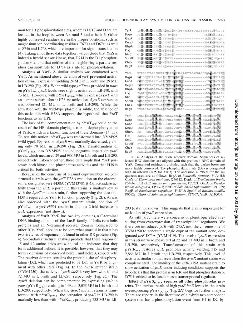
Analysis of YsrR. YsrR has two key domains, a C-terminalDNA-binding domain of the LuxR family of helix-turn-helixproteins and an N-terminal receiver domain. Compared toother RRs, YsrR appears to be somewhat unusual in that it hastwo stretches of sequence not found in other RR proteins (Fig.4). Secondary structural analysis predicts that these regions of15 and 12 amino acids are �-helical and indicates that theyform additional helices. It is possible, however, that they mayform extensions of conserved helix 1 and helix 3, respectively.The receiver domain contains the probable site of phosphory-lation (D2), which was predicted to be D75 in YsrR by align-ment with other RRs (Fig. 4). In the �ysrR mutant strain(YVM1250), the activity of ysaE-lacZ is very low, with 64 and72 MU in L broth and LB-290, respectively (Fig. 2C). The�ysrR deletion can be complemented by expressing ysrR intrans (pYsrRWT), resulting in 149 and 3,053 MU in L broth andLB-290, respectively. When the �ysrR mutant strain is trans-formed with pYsrRD75A, the activation of ysaE in LB-290 ismarkedly less than with pYsrRWT, producing 733 MU in LB-
290 (data not shown). This suggests that D75 is important foractivation of ysaE expression.
As with ysrT, there were concerns of pleiotropic effects re-sulting from overexpression of transcriptional regulators. Wetherefore introduced ysrR with D75A into the chromosome ofYVM1250 to generate a single copy of the mutant gene, des-ignated ysrR-D75A (YVM1419). The levels of ysaE expressionin this strain were measured at 32 and 33 MU in L broth andLB-290, respectively. Transformation of this strain withpYsrRWT restores ysaE promoter activity, yielding 315 and2,866 MU in L broth and LB-290, respectively. This level ofactivity is similar to that seen when the �ysrR mutant strain wascomplemented. The inability of the ysrR-D75A mutant strain toshow activation of ysaE under inducing conditions supports thehypotheses that this protein is an RR and that phosphorylation ofD75 is critical to its function as a transcriptional regulator.
Effect of pYsrSD714A requires all other phosphorelay pro-teins. The curious result of high ysaE-lacZ levels in the strainoverexpressing pYsrSD714A (Fig. 2A) begs for further analysis.There are reports in the literature of a hybrid two-componentsystem that has a phosphorylation event from H1 to D2, by-
*
FIG. 4. Analysis of the YsrR receiver domain. Sequences of se-lected REC domains are aligned with the predicted REC domain ofYsrR. Conserved residues are shaded such that the darker boxes aremore highly conserved. The phosphorylation site (D2) is designatedwith an asterisk (D75 for YsrR). The accession numbers for the se-quences used are as follows: BvgA of Bordetella pertussis, P0A4H2;CheY of Thermotoga maritima, Q56312; DegU of Brevibacillus brevis,P54662; FixJ of Bradyrhizobium japonicum, P23221; GacA of Pseudo-monas aeruginosa, Q51373; NtrC of Salmonella typhimurium, P41789;RegR of Rhodobacter capsulatus, P42508; Spo0F of Bacillus subtilis,P06628; UhpA of Salmonella typhimurium, P27667; YsrR, A1JQC4.
VOL. 192, 2010 UNIQUE PHOSPHORELAY SYSTEM FOR Ysa T3SS EXPRESSION 5893
on June 19, 2018 by guesthttp://jb.asm
.org/D
ownloaded from
passing the D1 and H2 residues (15, 42). Although this wassubsequently found to occur only when the respective proteinswere overexpressed (25), we wanted to address if YsrSD714A
acts independently of YsrT. The �ysrT mutant strain was trans-formed with pYsrSD714A, and ysaE-lacZ levels in this strainwere the same as with the vector, indicating that YsrT is re-quired for the constitutive on phenotype of pYsrSD714A (Fig.5). To determine if YsrR is also required and if phosphoryla-tion of the conserved residues is an important part of thisphenotype, we transformed pYsrSD714A into the ysrT-H38A,�ysrR, and ysrR-D75A mutant strains (Fig. 5). We observedthat YsrT and YsrR must be present and phosphorylatable. Ifeither gene is deleted or if the phosphorylation sites are mu-tated, ysaE levels are the same as the background (�25 to 60MU). These results indicate that the peculiar phenotype ob-served with pYsrSD714A requires wild-type copies of YsrT andYsrR, indicating that no step in the phosphorelay is bypassed.
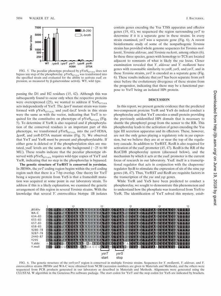
The genetic structure of YsrRST is conserved in yersiniae.In JB580v, the ysrT coding region begins within the ysrS codingregion such that there is a 7-bp overlap. One theory for YsrTbeing a separate protein from YsrS is that a frameshift muta-tion was acquired at some point in our laboratory strain. Toaddress if this is a likely explanation, we examined the geneticarrangement of this region in several Yersinia strains. With theknowledge that several Y. enterocolitica biotype 1B isolates
contain genes encoding the Ysa T3SS apparatus and effectorgenes (19, 41), we sequenced the region surrounding ysrT todetermine if it is a separate gene in these strains. In everystrain examined, ysrT was a separate gene (Fig. 6). A recentbioinformatic study of some of the nonpathogenic Yersiniastrains has provided whole genome sequences for Yersinia mol-laretii, Yersinia aldovae, and Yersinia ruckerii, among others (6).In these three species, genes with homology to TCS are locatedadjacent to remnants of what is likely the ysa locus. Closerexamination revealed that Y. aldovae and Y. mollaretii havegenes with reasonable similarity to ysrR, ysrS, and ysrT, and inthese Yersinia strains, ysrT is encoded as a separate gene (Fig.6). These results indicate that ysrT has been separate from ysrSsince before the evolutionary divergence of these strains fromthe progenitor, indicating that there may be a functional pur-pose to YsrT being an isolated HPt protein.
DISCUSSION
In this report, we present genetic evidence that the predictedtwo-component proteins YsrR and YsrS do indeed conduct aphosphorelay and that YsrT encodes a small protein providingthe previously unidentified HPt domain that is necessary toshuttle the phosphoryl group from the sensor to the RR. Thisphosphorelay leads to the activation of genes encoding the Ysatype III secretion apparatus and its effectors. These, however,are not the only genes playing a regulatory role in ysa expres-sion, but we believe they are at or near the top of the regula-tory cascade. In addition to YsrRST, RcsB is also required foractivation of the ysaE promoter (45, 47). RcsB is the RR of theRcsCDB phosphorelay system (discussed below), and themechanism by which it acts at the ysaE promoter is the currentfocus of research in our laboratory. YsaE itself is a transcrip-tional regulator that acts in conjunction with the chaperone/regulator SycB to stimulate the expression of six of the nine yspgenes (46, 47). Thus, YsrRST and RcsB are requisite factors inthe transcription of the ysa and ysp genes.
While YsrR and YsrS have been predicted to conduct aphosphorelay, we sought to demonstrate this phenomenon andto understand how the phosphate was transferred from YsrS toYsrR. The identification of YsrT solved this mystery, estab-
FIG. 5. The peculiar phenotype produced by pYsrSD714A does notbypass any step of the phosphorelay. pYsrSD714A was transformed intothe specified strain and evaluated for the ability to activate ysaE ex-pression, as measured by -galactosidase activity. WT, wild type.
FIG. 6. The genetic structure of the ysrS-ysrT region is conserved in multiple Yersinia strains. Sequences for Y. mollaretii, Y. aldovae, and Y.enterocolitica strains JB580v and WA-C were obtained from NCBI (accession numbers are given in Materials and Methods), and the others weresequenced from PCR products generated in our laboratory as described in Materials and Methods. Alignments were generated using theCLUSTAL W algorithm in the Geneious Pro software package. The start codon for YsrT and the stop codon for YsrS are indicated by brackets.
5894 WALKER ET AL. J. BACTERIOL.
on June 19, 2018 by guesthttp://jb.asm
.org/D
ownloaded from
lishing that YsrRST contain all of the predicted domainsknown to be required for phosphorelay. To determine ifYsrRST are indeed phosphorelay proteins, we mutated all fourpredicted phosphorylation sites. Each mutation ablated thenormal function of the proteins, and all but YsrSD714A (D2)produced the expected phenotype, with an inability to activateysaE expression. While the phenotype produced by the YsrRD56A mutation was exactly as predicted, there are two struc-tural deviations that indicate that the mechanism of how YsrRresponds to phosphorylation may be atypical. First, there aretwo insertions of 12 and 15 amino acids, respectively, andsecond, a highly conserved proline residue located four residesfrom the phosphorylation site is absent. Both features maycontribute to an altered conformation that could confer aunique function(s).
The constitutive on phenotype of the YsrSD714A mutant isnot easily understood but is very intriguing. It has been pro-posed that the evolutionary advantage of hybrid sensors is theincreased number of regulatory checkpoints to minimize phos-phorylation of the REC under noninducing conditions (13, 20,34). In addition to kinase activity, some HKs also appear topossess phosphatase activities (7, 14). One plausible explana-tion is that this mutation may have impaired the phosphatasefunction of YsrS without impairing the kinase activity. How-ever, one would have to assume that a neighboring residuecould substitute for D714 for the kinase activity but not for thephosphatase activity. YsrS has D resides at positions 718 and721, but these are unlikely to become phosphorylated due totheir locations just outside the active site. A curious feature ofthis region of YsrS is the absence of a highly conserved prolinefour residues from D714. It is tempting to speculate that thearchitecture of this loop may be altered and confer an atypicalfunction. We are continuing experimentation with this mutantin an effort to understand this unusual phenotype. However,biochemical analyses often used to complement the geneticexperiments and more definitively show transfer of phosphorylgroups have proven to be challenging for YsrS.
Two-component and phosphorelay systems are commonmechanisms by which prokaryotes and fungi can rapidly adaptto their environments, and many bacterial strains contain 20 to30 such systems (MiST2 database; 43). The majority of thesesystems are composed of two proteins, a sensor kinase and anRR, but in a few systems, multiple proteins are involved in aphosphorelay cascade. The first phosphorelay system describedconsisted of the sporulation regulators KinABC, Spo0F,Spo0B, and Spo0A of Bacillus subtilis, which have each domainencoded in a separate protein (5). More common among phos-phorelay systems are the hybrid two-component systems whichare composed of two proteins. However, many examples existwhere the phosphorelay requires three proteins: a sensor ki-nase containing the HK (H1) and REC (D1) domains, an HPt(H2) protein, and an RR (D2). One such system is the Rcssystem. In this system, RcsC is the sensor, RcsD has the HPtdomain, and RcsB is the DNA-binding RR (reviewed in ref-erence 26). In this case, the HPt protein is a large membrane-bound protein that has features suggesting that at one time itmay have been a complete hybrid sensor (39). In the Vibrioharveyi Lux system, the HPt-containing protein, LuxU, is en-coded as a small cytosolic protein (11). Intriguingly, LuxUserves as the HPt for two sensors (LuxN and LuxQ), allowing
cross communication and versatility in these important quo-rum-sensing regulators. While these two examples are cases ofindependent HPt domains, this is a relatively rare situation.Examination of the MiST2 database (43) shows that very feworganisms have proteins that contain only an HPt domain.Pseudomonas aeruginosa PAO1 is a rare example of an organ-ism that has several such free HPt proteins, with four. Y.enterocolitica has an average number of HPt-containing pro-teins (six) compared to other bacteria, and all are part ofhybrid HKs. This database does not identify YsrT as an HPtprotein (search last performed on 24/6/2010), perhaps becauseit is such a small ORF. This suggests that there may indeed bemore such proteins, but since the similarity between the knownHPt domains is weak, they may be difficult to identify in silico.
The location of ysrT led us to question if a frameshift mu-tation may have occurred in our strain, resulting in YsrT as aprotein independent of YsrS. Sequence analysis indicates thatthis genetic organization is conserved not only in Y. enteroco-litica 1B isolates but in more distantly related nonpathogenicYersinia strains. Whole-genome sequencing has recently beenperformed for several nonpathogenic Yersinia strains, and Y.ruckerii and Y. mollaretii have reasonably well-conserved ysrRand ysrS sequences and Y. mollaretii has a gene that is likelyysrT (6). The region downstream of ysrS is more divergent in Y.ruckerii, and the protein sequence for the ORF downstream ofysrS is quite different from YsrT but still has homology to HPtdomains (not shown). Y. aldovae appears to contain this locus,but the sequence suggests that a number of frameshift muta-tions have been acquired; the DNA sequence is well conserved,however, and indicates that ysrT would have encoded a sepa-rate protein. Thus, the conservation of the genetic structure ofthe ysrRST genes among not only the pathogenic 1B isolatesbut also environmental isolates implies that there is some func-tional importance to YsrT being produced as a separate pro-tein. However, one should be careful about assuming that thepresence of a ysrRST locus, as well as the ysa and ysp genes, willresult in secretion of Ysps. Although we have shown here thatbiotype 1B isolates of Y. enterocolitica have ysrT and Howard etal. previously demonstrated by microarray analysis that thesebiotype 1B isolates have the ysa and ysp genes (19), we havefound significant differences in the abilities of these samestrains to secrete Ysps under laboratory conditions (K. A.Walker, S. E. Witowski, and V. L. Miller, unpublished results).
This body of research serves to define genetically that theYsrRST system comprises a phosphorelay system and thatphosphorylation is a requisite element in the normal functionof these proteins in their role as activators of the ysa type IIIsecretion genes. Several elements of this system are unusual inthat (i) the hybrid sensor lacks an HPt domain, (ii) the HPtdomain is provided on the small cytosolic protein YsrT, (iii)ysrT is encoded immediately downstream of ysrS, and (iv) YsrTis unique to Y. enterocolitica and nonpathogenic Yersinia spe-cies. In addition, YsrR contains stretches of amino acids thatappear to be insertions not found in similar RRs, indicatingthat it may have some unique properties/functions as well. Likemany Gram-negative bacteria, Y. enterocolitica has about 30TCS, with 24 HKs, 6 of which are hybrid HKs, and 31 RRs. Ofthe six hybrid HKs, two are unique to yersiniae: YsrRST andYE3578-YE3579. Curiously, YE3578 and YE3579 are nearlyidentical to YsrR and YsrS, respectively, on both the amino
VOL. 192, 2010 UNIQUE PHOSPHORELAY SYSTEM FOR Ysa T3SS EXPRESSION 5895
on June 19, 2018 by guesthttp://jb.asm
.org/D
ownloaded from
acid and DNA levels. Adding to the intrigue of this secondsystem is that there is no HPt-containing protein associatedwith YE3578-YE3579. In analogy to the LuxNUO andLusQUO systems, it is tempting to speculate that YsrT couldfunction as the HPt for both YsrS and YE3579, thus explainingthe significance of ysrT encoding a protein detached from itscognate sensor. Efforts to elucidate whether YE3578-YE3579utilizes YsrT for regulating ysaE or other promoters are on-going in our laboratory.
ACKNOWLEDGMENTS
We thank Ruth E. Silversmith for critical reading of the manuscriptand Ruth E. Silversmith, Bob Bourret, Joshua Hall, and MatthewLawrenz for stimulating discussions. We are indebted to BrittanyFogarty and R. Patrick Summers for technical assistance.
This research was supported by National Institutes of Health grantAI063299 awarded to V. L. Miller.
REFERENCES
1. Altschul, S. F., W. Gish, W. Miller, E. W. Myers, and D. J. Lipman. 1990.Basic local alignment search tool. J. Mol. Biol. 215:403–410.
2. Appleby, J. L., J. S. Parkinson, and R. B. Bourret. 1996. Signal transductionvia the multi-step phosphorelay: not necessarily a road less traveled. Cell86:845–848.
3. Bottone, E. J. 1997. Yersinia enterocolitica: the charisma continues. Clin.Microbiol. Rev. 10:257–276.
4. Bourret, R. B. 2010. Receiver domain structure and function in responseregulator proteins. Curr. Opin. Microbiol. 13:142–149.
5. Burbulys, D., K. A. Trach, and J. A. Hoch. 1991. Initiation of sporulation inB. subtilis is controlled by a multicomponent phosphorelay. Cell 64:545–552.
6. Chen, P. E., C. Cook, A. C. Stewart, N. Nagarajan, D. D. Sommer, M. Pop,B. Thomason, M. P. Thomason, S. Lentz, N. Nolan, S. Sozhamannan, A.Sulakvelidze, A. Mateczun, L. Du, M. E. Zwick, and T. D. Read. 2010.Genomic characterization of the Yersinia genus. Genome Biol. 11:R1.
7. Clarke, D. J., S. A. Joyce, C. M. Toutain, A. Jacq, and I. B. Holland. 2002.Genetic analysis of the RcsC sensor kinase from Escherichia coli K-12. J.Bacteriol. 184:1204–1208.
8. Cover, T. L., and R. C. Aber. 1989. Yersinia enterocolitica. N. Engl. J. Med.321:16–24.
9. Foussard, M., S. Cabantous, J. Pedelacq, V. Guillet, S. Tranier, L. Mourey,C. Birck, and J. Samama. 2001. The molecular puzzle of two-componentsignaling cascades. Microbes Infect. 3:417–424.
10. Fredriksson-Ahomaa, M., M. Bucher, C. Hank, A. Stolle, and H. Korkeala.2001. High prevalence of Yersinia enterocolitica 4:O3 on pig offal in southernGermany: a slaughtering technique problem. Syst. Appl. Microbiol. 24:457–463.
11. Freeman, J. A., and B. L. Bassler. 1999. A genetic analysis of the function ofLuxO, a two-component response regulator involved in quorum sensing inVibrio harveyi. Mol. Microbiol. 31:665–677.
12. Freeman, J. A., and B. L. Bassler. 1999. Sequence and function of LuxU: atwo-component phosphorelay protein that regulates quorum sensing inVibrio harveyi. J. Bacteriol. 181:899–906.
13. Gao, R., and A. M. Stock. 2009. Biological insights from structures of two-component proteins. Annu. Rev. Microbiol. 63:133–154.
14. Georgellis, D., O. Kwon, P. De Wulf, and E. C. Lin. 1998. Signal decaythrough a reverse phosphorelay in the Arc two-component signal transduc-tion system. J. Biol. Chem. 273:32864–32869.
15. Georgellis, D., A. S. Lynch, and E. C. Lin. 1997. In vitro phosphorylationstudy of the arc two-component signal transduction system of Escherichiacoli. J. Bacteriol. 179:5429–5435.
16. Guex, N., and M. C. Peitsch. 1997. SWISS-MODEL and the Swiss-PdbViewer: an environment for comparative protein modeling. Electro-phoresis 18:2714–2723.
17. Haller, J. C., S. Carlson, K. J. Pederson, and D. E. Pierson. 2000. A chro-mosomally encoded type III secretion pathway in Yersinia enterocolitica isimportant in virulence. Mol. Microbiol. 36:1436–1446.
18. Ho, S. N., H. D. Hunt, R. M. Horton, J. K. Pullen, and L. R. Pease. 1989.Site-directed mutagenesis by overlap extension using the polymerase chainreaction. Gene 77:51–59.
19. Howard, S. L., M. W. Gaunt, J. Hinds, A. A. Witney, R. Stabler, and B. W.Wren. 2006. Application of comparative phylogenomics to study the evolu-tion of Yersinia enterocolitica and to identify genetic differences relating topathogenicity. J. Bacteriol. 188:3645–3653.
20. Inclan, Y. F., S. Laurent, and D. R. Zusman. 2008. The receiver domain ofFrzE, a CheA-CheY fusion protein, regulates the CheA histidine kinaseactivity and downstream signalling to the A- and S-motility systems of Myxo-coccus xanthus. Mol. Microbiol. 68:1328–1339.
21. Iwobi, A., A. Rakin, E. Garcia, and J. Heesemann. 2002. Representationaldifference analysis uncovers a novel IS10-like insertion element unique topathogenic strains of Yersinia enterocolitica. FEMS Microbiol. Lett. 210:251–255.
22. Jayarao, B. M., and D. R. Henning. 2001. Prevalence of foodborne patho-gens in bulk tank milk. J. Dairy Sci. 84:2157–2162.
23. Kelley, L. A., and M. J. Sternberg. 2009. Protein structure prediction on theWeb: a case study using the Phyre server. Nat. Protoc. 4:363–371.
24. Kinder, S. A., J. L. Badger, G. O. Bryant, J. C. Pepe, and V. L. Miller. 1993.Cloning of the YenI restriction endonuclease and methyltransferase fromYersinia enterocolitica serotype O8 and construction of a transformableR-M� mutant. Gene 136:271–275.
25. Kwon, O., D. Georgellis, and E. C. Lin. 2000. Phosphorelay as the solephysiological route of signal transmission by the arc two-component systemof Escherichia coli. J. Bacteriol. 182:3858–3862.
26. Majdalani, N., and S. Gottesman. 2005. The Rcs phosphorelay: a complexsignal transduction system. Annu. Rev. Microbiol. 59:379–405.
27. Matsumoto, H., and G. M. Young. 2006. Proteomic and functional analysis ofthe suite of Ysp proteins exported by the Ysa type III secretion system ofYersinia enterocolitica Biovar 1B. Mol. Microbiol. 59:689–706.
28. McNally, A., T. Cheasty, C. Fearnley, R. W. Dalziel, G. A. Paiba, G. Man-ning, and D. G. Newell. 2004. Comparison of the biotypes of Yersinia entero-colitica isolated from pigs, cattle and sheep at slaughter and from humanswith yersiniosis in Great Britain during 1999–2000. Lett. Appl. Microbiol.39:103–108.
29. Merriam, J. J., R. Mathur, R. Maxfield-Boumil, and R. R. Isberg. 1997.Analysis of the Legionella pneumophila fliI gene: intracellular growth of adefined mutant defective for flagellum biosynthesis. Infect. Immun. 65:2497–2501.
30. Mildiner-Earley, S., K. A. Walker, and V. L. Miller. 2007. Environmentalstimuli affecting expression of the Ysa type three secretion locus. Adv. Exp.Med. Biol. 603:211–216.
31. Miller, J. H. 1992. A short course in bacterial genetics. Cold Spring HarborLaboratory Press, Cold Spring Harbor, NY.
32. Miller, V. L., J. J. Farmer III, W. E. Hill, and S. Falkow. 1989. The ail locusis found uniquely in Yersinia enterocolitica serotypes commonly associatedwith disease. Infect. Immun. 57:121–131.
33. Miller, V. L., and J. J. Mekalanos. 1988. A novel suicide vector and its usein construction of insertion mutations: osmoregulation of outer membraneproteins and virulence determinants in Vibrio cholerae requires toxR. J.Bacteriol. 170:2575–2583.
34. Parkinson, J. S., and E. C. Kofoid. 1992. Communication modules in bac-terial signaling proteins. Annu. Rev. Genet. 26:71–112.
35. Pena-Sandoval, G. R., O. Kwon, and D. Georgellis. 2005. Requirement of thereceiver and phosphotransfer domains of ArcB for efficient dephosphoryla-tion of phosphorylated ArcA in vivo. J. Bacteriol. 187:3267–3272.
36. Posas, F., S. M. Wurgler-Murphy, T. Maeda, E. A. Witten, T. C. Thai, andH. Saito. 1996. Yeast HOG1 MAP kinase cascade is regulated by a multistepphosphorelay mechanism in the SLN1-YPD1-SSK1 “two-component” osmo-sensor. Cell 86:865–875.
37. Robins-Browne, R. M., and J. K. Prpic. 1985. Effects of iron and desferriox-amine on infections with Yersinia enterocolitica. Infect. Immun. 47:774–779.
38. Smith, R. E., A. M. Carey, J. M. Damare, F. M. Hetrick, R. W. Johnston, andW. H. Lee. 1981. Evaluation of iron dextran and mucin for enhancement ofthe virulence of Yersinia enterocolitica serotype O:3 in mice. Infect. Immun.34:550–560.
39. Takeda, S., Y. Fujisawa, M. Matsubara, H. Aiba, and T. Mizuno. 2001. Anovel feature of the multistep phosphorelay in Escherichia coli: a revisedmodel of the RcsC3YojN3RcsB signalling pathway implicated in capsularsynthesis and swarming behaviour. Mol. Microbiol. 40:440–450.
40. Thompson, J. D., D. G. Higgins, and T. J. Gibson. 1994. CLUSTAL W:improving the sensitivity of progressive multiple sequence alignment throughsequence weighting, position-specific gap penalties and weight matrix choice.Nucleic Acids Res. 22:4673–4680.
41. Thomson, N. R., S. Howard, B. W. Wren, M. T. Holden, L. Crossman,G. L. Challis, C. Churcher, K. Mungall, K. Brooks, T. Chillingworth, T.Feltwell, Z. Abdellah, H. Hauser, K. Jagels, M. Maddison, S. Moule, M.Sanders, S. Whitehead, M. A. Quail, G. Dougan, J. Parkhill, and M. B.Prentice. 2006. The complete genome sequence and comparative genomeanalysis of the high pathogenicity Yersinia enterocolitica strain 8081. PLoSGenet. 2:e206.
42. Tsuzuki, M., K. Ishige, and T. Mizuno. 1995. Phosphotransfer circuitry of theputative multi-signal transducer, ArcB, of Escherichia coli: in vitro studieswith mutants. Mol. Microbiol. 18:953–962.
43. Ulrich, L. E., and I. B. Zhulin. 2010. The MiST2 database: a comprehensivegenomics resource on microbial signal transduction. Nucleic Acids Res.38:D401–D407.
44. Vantrappen, G., E. Ponette, K. Geboes, and P. Bertrand. 1977. Yersiniaenteritis and enterocolitis: gastroenterological aspects. Gastroenterology 72:220–227.
5896 WALKER ET AL. J. BACTERIOL.
on June 19, 2018 by guesthttp://jb.asm
.org/D
ownloaded from
45. Venecia, K., and G. M. Young. 2005. Environmental regulation and virulenceattributes of the Ysa type III secretion system of Yersinia enterocolitica biovar1B. Infect. Immun. 73:5961–5977.
46. Walker, K. A., and V. L. Miller. 2004. Regulation of the Ysa type IIIsecretion system of Yersinia enterocolitica by YsaE/SycB and YsrS/YsrR. J.Bacteriol. 186:4056–4066.
47. Walker, K. A., and V. L. Miller. 2009. Synchronous gene expression of theYersinia enterocolitica Ysa type III secretion system and its effectors. J.Bacteriol. 191:1816–1826.
48. Wang, R. F., and S. R. Kushner. 1991. Construction of versatile low-copy-number vectors for cloning, sequencing and gene expression in Escherichiacoli. Gene 100:195–199.
49. West, A. H., and A. M. Stock. 2001. Histidine kinases and response regulatorproteins in two-component signaling systems. Trends Biochem. Sci. 26:369–376.
50. Young, B. M., and G. M. Young. 2002. YplA is exported by the Ysc, Ysa, andflagellar type III secretion systems of Yersinia enterocolitica. J. Bacteriol.184:1324–1334.
VOL. 192, 2010 UNIQUE PHOSPHORELAY SYSTEM FOR Ysa T3SS EXPRESSION 5897
on June 19, 2018 by guesthttp://jb.asm
.org/D
ownloaded from