Embed Size (px)
Citation preview

Corrections
ENVIRONMENTAL SCIENCESCorrection for “Unexpectedly high indoor hydroxyl radicalconcentrations associated with nitrous acid,” by Elena GómezAlvarez, Damien Amedro, Charbel Afif, Sasho Gligorovski,Coralie Schoemacker, Christa Fittschen, Jean-Francois Doussin,and Henri Wortham, which appeared in issue 33, August 13,2013, of Proc Natl Acad Sci USA (110:13294–13299; first pub-lished July 29, 2013; 10.1073/pnas.1308310110).The authors note that the author name Coralie Schoemacker
should instead appear as Coralie Schoemaecker.The authors note also that, in the affiliations, the text, “bLa-
boratoire de Physicochimie des Processus de la Combustionet de l’Atmosphère, Centre National de la Recherche Scientifi-que Unité Mixte de Recherche 8522, Université des Scienceset Technologies de Lille, 59655 Villeneuve d’Ascq Cedex, Lille,France” should appear as “
bUniversité Lille 1 Scienceset Technologies, Physicochimie des Processus de Combustion etde l’Atmosphère, Centre National de la Recherche Scientifique,Unité Mixte de Recherche 8522, Cité Scientifique Bât. C11,F-59655 Villeneuve d’Ascq, France.”“dFaculté des Sciences 61, Université Paris-Est Créteil Val de
Marne, Institut Pierre Simon Laplace, Equipe Mesure Et Ré-activité des Espèces d’Intérêt Atmosphérique, Laboratoire In-teruniversitaire des Systèmes Atmosphériques, 94010 CréteilCedex, Paris, France; and eFaculty of Sciences, Université SaintJoseph de Beyrouth, 11-514 Riad El Solh, Beirut 1107 2050,Lebanon” should appear as “dLaboratoire Interuniversitaire desSystèmes Atmosphériques, Unité Mixte de Recherche 7583,Centre National de la Recherche Scientifique, Université Paris-Est-Créteil et Université Paris Diderot, Institut Pierre SimonLaplace, 94010 Créteil, France; and eCentre d’Analyses et deRecherche, Faculty of Sciences, Université Saint-Joseph de Be-yrouth, Beirut 1107 2050, Lebanon.”The corrected author and affiliation lines appear below. The
online version has been corrected.
Elena Gómez Alvareza, Damien Amedrob,c, CharbelAfifd,e, Sasho Gligorovskia, Coralie Schoemaeckerb,Christa Fittschenb, Jean-Francois Doussind, andHenri Worthama
aAix Marseille University, Centre National de la Recherche Scientifique,Formation de Recherche en Evolution 3416, 13331 Marseille, France;bUniversité Lille 1 Sciences et Technologies, Physicochimie des Processus deCombustion et de l’Atmosphère, Centre National de la RechercheScientifique, Unité Mixte de Recherche 8522, Cité Scientifique Bât. C11,F-59655 Villeneuve d’Ascq, France; cDepartment of Atmospheric Chemistry,Max Planck Institute for Chemistry, Hahn-Meitner Weg 1, 55128 Mainz,Germany; dLaboratoire Interuniversitaire des Systèmes Atmosphériques,Unité Mixte de Recherche 7583, Centre National de la RechercheScientifique, Université Paris-Est-Créteil et Université Paris Diderot, InstitutPierre Simon Laplace, 94010 Créteil, France; and eCentre d’Analyses et deRecherche, Faculty of Sciences, Université Saint-Joseph de Beyrouth, Beirut1107 2050, Lebanon
www.pnas.org/cgi/doi/10.1073/pnas.1314629110
MICROBIOLOGYCorrection for “Identification of a small molecule with activityagainst drug-resistant and persistent tuberculosis,” by Feng Wang,Dhinakaran Sambandan, Rajkumar Halder, Jianing Wang,Sarah M. Batt, Brian Weinrick, Insha Ahmad, Pengyu Yang, YongZhang, John Kim, Morad Hassani, Stanislav Huszar, ClaudiaTrefzer, Zhenkun Ma, Takushi Kaneko, Khisi E. Mdluli, ScottFranzblau, Arnab K. Chatterjee, Kai Johnson, Katarina Mikusova,Gurdyal S. Besra, Klaus Fütterer, William R. Jacobs, Jr., andPeter G. Schultz, which appeared in issue 27, July 2, 2013, of ProcNatl Acad Sci USA (110:E2510–E2517; first published June 17,2013; 10.1073/pnas.1309171110).The authors note that Scott H. Robbins, S. Whitney Barnes,
and John R. Walker should be added to the author list betweenKlaus Fütterer and William R. Jacobs, Jr. Scott H. Robbinsshould be credited with designing research and performing re-search. S. Whitney Barnes and John R. Walker should be creditedwith analyzing data.The authors also note that the author name Kai Johnson should
instead appear as Kai Johnsson.The corrected author line, affiliation line, and author con-
tributions appear below. The online version has been corrected.
Feng Wanga,b,1, Dhinakaran Sambandanc,1, RajkumarHaldera,1, Jianing Wanga, Sarah M. Battd, BrianWeinrickc, Insha Ahmada, Pengyu Yanga, Yong Zhanga,John Kimc, Morad Hassanic, Stanislav Huszare, ClaudiaTrefzerf, Zhenkun Mag, Takushi Kanekog, Khisi E.Mdlulig, Scott Franzblauh, Arnab K. Chatterjeeb, KaiJohnssonf, Katarina Mikusovae, Gurdyal S. Besrad, KlausFüttererd, Scott H. Robbinsi, S. Whitney Barnesj, John R.Walkerj, William R. Jacobs, Jr.c,2, and Peter G. Schultza,b,2
aDepartment of Chemistry and The Skaggs Institute for Chemical Biology,The Scripps Research Institute, La Jolla, CA 92037; bCalifornia Institute forBiomedical Research, La Jolla, CA 92037; cHoward Hughes Medical Institute,Department of Microbiology and Immunology, Albert Einstein College ofMedicine, Bronx, NY 10461; dSchool of Biosciences, University ofBirmingham, Birmingham B15 2TT, United Kingdom; eDepartment ofBiochemistry, Faculty of Natural Sciences, Comenius University, 842 15Bratislava, Slovakia; fInstitute of Chemical Sciences and Engineering (ISIC),Institute of Bioengineering, Ecole Polytechnique Fédérale de Lausanne(EPFL), 1015 Lausanne, Switzerland; gGlobal Alliance for Tuberculosis DrugDevelopment, New York, NY 10005; hInstitute for Tuberculosis Research,College of Pharmacy, University of Illinois, Chicago, IL 60612; iMedImmuneLLC, Gaithersburg, MD 20878; and jGenomics Institute of the NovartisResearch Foundation, San Diego, CA 92121
Author contributions: F.W., D.S., P.Y., S.H.R., W.R.J., and P.G.S. designed research; F.W., D.S.,R.H., J.W., S.M.B., B.W., I.A., P.Y., Y.Z., J.K., M.H., S.H., C.T., K.F., and S.H.R. performed re-search; F.W., D.S., J.W., B.W., I.A., P.Y., Z.M., T.K., K.E.M., S.F., A.K.C., K.J., K.M., G.S.B., K.F.,S.W.B., J.R.W., W.R.J., and P.G.S. analyzed data; and F.W., D.S., K.M., K.F., W.R.J., and P.G.S.wrote the paper.
www.pnas.org/cgi/doi/10.1073/pnas.1315914110
15848–15849 | PNAS | September 24, 2013 | vol. 110 | no. 39 www.pnas.org

IMMUNOLOGYCorrection for “14-3-3σ regulates B-cell homeostasis throughstabilization of FOXO1,” by Yu-Wen Su, Zhenyue Hao, AtsushiHirao, Kazuo Yamamoto, Wen-Jye Lin, Ashley Young, GordonS. Duncan, Hiroki Yoshida, Andrew Wakeham, Philipp A. Lang,Kiichi Murakami, Heiko Hermeking, Bert Vogelstein, PamelaOhashi, and Tak W. Mak, which appeared in issue 4, January 25,
2011, of Proc Natl Acad Sci USA (108:1555–1560; first publishedJanuary 4, 2011; 10.1073/pnas.1017729108).The authors note that Fig. 5 appeared incorrectly. The figure
has been revised to indicate where gels were rearranged andwhere extraneous data has been removed. The corrected figureand its legend appear below.
www.pnas.org/cgi/doi/10.1073/pnas.1316144110
Fig. 5. FOXO functions. (A) Increased FOXO target gene (Bim) mRNA. Splenic B cells were left untreated (non) or stimulated for 2 or 6 h with 10 μg/mL anti-IgM or 10 μg/mL anti-IgM plus 1 μg/mL CD40L (IgM/CD40). Bim mRNA was determined by qRT-PCR. Data are Bim mRNA relative to 18S RNA. ΔΔCt values werenormalized to WT values in untreated controls (fold change, 1). *P < 0.05. (B) Altered proteins. Western blot of FOXO1, p27, Bim, and β-actin proteins inresting WT and KO B cells. (C) Normal FOXO1 mRNA. qRT-PCR of FOXO1 mRNA in resting WT and KO B cells. (D) Restoration of FOXO1 protein by proteasomeinhibition. Purified WT and KO B cells were left untreated (–) or treated with MG132 (20 μM) for 1 h at 37 °C. Nuclear (N) and cytosolic (C) fractions of cellswere immunoblotted to detect FOXO1 protein, P-S6 ribosomal protein (Ser240/244; p-rpS6), and total S6 ribosomal protein (rpS6; loading control for cytosol).(E) FOXO1 interacts directly with 14-3-3σ protein under overexpression conditions. 293T cells were transiently transfected with plasmid encoding His-tagged14-3-3σ, without (–) or with (+) plasmid encoding Flag-tagged FOXO1. At 2 d after transfection, total lysates were immunoprecipitated with beads conjugatedto anti-His (IP: His) or anti-Flag (IP: Flag) and immunoblotted to detect FOXO1 and 14-3-3σ. (F) FOXO1 interacts directly with endogenous 14-3-3σ in thecytosol. Ramos B cells were left untreated (0) or stimulated with anti-human IgM for 5 or 30 min. Nuclear (N) and cytosolic (C) fractions of cells were im-munoprecipitated with anti–14-3-3σ or anti-FOXO1 and immunoblotted to detect 14-3-3σ and FOXO1. (G) Altered signaling downstream of BCR engagement.Purified WT and KO B cells were left untreated (0) or stimulated with 10 μg/mL anti-IgM for the indicated times. Lysates were immunoblotted to detect P-Erk1/2,total Erk1/2, P-JNK1/2, P-Akt (S473), and total Akt.
PNAS | September 24, 2013 | vol. 110 | no. 39 | 15849
CORR
ECTIONS
Identification of a small molecule with activity againstdrug-resistant and persistent tuberculosisFeng Wanga,b,1, Dhinakaran Sambandanc,1, Rajkumar Haldera,1, Jianing Wanga, Sarah M. Battd, Brian Weinrickc,Insha Ahmada, Pengyu Yanga, Yong Zhanga, John Kimc, Morad Hassanic, Stanislav Huszare, Claudia Trefzerf,Zhenkun Mag, Takushi Kanekog, Khisi E. Mdlulig, Scott Franzblauh, Arnab K. Chatterjeeb, Kai Johnssonf,Katarina Mikusovae, Gurdyal S. Besrad, Klaus Füttererd, Scott H. Robbinsi, S. Whitney Barnesj, John R. Walkerj,William R. Jacobs, Jr.c,2, and Peter G. Schultza,b,2
aDepartment of Chemistry and The Skaggs Institute for Chemical Biology, The Scripps Research Institute, La Jolla, CA 92037; bCalifornia Institute forBiomedical Research, La Jolla, CA 92037; cHoward Hughes Medical Institute, Department of Microbiology and Immunology, Albert Einstein College ofMedicine, Bronx, NY 10461; dSchool of Biosciences, University of Birmingham, Birmingham B15 2TT, United Kingdom; eDepartment of Biochemistry, Faculty ofNatural Sciences, Comenius University, 842 15 Bratislava, Slovakia; fInstitute of Chemical Sciences and Engineering (ISIC), Institute of Bioengineering, EcolePolytechnique Fédérale de Lausanne (EPFL), 1015 Lausanne, Switzerland; gGlobal Alliance for Tuberculosis Drug Development, New York, NY 10005;hInstitute for Tuberculosis Research, College of Pharmacy, University of Illinois, Chicago, IL 60612; iMedImmune LLC, Gaithersburg, MD 20878; and jGenomicsInstitute of the Novartis Research Foundation, San Diego, CA 92121
Contributed by William R. Jacobs, Jr., May 15, 2013 (sent for review April 19, 2013)
A cell-based phenotypic screen for inhibitors of biofilm formationin mycobacteria identified the small molecule TCA1, which has bac-tericidal activity against both drug-susceptible and -resistant My-cobacterium tuberculosis (Mtb) and sterilizesMtb in vitro combinedwith rifampicin or isoniazid. In addition, TCA1 has bactericidal ac-tivity against nonreplicatingMtb in vitro and is efficacious in acuteand chronicMtb infection mouse models both alone and combinedwith rifampicin or isoniazid. Transcriptional analysis revealed thatTCA1 down-regulates genes known to be involved in Mtb persis-tence. Genetic and affinity-based methods identified decaprenyl-phosphoryl-β-D-ribofuranose oxidoreductase DprE1 and MoeW,enzymes involved in cell wall andmolybdenum cofactor biosynthe-sis, respectively, as targets responsible for the activity of TCA1.These in vitro and in vivo results indicate that this compound func-tions by a unique mechanism and suggest that TCA1 may lead tothe development of a class of antituberculosis agents.
drug resistance | dual mechanism
One of the greatest needs in global health is the developmentof new drugs against tuberculosis (TB) that shorten the
duration of TB chemotherapy and that are potent against drug-resistant strains of Mycobacterium tuberculosis (Mtb), for whichcurrent therapies are no longer effective (1). TB is exceptionalamong bacterial infections in that even drug-susceptible strains aredifficult to treat rapidly and effectively. This challenge is, in part,because of the phenomenon of Mtb persistence, a state of phe-notypic drug tolerance that is attributed to a quiescent or non-replicating population of bacilli. Long treatment regimes makecompliance problematic and lead to the emergence of drug-resistant mutants. Indeed, multidrug-resistant (MDR) and exten-sively drug-resistant (XDR) Mtb strains are becoming widespread,resulting in high failure rates, despite the use of second- and third-line antibiotics and longer treatment times (up to 2 y). A new drugin the drug regimen should shorten chemotherapy and overcomethe emergence of resistance to have a real impact on TB.Although numerous cell-based screens against Mtb have been
performed, to date, most screens are designed to identify moleculesthat are active against rapidly growing mycobacteria under growth-optimal laboratory conditions and inherently biased to identifyingbactericidal or bacteriostatic compounds against replicating Mtb(2). However, it is becoming apparent that the culture conditionsused in a screen very much affect our ability to identify inhibitorsthat will be active in vivo (2, 3). This issue is a particular concernin the development of drugs targeting persistentMtb. Both target-and cell-based screens have been carried out under conditions thatare thought to simulate those Mtb encounters during a chronicinfection (4, 5). For example, it has been shown that oxygen
deprivation or nutrient starvation inMtb cultures triggers metabolicchanges, resulting in nonreplicating, phenotypically drug-resistantbacilli in vitro (6, 7). Indeed, anaerobicMtb cultures are resistant toisoniazid (INH) and partly resistant to rifampicin (RIF) but highlysensitive to pyrazinamide (8), underscoring the differing drug sen-sitivities ofMtb in different metabolic states. Given the lack of clearconsensus on cell culture conditions that best reflect the in vivobiology of Mtb, we carried out a screen based on in vitro biofilmformation in the hope of identifying compounds with new mecha-nisms of action that may be effective against drug-resistant and-persistentMtb. Here, we report that the molecule TCA1 identifiedthrough this screen not only shows bactericidal activity against bothreplicating (WT and drug resistant) and nonreplicating Mtb butalso, is efficacious in acute and chronicMtb infection mouse modelsboth alone and combined with INH or RIF. Moreover, genetic andbiochemical studies show that TCA1 functions by inhibiting twodistinct biosynthetic pathways with concomitant down-regulationof genes known to be involved in mycobacterial persistence.
Results and DiscussionHigh-Throughput Screen Under Biofilm Culture Conditions. Patho-genic Mtb is not conducive to high-throughput screens involving
Significance
The global problem of TB has worsened in recent years with theemergence of drug-resistant organisms, and new drugs are clearlyneeded. In a cell-based high-throughput screen, a small molecule,TCA1, was discovered that has activity against replicating andnonreplicatingMycobacterium tuberculosis. It is also efficacious inacute and chronic rodent models of TB alone or combined withfrontline TB drugs. TCA1 functions by a unique mechanism, inhib-iting enzymes involved in cell wall and molybdenum cofactorbiosynthesis. This discovery represents a significant advance in thesearch for new agents to treat persistent and drug-resistant TB.
Author contributions: F.W., D.S., P.Y., S.H.R., W.R.J., and P.G.S. designed research; F.W., D.S.,R.H., J.W., S.M.B., B.W., I.A., P.Y., Y.Z., J.K., M.H., S.H., C.T., K.F., and S.H.R. performed re-search; F.W., D.S., J.W., B.W., I.A., P.Y., Z.M., T.K., K.E.M., S.F., A.K.C., K.J., K.M., G.S.B., K.F.,S.W.B., J.R.W., W.R.J., and P.G.S. analyzed data; and F.W., D.S., K.M., K.F., W.R.J., and P.G.S.wrote the paper.
The authors declare no conflict of interest.
Data deposition: The atomic coordinates and structure factors reported in this paper havebeen deposited in the Protein Data Bank, www.pdb.org (PDB ID code 4KW5).1F.W., D.S., and R.H. contributed equally to this work.2To whom correspondence may be addressed. E-mail: [email protected] or [email protected].
This article contains supporting information online at www.pnas.org/lookup/suppl/doi:10.1073/pnas.1309171110/-/DCSupplemental.
E2510–E2517 | PNAS | Published online June 17, 2013 www.pnas.org/cgi/doi/10.1073/pnas.1309171110

automation, because these experiments would need to be carriedout in a biosafety level 3 facility. However, M. smegmatis, a sap-rophytic, nonpathogenic mycobacteria that also forms in vitrobiofilms (9) that induce drug tolerance (10), is amenable to high-throughput screening. Therefore, the primary cell-based screenwas based on the inhibition of biofilm formation inM. smegmatis.We found that the in vitro biofilm, visualized as a pellicle that growsat the air–liquid interface, covers the whole surface of the well ina 384-well plate after formed, affording a high signal-to-noise ratiofor positive hits. A diverse library of 70,000 heterocycles wasscreened (SI Materials and Methods), which afforded 17 compoundswith minimum inhibitory concentrations (MIC50 values) of less than10 μM in a biofilm inhibition assay. Two classes of compounds wereidentified: the first class inhibits the growth of mycobacteria underbiofilm culture conditions, whereas the second class inhibits theformation of biofilms without significant growth inhibition. Thesehit compounds from the primary screen were then tested for theirability to inhibit in vitro biofilm growth in virulent Mtb H37Rvusing a scaled-up 24-well assay as previously described (11). Twocompounds, C7 and TCA1, were found to also inhibit biofilm for-mation by Mtb H37Rv (Fig. 1A). TCA1, which displayed potentinhibitory activity against Mtb under both biofilm and planktonicculture conditions, was selected for additional studies.
In Vitro Bactericidal Activity. TCA1 shows selective inhibitory ac-tivity against bacterial growth—it is inactive against Escherichiacoli, Staphylococcus aureus, and Pseudomonas aeruginosa, suggest-ing that the target for its bactericidal activity is specific to the genus
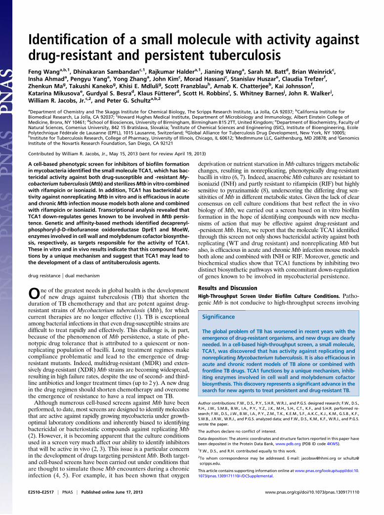
Mycobacterium (Fig. 1B). Interestingly, the activities of TCA1against M. smegmatis, M. bovis bacillus Calmette–Guérin, and Mtbare 20- to 150-fold higher in biofilm medium (MIC50 = 0.03, 0.04,and 0.01 μg/mL, respectively) than 7H9 medium (MIC50 = 4.5, 3,and 0.19 μg/mL, respectively). This observation underscores thevariable efficacy of a drug in different growth media (3), which inpart, may result from the expression of distinct target genes andmetabolic pathways. TCA1 is bactericidal with an MIC99 values of2.1 μg/mL in solid medium. To evaluate the bactericidal activity ofTCA1 againstMtb compared with the two frontline TB drugs INHand RIF, we performed a 21-d kinetic killing assay using compa-rable levels of each of the three drugs (20× MIC50 of each of thethree drugs). TCA1 is active by itself against exponentially growingvirulentMtb in 7H9 media, with a more than 3 log reduction in thenumber of bacilli over a treatment period of 21 d. Treatment withINH or RIF resulted in a comparable drop in cfu over the first 7 dof treatment, but the subsequent outgrowth of bacilli detected inINH- and RIF-treated cultures is absent in TCA1-treated cultures.Furthermore, TCA1 combined with either RIF or INH is able tosterilize an Mtb culture in ∼3 wk (Fig. 2A); removal of drug after3 wk of combination drug treatment did not lead toMtb outgrowth.We also tested the activity of TCA1 on drug-resistant Mtb.
RIF resistance is a marker for MDR-TB (90% of RIF-resistantstrains are also MDR) and typically requires 18–24 mo oftreatment. TCA1 by itself was active against a clinical strain thatis resistant to RIF (because of a mutation in rpoB), and moreimportantly, combined with INH, it sterilized the cultures within 1wk (Fig. 2B). Removal of both drugs after 3 wk of treatment did
Fig. 1. Chemical structures of the affinity resin (TCAP1) and the photo-affinity probe (TCAP2) used in pull-down experiments. Hit compound from screenunder biofilm culture condition. (A) Chemical structure of active compound. (B) TCA1 has selective activity against mycobacteria. (C) Chemical structures ofthe affinity resin and the photo-affinity probe used in pull-down experiments.
Wang et al. PNAS | Published online June 17, 2013 | E2511
MICRO
BIOLO
GY
PNASPL
US
not result in outgrowth. TCA1 was also found to be bactericidalagainst a strain with a mutation in katG (resulting in resistance toINH) (Fig. 2C). Finally, we also tested TCA1 against an XDR-TBstrain, mc28013, which is resistant to 10 TB drugs, including allfrontline drugs (Table S1). TCA1 showed potent bactericidalactivity against the XDR-TB strain (5 log cfu reduction in 3 wk)(Fig. 2D). The lack of cross-resistance to TCA1 in any of thesedrug-resistant strains suggests that TCA1 functions by a dis-tinct mechanism.We next tested the activity of TCA1 against nonreplicating
Mtb in a nutrient starvation assay, a widely used in vitro model ofthe Mtb dormancy phenotype (12, 13). Under these conditions,Mtb enters a nonreplicating state and has been shown to becometolerant to drugs without acquiring heritable drug resistance-inducing mutations (13). TCA1 shows bactericidal activity againstnonreplicatingMtb at a concentration of 7.5 μg/mL (40×MIC50 in7H9 medium), reducing cfu by 3 logs in 3 wk (Fig. 2E). Under thesame assay conditions, RIF (40× MIC50 in 7H9 medium) showedless bactericidal activity than TCA1. We also tested the activity ofTCA1 in an intramacrophage cell culture system to determinewhether it is active against intracellular mycobacteria, because inthe mouse model of infection and in humans, Mtb is believed toreside mainly in macrophages. TCA1 was found to be quite potentin an intracellular cfu assay with an MIC50 value of 0.6 μg/mL[MIC50 (RIF) = 2.7 μg/mL, MIC50 (INH) = 0.2 μg/mL] (SIMaterials and Methods). Finally, TCA1 shows no cytotoxicityagainst five mammalian cell lines (Huh7, 293T, K562, HepG2,and Vero cells) at the highest concentration tested (100 μM forVero cells and 25 μM for others); an hERG assay indicated thatTCA1 has no activity at 30 μM (Tables S2 and S3).
TCA1 Is Efficacious in Acute and Chronic Mtb Infection Mouse Models.We next examined the activity of TCA1 in a mouse model of Mtbinfection. We first determined the physical and pharmacokineticcharacteristics of TCA1. It is stable to proteolytic activity in hu-man or mouse plasma for up to 4 h. Moreover, a GSH trappingassay indicated that no GSH adduct was formed, and TCA1 hasno inhibitory activity against four CYP enzymes. After i.v. ad-ministration, TCA1 exhibited a low clearance and steady-statevolume of distribution, with an elimination half-life of 0.73 h.After oral administration of 20 and 50 mg/kg in solution formu-lation, TCA1 showed a high Cmax (2,122 and 5,653 nM, re-spectively), moderate exposure with oral bioavailability rangingfrom 19% to 46%, and a half-life of 1.8 h (Tables S2 and S3).We first performed the in vivo efficacy experiments in an
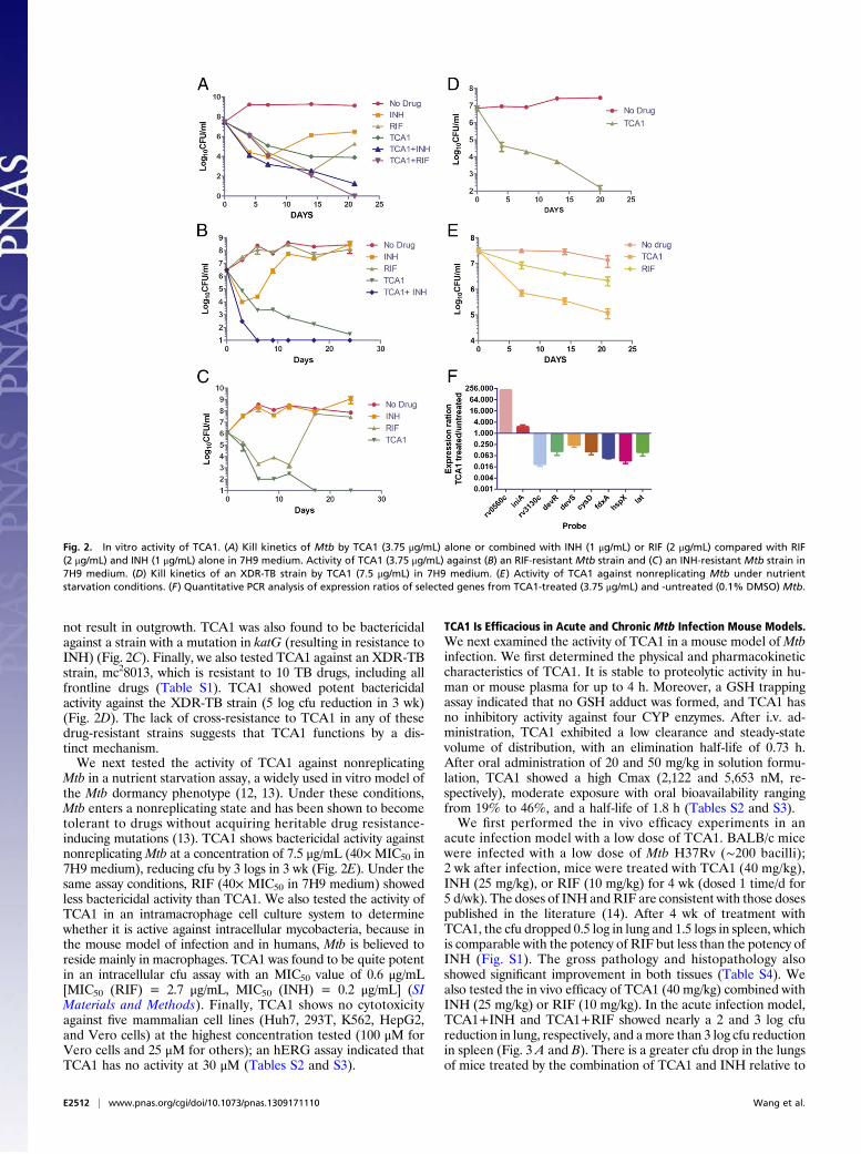
acute infection model with a low dose of TCA1. BALB/c micewere infected with a low dose of Mtb H37Rv (∼200 bacilli);2 wk after infection, mice were treated with TCA1 (40 mg/kg),INH (25 mg/kg), or RIF (10 mg/kg) for 4 wk (dosed 1 time/d for5 d/wk). The doses of INH andRIF are consistent with those dosespublished in the literature (14). After 4 wk of treatment withTCA1, the cfu dropped 0.5 log in lung and 1.5 logs in spleen, whichis comparable with the potency of RIF but less than the potency ofINH (Fig. S1). The gross pathology and histopathology alsoshowed significant improvement in both tissues (Table S4). Wealso tested the in vivo efficacy of TCA1 (40 mg/kg) combined withINH (25 mg/kg) or RIF (10 mg/kg). In the acute infection model,TCA1+INH and TCA1+RIF showed nearly a 2 and 3 log cfureduction in lung, respectively, and amore than 3 log cfu reductionin spleen (Fig. 3 A and B). There is a greater cfu drop in the lungsof mice treated by the combination of TCA1 and INH relative to
Fig. 2. In vitro activity of TCA1. (A) Kill kinetics of Mtb by TCA1 (3.75 μg/mL) alone or combined with INH (1 μg/mL) or RIF (2 μg/mL) compared with RIF(2 μg/mL) and INH (1 μg/mL) alone in 7H9 medium. Activity of TCA1 (3.75 μg/mL) against (B) an RIF-resistant Mtb strain and (C) an INH-resistant Mtb strain in7H9 medium. (D) Kill kinetics of an XDR-TB strain by TCA1 (7.5 μg/mL) in 7H9 medium. (E) Activity of TCA1 against nonreplicating Mtb under nutrientstarvation conditions. (F) Quantitative PCR analysis of expression ratios of selected genes from TCA1-treated (3.75 μg/mL) and -untreated (0.1% DMSO) Mtb.
E2512 | www.pnas.org/cgi/doi/10.1073/pnas.1309171110 Wang et al.
the combination of INH and RIF. We next tested the drug ina mouse model of chronic TB infection. Mice were challengedwith a low-dose aerosol infection, and treatment was initiated 4 wkafter infection. Similar combination treatments were efficacious inthe chronic infection model as well (Fig. 3 C and D).Because the mice were able to tolerate 40 mg/kg TCA1, we
increased the dose to 100 mg/kg using a similar protocol in theacute infection model. After 4 wk, the cfu dropped nearly 2 logsin lungs and more than 3 logs in spleen, showing that the in vivobactericidal activity of TCA1 is dose-dependent (Fig. 3 A and B).The mice again showed no obvious adverse effects or weight lossafter 4 wk of treatment. We also tested the drug in the chronicinfection model at 100 mg/kg. Again, TCA1 showed efficacy inboth lung (1 log cfu reduction) and spleen (1.4 log cfu reduction)(Fig. 3 C and D). These results show that the in vitro efficacy ofTCA1 is recapitulated in vivo, suggesting that the in vitro myco-bacterial biofilm is a useful phenotype to identify compounds ef-fective against Mtb in vivo either alone or combined with existingTB drugs.
Mechanism of Action Studies of TCA1. To gain insight into themechanism of action of TCA1, we treatedMtbH37Rv with TCA1(3.75 μg/mL) in 7H9 media and carried out genome-wide tran-scriptional analysis. Similar to INH and ethambutol (15), cellwall and fatty acid biosynthetic genes are affected by TCA1treatment, suggesting that TCA1 likely interferes with these path-ways. Unlike other known TB drugs, 10 of 86 genes differentiallydown-regulated compared with the DMSO control are genes pre-
viously implicated in TB dormancy, stress response, and RIF sus-ceptibility. These genes include rv3130c–rv3134c, fdxA, and hspX(members of the dos regulon), cysD, and rv3288c–rv3290c (mem-bers of the sigF regulon). The microarray results were confirmedby quantitative PCR (Fig. 2F). Most of these genes are part of thedormancy regulon controlled by dosR (16) and up-regulated underhypoxic conditions or by nitric oxide exposure. For example,fdxA, a low-redox potential electron carrier, is highly up-regulatedin Mtb under hypoxic conditions (17) but significantly down-regulated (>20-fold) in response to TCA1 treatment. Likewise,rv3130c is induced (>300-fold) under multiple stress condi-tions (18) but down-regulated (>30-fold) by TCA1. This down-regulation of genes involved in dormancy and drug toleranceseems to be unique to TCA1 and suggests that TCA1 may po-tentially sensitize Mtb to killing by antibiotics.To further explore the mechanism of action of TCA1, a
TCA1-resistant mutant that carries the cosmid (MSMEG_6379–MSMEG_6384) was isolated by selection of M. smegmatis trans-formed with a genomic cosmid library and grown in biofilmformation medium. Overexpression of each gene in this cosmidrevealed that MSMEG_6382, which is homologous to rv3790in the Mtb genome, confers high-level resistance to TCA1 (>20×MIC50) in both M. smegmatis and Mtb. We also managed toisolate spontaneous resistant mutants of M. smegmatis and Mtb,although the spontaneous mutation rate to TCA1 resistance isextremely low (10−8 to 10−9). Whole-genome sequencing of thegenomic DNA of the resistant mutants revealed that they all have
Fig. 3. In vivo efficacy of TCA1 in mouse models. In an acute Mtb infection mouse model (2 wk infection) followed by 4 wk of drug treatment, TCA1 showedsignificant bactericidal activity in (A) lungs and (B) spleen both alone (100 mg/kg) and combined with (40 mg/kg) INH (25 mg/kg) or RIF (10 mg/kg). The lowactivity of RIF as a monotherapy in this model is consistent with what has been observed in a previous study (14). In a chronic TB infection mouse model (4 wklow-dose infection) followed by 4wk of drug treatment, TCA1 showed activity in (C) lungs and (D) spleen both alone (100mg/kg) and combinedwith (40mg/kg)INH (25 mg/kg) or RIF (10 mg/kg; P value < 0.05). Mice were gavaged with TCA1 1 time/d for 5 d/wk. RIF and INH were administered in drinking water.
Wang et al. PNAS | Published online June 17, 2013 | E2513
MICRO
BIOLO
GY
PNASPL
US

a single-point mutation, resulting in the amino acid replacementTyr321Cys in MSMEG_6382 and Tyr314Cys in rv3790 (Fig. 4A).Rv3790 encodes DprE1, a component of the essential decap-renyl-phosphoryl-β-D-ribofuranose 2′-epimerase DprE1/DprE2required for cell wall arabinan biosynthesis. Indeed, TCA1 sup-pressed the activity of M. smegmatis DprE1 in membrane andcell envelope enzymatic fractions in a dose-dependent manner(Fig. 4B). DprE1 was previously identified as the target of thebenzothiazinones (BTZs) and a nitro-triazole molecule (3, 14).Both scaffolds contain active nitro-moieties and are believed tocovalently modify Cys387 on activation. We performed a com-petitive binding assay using a fluorescently labeled BTZ analog.TCA1 potently competed with BTZ in binding to DprE1, sug-gesting that the binding site of TCA1 overlaps with BTZ (Fig.S2). However, TCA1 does not have an active nitro-moiety, andthe Tyr314Cys mutant strain that is resistant to TCA1 is sensitiveto BTZ, suggesting that the binding mechanism of TCA1 isdifferent from these nitro-heterocycles.To determine the molecular basis by which TCA1 inhibits
DprE1, we determined the crystal structure of the enzyme boundto TCA1 (Table S5). The overall structure of the DprE1–TCA1
complex is largely unaltered compared with the structure of theligand-free protein with the same crystal symmetry (19). Theenzyme, which is structurally related to the vanillyl-alcohol oxi-dase family of flavoproteins (20), consists of an FAD binding anda substrate binding domain, with the flavin moiety of FAD posi-tioned at the interface between the two domains (Fig. 4C, Left).The substrate binding domain includes two disordered loop re-gions (residues 269–283 and 316–330) that leave the active siteopen and accessible for inhibitors. TCA1 binds in the centralcavity of the enzyme, adjacent to the isoalloxazine ring of FAD,in a boomerang-like conformation, with the thiophene moietyinserted deeply into the bottom of the active site (Fig. 4C, Right).The benzothiazole ring is oriented roughly parallel to the iso-alloxazine of FAD. Noncovalent interactions between TCA1 andthe enzyme are dominated by hydrophobic and van der Waalsinteractions (Fig. 4C), with the flavin contributing a large fractionof the total contact surface. Polar contacts are sparse but includethe H bonds between the carboxamido group and thiazole ni-trogen of TCA1 and Nζ of Lys418 (3.0 and 3.1 Å, respectively).The carbamate moiety makes van der Waals interactions withthe phenyl ring of Tyr314, consistent with the observation that
Fig. 4. TCA1 is a DprE1 inhibitor. (A) Sequence alignment of DprE1 of M. smegmatis and Mtb. A Y321C (Y314C in Mtb) mutation was identified in bothM. smegmatis and Mtb strains resistant to TCA1. (B) Inhibition of DprE1 by TCA1 in the cell-free assay for decaprenylphosphoryl arabinose (DPA) productionanalyzed by TLC and autoradiography. M. smegmatis membrane or cell envelope fractions were incubated with (Left) phospho-[14C]-ribose diphosphate and25 μg/mL TCA1 or BTZ043 or (Right) TCA1 in a dose–response fashion. Both TCA1 and BTZ043 potently inhibit conversion of the substrate DPR to the productDPA by DprE1/DprE2 epimerase. (C) Molecular surface ofMtb DprE1 with the FAD domain in light blue and the substrate binding domain in beige. The surfaceareas in pale green andmagenta indicate the positions of (Left) Cys387 and Tyr314. (Right) Noncovalent contacts between TCA1 and DprE1 are shown. Residueswithin a 4-Å radius of the inhibitor (violet) are shown as sticks, with FAD in yellow. Dashed lines indicate the shortest contacts (yellow, hydrophobic/van derWaals; orange, polar) between the residues and the inhibitor. Trp230, located within 4 Å of the carbamate moiety of TCA1, has been omitted for clarity.
E2514 | www.pnas.org/cgi/doi/10.1073/pnas.1309171110 Wang et al.

substituting this tyrosine with cysteine renders DprE1 insensitiveto TCA1. Superimposition of the structures of DprE1 bound tothe BTZ analog (CT325) (19) and TCA1 showed that the bindingsites of these two inhibitors overlap significantly.The above results suggest that DprE1 is a relevant target for the
bactericidal activity of TCA1 against replicating bacteria, similarto BTZ. However, there are some clear distinctions betweenTCA1 and BTZ. First, BTZ is not active against nonreplicatingMtb (14), whereas TCA1 is active against replicating and non-replicating Mtb. Second, the gene expression profiles of Mtbtreated by two compounds are also very different—TCA1 down-regulates persistence genes that are usually up-regulated in Mtb-dormant models, whereas BTZ does not (14). The Mtb strainoverexpressing DprE1 is resistant to TCA1 in 7H9 medium butstill sensitive to TCA1 in the nutrient starvation model (Fig. 5A).Moreover, TCA1 still potentiates INH or RIF on this DprE1-overexpressing strain (Fig. 5B). These results suggest that TCA1could potentially act on an additional mycobacterial target.Because TCA1 had diminished activity against the DprE1
(Y314C) mutant in normal growth medium, it is possible that asecond TCA1 target is not essential for Mtb growth under con-ditions of optimal growth. This complication makes the selectionof relevant mutants more difficult, and therefore, affinity-basedmethods were used to identify additional potential targets. Among
a group of analogs of TCA1, we found that a pyridyl analog,TCA17, has very similar in vitro activity to the activity of TCA1.TCA17 was immobilized on a resin through a linker moiety(TCAP1) (Fig. 1C) and used in a pull-down experiment with celllysates fromMtb. A 35-kDa band was identified on an SDS/PAGEafter silver staining, which disappeared in the presence of 50 μMTCA1 as a competitor. MS identified the band asMoeW, a proteininvolved in the biosynthesis of the molybdenum cofactor (MoCo)(Fig. S3) with homologs in only a few bacterial genomes. To con-firm the binding of TCA1 to MoeW, we overexpressed moeW inE. coli (which lacks an moeW gene homolog in its genome) andtreated this strain with a photoaffinity probe analogous to TCA1(TCAP2) (Fig. 1C) followed by UV irradiation and cell lysis. Asshown in Fig. S4, a band with the size of MoeW is present on anSDS/PAGE gel for the sample from the moeW-induced strain andabsent in the uninduced control sample. These results show thatTCA1 scaffold directly binds to MoeW.MoeW is predicted to contain an FAD/NAD binding domain
by protein sequence analysis, but its function has yet to be de-termined. The gene encodingMoeW is only conserved inMtb andbacillus Calmette–Guérin and not M. smegmatis or other myco-bacterial species, although it is homologous to moeB, anothergene involved in MoCo biosynthesis pathway and conservedin all mycobacteria species and many other bacteria (21). All
Fig. 5. TCA1 inhibits MoCo biosynthesis. (A) Mtb overexpressing dprE1 is sensitive to TCA1 (7.5 μg/mL) under nutrient starvation conditions. (B) Mtboverexpressing dprE1 conferred resistance to TCA1 (3.75 μg/mL) in 7H9 medium. In the meantime, TCA1 acts synergistically with INH (1 μg/mL) or RIF (2 μg/mL)against the same strain. (C–F) MoCo inhibition assay. HPLC profiles of MoCo Form A dephospho standard and sample extracted fromMtb. Arrow indicates theposition of MoCo Form A dephospho. (C) MoCo Form A dephospho standard from synthetic source. (D) Extracted sample fromMtb in the absence of TCA1. (E)Extracted sample from Mtb after treatment with TCA1 (7.5 μg/mL; 18 h). (F) Extracted sample from Mtb in the absence of TCA1 spiked with an MoCo Form Adephospho standard. (G) Kill kinetics of WT Mtb and Mtb overexpressing moeW by 7.5 μg/mL TCA1 in media using nitrate as the only nitrogen source. (H) Killkinetics of WT Mtb and Mtb overexpressing moeW by 7.5 μg/mL TCA1 under nutrient starvation conditions.
Wang et al. PNAS | Published online June 17, 2013 | E2515
MICRO
BIOLO
GY
PNASPL
US
molybdenum-using enzymes identified to date contain MoCo.MoCo is essential for the nitrate respiratory and assimilatoryfunction of Mtb nitro-reductase. Some of these nitro-reductaseshave been found to be involved in the response ofMtb to hypoxiaand nitric oxide (21, 22). To determine if TCA1 can block thebiosynthesis of MoCo, we analyzed cell extracts fromMtb treatedwith TCA1 by detection of dephosphorylated MoCo Form Ausing HPLC with fluorescence detection. Indeed, TCA1 (7.5 μg/mL) completely abolished the formation of MoCo in Mtb (Fig. 5C–F). It is known that MoCo is indispensable for nitrate assimi-lation by Mtb and thus, essential for Mtb to survive in media thatuses nitrate as the only nitrogen source (designated as nitratemedia). We generated an Mtb strain overexpressing moeW andfound it to confer resistance to TCA1 in nitrate media over 30 dof treatment (Fig. 5G). These results clearly show that TCA1asserts its activity against Mtb by inhibition of MoCo biosynthesisthrough interaction with MoeW. The Mtb strain overexpressingmoeW conferred resistance to TCA1 in a nutrient starvationmodel over 21 d of treatment as well (Fig. 5H), but the lower levelof resistance to TCA1 in the nutrient starvation model than ni-trate media suggests that the mechanism of action of TCA1 ismore complicated under the former condition. Nevertheless,the biochemical and genetic results clearly show that MoeW is arelevant target of TCA1.
ConclusionWe have developed a cell-based screen involving the growth ofmycobacteria as an in vitro biofilm (a pellicle). The natural modeof growth ofMtb in liquid culture in the absence of detergent is asa pellicle at the liquid–air interface. Indeed, bacillus Calmette–Guérin is grown as a pellicle for vaccine production. This assayallowed us to identify a potent inhibitor TCA1 against both rep-licating and nonreplicating Mtb as well as drug-resistant Mtb.TCA1 functions by a unique mechanism involving down-regula-tion of persistence genes and inhibition of both cell wall andMoCobiosynthesis. Moreover, TCA1 showed excellent in vivo efficacy inboth acute and chronic TB infection mouse models, suggestingthat this compound may serve as a lead for the development ofa class of drugs against persistent and drug-resistant Mtb. Indeed,we have subsequently identified a compound with good serumhalf-life that has excellent activities under both aerobic and an-aerobic conditions (MIC50 values are 0.3 and 1.5 μg/mL, respec-tively). Future work will focus on additional improvements in thein vivo activity of this molecule and detailed mechanistic studies,including attempts to isolate additional resistant mutants undervaried growth conditions. This work underscores the power ofcell-based phenotypic screens to uncover molecules with mech-anisms of action that provide unique approaches to the treatmentof human disease.
Materials and MethodsHigh-Throughput Screen for Inhibitors of Biofilm Formation Inhibition. A di-verse chemical library (∼70,000 compounds) was used for the primary screen.This in-house compound library was created based on a chemoinformaticanalysis of scaffold chemical diversity, historical proprietary screen hit rates[>300 Mio data points from the high throughput (HTS) database], andcommercial availability; 105/mL M. smegmatis cells were plated in 384-wellplates in biofilm formation medium of M63 salts minimal medium supple-mented with 2% glucose, 0.5% Casamino Acids, 1 mM MgSO4, and 0.7 mMCaCl2. RIF and TMC207 were used as positive controls, and DMSO (0.1%) wasused as a negative control. Cells were treated with 10 μM compound andincubated for 3 d, and the OD of each well was determined with an EnVisionMultilabel Reader. The average Z′ and coefficient values are 0.512 and 8.7%,respectively. We used a high-stringency cutoff (threefold inhibition) to pickhits that are most likely growth inhibitors (hit rate = 0.03%) and a low-stringency cutoff (twofold inhibition) to include hits that inhibit biofilmformation without significant growth inhibition (hit rate = 0.17%).
In Vitro Activity Assays. For kinetic killing assays, exponentially growing cul-tures of mycobacteria were diluted in fresh media to an OD600 of 0.1–0.2.Various drugs were added to the culture at the indicated concentrations. Thenumber of cfus at the start of the experiment was estimated by plating ap-propriate dilutions of the culture onto 7H10 agar plates. The effect of drugwas monitored by plating for cfus at the indicated time points. All experi-ments were carried out in triplicate. MICs were determined by a turbidityassay. Threefold serial dilutions in DMSO were prepared for each compound.Mtb cultures (OD = 0.04) were incubated with compounds at 37 °C for 5 d,and OD600 was determined with an Envision plate reader. All experimentswere carried out in duplicate. For assays under starvation conditions, a log-phase growing Mtb culture was centrifuged, and the cell pellet was washedtwo times with PBS, resuspended in PBS with Tyloxapol (0.05%; OD = 0.3),and incubated with DMSO, TCA1 (7.5 μg/mL), and RIF (2 μg/mL). All experi-ments were carried out in triplicate. For intracellular macrophage assays,J744.1 murine macrophage cells were infected with Mtb at a multiplicity ofinfection (MOI) of 1:3 and incubated for 2 h at 37 °C. After washing the cellmonolayer three times, 20 μM amikacin was added, and the culture was in-cubated for an additional 2 h to kill the remaining extracellular bacteria.Infected cells were then incubated in the presence of serial dilutions ofcompounds for 5 d. Cells were washed three times and lysed in each well;the lysate was transferred to a 96-well plate for serial dilution and thenplated on 7H11 agar medium for cfu assays. All experiments were carriedout in triplicate.
In Vivo Efficacy Experiments. Six- to eight-week-old female BALB/c mice (USNational Cancer Institute) were infected by aerosol with a low dose (∼50bacilli) of Mtb H37Rv. Infection dose was verified by plating the inoculumand the whole-lung homogenates onto 7H10 plates at 24 h postinfection.Treatment of BALB/c mice began at either 2 or 4 wk postinfection, with RIF(10 mg/kg) and INH (25 mg/kg) administered ad libitum in drinking water(changed one time every 2 d). TCA1 was administered by oral gavage onetime daily for 5 d/wk at a dosage of either 40 or 100 mg/kg for the indicateddurations. At predetermined time points or humane endpoints, animalswere heavily sedated and euthanized, and tissues were collected for cultureand pathology. Treatment efficacy was assessed on the basis of cfu in thelungs and spleen of treated mice compared with untreated controls andbacterial burden in these organs before treatment start. Organs were ho-mogenized in PBS containing Tween-80 [0.05% (vol/vol)], and various dilu-tions were placed on 7H10 plates. Plates were incubated at 37 °C for 3 wk,and cfus on the various plates were recorded. All animal experimentalprotocols were approved by the Animal Care and Use Committee of AECOM.
Genome-Wide Transcriptional Analysis. Triplicate 10-mL cultures of mycobac-teria were grown to log phase for transcriptional profiling of planktonic cellsor for 3 wk in pellicle media for transcriptional profiling of pellicle cells. ForTCA1 treatment, log-phase cultures were treated with 3.75 μg/mL TCA1 orDMSO vehicle for 12 h. Cells were harvested, washed, and resuspended in1 mL RNA Protect reagent (Qiagen) and incubated 4 h at room temperature(21 °C). All transcriptional profiling procedures, including RNA extraction,DNase treatment, cDNA synthesis, labeling, microarray hybridization, wash-ing, scanning, and data analysis, were performed as previously described (23).Microarray data have been deposited in the US National Center for Bio-technology Information Gene Expression Omnibus (accession no. GSE42151).For quantitative PCR experiments, diluted cDNA was used as a template at50 ng per reaction for real-time PCR reactions containing primer sets designedby Primer 3 and SYBR Green PCRMasterMix (Applied Biosystems) in accordancewith the manufacturers’ instructions. These reactions were carried out on anABI 9700HT real-time PCR cycler (Applied Biosystems).
DprE1 Competition Assay. DprE1 was incubated with serial dilutions of TCA1for 15 min. BTZ-BODIPY was added, and the sample was incubated for 1 h at37 °C. BTZ-BODIPY is a fluorescent BTZ derivative that reacts in the presenceof farnesylphosphoribose, with DprE1 forming a covalent bond. Samples arethen analyzed by SDS/PAGE (fluorescence and Coomassie staining).
Crystallization and Structure Determination. Mtb DprE1 (Rv3790) was pre-pared for crystallization as described in the work by Batt et al. (19). Beforesetting up crystallization experiments, the TCA1 inhibitor (in DMSO) was in-cubated with concentrated protein (∼35 mg/mL) for 30 min at a molar ratioof 3 TCA1:1 DprE1. Crystals were grown by sitting drop vapor diffusion andappeared over a reservoir consisting of 40–43% (wt/vol) polypropylene glycol400 and 0.1 M imidazole, pH 7.0. Crystals were mounted into nylon loopsdirectly from the drop and frozen in liquid nitrogen. X-ray diffraction datato 2.6 Å resolution were recorded on beamline I02 of the Diamond Light
E2516 | www.pnas.org/cgi/doi/10.1073/pnas.1309171110 Wang et al.

Source (Table S5). The crystals were in space group P21, with twomolecules ofthe complex in the crystallographic asymmetric unit. Initial phases wereobtained by molecular replacement (PHASER) (24) using the apo structureof DprE1 (Protein Data Bank ID code 4FDP) (19) as a search model. Afterrefinement of the molecular replacement solution, density for TCA1 wasclearly visible in the active sites of the two crystallographically distinctcopies of DprE1. Density shape and stereochemical constraints allowed us tounequivocally place the inhibitor in the active site of DprE1. Model re-building and structure refinement (COOT) (25), REFMAC5 (26), and PHENIX.REFINE (27) led to final R factors of 23.7% and 17.6% for the test andworking sets, respectively.
Affinity-Based Proteomics and Photo-Affinity Labeling. H37Ra cells were lysedwith homogenization buffer (60 mM β-glycerophosphate, 15 mM p-nitro-phenyl phosphate, 25 mM Mops, pH 7.2, 15 mM MgCl2, 1 mM DTT, proteaseinhibitors, 0.5% Nonidet P-40). Cell lysates were centrifuged at 16,000 × gfor 20 min at 4 °C, and the supernatant was collected. Total protein con-centration in the supernatant was determined by a BCA protein assay kit(Pierce). The lysates (1 mg) were then added to the affinity resin (30 μL), andthe loading buffer (50 mM Tris·HCl, pH 7.4, 5 mM NaF, 250 mM NaCl, 5 mMEDTA, 5 mM EGTA, protease inhibitors, 0.1% Nonidet P-40) was added to afinal volume of 1 mL (for the competition experiment, TCA1 was added toa final concentration of 50 μM). After rotating at 4 °C for 1 h, the mixturewas centrifuged at 16,000 × g for 1 min at 4 °C, and the supernatant wasremoved. The affinity resin was then washed five times with cold loadingbuffer and eluted by boiling with Laemmli sample buffer (Invitrogen) at95 °C for 3 min. Samples were loaded and separated on a 4–20% Tris·glycinegel (Invitrogen). The gel band was extracted and analyzed by proteomics.For the photo-affinity experiments, E. coli cells overexpressing MoeW andnative cells were lysed, and the photo-affinity probe was added to celllysates (1 mg) in 50 μL PBS and incubated for 2 h at room temperaturefollowed by UV irradiation with a UV lamp for 20 min. The reaction mixtureswere then subjected to click chemistry with rhodamine-azide (100 μM) andincubated for 2 h at room temperature with gentle mixing. The reactionswere terminated by the addition of prechilled acetone (0.5 mL), placedat −20 °C for 30 min, and centrifuged at 16,000 × g for 10 min at 4 °C toprecipitate proteins. The pellet was washed two times with 200 μL pre-chilled methanol, resuspended in 25 μL 1× standard reducing SDS loading
buffer, and heated for 10 min at 95 °C; samples were loaded for sepa-ration by SDS/PAGE and then visualized by in-gel fluorescent scanning.
In Vitro Assays in Nitrate-Only Media. An Mtb culture was resuspended undernitrogen-limiting conditions (22) (a basal medium [1 L basal medium con-tains 1 g KH2PO4, 2.5 g Na2HPO4, 2 g K2SO4, 2 mL trace elements; 1 L traceelements contained 40 mg ZnCl2, 200 mg FeCl30.6H2O, 10 mg CuCl20.4H2O,10 mg MnCl20.4H2O, 10 mg Na2B4O70.10H2O, 10 mg (NH4)6Mo7O240.4H2O]supplemented with NaNO3 as the sole source of nitrogen, 0.5 mM MgCl2,0.5 mM CaCl2, 10% ADS, 0.2% glycerol and 0.05% Tween 80) and incubatedfor 24 h; 7.5 μg/mL TCA1 was then added to the culture and incubated for30 d. Cfu assay was used to determine the bacterial viability at each time point.
All experiments were carried out in triplicate.
MoCo Inhibition Assay. The synthesis of MoCo Form A dephospho was carriedout according to the procedures described. The 1H-NMR spectrum matcheswhat has been reported in the literature (28, 29). Conversion of all sources ofmolybdopterin to Form A dephospho was performed by the methods pre-viously reported with slight modifications (21, 30); 100 mL Mtb culture wereharvested, and the pellet was resuspended in extraction solution (2 mL;10 mM sodium ascorbate). The cells were lysed and centrifuged at 16,000 × g;the supernatant was collected and treated with acidic iodine solution at 95 °Cfor 25 min, and the excess iodine was removed by adding sodium ascorbate.After centrifugation, the solution was neutralized with ammonium hydroxideand then concentrated and dephosphorylated using calf intestinal phosphatase(NEB) at 37 °C for 3 h. HPLC analysis was performed using Agilent C18 column(150 × 4.6 mm, 10-μm particle size) with gradient elution by buffer A (50 mMammonium acetate) and buffer B (MeOH; 97% A to 93% A in 14 min and 97%B wash from 15 to 22 min). Fluorescence detection was at 370/450 nm.
ACKNOWLEDGMENTS. We thank Drs. Kelli Kuhen, Scott Robins, and ValerieMizrahi for helpful discussions, Chun Li and Johnathan Chang for pharma-cokinetic experiments, and Zhong Chen, Ivana Centarova, Emoke Kilacskova,and Dr. Baojie Wan for technical support. We also thank Dr. Stewart Cole andDr. Vadim Makarov for providing BTZ043. This work was supported, in part,by European Community’s Seventh Framework Programme Grant 260872 (toK.M.), the Global Alliance for TB Drug Development (P.G.S.), the NationalInstitutes of Health Grants AI26170 and A10-97548, and the Albert EinsteinCollege of Medicine Center for AIDS Research Grant AI0–51519.
1. Gandhi NR, et al. (2010) HIV coinfection in multidrug- and extensively drug-resistanttuberculosis results in high early mortality. Am J Respir Crit Care Med 181(1):80–86.
2. Pethe K, et al. (2010) A chemical genetic screen in Mycobacterium tuberculosisidentifies carbon-source-dependent growth inhibitors devoid of in vivo efficacy. NatCommun 1:57.
3. Stanley SA, et al. (2012) Identification of novel inhibitors of M. tuberculosis growthusing whole cell based high-throughput screening. ACS Chem Biol 7(8):1377–1384.
4. Mak PA, et al. (2012) A high-throughput screen to identify inhibitors of ATPhomeostasis in non-replicating Mycobacterium tuberculosis. ACS Chem Biol 7(7):1190–1197.
5. Gold B, et al. (2012) Nonsteroidal anti-inflammatory drug sensitizes Mycobacteriumtuberculosis to endogenous and exogenous antimicrobials. Proc Natl Acad Sci USA109(40):16004–16011.
6. Wayne LG, Hayes LG (1996) An in vitro model for sequential study of shiftdown ofMycobacterium tuberculosis through two stages of nonreplicating persistence. InfectImmun 64(6):2062–2069.
7. Wayne LG, Sohaskey CD (2001) Nonreplicating persistence of mycobacteriumtuberculosis. Annu Rev Microbiol 55:139–163.
8. Mitchison DA, Coates AR (2004) Predictive in vitro models of the sterilizing activity ofanti-tuberculosis drugs. Curr Pharm Des 10(26):3285–3295.
9. Ojha A, et al. (2005) GroEL1: A dedicated chaperone involved in mycolic acidbiosynthesis during biofilm formation in mycobacteria. Cell 123(5):861–873.
10. Teng R, Dick T (2003) Isoniazid resistance of exponentially growing Mycobacteriumsmegmatis biofilm culture. FEMS Microbiol Lett 227(2):171–174.
11. Ojha AK, et al. (2008) Growth of Mycobacterium tuberculosis biofilms containing freemycolic acids and harbouring drug-tolerant bacteria. Mol Microbiol 69(1):164–174.
12. Gengenbacher M, Rao SP, Pethe K, Dick T (2010) Nutrient-starved, non-replicatingMycobacterium tuberculosis requires respiration, ATP synthase and isocitrate lyase formaintenance of ATP homeostasis and viability. Microbiology 156(Pt 1):81–87.
13. Betts JC, Lukey PT, Robb LC, McAdam RA, Duncan K (2002) Evaluation of a nutrientstarvation model of Mycobacterium tuberculosis persistence by gene and proteinexpression profiling. Mol Microbiol 43(3):717–731.
14. Makarov V, et al. (2009) Benzothiazinones kill Mycobacterium tuberculosis byblocking arabinan synthesis. Science 324(5928):801–804.
15. Boshoff HI, et al. (2004) The transcriptional responses of Mycobacterium tuberculosisto inhibitors of metabolism: Novel insights into drug mechanisms of action. J BiolChem 279(38):40174–40184.
16. Voskuil MI, et al. (2003) Inhibition of respiration by nitric oxide induces aMycobacterium tuberculosis dormancy program. J Exp Med 198(5):705–713.
17. Muttucumaru DG, Roberts G, Hinds J, Stabler RA, Parish T (2004) Gene expressionprofile of Mycobacterium tuberculosis in a non-replicating state. Tuberculosis (Edinb)84(3–4):239–246.
18. Deb C, et al. (2009) A novel in vitro multiple-stress dormancy model for Mycobacteriumtuberculosis generates a lipid-loaded, drug-tolerant, dormant pathogen. PLoS One 4(6):e6077.
19. Batt SM, et al. (2012) Structural basis of inhibition of Mycobacterium tuberculosisDprE1 by benzothiazinone inhibitors. Proc Natl Acad Sci USA 109(28):11354–11359.
20. Mattevi A, Fraaije MW, Coda A, van Berkel WJ (1997) Crystallization and preliminaryX-ray analysis of the flavoenzyme vanillyl-alcohol oxidase from Penicillium simplicissimum.Proteins 27(4):601–603.
21. Williams MJ, Kana BD, Mizrahi V (2011) Functional analysis of molybdopterinbiosynthesis in mycobacteria identifies a fused molybdopterin synthase in Mycobacteriumtuberculosis. J Bacteriol 193(1):98–106.
22. Malm S, et al. (2009) The roles of the nitrate reductase NarGHJI, the nitrite reductaseNirBD and the response regulator GlnR in nitrate assimilation of Mycobacteriumtuberculosis. Microbiology 155(Pt 4):1332–1339.
23. Vilchèze C, Weinrick B, Wong KW, Chen B, Jacobs WR, Jr. (2010) NAD+ auxotrophy isbacteriocidal for the tubercle bacilli. Mol Microbiol 76(2):365–377.
24. McCoy AJ, et al. (2007) Phaser crystallographic software. J Appl Cryst 40(Pt 4):658–674.25. Emsley P, Lohkamp B, Scott WG, Cowtan K (2010) Features and development of Coot.
Acta Crystallogr D Biol Crystallogr 66(Pt 4):486–501.26. Murshudov GN, et al. (2011) REFMAC5 for the refinement of macromolecular crystal
structures. Acta Crystallogr D Biol Crystallogr 67(Pt 4):355–367.27. Adams PD, et al. (2010) PHENIX: A comprehensive Python-based system for
macromolecular structure solution. Acta Crystallogr D Biol Crystallogr 66(Pt 2):213–221.
28. Taylor EC, Ray PS, Darwish IS (1989) Studies on the molybdenum cofactor.Determination of the structure and absolute configuration of form A. J AmChem Soc 111(19):7664–7665.
29. Mohr D, Kazimierczuk Z, Pfleiderer W (1992) Pteridines. Part XCVII. Synthesis andproperties of 6-thioxanthopterin and 7-thioisoxanthopterin. Helv Chim Acta 75(7):2317–2326.
30. Johnson ME, Rajagopalan KV (1987) Involvement of chlA, E, M, and N loci inEscherichia coli molybdopterin biosynthesis. J Bacteriol 169(1):117–125.
Wang et al. PNAS | Published online June 17, 2013 | E2517
MICRO
BIOLO
GY
PNASPL
US
![Identification of a Protein Network Interacting with TdRF1 ... · Protective Role against Cellular Dehydration1[C][W] Davide Guerra, ... TEOSTRESS” Project), ... The construct](https://img.pdfslide.us/doc/110x75/5ac8e6e07f8b9aa1298c9fa1/identication-of-a-protein-network-interacting-with-tdrf1-role-against-cellular.jpg)