Embed Size (px)
Citation preview

Proc. Nati. Acad. Sci. USAVol. 88, pp. 9897-9901, November 1991Cell Biology
Hypoxia induces endothelial cell synthesis of membrane-associated proteins
(hypoxia/endothelium/coagulation)
SATOSHI OGAWA*, MATTHIAS CLAUSS, KEISUKE KUWABARA, REVATI SHREENIWAS, CAESAR BUTURA,SHIN KOGA, AND DAVID STERNDepartment of Physiology and Cellular Biophysics, College of Physicians and Surgeons, Columbia University, New York, NY 10032
Communicated by George E. Palade, July 3, 1991
ABSTRACT Hypoxemia is associated with a prothrom-botic tendency. In this study we report the purification andpartial characterization of an activator of a central coagulationcomponent, factor X, induced in endothelium by exposure tohypoxia (hypoxia-induced factor X activator or Xt). Expres-sion of Xact occurred in a reversible manner when endothelialcell cultures were exposed to hypoxia or sodium azide but notin response to a variety of other alterations in the cellularmilieu, such as heat shock or glucose deprivation. The activityof X,,t, which was not detected in normoxic endothelial cells,was maximal under acidic conditions, pH 6.0-6.8, which oftencoexist with hypoxia in an ischemic milieu. By sequentialisoelectric focusing and preparative SDS/PAGE of endothelialmembrane-rich fractions, Xad was purified 419,000-fold andfound to be a single-chain, -100-kDa polypeptide with pI -5.0.Activation of factor X by purified Xt was not affected byblocking antibodies to other coagulation proteins or by phe-nylmethylsulfonyl fluoride or leupeptin but was prevented bymercury chloride or iodoacetamide. In addition to the induc-tion of Xad,, two-dimensional gel analysis of membrane frac-tions from metabolically labeled hypoxic endothelial culturesrevealed two groups of -10 additional spots: (i) a group forwhich expression was maximal after 24 hr and (i) a group forwhich expression continued to increase up to 48 hr. The patternof hypoxia-mediated modulation of protein expression wasdistinct from that seen with other cellular stimuli but could beduplicated, in part, by sodium azide. These results indicate thathypoxia elicits a specific biosynthetic response, including theexpression of endothelial cell-surface molecules that can altercellular function and may potentially serve as markers ofhypoxemic vessel-wall injury.
Exposure of endothelium to environments of low oxygentension is a frequent occurrence in various disorders, espe-cially those associated with compromise of the circulation.Two crucial functions of endothelium, maintenance of apermeability barrier and preservation of the fluidity of blood,are adversely affected by levels of hypoxia that occur inischemic syndromes (1, 2). Based on studies in cell culture,such hypoxia-mediated perturbation of endothelial cell (EC)functions results from alterations in metabolic pathways, notfrom a change in viability with death of the cell monolayer (3,4). For example, exposure of bovine aortic ECs to hypoxialed to an increase in their permeability to solutes, but thesechanges were reversible on restoration of cultures to nor-moxia (4).Hypoxemia has long been associated with a prothrombotic
tendency, especially in the setting of deep vein thrombosis,where stasis of an extremity in an animal model results inextreme local hypoxemia and fibrin deposition on the cusps
of vein valves (2). In a previous study (4), we showed thathypoxic cultured ECs expressed an apparently unusual pro-coagulant activity, allowing them to activate directly a centralcoagulation component, factor X. We now report furthercharacterization of the hypoxia-induced factor X activator(Xact) and the identification of a group of EC surface-associated proteins, the expression of which is induced/enhanced by hypoxia (termed oxygen-regulated proteins orORPs) (5, 6).
METHODS
Induction of Hypoxia. Bovine aortic ECs were grown andrendered hypoxic, as described (4) (smooth muscle cells wereobtained from the same calfaortas by further scraping vesselsafter removal of ECs). During these experiments, the P02 ofthe culture medium was -14 mmHg (1 mmHg = 133 Pa), andthe pH of the medium remained constant (when Hepes wasomitted from the medium, pH fell). By 72 hr ofEC incubationunder hypoxic conditions, glucose concentration in the me-dium fell from 5 to 2.5 mM. This degree of glucose depletiondid not affect Xact expression and did not induce synthesis ofany of the proteins seen when cultures were labeled inglucose-free medium (see below). ATP levels were deter-mined by using the luciferase assay (7), and protein synthesiswas assessed by the incorporation of radiolabeled aminoacids (8).
Activation of Factor X by Hypoxic ECs. Intact monolayers,cell suspensions, membrane-rich fractions, or samples fromthe purification procedure described below were incubatedwith purified bovine factor X (1 AM or as stated) at 370C forthe indicated time, and then the presence of factor Xa wasassessed by chromogenic substrate, coagulant, or radiomet-ric assays (4, 9, 10).
Purification of Xa. This purification included preparativeisoelectric focusing and SDS/PAGE (11). ECs exposed tohypoxia for 48 hr (5 x 10⁹ cells) were washed in buffer,scraped from the growth surface, and resuspended in Veronalbuffer (20 mM; pH 7.8) containing phenylmethylsulfonylfluoride (2 mM) to elute Xact. The eluate was diluted in 50 mlof 1.5% Ampholyte (pH range 3-10, Bio-Rad)/0.1% octylf3-glucoside, and isoelectric focusing (Rotofor Cell, Bio-Rad)was done. Fractions were dialyzed against 0.4 M Tris/HCl,pH 7.5/0.2 M NaCl/0.1% octyl 83-glucoside, diluted inVeronal-buffered saline, and tested for Xact activity. Next,preparative nonreduced SDS/PAGE (10%) was done, andproteins in the gel were either visualized by silver staining or
Abbreviations: ORP, oxygen-regulated protein; Xact, hypoxia-induced factor X activator; TNF, tumor necrosis factor/cachectin;EC, endothelial cell.*To whom reprint requests should be addressed at: Rover PhysiologyLaboratory, Department of Physiology and Cellular Biophysics,Columbia University College of Physicians and Surgeons, 630 West168th Street, New York, NY 10032.
9897
The publication costs of this article were defrayed in part by page chargepayment. This article must therefore be hereby marked "advertisement"in accordance with 18 U.S.C. §1734 solely to indicate this fact.
Dow
nloa
ded
by g
uest
on
Feb
ruar
y 6,
202
0
Proc. Natl. Acad. Sci. USA 88 (1991)
electroeluted from the gel and assessed for Xact activity [thelatter after SDS removal (12)].
Metabolic Labeling. EC metabolic labeling was done byincubating cultures in methionine-poor minimal essentialmedium/5% dialyzed fetal calf serum/[35S]methionine at 0.2mCi/ml (1 Ci = 37 GBq), the latter added 8 hr before the endof an experiment.Two-Dimensional Gel Analysis. This was done after expo-
sure of EC monolayers to the test conditions by suspendingthe cells in (10 mM Tris, pH 7.2)/aprotonin (at 100 units/ml),Dounce homogenizing, adding additional buffer (10 mMTris/10 mM NaCl/10 mM KCl/5 mM CaCl2/2 mM MgCl2/0.5 M sucrose/aprotinin at 100 units/ml), and centrifuging atlow speed to remove debris/nuclei. The supernatant was thenultracentrifuged (100,000 x g for 2 hr), and the membrane-rich pellet (from -106 cells) was solubilized in SDS/gel bufferand subjected to two-dimensional gel analysis (13). Gels wererun by Protein DataBase (Huntington Station, NY) andanalyzed with the PDQuest system (14).
RESULTSHypoxia-Induced, EC-Dependent Factor X Activation. EC
monolayers grown to confluence in normoxia and then placedin hypoxia for 3 days maintained their viability: (i) productionofATP continued at levels of -75% that seen in normoxia (7),(it) protein synthesis also continued with only a decrease by20-30% in incorporation of radiolabeled amino acids intoprotein as compared with normoxia (4), (ii) uptake of thevital dye trypan blue was not increased (4), and (iv) afterrestitution to normoxia, the cells proliferated on furthersubculturing.
In contrast to this general maintenance of "housekeeping"functions necessary for cell viability, there were subtlealterations in properties of the hypoxic EC, which hadimplications for their central role in vascular homeostasis.Previously, we had observed that on exposure to hypoxia,ECs acquired the ability to shorten the clotting time ofrecalcified plasma, due to the expression of a cell-surfaceprocoagulant activity that directly activated factorX (4). Thishypoxia-induced Xact was not seen in intact cell monolayersor subcellular fractions of normoxic cultures, and its expres-sion in hypoxic ECs was blocked by cycloheximide additionto culture medium, suggesting a role for de novo proteinsynthesis. Furthermore, mercuric chloride blocked factor Xactivation by hypoxic ECs, whereas phenylmethylsulfonylfluoride had no effect, suggesting that a sulfhydryl group wasnecessary for Xact activity. In contrast to the effect ofhypoxia, exposure ofECs to heat shock, glucose deprivation,or the cytokine tumor necrosis factor (TNF) did not inducean activity similar to Xact (data not shown). Furthermorestudies with metabolic inhibitors showed that neither2-deoxy-glucose nor fluoride (which inhibit glycolysis) induced Xact,whereas sodium azide, an inhibitor of the electron-transportchain, did (data not shown). Activator expression was pre-dominately on the cell surface, as experiments after freeze-thaw lysis ofhypoxic ECs did not demonstrate additional Xatactivity.Our culture system was set up to achieve selective hypoxia
(for example, pH did not vary beyond 7.3-7.6), but in theischemic milieu hypoxemia is closely associated with acido-sis. In this context, when ECs in medium without Hepes weremaintained for 48 hr in hypoxia, pH of the medium fell to6.5-6.8. This change led us to examine pH effect on factor Xactivation by hypoxic EC cultures. Although the pH optimumfor activation of factor X by the extrinsic and intrinsicsystems is at or about neutral pH, which is not surprisingbecause neutral proteases are involved (15), Xat was moreeffective in a lower pH range (Fig. 1 A-B). For example, atpH 6.8, Vmax for factor Xa formation was increased =4-fold
.c SE0
0 4
cm
C 3
C
0
.01x0
Ui. I
500
400
2 300
0 200
100
0
F 200
E° 100x
0
LL0U-
06
L0
too-0,-
x
L L.
tsCLco'
is60
03.2010AI
54 10 150Factor X (jig/mi)
200 2!
5.2 6.0 6.S 7.4 $AlpH
FI
0 20 40 60
D~~~~Ir I
-n-
if
V~~~72N 72H 6R 12R 48R
FIG. 1. FactorX activation by hypoxic endothelial cells: effect ofpH and reversibility after exposure to ambient air. (A) EC mono-layers grown in normoxia were incubated in hypoxia for 48 hr andincubated in buffer with pH 6.8 (A) or 7.4 (*) in the presence offactorX. Factor Xa formation is shown as ng/ml per min. (Inset) Doublereciprocal plots. (B) EC monolayers were made hypoxic for 48 hr,incubated with 1 ,uM factor X at the indicated pH for 30 min at 37°C.Factor X activation is shown as a percentage of that seen at pH 7.4.(C) EC monolayers were incubated for 72 hr in hypoxia and resustpended in either plasma deficient in factors II/VII/X (open bars) orVeronal-buffered saline (hatched bars) supplemented with 3H-labeled factor X (100 ,g/ml). Then, samples were removed for theradiometric assay to detect factor Xa formation. (D) EC monolayerswere incubated for 72 hr in hypoxia or normoxia and then exposedto ambient air, and factorXaformation was studied after adding 1 .Mfactor X for 30 min at 37°C. 72N or 72H, normoxia or hypoxia for 72hr, respectively; R, remainder, which are hypoxia for 72 hr followedby the indicated time in hr for normoxia (i.e., reoxygenation).
(5.1 versus 1.2 ng/ml per min) and Km fell =2-fold (28 versus13 ,ug/ml) compared with these parameters at pH 7.4 (Fig.1A). Over a broader pH range, it was evident that theoptimum pH for factor X activation was 6.0-6.8, whereasoutside these pHs factor Xa formation decreased (Fig. 1B).
=7.4
=6.5
'4
9898 Cell Biology: Ogawa et al.
A IA
I I IAL
4 -
pH = 6.8 1 PN=7 ,2 .
pi =
-100 0 100PN = 7.4
I&
I I0Ik
Dow
nloa
ded
by g
uest
on
Feb
ruar
y 6,
202
0
Proc. Natl. Acad. Sci. USA 88 (1991) 9899
These data indicated that other environmental factors likelyto occur during hypoxemia, such as acidosis, could modulatefunctional activity of Xact. In this context, experiments inwhich hypoxic ECs were incubated with 3H-labeled factor Xin the presence of plasma showed that factor Xa formationdid occur in a complex, more physiologically relevant system(Fig. 1C). In fact, factor Xa formation by hypoxic ECs wasmore effective in the plasma-based system than in buffer withonly factor X. Normoxic endothelial cells, under the sameconditions, did not activate factor X (data not shown).Induction of Xact was reversible, as after exposure to nor-moxia, its activity diminished back to baseline (Fig. iD).
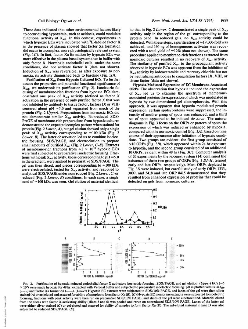
Purification of X,,t from Hypoxic Cultured ECs. To furtherassess the properties and potential functional significance ofXact, we undertook its purification (Fig. 2). Isoelectric fo-cusing of membrane-rich fractions from hypoxic ECs dem-onstrated one peak of Xact activity (defined as factor Xactivation in the presence of only purified factor X that wasnot inhibited by antibody to tissue factor, factors IX or VIII)centered about pH -5.0 and separated from much of theprotein (Fig. 2 Upper). Preparations from normoxic ECs didnot demonstrate similar Xact activity. Nonreduced SDS/PAGE of membrane-rich preparations from hypoxic culturesdemonstrated the expected complex pattern when stained forprotein (Fig. 2 Lower, A), but gel elution showed only a singlepeak of Xac, activity corresponding to -100 kDa (Fig. 2Lower, B). The latter observation led us to combine isoelec-tric focusing, SDS/PAGE, and electroelution to preparesmall amounts of purified Xact (Fig. 2 Lower, C-E). Extractsof membrane-rich fractions from -2 x 10¹⁰ hypoxic ECswere first subjected to preparative isoelectric focusing. Frac-tions with peak Xact activity, those corresponding to pH -5.0in the gradient, were applied to preparative SDS/PAGE. Thegel was then sliced, and pieces corresponding to -100 kDawere electroeluted, tested for Xact activity, and reapplied toanalytical SDS/PAGE under nonreduced (Fig. 2 Lower, C) orreduced (Fig. 2 Lower, E) conditions. In each case, a singleband of =100 kDa was seen. Gel elution of material identical
E
Cal6X
765432
0
A
to that in Fig. 2 Lower, C demonstrated a single peak of Xactactivity only in the region of the gel corresponding to theprotein band. In reduced gels, no Xact activity could bedetected. With these steps, a purification of -19,000-fold wasachieved, and 160 ng of homogeneous activator was recov-ered with a total yield of "12% (data not shown). The sameprocedure applied to membrane-rich fractions extracted fromnormoxic cultures resulted in no recovery of Xact activity.The similarity of purified Xact to the procoagulant activityobserved in hypoxic ECs was apparent from the inhibition ofXact activity by iodoacetamide and mercury chloride but notby neutralizing antibodies to coagulation factors IX, VIII, ortissue factor (data not shown).Hypoxia-Mediated Expression of EC Membrane-Associated
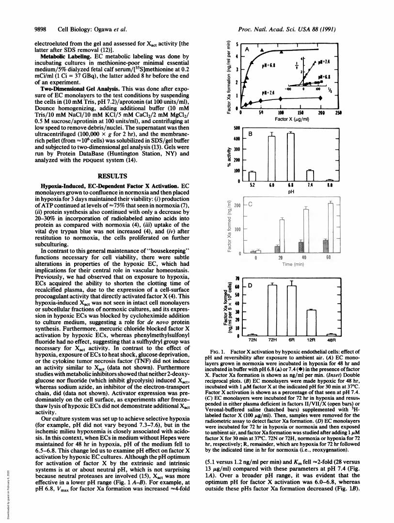
ORPs. The observation that hypoxia induced the expressionof Xact led us to examine the spectrum of membrane-associated proteins the expression ofwhich was modulated inhypoxia by two-dimensional gel electrophoresis. With thisapproach, it was apparent that hypoxia modulated proteinexpression: certain spots/proteins were suppressed, the in-tensity of another group of spots was enhanced, and a thirdset of spots appeared to be induced de novo. The autora-diograms in Fig. 3 focus on the ORPs or pattern of spots theexpression of which was induced or enhanced by hypoxia,compared with the normoxic control (Fig. 3A), based on timecourse of their appearance after initiation of hypoxic condi-tions. Two groups are evident: the first group consisted of"10 ORPs (Fig. 3B), which appeared within 24-hr exposureto hypoxia, and the second group consisted of an additional10 ORPs, evident within 48 hr (Fig. 3C). Computer analysisof 20 experiments by the PDQuest system (14) confirmed theexistence of these two groups of ORPs (Fig. 3 DI-II, termedearly and late ORPs, respectively). Most ORPs depicted inFig. 3D were induced, but careful study of early ORPs 1333,3009, and 5418 and late ORP 8415 demonstrated that theyresulted from enhanced expression of proteins that could bedetected on gels from normoxic cultures.
-' 'I I11 I.,, .
\1\ e 0.4/I - 0.3
/\ 0.3I I
0.°D28022j@1~I
3.5 4.5 5.5 6.5 1.5 8.5 9. 0pH
B C 0 E2 3~~~~~--- _ T7
"N 2
n6'
10412L ,
2 4 6 8 10FACTORX- FORMED Ingmi
-200k
- 90k- 70k
- 30k--0 2 4 6 8 10
FACTOR Xa FORMED 4ng/ml
FIG. 2. Purification of hypoxia-induced endothelial factor X activator: isoelectric focusing, SDS/PAGE, and gel elution. (Upper) ECs (-5x 10⁹) were made hypoxic for 48 hr, extracted with Veronal buffer and subjected to preparative isoelectric focusing. pH is plotted versus OD280(---) and factor Xa formation (-). (Lower) Hypoxic EC extracts were subjected to SDS/l0o PAGE, and lanes of the gel were then silverstained (A) or gel eluted and assayed for ability ofsamples to form factorXa (B). (C) Hypoxic EC membrane extracts were subjected to isoelectricfocusing, fractions with peak activity were then run on preparative SDS/1Oo PAGE, and slices of the gel were electroeluted. Material elutedfrom the slices with factor X-activating ability (slices 5 and 6) was pooled and rerun on nonreduced SDS/10%o PAGE. Lanes of the latter gelwere either silver-stained (C) or gel-eluted and assayed for ability of samples to form factor Xa (D). The gel-eluted material in lane D was alsosubjected to reduced SDS/PAGE (E).
Cell Biology: Ogawa et al.
Dow
nloa
ded
by g
uest
on
Feb
ruar
y 6,
202
0

Proc. Natl. Acad. Sci. USA 88 (1991)
kDaA 140
th
:80
60
40 -
30301-
4.0 50 60 65 pH
B E rr
1 .1*
I
.
ON- '' "o*A,~'
It
4.0 5.0 6.0 6.5 pH
kDa140
80
601
40 -
C r.1 l:
c 't
\ .
.01
4
I I I i I
301-
40 5.0 60 65 pH
15 - 27 18 ' I I
- Di#1530 #5417
*1333 #5418#311 #2326 #3328
#3009 09aI154.2 4.6 5.0 5.4 5.8 6.2 6.6
pi
co
id
a4.2 4.6 5.0 5.4 5.8pi
FIG. 3. Autoradiograms and difference maps from two-dimensional gels of membranes derived from hypoxic and normoxic endothelialcultures. ECs were grown to confluence in normoxia and then either maintained in normoxia for 24 hr (A), or placed in hypoxia (PO2 -14 mmHg)for 24 hr (B) or 48 hr (C). [35S]Methionine was added to cultures 8 hr before harvesting samples, EC membranes were prepared, and sampleswere processed for SDS/PAGE. Arrowheads (B and C) in hypoxic gels denote spots enhanced (>five times intensity in normoxic controls) orinduced in hypoxia. (D) Difference map, based on PDQUEST analysis and visual inspection of 20 two-dimensional gel experiments: early ORPsare shown in DI, and late ORPs are shown in DII.
To know whether hypoxia was the specific stimulus thatmodulated ORP expression, it was important to know whetherORPs were expressed in response to other recognized cellularperturbations, such as heat shock, glucose deprivation, orTNF exposure (in the latter case, data not shown). Two-dimensional gel analysis of EC membranes after each of thelatter three perturbations demonstrated a complex pattern.With the PDQuest system, difference maps of proteins theexpression of which was enhanced or induced were con-structed (Fig. 4A-B); no overlap with hypoxia was seen.To examine metabolic pathways that could be involved in
the production of ORPs, ECs were incubated with eitherfluoride, 2-deoxyglucose, or azide, and two-dimensional gelanalysis was done (Fig. 4 C-E). Each inhibitor induceddifferent patterns of membrane-associated proteins, but onlyazide (Fig. 4E) induced spots identical to ORPs (311, 403,608, and 7420), suggesting that inhibition of the respiratorychain may be involved in the expression of these ORPs.
DISCUSSIONHypoxemia, which is frequently associated with a range ofcardiovascular and pulmonary disorders, is a pathophysio-logically relevant example of a common perturbation of theendothelial microenvironment. We report here the purifica-tion of an activator of coagulation factor X that is induced byhypoxia, termed tentatively Xact. From our current evidence,Xact appears distinct from tissue factor and the factor IXa-VIIIa complex. Furthermore, migration of the 100-kDa Xcton SDS/PAGE is distinct from that of the 68-kDa tumorprocoagulant (16), an activator of factor X present in malig-nant tissue that bears certain similarities to Xact, including
inhibition by mercury chloride and iodoacetamide, and abil-ity to be extracted from membranes by low-ionic strengthVeronal buffer. Although insufficient Xact was available forformal kinetic experiments, its effectiveness for activation offactor X at pH 7.4 appeared low; Xact is about two orders ofmagnitude less efficient than tissue factor-mediated factor Xactivation on thrombin-perturbed endothelium (17). The lowactivity of Xact suggests that other coagulation factors orplasma components may be more effective substrates for thisenzyme. One means of enhancing the reactivity of Xact forfactor X involved reduction ofthe pH ofthe reaction mixture,suggesting that Xact may function more effectively in anischemic milieu where acidosis and, perhaps, other compo-nents of the altered environment may further promote itsactivity. In addition, when factorX activation was studied onhypoxic ECs in the presence of otter plasma proteins,3-fold enhancement in factor Xa formation occurred, im-plying that other components of the plasma may regulateactivity of Xact.Work by other investigators indicating that anoxia induced
the synthesis ofORPs by other cell types (5, 6) led us to defineORPs in endothelium. From analysis of many two-dimensional gels using the PDQuest system, our studiesdemonstrate that hypoxia induces the expression of at least16 membrane-associated proteins in ECs, compared withnormoxic controls. Studies of total cell lysates and releasedproducts of anoxic fibroblasts (6), with methods similar toours, did not identify spots/proteins resembling those seenwith endothelium. In addition, we note (i) the absence of anysimilarity between the pattern of membrane-associated pro-teins induced in ECs subjected to heat shock, glucose dep-
kDa140
80
60
401-
aa
-DII #1605#608
#0 #1311 #4234 #7420#4420 #8415#2227 #62
*2708Iq Io -1 . I ..I .1I .-
6.2 6.6
9900 Cell Biology: Ogawa et al.
Dow
nloa
ded
by g
uest
on
Feb
ruar
y 6,
202
0

Proc. Natl. Acad. Sci. USA 88 (1991) 9901
A #1653#25S
#1414o D -#2225 #52#4121#3116
#151NIS4.2 4.6 5. 5.4 i5 6.2 6.6
P1
s #486 #5216
#4311#818 -
15 .4.2 46 5i 5.4 5S 6.2 6.
p.
#811
0 #5422#4526
#2331-#4211
#1127
154.2 4.6 5. 5.4 5.8 6.2 6.6
0 ~~~~~~~#8611D #6506"1#3584 #5422#3505
#1213
154.2 4.6 5.0 5.4 5.8 6.2 6.6
EkDa
120 r
80 1-.
60 -
40 F
*~~~
. ,
.
it ado..8P..
44. 4iI
30 I
.404 -*48013*-,T821 422 -
CZ1 2 420.311 *.44
Is412 446 0 5 4 4 4i2 964
p1
4 5 6 7 pH
FIG. 4. Comparison of hypoxia-induced endothelial membrane-associated proteins with those induced by heat shock, glucose deprivation,TNF, and metabolic inhibitors. ECs were exposed to heat shock (A; 420C for 3 hr), glucose-free medium (B; 16 hr), fluoride (C; 1 mM for 16hr), 2-deoxyglucose (D; 25 mM for 16 hr), or azide (E; 1 mM for 16 hr) and subjected to two-dimensional gel analysis. Difference maps of spotsenhanced in expression (>five times) or induced are shown. Because spots with identical mobility to ORPs were found in E, the autoradiogramis displayed (arrows indicate enhanced spots) as well as a difference map (Inset).
rivation, or TNF and those observed in hypoxia; and (it)hypoxia of vascular smooth muscle cells did not induceproteins that comigrated with the EC membrane-associatedORPs (data not shown), both suggesting that a component ofthe EC biosynthetic response to hypoxia appears to involvethe production of distinctive proteins.Our studies indicate that a part of the EC response to
hypoxia includes induction of the synthesis of a range ofadditional or modified proteins that can contribute to theperturbed functional phenotype of hypoxic cultures. Xact isan example of a protein induced in the presence of only lowoxygen concentrations that could potentially locally activatethe coagulation system in ischemic vasculature and markhypoxemic vessels.
This work was supported by grants from the U.S. Public HealthService (HL34625, HL42833, HL42507), Council for Tobacco Re-search (1971 and 2101R1), Schultz and Stony-Wold Foundations, andNew York Lung Association.
1. Stelzner, T., O'Brien, R., Sato, K. & Weil, J. (1988) J. Clin.Invest. 82, 1840-1847.
2. Hamer, J., Malone, P. & Silver, I. (1981) Br. J. Surg. 68,166-170.
3. Lee, S.-L. & Fanburg, B. (1987) Circ. Res. 60, 653-688.4. Ogawa, S., Gerlach, H., Esposito, C., Pasagian-Macaulay, A.,
Brett, J. & Stern, D. (1990) J. Clin. Invest. 85, 1090-1098.5. Subjeck, J. & Thung-Tai, S. (1986) Am. J. Physiol. 250,
C1-C17.6. Anderson, G., Stoler, D. & Scarcello, L. (1989) J. Biol. Chem.
264, 14885-14892.7. Loike, J., Kozler, V. & Silverstein, S. (1979) J. Biol. Chem.
254, 9558-9564.8. Madri, J., Partt, B. & Tucker, A. (1988) J. Cell Biol. 106,
1375-1384.9. Bajaj, P. & Mann, K. (1973) J. Biol. Chem. 248, 7729-7741.
10. Silverberg, S., Nemerson, Y. & Zur, M. (1977) J. Biol. Chem.252, 8481-8488.
11. Laemmli, U. (1970) Nature (London) 227, 680-685.12. Henderson, L. & Konigsberg, W. (1979) Anal. Biochem. 93,
153-157.13. O'Farrell, P. (1975) J. Biol. Chem. 250, 4007-4021.14. Garrels, J. (1989) J. Biol. Chem. 264, 5283-5298.15. Mann, K. (1984) Prog. Hemostasis Thromb. 2, 1-23.16. Falanga, A. & Gordon, S. (1985) Biochemistry 24, 5558-5567.17. Almus, F., Rao, L., Mohan, V. & Rapaport, S. I. (1989)
Thromb. Haemostasis 62, 1067-1073.
_~~~~~~~ _ I_ _
Cell Biology: Ogawa et al.
Dow
nloa
ded
by g
uest
on
Feb
ruar
y 6,
202
0