Embed Size (px)
Citation preview
HYPOXIA-INDUCED PULMONARY HYPERTENSION AND
CARDIAC DYSFUNCTION; THE ROLE OF
INFLAMMASOMES AND RELATED CYTOKINES
FADILA TELAREVIC CERO
DISSERTATION FOR THE DEGREE OF PHILOSOPHIAE DOCTOR
DEPARTMENT OF PULMONARY MEDICINE AND
INSTITUTE FOR EXPERIMENTAL MEDICAL RESEARCH
OSLO UNIVERSITY HOSPITAL ULLEVÅL
AND UNIVERSITY OF OSLO
© Fadila Telarevic Cero, 2019 Series of dissertations submitted to the Faculty of Medicine, University of Oslo ISBN 978-82-8377-524-2 All rights reserved. No part of this publication may be reproduced or transmitted, in any form or by any means, without permission. Cover: Hanne Baadsgaard Utigard. Print production: Reprosentralen, University of Oslo.

4
ACKNOWLEDGEMENTS
I would like to thank my main supervisor, Professor Ole Henning Skjønsberg, for seven years
of training and cooperation. Under his supervision I have learned to combine knowledge and
creativity when making decisions in research projects and I have learned that behind high-
quality research lies hard work and dedication. During these years he has showed me that it is
important to be structured and organized to lead a project group. I especially thank him for
always being available for questions and advice and for giving me comprehensive feedback
on each study and carefully considered criticism in the writing process.
Furthermore, I would like to thank my co-supervisor Professor Geir Christensen for his
important contribution in planning of our research projects, his carefully considered feedback
and inspiring cooperation. I have learned a lot from our work together.
Fredrik Borchsenius, Harald Mellem, Trond Bjørge and Department of Pulmonary Medicine
are responsible for giving me the research time needed to complete my work and financing it.
This has been a great opportunity and I am very thankful for it. Through Department of
Pulmonary Medicine I have also had the chance to visit and to present my work at both
national and international research conferences. I also received funding from Trelasthandler A.
Delphin og hustrus legat.
I would also like to thank Ole Sejersted, Ivar Sjaastad, Lisbeth Hagen Winer and the Institute
for Experimental Medical Research (IEMF) for allowing me perform my research in a
professional environment and for letting me use their facilities. Skilful co-workers and well
equipped labs make IEMF an inspiring place to work.
Finally, I am grateful to all my co-workers who have participated in the collection and
interpretation of data as well as writing of manuscripts. I have had a particular close
cooperation with Karl-Otto Larsen, Vigdis Hillestad and Camilla Udjus. Karl-Otto Larsen has
been involved in all the projects and has provided help with experimental procedures,
planning experiments and giving me helpful advice. Vigdis Hillestad and I have been working
together on three of the Papers included in this thesis, while I have had a close cooperation on
the Paper III with Camilla Udjus. Ivar Sjaastad has done all the echocardiographic recordings
and has provided useful advice about image analysis and interpretations. Skilful technical help
was provided by Dina Behmen, Almira Hasic, Solveig Sirnes, Hilde Dischington, Ulla Helene
Enger and Ingeborg Goverud. Else Marit Løberg deserves a special gratitude for interpretation

5
of all histopathological preparations. Arne Yndestad, Bente Halvorsen and Pål Aukrust, from
Institute for Clinical Research at Oslo University Hospital, Rikshospitalet, have been
important collaborators in Paper II and III. You have all been important contributors and I am
very lucky to have had the opportunity to work with all of you.
Finally, I give my warmest thanks to my dearest husband, Senad, father to our daughter Sara.
You have been supportive during this process and you have motivated me all the time do my
best. You have been responsible for keeping me fed and happy, so I have been able to focus
on work. Thank you so much for always being there for me.
6
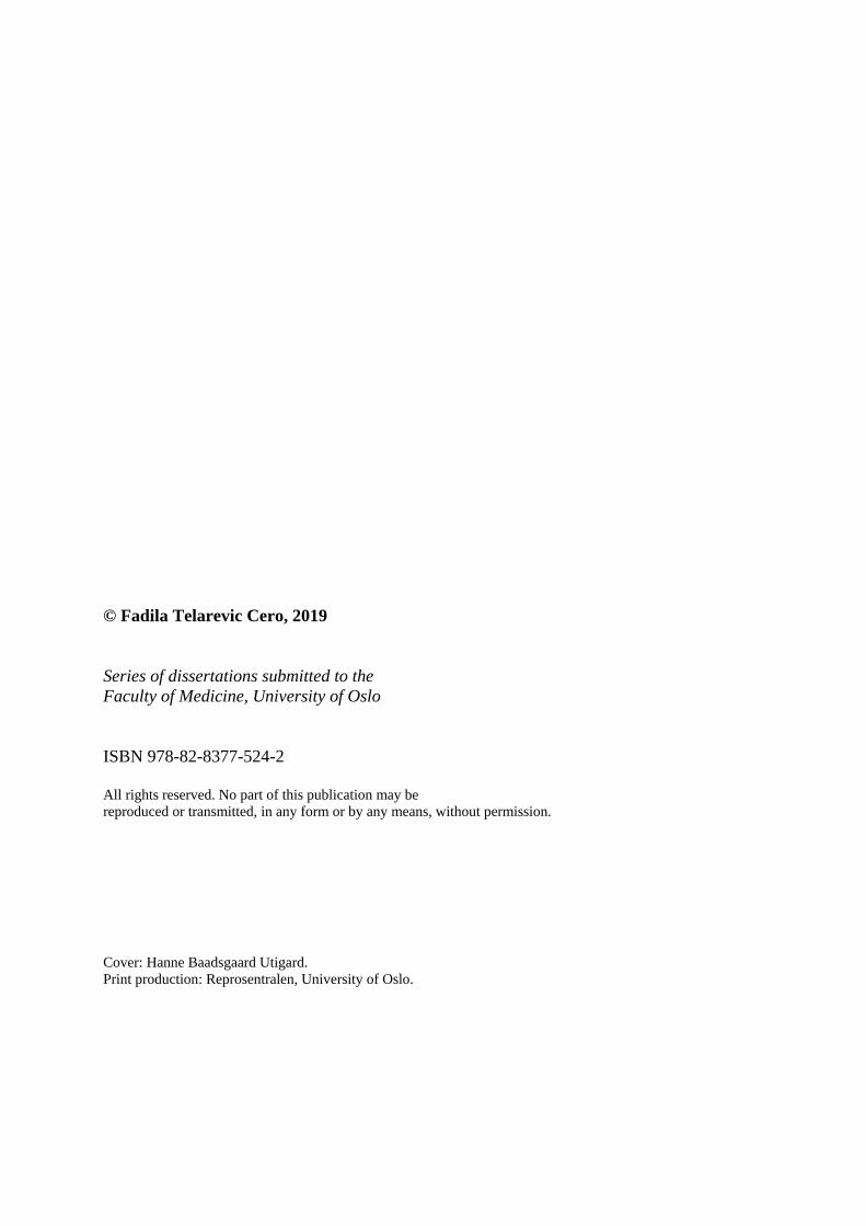
TABLE OF CONTENTS
LIST OF PAPERS .................................................................................................................... 8
SELECTED ABBREVATIONS .............................................................................................. 9
INTRODUCTION .................................................................................................................. 12
Inflammatory processes in the lung ......................................................................................... 12
Alveolar hypoxia ...................................................................................................................... 14
Innate immunity and inflammasomes ...................................................................................... 15
Activation of the inflammasome and innate immunity in lung disease
(IL-18, IL-1β and IL-12) .......................................................................................................... 18
Pulmonary hypertension ........................................................................................................... 20
Innate immunity in pulmonary hypertension and cardiac diastolic dysfunction...................... 23
AIMS OF THE STUDY ......................................................................................................... 25
METHODOLOGICAL CONSIDERATIONS .................................................................... 26
Animal models ......................................................................................................................... 26
Hemodynamic measurements .................................................................................................. 27
Magnetic resonance imaging of the heart................................................................................. 28
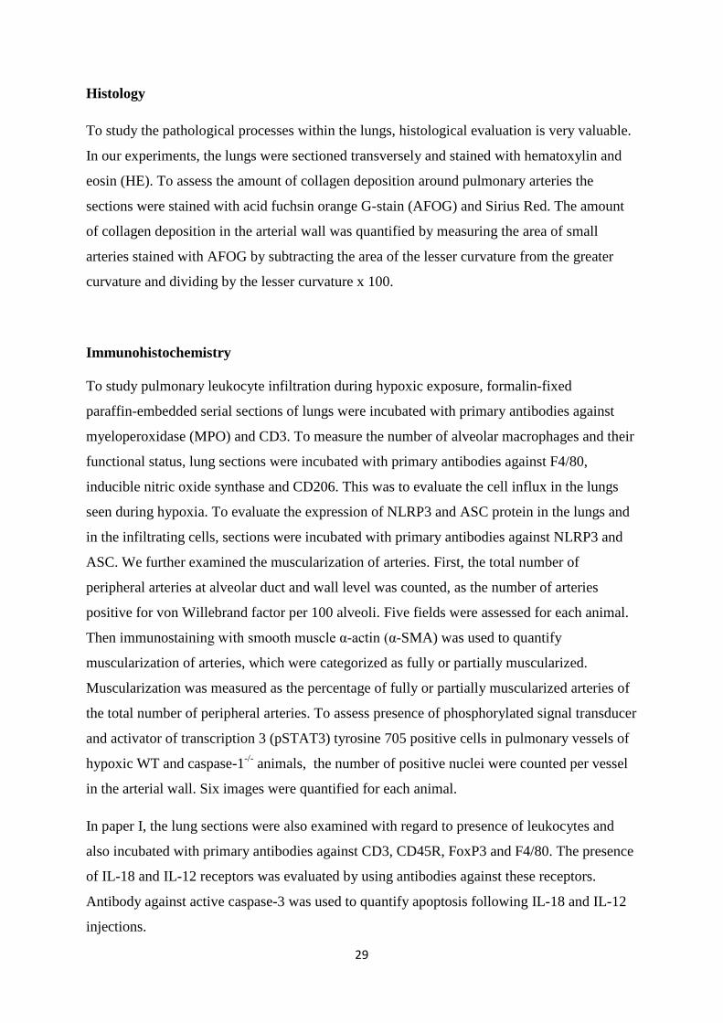
Histology .................................................................................................................................. 29
Immunohistochemistry ............................................................................................................. 29
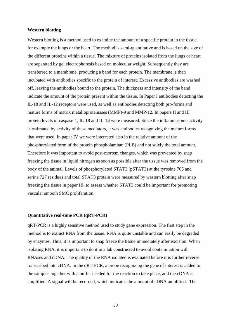
Western blotting ....................................................................................................................... 30
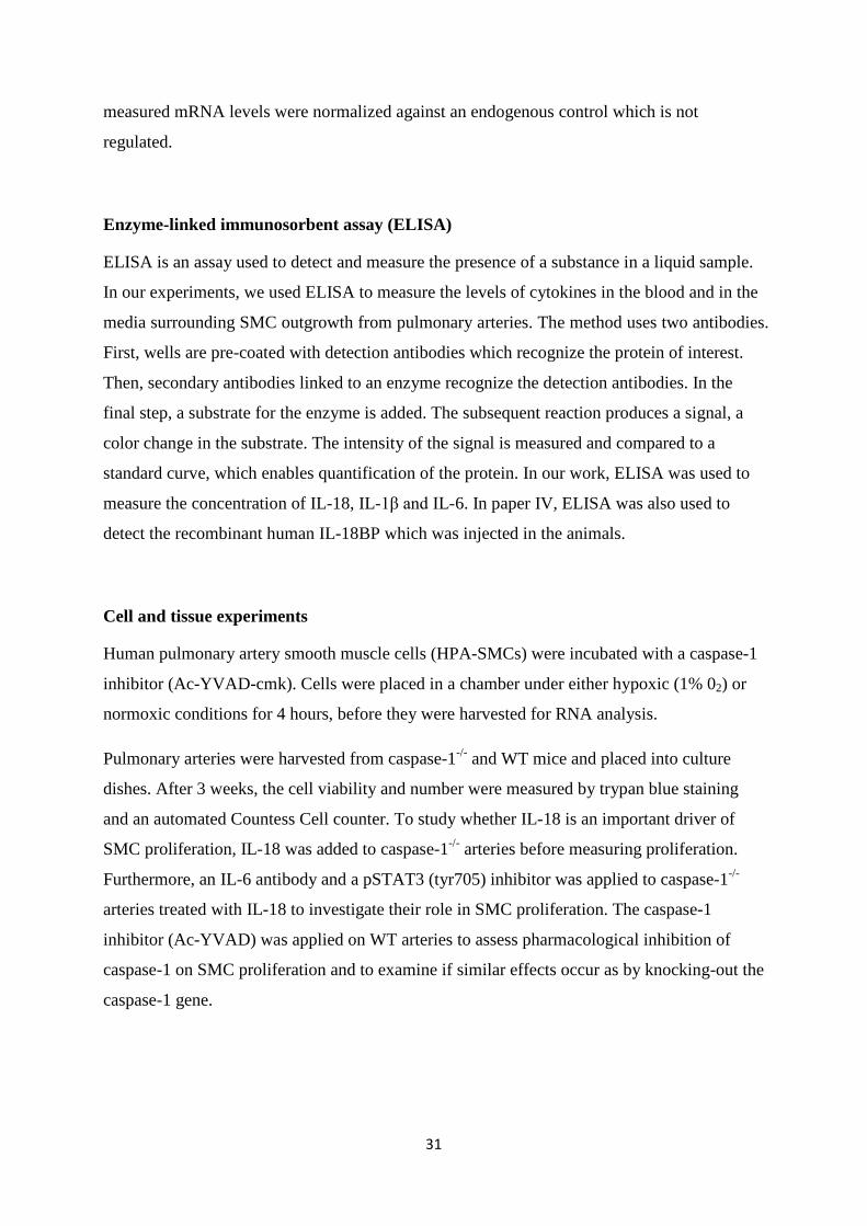
Quantitative real-time PCR (qRT-PCR) .................................................................................. 30
Enzyme-linked immunosorbent assay (ELISA) ....................................................................... 31
Cell and tissue experiments ...................................................................................................... 31
SUMMARY OF RESULTS ................................................................................................... 32
DISCUSSION ......................................................................................................................... 36
Innate immunity in hypoxia-induced inflammation and pulmonary hypertension .................. 36
Innate immunity in cardiovascular disease .............................................................................. 40

7
Anti-inflammatory treatment in hypoxia-induced pulmonary hypertension ............................ 42
Anti-inflammatory treatment in hypoxia-induced right heart remodeling and left ventricular diastolic dysfunction .............................................................................................. 43
MAIN FINDINGS AND CONCLUSIONS .......................................................................... 45
REFERENCES ....................................................................................................................... 47
8
LIST OF PAPERS
PAPER I
Cero FT, Hillestad V, Løberg EM, Christensen G, Larsen KO, Skjønsberg OH
IL-18 and IL-12 synergy induces matrix degrading enzymes in the lung.
Exp Lung Res, 2012:38(8):406-419.
PAPER II
Cero FT, Hillestad, Sjaastad I, Yndestad A, Aukrust P, Ranheim T, Gjervold IL, Olsen MB,
Lien E, Zhang L, Haugstad SB, Løberg EM, Christensen G, Larsen KO, Skjønsberg OH
Absence of the inflammasome adaptor ASC reduces hypoxia-induced pulmonary
hypertension in mice.
Am J Physiol Lung Cell Mol Physiol, 2015:309:L378–L38.
PAPER III
Udjus C, Cero FT, Halvorsen B, Behmen D, Carlson CR, Bendiksen BA, Espe EK, Sjaastad I,
Løberg EM, Yndestad A, Aukrust P, Christensen G, Skjønsberg OH, Larsen KO
Caspase-1 induces smooth muscle cell growth in hypoxia-induced pulmonary hypertension.
In manuscript form.
PAPER IV
Hillestad V, Espe EK, Cero FT, Larsen KO, Sjaastad I, Nygard S, Skjønsberg OH,
Christensen G
IL-18 neutralization during alveolar hypoxia improves left ventricular diastolic function
in mice.
Acta Physiol, 2015:213:492–504.
9
SELECTED ABBREVATIONS
AECOPD: acute exacerbations of COPD
AH: alveolar hypoxia
AMS: acute mountain sickness
ASC: apoptosis-associated speck like protein containing a caspase recruitment domain
BALF: bronchoalveolar lavage fluid
CF: cystic fibrosis
COPD: chronic obstructive pulmonary disease
CTL: C-type lectins
DAMPs: danger-associated molecular patterns
ECM: extracellular matrix
FCAS: familial cold autoinflammatory syndrome
FEV1: forced expiratory volume in the first second
GOLD: Global Initiative for Chronic Obstructive Lung Disease
HACE: high-altitude cerebral edema
HAPE: high altitude pulmonary edema
HFNEF: heart failure with normal left ventricular ejection fraction
HIF: hypoxia-inducible factor
HPA-SMC: human pulmonary artery smooth muscle cells
HPV: hypoxic pulmonary vasoconstriction
IFN: interferon
IPF: idiopathic pulmonary fibrosis
IL: interleukin
IL-18BP: interleukin-18 binding protein
ILD: interstitial lung disease
LV: left ventricle
MMP: matrix metalloproteinases
10
mPAP: mean pulmonary artery pressure
NLR: nucleotide-binding oligomerization domain (NOD)-like receptor
NO: nitric oxide
PAAT: pulmonary artery acceleration time
PAMPs: pathogen-associated molecular patterns
PLB: phospholamban
PO2: partial pressure of oxygen
pSTAT3: phosphorylated signal transducer and activator of transcription 3
ROS: reactive oxygen species
PRR: pattern recognition receptor
RV: right ventricle
RVOT: right ventricular outflow tract
RVSP: right ventricular systolic pressure
SERCA2: sarcoplasmic reticulum Ca2+ ATPase
SMC: smooth muscle cell
α-SMA: smooth muscle α-actin
SSc: systemic sclerosis
tau: time constant of isovolumic relaxation
TLR: toll-like receptor
TNF: tumor necrosis factor
TGF-β: transforming growth factor β

12
INTRODUCTION
Inflammatory processes in the lung
Inflammation is an essential component of lung diseases of various etiologies. Changes to the
airway structure have been described in patients with asthma, chronic obstructive pulmonary
disease (COPD) and cystic fibrosis (CF). It has been proposed that airway inflammation
drives these structural changes, and there has been a focus on investigating the involved
mechanisms in these inflammatory processes [1, 2]. Obstructive lung diseases, such as asthma
and COPD, are connected to both acute and chronic inflammation. Asthma is a disorder of the
airways involving various inflammatory mediators and cells and is characterized by bronchial
hyperresponsiveness and chronic inflammation [3]. Symptomatic attacks of asthma may be
caused by several factors, such as allergens, viruses and pollutants, which may induce an
additional, acute inflammatory response in the airways [4] and are characterized by sudden
worsening of respiratory symptoms. Acute exacerbations of COPD (AECOPD) are also
characterized by acute decline in airway function and worsening of the airway symptoms [5].
The common causes of AECOPD are viral and bacterial infections, but environmental stresses
are also involved [5, 6]. A hallmark of COPD is complex underlying chronic inflammation
that affects peripheral airways and lung parenchyma. This inflammatory response is thought
to be triggered by inhaled irritants such as smoke and air pollutants [7]. These inhaled irritants
can activate pattern recognition receptors such as Toll-like receptors (TLRs) on CD8+ T cells,
an important cell type which is increased in the central and peripheral airways in COPD
patients [8]. TLRs are key components of the innate immune system and acts by sensing
danger signals such as pathogen-associated molecular patterns on bacteria and viruses or
endogenous danger signals initiating inflammatory responses [9]. They are also a part of
adaptive immune responses involving T- and B lymphocytes [10, 11]. Chronic inhalation of
irritants initially activates TLRs in epithelial cells and macrophages. This leads to release of
chemotactic factors that attract neutrophils and monocytes in COPD patients [7]. In later
stages of the disease the adaptive immune responses are activated leading to increased number
and activation of T- and B lymphocytes, as well as dendritic cells [7]. Thus, the inflammatory
response in COPD is multifactorial involving both innate and adaptive immunity. Long-acting
bronchodilators are the basis of COPD therapy, and there are currently no effective anti-
inflammatory treatments. Inhaled corticosteroids have been shown to have limited clinical
benefits in COPD patients, and there is a need for new anti-inflammatory agents [12].
13
Interstitial lung diseases (ILDs) are characterized by inflammation and fibrosis of the
interstitium of the lungs. As the disease progresses, worsening hypoxemia and respiratory
failure may develop [13]. Previous research has shown that neutrophils and macrophages may
be important in driving the fibrotic processes in the lung, thus implicating a role for innate
immunity in the development of lung fibrosis [14]. ILDs are a large group of diffuse
parenchymal lung disorders, of which some, like idiopathic pulmonary fibrosis (IPF), one of
the idiopathic interstitial pneumonias, is associated with high morbidity and mortality [15].
Many IPF treatment trials have had negative outcome. During the last years, however, some
new compounds have shown beneficial effects on the progression of the disease [16].
Pirfenidone is a pyridone analogue which inhibits cytokines that play a key role in fibrosis
and inflammation. Nintedanib is a tyrosine kinase inhibitor targeting several growth factor
receptors resulting in reduced proliferation of human endothelial cells, vascular smooth
muscle cells and myofibroblasts, and also reduced collagen secretion [17]. However, currently
available therapies for IPF have limited effect, and the prognosis associated with this
condition remains poor [18]. Systemic sclerosis (SSc) has the highest fatality rate among
connective tissue diseases, and pulmonary involvement is considered to be the main cause of
mortality [19]. There is also evidence suggesting that morbidity and mortality rates are raising
for patients suffering from sarcoidosis in the USA, supporting the need to develop more
effective anti-inflammatory therapies for this condition, as well [20]. Altogether, additional
research is needed to develop more specific and effective therapies that could potentially
improve outcome of patients with inflammatory diseases related to lungs and airways [14].
The outcome of patients with chronic lung diseases such as ILD and COPD are dependent on
the severity of the disease and the related complications. Pulmonary hypertension is a serious
complication with a high prevalence (30-40%) in ILD patients, leading to reduced exercise
capacity and poor prognosis of these patients [21]. The prevalence of pulmonary hypertension
in stable COPD varies from 20 to 91% depending on the definition of pulmonary
hypertension (mean pulmonary artery pressure (mPAP) > 20 versus >25 mm Hg), the method
of measuring the PAP (echocardiography versus right heart catheterization), and the severity
of COPD (forced expiratory volume in the first second: FEV1) [22]. Pulmonary hypertension
in COPD patients also adversely affects survival and exercise capacity and is associated with
an increased risk of acute exacerbations [22]. Patients suffering from pulmonary hypertension
have increased resistance in their pulmonary vessels leading to increased work load on the
right ventricle (RV), a condition that may result in RV hypertrophy, and eventually dilatation
14
and failure [23-25]. During recent years, inflammatory processes have been claimed to be
important in the pathogenesis of pulmonary hypertension due to lung diseases and/or
hypoxia [26]. Furthermore it is shown that inflammatory processes are prominent features of
various forms of pulmonary hypertension, and inflammation is claimed to be of major
importance during the early phase of pulmonary hypertension [27]. The inflammatory cells
produce chemokines and cytokines leading to vascular remodeling by regulating growth,
migration, differentiation and metabolism of the different vascular cell types [27, 28]. Thus,
better understanding of these inflammatory processes in pulmonary hypertension may identify
innovative therapeutic strategies for the treatment of this devastating disease [27].
Alveolar hypoxia
Reduction of the alveolar partial pressure of oxygen (PO2) (alveolar hypoxia, AH) may occur
in chronic lung diseases of various etiologies, such as COPD and ILDs. Ventilation/perfusion
mismatch, resulting from progressive airflow limitation found in COPD patients, contributes
to the development of AH. It is well known that AH leads to hypoxic vasoconstriction in
pulmonary arteries, resulting in increased pulmonary artery pressure [29]. Hypoxic
constriction of the pulmonary arteries is a protective mechanism to reduce
ventilation/perfusion mismatch by directing the blood flow to better ventilated alveoli. When
AH is diffuse, such it is in severe COPD, it causes generalized pulmonary vasoconstriction
and consequently higher pulmonary artery pressure [22]. Chronic hypoxia leads to complex
pulmonary vascular remodeling [30], which further contributes to increased pulmonary
vascular resistance [22]. Elevated pulmonary artery pressure is also a consequence of
destruction of the pulmonary vascular bed by emphysema and hypoxic pulmonary
vasoconstriction caused by polycythemia seen in COPD patients [22]. Polycythemia causes a
local deficiency of nitric oxide (NO) and thus augments hypoxic pulmonary
vasoconstriction [22]. Severe emphysema with air-trapping and hyperinflation gives positive
alveolar pressure and contributes also to increased pulmonary artery pressure [22].
In addition to pulmonary arterial vasoconstriction, alveolar hypoxia leads to low oxygen
levels in the blood (hypoxemia), and uncorrected chronic hypoxemia is associated with
systemic inflammation and skeletal muscle dysfunction in COPD patients [29]. When
ascending to high altitudes, the barometric pressure falls with increasing altitude in a
logarithmic fashion. Thus, the partial pressure of oxygen (21% of barometric pressure) will
15
therefore also decrease and result in reduced alveolar PO2 [31]. Acute hypoxic exposure for
the first few days gives an increase in heart rate, myocardial contractility and cardiac output,
while acclimatization results in decreased left ventricular work and cardiac output and
increased right ventricular work [32].
Furthermore alveolar hypoxia causes hypoxic pulmonary vasoconstriction (HPV); this
increases the pulmonary arterial pressure, causing pulmonary hypertension. Pulmonary
hypertension together with “stress failure” of the pulmonary capillaries, causing leakage of
proteins and white blood cells and disturbed alveolar fluid clearance, are thought to be
responsible for high altitude pulmonary edema (HAPE) [31]. HAPE is a part of acute clinical
syndromes that are a consequence of hypobaric hypoxia due to rapid ascent to high altitude.
Acute mountain sickness (AMS) and high-altitude cerebral edema (HACE), both affecting the
brain, are also parts of this cluster of hypoxia related acute clinical syndromes [31].
Systemic effects are also observed in other diseases with alveolar hypoxia, such as
cardiovascular and metabolic dysfunctions of sleep-apnea, where inflammation is proposed to
influence the development and outcome of these conditions [33]. Our research group has
previously shown that alveolar hypoxia leads to increased circulating levels of the pro-
inflammatory cytokine IL-18 [34], implicating this cytokine in the inflammatory process
caused by alveolar hypoxia. Altogether it seems likely that inflammatory mediators may
contribute to the pathogenesis of various systemic pathological processes during alveolar
hypoxia [33].
Innate immunity and inflammasomes
There are two types of immunity used to protect the host from infections in vertebrates; innate
and adaptive immunity. The innate immunity is conserved across evolution and is present in
all multicellular organisms. It is the first line of defense and is able to trigger and guide the
slower but more specific adaptive immune response, which is evolved through time and of
newer evolutionary origin [35]. The innate immune system recognizes antigens that are
common to a wide range of pathogens, while the adaptive immune system has more diverse
recognition mechanisms where T-cell receptors and immunoglobulins are involved. This
results in immunological memory, which provides immunity to pathogens that our body has
previously been exposed to. Adaptive immune responses are mainly mediated through T- and
16
B-cell activation, while neutrophils, monocytes/macrophages, mast cells, natural killer cells
and dendritic cells are effector cells in the innate immune response. The complement system
and various soluble mediators secreted by these cells are also a part of innate immunity, and
together they make up the first line defense against microorganisms and xenobiotics [36].
Furthermore it is suggested that the innate responses are not only the first line of defense in an
immunological reaction against microorganisms, but also serves as a sophisticated system for
sensing danger signals from host-derived signals of cellular stress and DNA-damage [37].
These danger signals are detected through various cellular receptors called nucleotide-binding
oligomerization domain (NOD)-like receptors (NLRs), membrane bound Toll-like receptors
(TLRs) and C-type lectins (CTLs) [37, 38]. These receptors are also known as pattern
recognition receptors (PRRs), and several cytoplasmic PRRs are known to function in
inflammasome-based innate immunity. The NLR family constitutes the majority of PRRs that
function in inflammasome assembly. NLRs can be divided into four subfamilies; NLRA with
a transcriptional activation domain, NLRB with a baculovirus inhibitor of apoptosis repeat
(BIR) domain, NLRC with a caspase recruitment domain (CARD), and the largest NLRP
subfamily with a PYRIN (PYD) domain [39]. The PYRIN-CARD protein is also called
apoptosis-associated speck like protein containing a caspase-recruitment domain (ASC) and
functions as an adaptor which associates with procaspase-1 [40]. There are 23 NLR genes in
the human genome and 34 NLR genes are found present in the mouse genome [41]. There are
different activation patterns within the different inflammasomes and NLRs are involved in
diverse signaling pathways [37, 41].
Inflammasomes that are well characterized are NLRP1, NLRP3, NLRC4, NLRP6, NLRP7
and AIM2. They activate caspase-1 through the interaction between the sensor and the
adaptor protein called ASC [38, 42, 43]. The best characterized inflammasome is the NLRP3
inflammasome, and it consists of a sensor molecule NLR3, the adaptor protein ASC and
caspase-1 [37, 38]. During activation, the inflammasome assembles and triggers the activation
of caspase-1, which in turn cleaves pro-interleukin (IL)-18 and pro-IL-1β in the cytosol and
converts them to their mature forms. Mature IL-18 and IL-1β are then released to the
extracellular milieu where they can exert their effects. The NLRP3-inflammasome can be
activated upon exposure to a wide range of stimuli like whole pathogens, such as fungi
(e.g. Candida albicans), bacteria that produce toxins (e.g. Listeria monocytogenes and
Staphylococcus aureus), and various viruses (e.g. sendai virus, influenza virus and
17
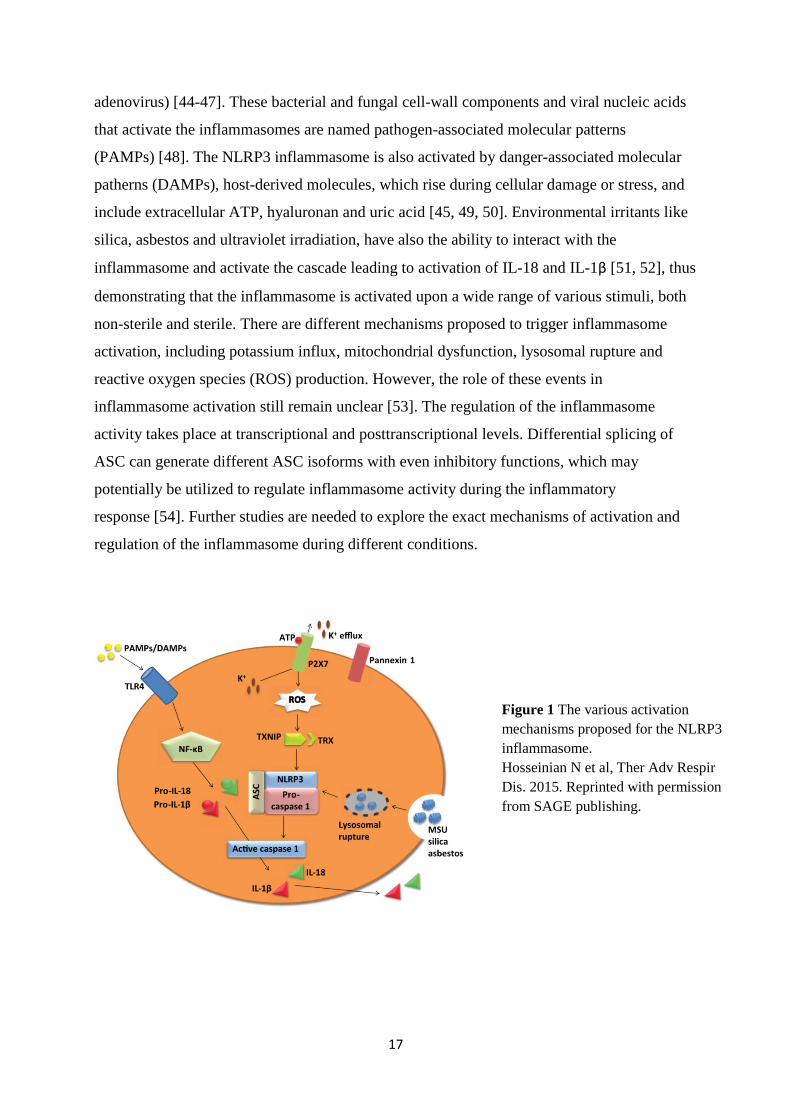
adenovirus) [44-47]. These bacterial and fungal cell-wall components and viral nucleic acids
that activate the inflammasomes are named pathogen-associated molecular patterns
(PAMPs) [48]. The NLRP3 inflammasome is also activated by danger-associated molecular
patherns (DAMPs), host-derived molecules, which rise during cellular damage or stress, and
include extracellular ATP, hyaluronan and uric acid [45, 49, 50]. Environmental irritants like
silica, asbestos and ultraviolet irradiation, have also the ability to interact with the
inflammasome and activate the cascade leading to activation of IL-18 and IL-1β [51, 52], thus
demonstrating that the inflammasome is activated upon a wide range of various stimuli, both
non-sterile and sterile. There are different mechanisms proposed to trigger inflammasome
activation, including potassium influx, mitochondrial dysfunction, lysosomal rupture and
reactive oxygen species (ROS) production. However, the role of these events in
inflammasome activation still remain unclear [53]. The regulation of the inflammasome
activity takes place at transcriptional and posttranscriptional levels. Differential splicing of
ASC can generate different ASC isoforms with even inhibitory functions, which may
potentially be utilized to regulate inflammasome activity during the inflammatory
response [54]. Further studies are needed to explore the exact mechanisms of activation and
regulation of the inflammasome during different conditions.
Figure 1 The various activation mechanisms proposed for the NLRP3 inflammasome. Hosseinian N et al, Ther Adv Respir Dis. 2015. Reprinted with permission from SAGE publishing.
18
Activation of the inflammasome and innate immunity in lung disease (IL-18,
IL-1β and IL-12)
Familial cold autoinflammatory syndrome (FCAS) and Muckel-Well syndrome are
inflammatory systemic diseases characterized by episodes of rash, arthralgia, fever and
conjunctivitis. The differences are that FCAS symptoms are precipitated by cold exposure,
while Muckle-Well syndrome is often associated with sensorineural hearing loss. These two
diseases were the first to be linked to mutations in the inflammasome, and mutations in NLR
genes were thought to be the cause of the diseases [55]. The role of the inflammasome is also
important in lung diseases of other etiology. Several bacteria that can cause pneumonia have
been shown to activate the NLRP3 inflammasome, like Streptococcus pneumoniae, Listeria
monocytogenes and Staphylococcus aureus, by their secretion of toxins [45, 56, 57].
Mycobacterium tuberculosis also has the ability to activate the NLRP3 inflammasome [58], as
well as different types of viruses like influenza A virus [59], demonstrating that infections of
various etiologies affecting the respiratory system activate the innate immune system through
activation of the inflammasome.
A role for the inflammasome is also proposed in chronic airway inflammation where IL-1β
and IL-18 have been suggested to be involved in the development of COPD, and caspase-1
levels are found to be increased in lung tissue from these patients [60-62]. Increased levels of
IL-1β have been found in COPD patients and correlates with the severity of COPD [63].
Induction of IL-1β production in the lungs of adult mice caused pulmonary inflammation,
enlargement of distal airspaces, mucous cell metaplasia and airway fibrosis, a phenotype that
resembles many of the features of COPD [64].
IL-18 protein has been shown to be strongly expressed in alveolar macrophages, CD8+ T-cells,
and in both the bronchiolar and alveolar epithelia in the lungs of COPD patients [61].
Furthermore, this cytokine was significantly higher in the serum of patients with Global
Initiative for Chronic Obstructive Lung Disease (GOLD) stage III and IV, compared to
smokers and nonsmokers without COPD, and a negative correlation between serum IL-18
levels and the forced expiratory volume in one second has been found, indicating that the
level of IL-18 is related to the severity of COPD [61]. Transgenic IL-18 mice that
constitutively overproduced mature IL-18 in the lungs showed chronic pulmonary lung
inflammation with increased appearance of CD8+ T-cells, neutrophils, macrophages and
eosinophils, in addition to increased production of interferon (IFN)-γ, IL-5, and IL-13.
19
Furthermore IL-18 overproduction led to severe emphysematous changes, dilatation of the
right ventricle, and mild pulmonary hypertension [65]. These findings indicate that IL-18 may
be an important mediator in pulmonary inflammation and features characteristic for COPD,
raising the question whether IL-18 inhibition may be a feasible treatment in COPD [65]. Mice
exposed to cigarette smoke have increased caspase-1 activity in their lung tissue, and also
increased IL-18 and IL-1β in bronchoalveolar lavage fluid (BALF), compared to mice
breathing normal air [62]. Caspase-1 activity is also found to be higher in lungs of COPD
patients and smokers compared to non-smokers, further indicating the inflammasome to be
involved in the inflammatory process of this disease [62].
Asthma is another chronic airway inflammatory disease where inflammasome activation is
proposed to play a role in the pathogenesis. Elevated levels of the IL-1β protein have been
shown to be present in the airways of patients with asthma [66], and there is also evidence
supporting a role for IL-1β in modulating airway constriction and relaxation via effects on
airway smooth muscle [67]. Furthermore it has been observed that IL-18 and its receptor are
strongly expressed in the lungs of patients with fatal asthma [68]. Circulating IL-18 levels are
found to be significantly higher in patients with moderate or severe asthma compared to
healthy controls [69]. These findings support a role of IL-1β and IL-18 in the pathophysiology
of asthma [69, 70].
In interstitial lung diseases, excessive accumulation of collagen and other extracellular matrix
components in the lung interstitium and basement membranes are responsible for the impaired
ventilatory function, which may lead to respiratory failure and death [71]. There are
indications that the inflammasome is involved in the pathogenesis of fibrosis since
particulates of asbestos, silica, bleomycin and statins, agents known to be able to initiate the
fibrotic process, can activate the inflammasome and the production of active IL-1β [72, 73].
Overexpression of IL-1β in rat lung stimulates the production of transforming growth factor
(TGF)-β, which is associated with progressive fibrosis in the lung [74]. The NLRP3
inflammasome is further claimed to be implicated in the pathogenesis of lung fibrosis in IPF
and rheumatoid arthritis with histopathological pattern of usual interstitial pneumonia [75].
IL-12 is proposed to be an important regulator of both innate and adaptive immunity [76].
IL-12 is produced by macrophages and activated dendritic cells within hours after encounter
with pathogens. IL-12 drives production of IFN-γ, and is capable of regulating T-cell
development and natural killer cell function together with the function of antigen-presenting
20
cells [76], involving this cytokine in both innate and adaptive immune responses. Previous
studies have shown that inflammatory effects of IL-18 are potentiated by IL-12 [77, 78], and
with regard to lung disease, both IL-12 and IL-18 are increased in COPD patients, suggesting
these two cytokines to be involved in the inflammatory pathways leading to COPD [61].
Furthermore, IL-12 may be involved in conditions involving hypoxia, since it has been shown
that murine macrophages exposed to hypoxic condition produced higher levels of this
cytokine [79].
Pulmonary hypertension
Pulmonary hypertension is defined as an increase in the mean pulmonary arterial pressure
(PAP) ≥25 mmHg at rest, as assessed by right heart catheterization [80]. The underlying
pathophysiological mechanisms are multifactorial, and the first clinical classification of
pulmonary hypertension was established in 1998 when it was divided in two categories,
primary pulmonary hypertension and secondary pulmonary hypertension according to the
presence of identified causes or risk factors. Five categories of pulmonary hypertension exist
currently, based on the underlying pathophysiology [80]. Current classification is presented in
Table 1 (next page).
21
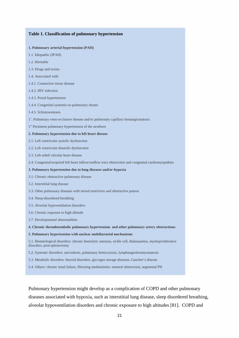
Table 1. Classification of pulmonary hypertension
1. Pulmonary arterial hypertension (PAH)
1.1. Idiopathic (IPAH)
1.2. Heritable
1.3. Drugs and toxins
1.4. Associated with:
1.4.1. Connective tissue disease
1.4.2. HIV infection
1.4.3. Portal hypertension
1.4.4. Congenital systemic-to-pulmonary shunts
1.4.5. Schistosomiasis
1’. Pulmonary veno-occlusive disease and/or pulmonary capillary hemangiomatosis
1” Persistent pulmonary hypertension of the newborn
2. Pulmonary hypertension due to left heart disease
2.1. Left ventricular systolic dysfunction
2.2. Left ventricular diastolic dysfunction
2.3. Left-sided valvular heart disease
2.4. Congenital/acquired left heart inflow/outflow tract obstruction and congenital cardiomyopathies
3. Pulmonary hypertension due to lung diseases and/or hypoxia
3.1. Chronic obstructive pulmonary disease
3.2. Interstitial lung disease
3.3. Other pulmonary diseases with mixed restrictive and obstructive pattern
3.4. Sleep-disordered breathing
3.5. Alveolar hypoventilation disorders
3.6. Chronic exposure to high altitude
3.7. Developmental abnormalities
4. Chronic thromboembolic pulmonary hypertension and other pulmonary artery obstructions
5. Pulmonary hypertension with unclear multifactorial mechanisms
5.1. Hematological disorders: chronic hemolytic anemias, sickle cell, thalassaemia, myeloproliferative disorders, post-splenectomy
5.2. Systemic disorders: sarcoidosis, pulmonary histiocytosis, lymphangioleiomyomatosis
5.3. Metabolic disorders: thyroid disorders, glycogen storage diseases, Gaucher’s disease
5.4. Others: chronic renal failure, fibrosing mediastinitis. tumoral obstruction, segmental PH
Pulmonary hypertension might develop as a complication of COPD and other pulmonary
diseases associated with hypoxia, such as interstitial lung disease, sleep disordered breathing,
alveolar hypoventilation disorders and chronic exposure to high altitudes [81]. COPD and
22
other pulmonary diseases mentioned are often followed by low oxygen tension in the blood. It
is known that acute alveolar hypoxia leads to a vasoconstrictor response in the pulmonary
vascular bed, redirecting the blood to the best ventilated areas of the lungs [82]. Pulmonary
arteries constrict to moderate to severe (2,7-8 kPa) hypoxia, whereas systemic arteries
vasodilate. As previously mentioned, this phenomenon is called hypoxic pulmonary
vasoconstriction (HPV), and is responsible for optimizing the ventilation–perfusion ratio
during localized alveolar hypoxia. However, more widespread alveolar hypoxia results in
generalized constriction of pulmonary arteries, resulting in increased load on the right side of
the heart [83].
Chronic hypoxic exposure induces alterations in the structure of pulmonary arteries, in both
biochemical and functional phenotypes of each of the cell types in the vascular wall [30].
Structural changes related to hypoxic exposure in mammals include the appearance of smooth
muscle cells in previously non-muscularized vessels of the alveolar wall. There is also medial
and adventitial thickening of the muscular and elastic vessels present [30]. It is believed that
medial thickening is due to hypertrophy and increased accumulation of smooth muscle cells
(SMCs) and increased deposition of extracellular matrix (ECM) components, mainly collagen
and elastin. The increased deposition of ECM proteins, together with accumulation of
fibroblasts and myofibroblasts, are responsible for the thickening of adventitial layer of
pulmonary arteries. Collagen and elastin are also the main ECM components deposited in
adventitia, together with fibronectin. In the larger pulmonary arteries, the media and
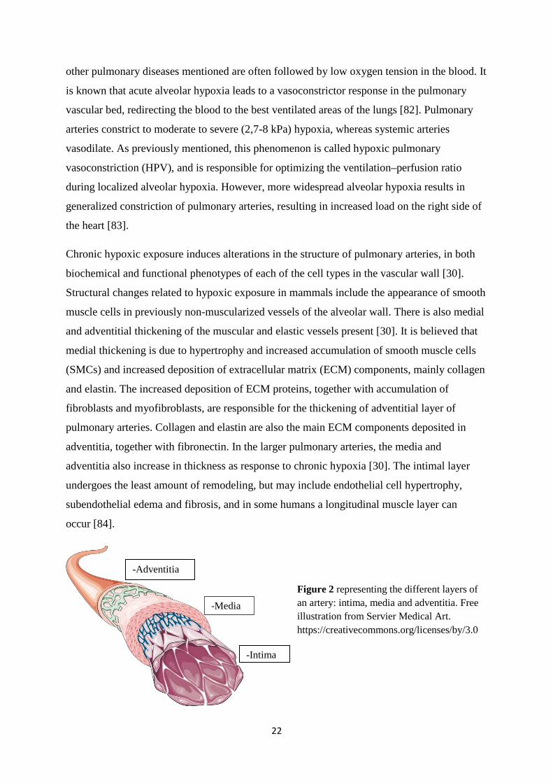
adventitia also increase in thickness as response to chronic hypoxia [30]. The intimal layer
undergoes the least amount of remodeling, but may include endothelial cell hypertrophy,
subendothelial edema and fibrosis, and in some humans a longitudinal muscle layer can
occur [84].
-Intima
-Media
-Adventitia
Figure 2 representing the different layers of an artery: intima, media and adventitia. Free illustration from Servier Medical Art. https://creativecommons.org/licenses/by/3.0
23
There is increasing evidence that both acute and chronic hypoxic exposure results in increased
expression of inflammatory cytokines, chemokines, adhesion molecules, and accumulation of
leukocytes within the lung and in particular around pulmonary vessels [30, 84], thus probably
playing an important role in driving the vascular remodeling observed.
Innate immunity in pulmonary hypertension and cardiac diastolic dysfunction
Hypoxia is a common feature of chronic pulmonary diseases, and our research group has
previously linked alveolar hypoxia to inflammasome activation through discovery of
increased levels of mature IL-18 during hypoxic exposure [34]. In addition, Villegas et al.
suggested ROS to be involved in the pathogenesis of hypoxia induced pulmonary
hypertension through the NLRP3 inflammasome [85]. Clinically, increased levels of IL-1β
and IL-18 have been observed in patients with pulmonary arterial hypertension [86, 87]. IL-18
transgenic mice which have overproduction of IL-18 in their lungs, develop severe
emphysema, mild pulmonary hypertension and dilatation of the right ventricle [65], further
implicating IL-18 in the pathological processes involving the lungs. These studies support a
possible role of the inflammasomes in the pathogenesis of pulmonary hypertension, but this
mechanism has not been properly documented.
IL-18 has been suggested as an important mediator in diastolic dysfunction of the heart
related to alveolar hypoxia [34]. Heart failure may be due to either systolic or diastolic
dysfunction, or a combination. Diastolic heart failure is also referred to as heart failure with
normal left ventricular ejection fraction (HFNEF) [88]. Diastolic dysfunction may have two
underlying mechanisms, an abnormal active relaxation of the cardiomyocytes or increased
ventricular stiffness [89]. Parameters used to diagnose diastolic left ventricular (LV)
dysfunction can be obtained either invasively during cardiac catheterization or non-invasively
by echocardiographic techniques. LV end-diastolic pressure, time constant of isovolumic
relaxation (tau) and pulmonary capillary wedge pressure are invasive measures used to
diagnose diastolic dysfunction [88]. Another parameter derived from pressure curves, the
maximum negative rate of change of left ventricular pressure (-dP/dtmax), describes diastolic
function, but is more dependent on the prevailing load whereas tau is relatively independent
of both load and heart rate [90]. There are also several non-invasive measures that can be used
to assess diastolic function, such as mitral valve flow velocity and tissue velocities [88].
24
The diastolic dysfunction related to alveolar hypoxia, as described in an experimental study
by Larsen et al, was related to impaired relaxation of the myocardium due to reduced
phosphorylation of the calcium handling protein phospholamban [91]. COPD and pulmonary
hypertension, both conditions in which increased levels of IL-18 have been found, can lead to
diastolic dysfunction, worsening the outcome of these patients [25, 92]. Daily administration
of IL-18 to healthy mice induced interstitial fibrosis in the heart and myocyte hypertrophy
resulting in increased ventricular stiffness. These results implicate IL-18 in the pathogenesis
of left ventricular diastolic dysfunction [93].
In the present thesis, we have explored the role of the innate immune system in pulmonary
inflammation, pulmonary hypertension and diastolic dysfunction. We have specifically
examined the role of the inflammasome components NLRP3, ASC and caspase-1 in
development of pulmonary hypertension and right ventricular remodeling. By administrating
IL-18 and IL-12, effects on mediators related to lung inflammation and emphysema were
studied, as well as apoptosis. We have further focused on inhibition of IL-18 using a natural
occurring antagonist and examined the effects on cardiac function.
25
AIMS OF THE STUDY
The main aim of the thesis was to explore mechanisms of pulmonary inflammation,
pulmonary hypertension, and cardiac function and morphology related to hypoxia and innate
immunity.
The specific aims were:
Paper I
To examine the effect of the cytokines IL-18 and IL-12 on inflammatory processes in the
lungs.
Paper II
To investigate whether the inflammasome is activated during alveolar hypoxia and involved
in the development of hypoxia induced pulmonary hypertension.
Paper III
To study the role of the inflammasome component caspase-1 in hypoxia-induced pulmonary
hypertension and explore mechanisms for increased pulmonary artery pressure.
Paper IV
To examine whether inhibition of IL-18, a product of inflammasome activation, during
alveolar hypoxia would prevent development of pulmonary hypertension and improve left
ventricular (LV) diastolic function.
26
METHODOLOGICAL CONSIDERATIONS
Animal models
Mouse models are used extensively in the field of pulmonary and cardiac research to increase
the knowledge on molecular mechanisms underlying various pulmonary and cardiac
conditions. The aim is to achieve new understanding and often to develop new therapeutic
principles for treating these diseases in humans. There are several advantages when using
mouse models. Mice are small in size, which makes them easy to handle in animal facilities. It
is also possible to acquire tissue for examination, which would not be as easily acquired from
human subjects. Another important reason for using mice is the possibility to create
genetically modified mouse models, where function of one particular gene can be either
removed (knocked-out, KO) or added (overexpressed) to the mouse genome. In this way, we
can study the phenotype of the respective genes.
In paper II and III we utilized mice lacking the gene for either NLRP3, ASC or caspase-1 to
study the function of these components of the inflammasome. The details on how these
knock-outs were created are described previously [94, 95]. They were made on C57Bl/6
background and we used C57Bl/6 mice as controls. This is the most widely used inbred strain
which features low genetic variability and thus highly reproducible results in experimental
studies. Here we used a model of chronic hypoxia to study the role of IL-18 and inflammsome
components in the development of pulmonary hypertension, right ventricular hypertrophy and
diastolic dysfunction of the heart. In our experiments, C57Bl/6 wild type mice and NLRP3,
ASC and caspase-1 KO mice were 8 weeks old when placed in a tightly sealed chamber and
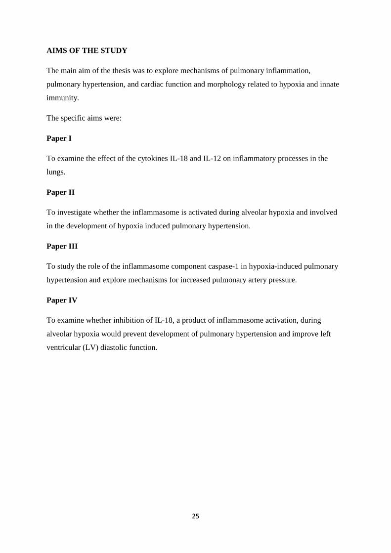
exposed to 10% oxygen for 3 days and up to 3 months (Figure 3). The carbon dioxide
concentration was monitored and kept under 0.4%, and humidity was measured daily. The
control groups were breathing room air.
Figure 3 Mice in hypoxia chambers breathing 10% O2 and mice breathing room air.
27
In paper I C57Bl/6 mice were utilized, and they received intraperitoneal (i.p.) injections with
IL-18, IL-12 or both of these cytokines combined. Control mice received i.p. injections with
phosphate-buffered saline (PBS). The purpose was to evaluate the inflammatory response in
the lungs induced by these two cytokines.
A model of chronic alveolar hypoxia was also used in paper IV, where C57Bl/6 mice were
placed in a hypoxia chamber, as previously described, for 2 weeks. They were treated with
either IL-18 binding protein (IL-18BP) or vehicle (i.p.). Since IL-18 BP is a natural inhibitor
of IL-18, the aim was to investigate whether inhibiton of IL-18 during alveolar hypoxia would
prevent development of pulmonary hypertension and improve LV diastolic function.
During all invasive procedures anesthesia by inhalation of isoflurane was used. Blood was
drawn from inferior vena cava, and the heart and lungs were rapidly excised. The atria, right
ventricular free wall, left ventricle and lungs were weighed and immediately snap frozen in
liquid nitrogen and stored at -70°C. The blood samples were centrifuged to obtain serum or
plasma. The blood and organs were used for ELISA, Western blot, PCR, histology and
immunohistochemical analyses.
There are many advantages in using mice models, as mentioned previously, but there are also
limitations. One of the most important questions is whether our results and findings are
transferable to humans. There are genetically differences between the two species, but at the
same time 99% of the mouse genes have a human homolog gene [96]. The biological
processes are often similar, but may differ between species. Therefore, it is important to
evaluate the findings in animal experiments thoroughly and underline that these findings are
not necessarily relevant for humans.
Hemodynamic measurements
Our institute has access to specialized equipment used to investigate cardiac function in small
animals such as high-frequency probes in echocardiography and pressure catheters of small
size, which can be used to access the ventricles through the vessels on the neck. Cardiac
catheterization is the gold standard for pressure measurements in the heart. Pressure
measurements in the RV were performed with intact chest by catheterization of the right
external jugular vein at 30 seconds, 2 minutes, 5 minutes, 2 weeks, 3 weeks, 1 month and 3
months of hypoxia with a 1.1 Fr Samba Preclin 420 LP micro pressure transducer (Samba
28
Sensors, Sweden) (papers II, III and IV). Pressure measurements were also performed in the
LV (Paper IV), using the same Samba catheter for catheterization of the right carotid artery.
Left ventricular systolic pressure, end-diastolic pressure and positive/minimum derivative of
the pressure curve (dP/dtmin) were registered. The time constant of isovolumic relaxation (tau)
was calculated using a custom made script fitting the pressure curve during relaxation phase.
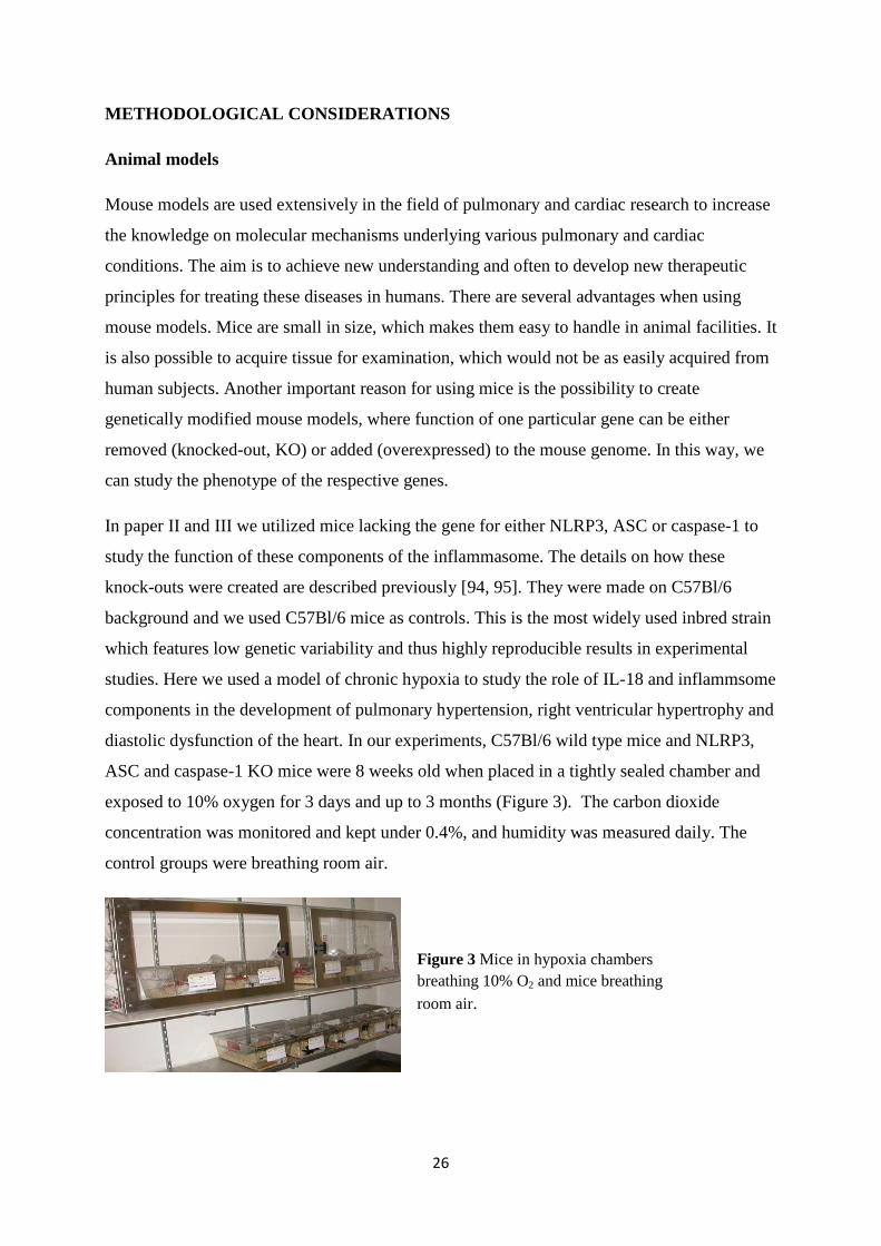
Echocardiography allows non-invasive, relatively fast and repeated examinations in each
animal. Echocardiopgraphy was performed with VEVO 21000 (Visual Sonics, Toronto,
Canada, Figure 4) to examine pulmonary artery acceleration time (PAAT), which is an
indirect measurement of PAP. Mitral flow and tissue velocities were studied to assess LV
diastolic function in paper IV.
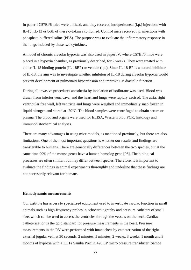
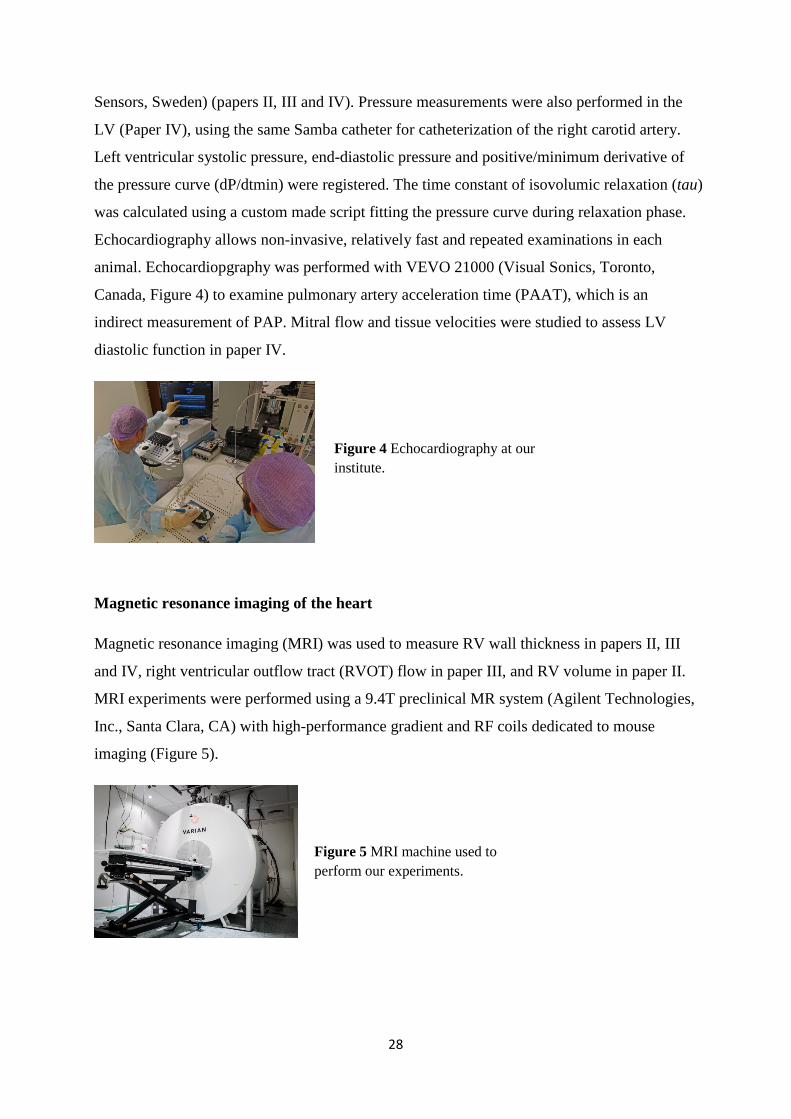
Magnetic resonance imaging of the heart
Magnetic resonance imaging (MRI) was used to measure RV wall thickness in papers II, III
and IV, right ventricular outflow tract (RVOT) flow in paper III, and RV volume in paper II.
MRI experiments were performed using a 9.4T preclinical MR system (Agilent Technologies,
Inc., Santa Clara, CA) with high-performance gradient and RF coils dedicated to mouse
imaging (Figure 5).
Figure 5 MRI machine used to perform our experiments.
Figure 4 Echocardiography at our institute.
29
Histology
To study the pathological processes within the lungs, histological evaluation is very valuable.
In our experiments, the lungs were sectioned transversely and stained with hematoxylin and
eosin (HE). To assess the amount of collagen deposition around pulmonary arteries the
sections were stained with acid fuchsin orange G-stain (AFOG) and Sirius Red. The amount
of collagen deposition in the arterial wall was quantified by measuring the area of small
arteries stained with AFOG by subtracting the area of the lesser curvature from the greater
curvature and dividing by the lesser curvature x 100.
Immunohistochemistry
To study pulmonary leukocyte infiltration during hypoxic exposure, formalin-fixed
paraffin-embedded serial sections of lungs were incubated with primary antibodies against
myeloperoxidase (MPO) and CD3. To measure the number of alveolar macrophages and their
functional status, lung sections were incubated with primary antibodies against F4/80,
inducible nitric oxide synthase and CD206. This was to evaluate the cell influx in the lungs
seen during hypoxia. To evaluate the expression of NLRP3 and ASC protein in the lungs and
in the infiltrating cells, sections were incubated with primary antibodies against NLRP3 and
ASC. We further examined the muscularization of arteries. First, the total number of
peripheral arteries at alveolar duct and wall level was counted, as the number of arteries
positive for von Willebrand factor per 100 alveoli. Five fields were assessed for each animal.
Then immunostaining with smooth muscle α-actin (α-SMA) was used to quantify
muscularization of arteries, which were categorized as fully or partially muscularized.
Muscularization was measured as the percentage of fully or partially muscularized arteries of
the total number of peripheral arteries. To assess presence of phosphorylated signal transducer
and activator of transcription 3 (pSTAT3) tyrosine 705 positive cells in pulmonary vessels of
hypoxic WT and caspase-1-/- animals, the number of positive nuclei were counted per vessel
in the arterial wall. Six images were quantified for each animal.
In paper I, the lung sections were also examined with regard to presence of leukocytes and
also incubated with primary antibodies against CD3, CD45R, FoxP3 and F4/80. The presence
of IL-18 and IL-12 receptors was evaluated by using antibodies against these receptors.
Antibody against active caspase-3 was used to quantify apoptosis following IL-18 and IL-12
injections.
30
Western blotting
Western blotting is a method used to examine the amount of a specific protein in the tissue,
for example the lungs or the heart. The method is semi-quantitative and is based on the size of
the different proteins within a tissue. The mixture of proteins isolated from the lungs or heart
are separated by gel electrophoresis based on molecular weight. Subsequently they are
transferred to a membrane, producing a band for each protein. The membrane is then
incubated with antibodies specific to the protein of interest. Excessive antibodies are washed
off, leaving the antibodies bound to the protein. The thickness and intensity of the band
indicate the amount of the protein present within the tissue. In Paper I antibodies detecting the
IL-18 and IL-12 receptors were used, as well as antibodies detecting both pro-forms and
mature forms of matrix metalloproteinases (MMP)-9 and MMP-12. In papers II and III
protein levels of caspase-1, IL-18 and IL-1β were measured. Since the inflammasome activity
is estimated by activity of these mediators, it was antibodies recognizing the mature forms
that were used. In paper IV we were interested also in the relative amount of the
phosphorylated form of the protein phospholamban (PLB) and not solely the total amount.
Therefore it was important to avoid post-mortem changes, which was prevented by snap
freezing the tissue in liquid nitrogen as soon as possible after the tissue was removed from the
body of the animal. Levels of phosphorylated STAT3 (pSTAT3) at the tyrosine 705 and
serine 727 residues and total STAT3 protein were measured by western blotting after snap
freezing the tissue in paper III, to assess whether STAT3 could be important for promoting
vascular smooth SMC proliferation.
Quantitative real-time PCR (qRT-PCR)
qRT-PCR is a highly sensitive method used to study gene expression. The first step in the
method is to extract RNA from the tissue. RNA is quite unstable and can easily be degraded
by enzymes. Thus, it is important to snap freeze the tissue immediately after excision. When
isolating RNA, it is important to do it in a lab constructed to avoid contamination with
RNAses and cDNA. The quality of the RNA isolated is evaluated before it is further reverse
transcribed into cDNA. In the qRT-PCR, a probe recognizing the gene of interest is added to
the samples together with a buffer needed for the reaction to take place, and the cDNA is
amplified. A signal will be recorded, which indicates the amount of cDNA amplified. The
31
measured mRNA levels were normalized against an endogenous control which is not
regulated.
Enzyme-linked immunosorbent assay (ELISA)
ELISA is an assay used to detect and measure the presence of a substance in a liquid sample.
In our experiments, we used ELISA to measure the levels of cytokines in the blood and in the
media surrounding SMC outgrowth from pulmonary arteries. The method uses two antibodies.
First, wells are pre-coated with detection antibodies which recognize the protein of interest.
Then, secondary antibodies linked to an enzyme recognize the detection antibodies. In the
final step, a substrate for the enzyme is added. The subsequent reaction produces a signal, a
color change in the substrate. The intensity of the signal is measured and compared to a
standard curve, which enables quantification of the protein. In our work, ELISA was used to
measure the concentration of IL-18, IL-1β and IL-6. In paper IV, ELISA was also used to
detect the recombinant human IL-18BP which was injected in the animals.
Cell and tissue experiments
Human pulmonary artery smooth muscle cells (HPA-SMCs) were incubated with a caspase-1
inhibitor (Ac-YVAD-cmk). Cells were placed in a chamber under either hypoxic (1% 02) or
normoxic conditions for 4 hours, before they were harvested for RNA analysis.
Pulmonary arteries were harvested from caspase-1-/- and WT mice and placed into culture
dishes. After 3 weeks, the cell viability and number were measured by trypan blue staining
and an automated Countess Cell counter. To study whether IL-18 is an important driver of
SMC proliferation, IL-18 was added to caspase-1-/- arteries before measuring proliferation.
Furthermore, an IL-6 antibody and a pSTAT3 (tyr705) inhibitor was applied to caspase-1-/-
arteries treated with IL-18 to investigate their role in SMC proliferation. The caspase-1
inhibitor (Ac-YVAD) was applied on WT arteries to assess pharmacological inhibition of
caspase-1 on SMC proliferation and to examine if similar effects occur as by knocking-out the
caspase-1 gene.
32
SUMMARY OF RESULTS
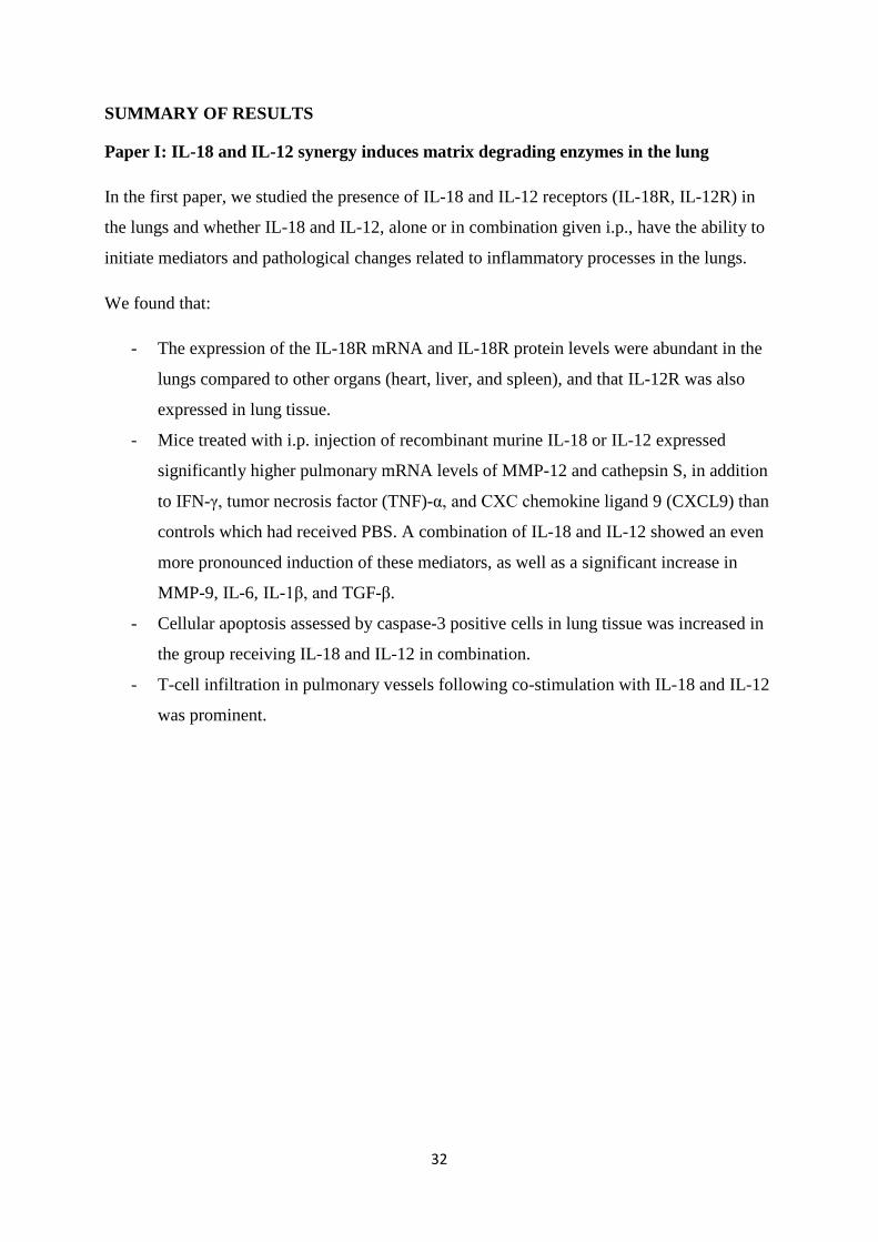
Paper I: IL-18 and IL-12 synergy induces matrix degrading enzymes in the lung
In the first paper, we studied the presence of IL-18 and IL-12 receptors (IL-18R, IL-12R) in
the lungs and whether IL-18 and IL-12, alone or in combination given i.p., have the ability to
initiate mediators and pathological changes related to inflammatory processes in the lungs.
We found that:
- The expression of the IL-18R mRNA and IL-18R protein levels were abundant in the
lungs compared to other organs (heart, liver, and spleen), and that IL-12R was also
expressed in lung tissue.
- Mice treated with i.p. injection of recombinant murine IL-18 or IL-12 expressed
significantly higher pulmonary mRNA levels of MMP-12 and cathepsin S, in addition
to IFN-γ, tumor necrosis factor (TNF)-α, and CXC chemokine ligand 9 (CXCL9) than
controls which had received PBS. A combination of IL-18 and IL-12 showed an even
more pronounced induction of these mediators, as well as a significant increase in
MMP-9, IL-6, IL-1β, and TGF-β.
- Cellular apoptosis assessed by caspase-3 positive cells in lung tissue was increased in
the group receiving IL-18 and IL-12 in combination.
- T-cell infiltration in pulmonary vessels following co-stimulation with IL-18 and IL-12
was prominent.
33
Paper II: Absence of the inflammasome adaptor ASC reduces hypoxia-induced
pulmonary hypertension in mice
The aim of this study was to investigate the role of the inflammasome in hypoxia-induced
pulmonary hypertension. Inflammasomes are part of the innate immune system and consist of
an enzyme caspase-1, a receptor, where NLRP3 is the best characterized, and the adaptor
protein ASC. We utilized mice deficient of NLRP3 and ASC and exposed them to 10%
oxygen for three days, one and three months to investigate whether these components played
a role in development of pulmonary hypertension. Control mice were breathing room air.
We showed that:
- Right ventricular systolic pressure (RVSP) of ASC-/- mice was significantly lower than
WT mice exposed to chronic hypoxia, indicating attenuation of pulmonary
hypertension in mice lacking ASC. Furthermore, ASC-/- mice displayed less
remodeling of the pulmonary vasculature, as shown by reduced degree of
muscularization and less fibrosis of the pulmonary arteries. RVSP of NLRP3-/- mice
exposed to hypoxia was not significantly altered compared to WT hypoxia.
- Magnetic resonance imaging supported these findings by demonstrating reduced right
ventricular remodeling in ASC-/- mice.
- Three days of hypoxic exposure demonstrated infiltration of leukocytes containing
NLRP3 and ASC components around pulmonary vessels.
- Hypoxic exposure increased protein levels of caspase-1, IL-18 and IL-1β in WT and
NLRP3-/- mice after three days and one month, showing inflammasome activation,
while this response was absent in ASC-/- mice.
34
Paper III: Caspase-1 deficiency reduces pulmonary hypertension
The aim of this study was to investigate the role of the inflammasome component caspase-1 in
hypoxia-induced pulmonary hypertension. Here we utilized mice deficient of caspase-1 and
subjected them to 10% oxygen to examine whether this enzyme influenced the development
of pulmonary hypertension and right ventricle remodeling. In addition, mechanisms leading to
hypoxia-induced pulmonary hypertension were explored.
We found that:
- Development of pulmonary hypertension in caspase-1 deficient mice was attenuated
compared to WT mice after 2 weeks of hypoxic exposure.
- Right ventricular weight and magnetic resonance imaging showed reduced right
ventricular remodeling in caspase-1-/- compared to WT mice in hypoxia, which is in
concordance with reduced pulmonary hypertension.
- Caspase-1-/- mice displayed less remodeling of the pulmonary vasculature, as shown
by reduced degree of muscularization of the pulmonary arteries.
- IL-18 and IL-1β levels did not increase significantly in caspase-1-/- mice after hypoxic
exposure, in contrast to WT mice. Furthermore, there was less perivascular infiltration
of leukocytes in caspase-1 deficient mice compared to WT, showing reduced
inflammatory response in these animals.
- Upregulation of the IL-18/IL-6/STAT3 pathway observed in hypoxic WT mice was
attenuated in caspase-1-/- mice.
- Isolated arteries from caspase-1-/- mice showed impaired smooth muscle cell
proliferation compared to WT. Levels of IL-18, IL-1β and IL-6 were also reduced in
the surrounding media of WT arteries treated with caspase-1 inhibitor and in caspase-1
deficient arteries.
- IL-18 induced vascular smooth muscle cell proliferation in caspase-1 deficient
pulmonary arteries, while IL-6 inhibition impaired proliferation significantly. pSTAT3
inhibition also showed a reduction in proliferation, although not reaching significant
level. These data indicate that caspase-1 mediates smooth muscle cell proliferation
through IL-18/IL-6/STAT3 signaling.
- Caspase-1 does not seem to be involved in acute hypoxic pulmonary vasoconstriction.
35
Paper IV: IL-18 neutralization during alveolar hypoxia improves left ventricular
diastolic function in mice.
In this paper, we examined whether inhibition of IL-18, a product of inflammasome activation,
would hamper the development of hypoxia-induced LV diastolic dysfunction. In addition, the
influence of IL-18 inhibition on calcium handling in the LV was studied. First, we
investigated whether IL-18BP given i.p. could inhibit the functional consequence of
circulating IL-18 produced in vivo using LPS as a stimulus.
We showed that:
- LPS induced production of IL-18, which in turn induced release of IFN –γ, as
previously known from other studies. IL-18BP efficiently inhibited production of
IFN-γ, indicating IL-18 inhibition.
- Exposure to hypoxia for two weeks induced LV diastolic dysfunction in mice, shown
by prolonged time constant of isovolumic relaxation (tau).
- Mice treated with IL-18BP during hypoxia had a decrease in tau compared to the
hypoxia vehicle group, indicating improved diastolic function.
- Hypoxic exposure induced decreased levels of serine16phophorylated PLB, which
were normalized by IL-18BP treatment, indicating a role for IL-18 in regulation of
calcium-handling proteins in diastolic dysfunction.
- Mice exposed to hypoxia developed pulmonary hypertension and RV hypertrophy.
MRI showed less increase in RV wall thickness in IL-18BP treated animals, showing a
reduction in RV remodeling. RV systolic pressure was not significantly reduced by
IL-18BP.
36
DISCUSSION
Chronic lung diseases of various etiologies are often associated with alveolar hypoxia, either
regionally or globally. Sleep apnea, obesity hypoventilation syndrome, COPD, asthma,
bronchial tumors, ILDs and neuromuscular and skeletal disorders affecting the chest wall are
examples of conditions were alveolar hypoxia may occur [30]. Alveolar hypoxia triggers the
development of pulmonary hypertension, which is a serious complication to the above
mentioned diseases. Our research group has previously reported increased levels of IL-18
during hypoxic exposure of mice, indicating that hypoxia may be linked to inflammasome
activation. While it is known that various danger signals, such as bacteria, virus and also
sterile stimuli activate the innate immune system through activation of the inflammasome, the
results of the current thesis have shown that hypoxia also seems to be perceived as a danger
signal activating the immune system through this pathway. Articles included in this thesis
support an important role for the innate immunity in development of hypoxia-induced
pulmonary inflammation, pulmonary hypertension and cardiac dysfunction.
Innate immunity in hypoxia-induced inflammation and pulmonary hypertension
Localized acute hypoxic exposure leads to regional vasoconstriction of the pulmonary arteries
to redirect blood to the better ventilated areas of the lungs, which is beneficial. More
widespread alveolar hypoxia leads to generalized vasoconstriction, increased pressure in the
pulmonary arteries and increased right ventricular afterload [83]. The link between alveolar
hypoxia and development of pulmonary hypertension has not been fully elucidated [83]. Our
previous finding of increased levels of IL-18 during alveolar hypoxia, prompted us to explore
the role of innate immunity in this process by studying whether the inflammasome is activated
during acute and chronic hypoxic exposure and how inflammasome activation may contribute
to the development of pulmonary hypertension. In paper II, we exposed WT mice to 3 days,
1 month and 3 months of hypoxia. We found that acute hypoxic exposure for 3 days induced
infiltration of leukocytes containing NLRP3 and ASC components around pulmonary vessels
and bronchi, implicating involvement of inflammatory cells with the inflammasome.
Activation of the inflammasome was shown by increased levels of active caspase-1, mature
IL-18 and IL-1β. This response was still present after 1 month, showing inflammasome
activation both during acute and chronic hypoxic exposure. Having in mind that we could
document high levels of IL-18 receptors in the lungs, indicating the lung as a target organ for
37
this cytokine, we wanted to study the effect of inflammasome activation on the development
of hypoxia-induced pulmonary hypertension further, by utilizing mice deficient of the
inflammasome components caspase-1, NLRP3 and ASC. We found decreased inflammasome
activation in hypoxic mice lacking caspase-1 and ASC, corresponding with a significantly
decreased RVSP compared to WT mice, both after 2 weeks and 3 months of hypoxia. Our
results show that hypoxia acts as a danger signal, activating the innate immune system by
inducing leukocyte infiltration and activation of the inflammasome. Furthermore, this thesis
suggests that activation of the inflammasome is of importance for development of pulmonary
hypertension, probably through caspase-1 mediated activation of the inflammatory cytokines
IL-18 and IL-1β.
Chronic hypoxia causes mainly medial and adventitial thickening of the pulmonary arteries
due to hypertrophy and increased number of smooth muscle cells and increased deposition of
ECM components, which lead to vascular remodeling, as previously mentioned. The
mechanisms behind these changes are not fully known, but in all cases of hypoxia-related
vascular remodeling and pulmonary hypertension, inflammation, and particularly persistent
inflammation, is thought to play an important role [84]. It is suggested that a combination of
hypoxia and local tissue factors/cytokines interact with SMCs, fibroblasts and macrophages
and promote proremodeling and a proinflammatory phenotype, which in turn promotes
transition to chronic nonresolving inflammation and vascular remodeling, leading to
pulmonary hypertension [84]. In the current thesis, it is shown that the innate immune system,
through activation of inflammasomes, seems to play an important role in inducing and
maintaining this inflammation.
Our articles confirm vascular remodeling of pulmonary arteries after hypoxic exposure. Two
weeks of hypoxia induced increased muscularization of pulmonary arteries, and after
3 months collagen deposition and fibrosis was also observed. Caspase-1-/- mice showed
reduced muscularization after 2 weeks of hypoxia, indicating a role for inflammasome
activation in hypoxia-induced proliferation of SMCs. This finding was supported by
decreased proliferation of hypoxia-exposed human SMCs in vitro after treatment with a
caspase-1 inhibitor. Attachment of isolated pulmonary arteries to culture dishes seemed to
trigger inflammasome activation, as documented by increased levels of IL-1β in the medium
surrounding WT arteries compared to caspase-1-/- arteries. Moreover, the level of IL-18 was
significantly reduced in the medium surrounding WT pulmonary arteries inhibited by a
38
selective caspase-1 inhibitor. Outgrowth of SMCs from arteries deficient in caspase-1 was
significantly decreased compared to WT. Decreased outgrowth was also observed in WT
pulmonary arteries treated with a caspase-1 inhibitor. Thus, hypoxia-induced proliferation of
vascular SMCs seems to be linked to inflammasome activation. In addition to SMC
proliferation, hypoxic exposure resulted in collagen deposition in the pulmonary artery wall of
WT mice. This process was attenuated in mice lacking the ASC gene after 3 months of
hypoxic exposure, while there were no significant differences in collagen deposition after 2
weeks of hypoxia between WT and mice lacking the caspase-1 gene. Our results indicate that
the fibrotic process is connected to activation of the innate immune system through the
inflammasome when exposed to long-term hypoxia.
After having substantiated that alveolar hypoxia activates the inflammasome, we tried to
establish a link between this component of the immune system and the remodeling of
pulmonary arteries leading to pulmonary hypertension. The proteolytic cleavage of the pro-
forms of IL-18 and IL-1β seems to be of major importance, leading to increased levels of the
biologically active form of these inflammatory cytokines. IL-18 has been most thoroughly
studied in the current thesis. Both of these cytokines are shown to be able to induce vascular
smooth muscle cell proliferation and migration [97-99], as well as increased collagen
production and proliferation of fibroblasts from various origins [100-102]. Therefore the
observed reduction of muscularization and fibrosis in ASC-/- and reduced muscularization in
caspase-1-/- mice may be due to the impaired activation of IL-18 and IL-1β. In our
experiments, IL-18 induced robust smooth muscle cell outgrowth from pulmonary arteries
lacking caspase-1, indicating IL-18 to be an important stimulator of smooth muscle cell
proliferation in pulmonary arteries. Our results may be relevant for pulmonary hypertension
of various etiologies since different types of pulmonary hypertension have increased IL-18
and IL-1β [86, 87, 103].
We further wished to explore possible downstream mediators of IL-18 and IL-1β and
analyzed a panel of proinflammatory mediators. IL-6 expression was significantly reduced in
mice lacking caspase-1 compared to WT exposed to hypoxia. IL-1β has previously been
shown to induce IL-6 production [104]. The role of IL-6 was further examined by adding an
IL-6 inhibitor to caspase-1 deficient arteries replenished with IL-18. In this experiment, we
observed that IL-6 inhibition impaired SMC proliferation. IL-6 can induce tyrosine and serine
phosphorylation of STAT3 [105], and STAT3 activation has been shown to be responsible for
39
modulation of the expression of several proteins already established in the pathogenesis of
pulmonary hypertension [106]. Our experiments confirmed activation of pSTAT3 in WT
animals during hypoxia, while this activation was absent in caspase-1 deficient mice.
Furthermore, when a STAT3 inhibitor was added to IL-18 replenished caspase-1 deficient
arteries, SMC proliferation was impaired. Taken together our results suggest the IL-6/STAT3
axis to be involved in vascular smooth muscle cell proliferation. Impaired IL-6 expression
was also demonstrated in ASC-/- mice exposed to hypoxia, strengthening the link between
IL-6 and the inflammasome in the development of pulmonary hypertension. IL-6 has been
found to be elevated in patients with pulmonary hypertension [86, 103, 107]. In addition,
transgenic mice with lung specific IL-6 overexpression exhibit elevated right ventricular
systolic pressure, right ventricular hypertrophy and increased muscularization of the
pulmonary arteries, all of which were exacerbated by chronic hypoxia [108]. A pathogenic
role for IL-6 in certain types of pulmonary hypertension is further supported by observations
that IL-6-/- mice are protected from pulmonary hypertension [109]. Previous studies, together
with our findings, support a role of IL-6 in the pathogenesis of pulmonary hypertension.
It has been shown that hypoxia and changes in wall stress induces generation of ROS,
activation of nuclear factor (NF)-κB signaling, production of IL-6 and activation of hypoxia-
inducible factor (HIF), which further activate downstream pathways leading to vascular
remodeling in all three layers [84]. Matrix metalloproteinases are also involved in vascular
remodeling [110], and the level of MMP-9 is positively correlated with the severity of
experimental pulmonary hypertension [111, 112]. Our research revealed reduced MMP-9 and
HIF-1α in ASC deficient mice during hypoxic exposure, indicating that these two mediators
may be involved in reduced pulmonary hypertension in mice depleted of ASC together with
IL-6. Furthermore, in paper III we found reduced pSTAT3 protein levels in caspase-1-/- mice
exposed to hypoxia, supporting a role for the IL-6/STAT3 pathway in hypoxic pulmonary
hypertension. Toll-like receptors (TLRs) and NOD-like receptors (NLRs) involved in
inflammasome formation can activate a series of signaling pathways, resulting in upregulation
of genes encoding pro-inflammatory cytokines, such as TNFα and IL-6 [113]. However, we
did not find changes in TNFα expression in WT mice exposed to hypoxia. Our work indicates
that inflammation and inflammasome activation is important in the development of hypoxic
pulmonary hypertension through changes in medial and adventitial layers of the arteries,
possibly through actions of IL-6/STAT3, in addition to MMP-9 and HIF-1α.
40
In our first study, mice received IL-18 i.p. We discovered that IL-18 had the ability to induce
inflammatory mediators and proteolytic enzymes known to participate in the pathogenesis of
pulmonary emphysema. This effect was enhanced by co-stimulation with IL-12. Moreover,
high levels of the IL-18R were present in the lungs compared to other organs, indicating the
lungs to be an important target organ for IL-18. Since both IL-18 and IL-12 are found in
COPD patients [61], it is possible that the effects of IL-18 and IL-12 are relevant for
development of pulmonary inflammation and emphysema seen in these patients. Furthermore,
pulmonary hypertension is a complication to COPD [81], and it is possible that the observed
effects are relevant for COPD associated pulmonary hypertension, as well. Co-stimulation
induced IL-6 expression, a cytokine shown to be involved in vascular remodeling in
pulmonary hypertension both by others and by our group. Previously, it has been shown that
pulmonary arterial hypertension patients have significantly higher levels of IL-12 compared
with healthy control subjects [103]. Therefore, we cannot rule out that IL-12 is involved in
vascular remodeling together with IL-18 through the actions of IL-6 in COPD patients.
Innate immunity in cardiovascular disease
IL-18 has previously been implicated in atherosclerotic plaque instability and is associated
with cardiovascular disease risk [114]. The first studies that suggested IL-18 to be involved in
human heart failure found increased circulating levels of IL-18 in patients with chronic heart
failure. The IL-18 levels were higher in patients with severe heart failure and in those who
died during follow-up compared to survivors [115, 116]. IL-1β is also implicated in both
patients and animal models of cardiovascular disease [117], and a significant correlation
between declining functional class of heart failure and increasing levels of IL-1β has been
found [118]. These studies show a link between inflammasome activity and cardiovascular
diseases of different etiology.
Patients suffering from pulmonary hypertension develop increased resistance in the
pulmonary vascular bed and increased work load on the RV. These changes can result in RV
dilatation and failure [25], a process relevant for chronic lung diseases associated with
hypoxia. In our studies, we showed that healthy mice exposed to chronic hypoxia develop RV
hypertrophy after 2 weeks and that they develop RV dilatation after 3 months. In Paper III
caspase-1-/- mice had reduced RV remodeling, as demonstrated by reduced RV weight and
41
RV free wall thickness, compared to WT mice after 2 weeks of hypoxic exposure. ASC-/- mice
showed no dilatation of the RV ventricle after 3 months of hypoxia, in contrast to WT mice
(Paper II). The right ventricle is exposed to increased afterload by chronic pulmonary
hypertension, and the initial adaptive response of myocardial hypertrophy is followed by a
progressive contractile dysfunction and dilatation of the ventricle [25]. Since both caspase-1-/-
and ASC-/- mice had reduced RV remodeling, it seems likely that these changes are due to
reduced right ventricular systolic pressure (RVSP) and hence reduced workload on the RV.
However, since both IL-18 and IL-1 β may promote myocardial changes like hypertrophy and
myocardial dysfunction [117, 119], in the absence of increased afterload, a more direct impact
of these cytokines in the process of cardiac remodeling is plausible, as well.
Diastolic dysfunction of the LV is a frequent echocardiographic finding in COPD patients,
and is associated with disease severity [92]. A role for IL-18 in left ventricular diastolic
dysfunction caused by alveolar hypoxia was proposed by our group in a study where mice
exposed to hypoxia developed LV diastolic dysfunction in parallel with elevated levels of
circulating IL-18 [34]. Larsen et al. showed that IL-18 plays a role in dephosphorylation of
PLB, a protein that regulates the activity of sarcoplasmic reticulum (SR) Ca2+ ATPase
(SERCA2). SERCA2 is located in the SR and is responsible for pumping Ca2+ into the SR
and regulating cardiac muscle relaxation and cardiac contractility. When PLB is
phosphorylated it dissociates from SERCA2, activating this Ca2+ pump, but in the
dephosphorylated state PLB inhibits SERCA2 activity [120].
Based on these previous findings, we wished to investigate whether diastolic dysfunction
induced by alveolar hypoxia could be improved by IL-18 inhibition, and if this inhibition was
related to the phosphorylation status of PLB. In Paper IV mice developed LV diastolic
dysfunction after 2 weeks of hypoxic exposure. This was shown by prolonged tau. When
mice received IL-18BP, the relaxation of the myocardium improved, as shown by
significantly reduced tau values. Furthermore, we found decreased levels of phosphorylated
PLB in hypoxia, which were normalized by IL-18BP treatment. The restored levels of
phosphorylated PLB can be a possible mechanism for the observed improvement in diastolic
function by decreased inhibition of SERCA2 activity. However, we did not find a significant
reduction in RVSP, although a trend towards both reduced RVSP and RV weight was found.
This is in contrast to ASC-/- and caspase-1-/- mice, which both had reduced RVSP and RV
42
hypertrophy, and ASC-/- mice did not develop RV dilatation after 3 months of hypoxia. Both
ASC-/- and caspase-1-/- mice lacked the increase in circulating IL-18, which suggests a role for
IL-18 in RV hypertrophy and remodeling. IL-18 and IL-18BP have been shown to play a role
in cardiac hypertrophy [97, 121]. Our results, together with these previous studies, indicate
that IL-18 may directly affect RV remodeling. However, we did not find a significant
reduction in RVSP and RV weight by inhibiting IL-18 with IL-18BP, even though we found a
trend. This may be due to incomplete inhibition of IL-18 activity by IL-18BP or that
inhibition of both IL-18 and IL-1β, as in mice lacking ASC and caspase-1, has synergistic
effects on the development of pulmonary hypertension.
Anti-inflammatory treatment in hypoxia-induced pulmonary hypertension
Our studies show an important role for innate immunity in pulmonary inflammation and
hypoxia-induced pulmonary hypertension. In addition, injection of IL-18 together with IL-12
in mice promoted expression of mediators involved in development of emphysema.
Interestingly, this treatment also induced IL-6, a cytokine that previously has been shown to
be involved in the development of pulmonary hypertension. Furthermore, by using mice
lacking ASC and caspase-1, we demonstrated inhibited activation of IL-18 and IL-1β during
chronic hypoxia and a beneficial effect on hypoxia-induced pulmonary hypertension. This
was shown by reduced RVSP, RV remodeling and reduced remodeling of the pulmonary
arteries in ASC-/- and caspase-1-/- mice. These results suggest that innate immunity, and
particularly components and mediators related to the inflammasome, could be
pharmacological targets in treatment of pulmonary hypertension. Inhibitors of caspase-1,
IL-18 and IL-1β already exist and could be potential candidates for treatment of pulmonary
hypertension in the future.
There are several therapies shown to be beneficial and approved for treating pulmonary
arterial hypertension, such as endothelin-1 receptor antagonists, phosphodiesterase-5
inhibitors, guanylate cyclase stimulators and prostacyclin analogues [122]. However,
currently there is no specific therapy for pulmonary hypertension associated with lung
diseases. Drugs used for treating pulmonary arterial hypertension have so far not improved
symptoms or outcome for this group of patients and are therefore not recommended for
patients with pulmonary hypertension due to lung disease [122, 123]. Long-term
43
O2-administration is shown to improve survival in stable COPD patients with resting
hypoxemia (PaO2< 7,3kPa) [124, 125], and long-term O2-administration is the only therapy
which has been shown to reduce progression of pulmonary hypertension in COPD patients
and which is recommended in hypoxic COPD patients with pulmonary hypertension [122].
Thus, more effective medical treatment of pulmonary hypertension associated with lung
disease is warranted [126].
Inflammation has been shown to play a role in hypoxia-related vascular remodeling and
pulmonary hypertension, and persistent inflammation is thought to be particularly
important [84]. Our findings support the theory that inflammation, and particularly
inflammation due to innate immunity, is important in hypoxia-induced pulmonary
hypertension.
Anti-inflammatory treatment in hypoxia-induced right heart remodeling and left
ventricular diastolic dysfunction
Pulmonary hypertension is a common cause of right heart failure [25]. With regard to medical
treatment, regression of RV hypertrophy has been demonstrated after 1 year of treatment with
high-dose calcium channel blockers [127]. Prostacyclin treatment also gave a modest RV
reverse remodeling with less dilatation and improved RV stroke volume in patients with
pulmonary arterial hypertension [128, 129]. Riociguat, a guanylate cyclase stimulator, has
decreased RV hypertrophy in rats which developed pulmonary vascular pathology similar to
pulmonary arterial hypertension when exposed to vascular endothelial growth receptor
antagonist and hypoxia [130]. However, since drugs used for treating pulmonary arterial
hypertension seem not to improve symptoms or outcome for patients with pulmonary
hypertension due to lung disease [122, 123], these treatment options are not available for this
group of patients. Therapy for RV dysfunction associated with chronic lung disease would
have to reduce both the increased pulmonary vascular resistance caused by hypoxic
vasoconstriction and pulmonary vascular remodeling. Therefore, current recommendations
only support long term oxygen therapy and the use of medication targeted at the underlying
pulmonary disease [131]. We showed reduced RV hypetrophy and improved LV diastolic
function by inhibiting circulating IL-18 (study IV). In study II and III circulating levels of
IL-18 were reduced in ASC-/- and caspase-1-/- mice, concomitant with diminished RV

44
remodeling. To our knowledge, we are the first to show that inhibition of inflammasome
components has a beneficial effect on RV remodeling. We are also the first to show improved
diastolic function in hypoxic mice treated with an IL-18 inhibitor. Currently, there is no
specific therapy to improve LV diastolic function directly, and treatment of underlying
disease is therefore the most important therapeutic approach [132]. Our findings in hypoxia
exposed mice suggest that inhibition of the inflammasome and related mediators could be an
interesting approach to improve RV remodeling and diastolic function in patients with lung
disease.
45
MAIN FINDINGS AND CONCLUSIONS
Paper I
Our research has shown a high concentration of receptors for the inflammatory cytokine IL-18
in mouse lungs, suggesting the lung as a target organ for this cytokine. Administration of
IL-18 together with IL-12, cytokines found to be increased in COPD patients, leads to an
inflammatory response demonstrated by increased proinflammatory cytokines, as well as
T-cell infiltration in the lungs. Stimulation with IL-18 or IL-12 induced MMP-12 and
cathepsin S, mediators known to be involved in the development of emphysema. These two
cytokines seemed to have a synergistic effect, inducing an additional increase in MMP-9 and
cellular apoptosis, implicating a role for IL-18 and IL-12 in emphysema development.
Paper II
In this study, we showed that mice lacking the inflammasome component ASC are partially
protected from development of pulmonary hypertension and right ventricle remodeling in
response to hypoxia. Hypoxia increased protein levels of caspase-1, IL-18 and IL-1β in WT
and NLRP3-/- mice, showing inflammasome activation. Inflammasome activation was absent
in ASC-/- mice. ASC-/- mice also displayed reduced muscularization and collagen deposition
around pulmonary arteries. These data suggest an important role for the inflammasome in
regulating vascular remodeling. NLRP3-/- mice did not show attenuated development of
pulmonary hypertension and they did not have reduced vascular remodeling of pulmonary
arteries, indicating NLRP3 to be less important for the development of hypoxia-induced
pulmonary hypertension. One possible explanation is that ASC and caspase-1 in combination
with other inflammasome receptors than NLRP3 are implicated in this process.
Paper III
Caspase-1 deficient mice subjected to hypoxic exposure demonstrated reduced pulmonary
hypertension and right ventricle remodeling compared to WT mice. They also displayed less
remodeling of pulmonary vasculature, as shown by reduced degree of muscularization.
Inflammasome activation was absent in caspase-1-/- mice. These data further support the
inflammasome to be important in hypoxia-induced pulmonary hypertension and cardiac
remodeling. We also showed a down regulation of IL-6 and pSTAT3 in caspase-1-/- mice
when exposed to hypoxia, suggesting the IL-18/IL-6/STAT3 pathway to be involved in the
development of hypoxia induced pulmonary hypertension.
46
Paper IV
In addition to pulmonary hypertension, alveolar hypoxia induces increased circulating levels
of IL-18 and left ventricular diastolic dysfunction. The results in paper IV show that
neutralization of IL-18 by IL-18BP leads to improved diastolic function of the left ventricle,
possibly mediated through increased phosphorylation of phospholamban. IL-18 neutralization
also attenuated hypoxia induced right ventricular remodeling. These data suggest that IL-18, a
product of inflammasome activation, is of importance for cardiac function and remodeling.
47
REFERENCES
1. Aoshiba K, Nagai A: Differences in airway remodeling between asthma and chronic obstructive pulmonary disease. Clin Rev Allergy Immunol 2004, 27(1):35-43.
2. Cantin AM, Hartl D, Konstan MW, Chmiel JF: Inflammation in cystic fibrosis lung disease: Pathogenesis and therapy. J Cyst Fibros 2015, 14(4):419-430.
3. Samitas K, Delimpoura V, Zervas E, Gaga M: Anti-IgE treatment, airway inflammation and remodelling in severe allergic asthma: current knowledge and future perspectives. Eur Respir Rev 2015, 24(138):594-601.
4. Bousquet J, Jeffery PK, Busse WW, Johnson M, Vignola AM: Asthma. From bronchoconstriction to airways inflammation and remodeling. Am J Respir Crit Care Med 2000, 161(5):1720-1745.
5. MacIntyre N, Huang YC: Acute exacerbations and respiratory failure in chronic obstructive pulmonary disease. Proc Am Thorac Soc 2008, 5(4):530-535.
6. Bafadhel M, McKenna S, Terry S, Mistry V, Reid C, Haldar P, McCormick M, Haldar K, Kebadze T, Duvoix A et al: Acute exacerbations of chronic obstructive pulmonary disease: identification of biologic clusters and their biomarkers. Am J Respir Crit Care Med 2011, 184(6):662-671.
7. Barnes PJ: New anti-inflammatory targets for chronic obstructive pulmonary disease. Nat Rev Drug Discov 2013, 12(7):543-559.
8. Nadigel J, Prefontaine D, Baglole CJ, Maltais F, Bourbeau J, Eidelman DH, Hamid Q: Cigarette smoke increases TLR4 and TLR9 expression and induces cytokine production from CD8(+) T cells in chronic obstructive pulmonary disease. Respir Res 2011, 12:149.
9. Grishman EK, White PC, Savani RC: Toll-like receptors, the NLRP3 inflammasome, and interleukin-1β in the development and progression of type 1 diabetes. Pediatr Res 2012, 71(6):626-632.
10. Kabelitz D: Expression and function of Toll-like receptors in T lymphocytes. Curr Opin Immunol 2007, 19(1):39-45.
11. Bernasconi NL, Onai N, Lanzavecchia A: A role for Toll-like receptors in acquired immunity: up-regulation of TLR9 by BCR triggering in naive B cells and constitutive expression in memory B cells. Blood 2003, 101(11):4500-4504.
12. Charron CE, Russell P, Ito K, Lea S, Kizawa Y, Brindley C, Singh D: RV568, a narrow-spectrum kinase inhibitor with p38 MAPK-α and -γ selectivity, suppresses COPD inflammation. Eur Respir J 2017, 50(4).
13. Troy L, Corte T: Interstitial lung disease in 2015: where are we now? Aust Fam Physician 2015, 44(8):546-552.
14. O'Dwyer DN, Ashley SL, Moore BB: Influences of innate immunity, autophagy, and fibroblast activation in the pathogenesis of lung fibrosis. Am J Physiol Lung Cell Mol Physiol 2016, 311(3):L590-601.
15. Antoniou KM, Margaritopoulos GA, Tomassetti S, Bonella F, Costabel U, Poletti V: Interstitial lung disease. Eur Respir Rev 2014, 23(131):40-54.
16. Bendstrup E, Hyldgaard C, Altraja A, Sjaheim T, Myllarniemi M, Gudmundsson G, Skold M, Hilberg O: Organisation of diagnosis and treatment of idiopathic pulmonary fibrosis and other interstitial lung diseases in the Nordic countries. Eur Clin Respir J 2015, 2.
17. Adegunsoye A, Strek ME: Therapeutic Approach to Adult Fibrotic Lung Diseases. Chest 2016, 150(6):1371-1386.
18. Daccord C, Maher TM: Recent advances in understanding idiopathic pulmonary fibrosis. F1000Res 2016, 5.
19. Giacomelli R, Liakouli V, Berardicurti O, Ruscitti P, Di Benedetto P, Carubbi F, Guggino G, Di Bartolomeo S, Ciccia F, Triolo G et al: Interstitial lung disease in systemic sclerosis: current and future treatment. Rheumatol Int 2017, 37(6):853-863.
48
20. Chiarchiaro J, Chen BB, Gibson KF: New molecular targets for the treatment of sarcoidosis. Curr Opin Pulm Med 2016, 22(5):515-521.
21. Behr J, Ryu JH: Pulmonary hypertension in interstitial lung disease. Eur Respir J 2008, 31(6):1357-1367.
22. Shujaat A, Bajwa AA, Cury JD: Pulmonary Hypertension Secondary to COPD. Pulm Med 2012, 2012:203952.
23. Archer SL, Weir EK, Wilkins MR: Basic science of pulmonary arterial hypertension for clinicians: new concepts and experimental therapies. Circulation 2010, 121(18):2045-2066.
24. Naeije R: Pulmonary hypertension and right heart failure in chronic obstructive pulmonary disease. Proc Am Thorac Soc 2005, 2(1):20-22.
25. Voelkel NF, Quaife RA, Leinwand LA, Barst RJ, McGoon MD, Meldrum DR, Dupuis J, Long CS, Rubin LJ, Smart FW et al: Right ventricular function and failure: report of a National Heart, Lung, and Blood Institute working group on cellular and molecular mechanisms of right heart failure. Circulation 2006, 114(17):1883-1891.
26. Chen T, Yang C, Li M, Tan X: Alveolar Hypoxia-Induced Pulmonary Inflammation: From Local Initiation to Secondary Promotion by Activated Systemic Inflammation. J Vasc Res 2016, 53(5-6):317-329.
27. Pullamsetti SS, Savai R, Janssen W, Dahal BK, Seeger W, Grimminger F, Ghofrani HA, Weissmann N, Schermuly RT: Inflammation, immunological reaction and role of infection in pulmonary hypertension. Clin Microbiol Infect 2011, 17(1):7-14.
28. Rabinovitch M, Guignabert C, Humbert M, Nicolls MR: Inflammation and immunity in the pathogenesis of pulmonary arterial hypertension. Circ Res 2014, 115(1):165-175.
29. Kent BD, Mitchell PD, McNicholas WT: Hypoxemia in patients with COPD: cause, effects, and disease progression. International journal of chronic obstructive pulmonary disease 2011, 6:199-208.
30. Stenmark KR, Fagan KA, Frid MG: Hypoxia-induced pulmonary vascular remodeling: cellular and molecular mechanisms. Circ Res 2006, 99(7):675-691.
31. Paralikar SJ, Paralikar JH: High-altitude medicine. Indian J Occup Environ Med 2010, 14(1):6-12.
32. Bartsch P, Gibbs JS: Effect of altitude on the heart and the lungs. Circulation 2007, 116(19):2191-2202.
33. Chao J, Wood JG, Gonzalez NC: Alveolar hypoxia, alveolar macrophages, and systemic inflammation. Respir Res 2009, 10:54.
34. Larsen KO, Lygren B, Sjaastad I, Krobert KA, Arnkværn K, Florholmen G, Larsen AK, Levy FO, Taskèn K, Skjønsberg OH et al: Diastolic dysfunction in alveolar hypoxia: a role for interleukin-18-mediated increase in protein phosphatase 2A. Cardiovasc Res 2008, 80(1):47-54.
35. Raberg L, Vestberg M, Hasselquist D, Holmdahl R, Svensson E, Nilsson JA: Basal metabolic rate and the evolution of the adaptive immune system. Proc Biol Sci 2002, 269(1493):817-821.
36. Kumar V, Sharma A: Neutrophils: Cinderella of innate immune system. Int Immunopharmacol 2010, 10(11):1325-1334.
37. Schroder K, Tschopp J: The inflammasomes. Cell 2010, 140(6):821-832. 38. Tschopp J, Schroder K: NLRP3 inflammasome activation: The convergence of multiple
signalling pathways on ROS production? Nat Rev Immunol 2010, 10(3):210-215. 39. Zhao Y, Shao F: The NAIP-NLRC4 inflammasome in innate immune detection of bacterial
flagellin and type III secretion apparatus. Immunol Rev 2015, 265(1):85-102. 40. Srinivasula SM, Poyet JL, Razmara M, Datta P, Zhang Z, Alnemri ES: The PYRIN-CARD protein
ASC is an activating adaptor for caspase-1. J Biol Chem 2002, 277(24):21119-21122. 41. Kanneganti TD, Lamkanfi M, Núñez G: Intracellular NOD-like receptors in host defense and
disease. Immunity 2007, 27(4):549-559.
49
42. Khare S, Dorfleutner A, Bryan NB, Yun C, Radian AD, de Almeida L, Rojanasakul Y, Stehlik C: An NLRP7-containing inflammasome mediates recognition of microbial lipopeptides in human macrophages. Immunity 2012, 36(3):464-476.
43. Rathinam VA, Vanaja SK, Fitzgerald KA: Regulation of inflammasome signaling. Nat Immunol 2012, 13(4):333-342.
44. Kanneganti TD, Body-Malapel M, Amer A, Park JH, Whitfield J, Franchi L, Taraporewala ZF, Miller D, Patton JT, Inohara N et al: Critical role for Cryopyrin/Nalp3 in activation of caspase-1 in response to viral infection and double-stranded RNA. J Biol Chem 2006, 281(48):36560-36568.
45. Mariathasan S, Weiss DS, Newton K, McBride J, O'Rourke K, Roose-Girma M, Lee WP, Weinrauch Y, Monack DM, Dixit VM: Cryopyrin activates the inflammasome in response to toxins and ATP. Nature 2006, 440(7081):228-232.
46. Muruve DA, Pètrilli V, Zaiss AK, White LR, Clark SA, Ross PJ, Parks RJ, Tschopp J: The inflammasome recognizes cytosolic microbial and host DNA and triggers an innate immune response. Nature 2008, 452(7183):103-107.
47. Gross O, Poeck H, Bscheider M, Dostert C, Hannesschlager N, Endres S, Hartmann G, Tardivel A, Schweighoffer E, Tybulewicz V et al: Syk kinase signalling couples to the Nlrp3 inflammasome for anti-fungal host defence. Nature 2009, 459(7245):433-436.
48. Iwasaki A, Medzhitov R: Control of adaptive immunity by the innate immune system. Nat Immunol 2015, 16(4):343-353.
49. Gasse P, Riteau N, Charron S, Girre S, Fick L, Pètrilli V, Tschopp J, Lagente V, Quesniaux VF, Ryffel B et al: Uric acid is a danger signal activating NALP3 inflammasome in lung injury inflammation and fibrosis. Am J Respir Crit Care Med 2009, 179(10):903-913.
50. Yamasaki K, Muto J, Taylor KR, Cogen AL, Audish D, Bertin J, Grant EP, Coyle AJ, Misaghi A, Hoffman HM et al: NLRP3/cryopyrin is necessary for interleukin-1beta (IL-1β) release in response to hyaluronan, an endogenous trigger of inflammation in response to injury. J Biol Chem 2009, 284(19):12762-12771.
51. Cassel SL, Eisenbarth SC, Iyer SS, Sadler JJ, Colegio OR, Tephly LA, Carter AB, Rothman PB, Flavell RA, Sutterwala FS: The Nalp3 inflammasome is essential for the development of silicosis. Proc Natl Acad Sci U S A 2008, 105(26):9035-9040.
52. Feldmeyer L, Keller M, Niklaus G, Hohl D, Werner S, Beer HD: The inflammasome mediates UVB-induced activation and secretion of interleukin-1β by keratinocytes. Curr Biol 2007, 17(13):1140-1145.
53. He Y, Hara H, Núñez G: Mechanism and Regulation of NLRP3 Inflammasome Activation. Trends Biochem Sci 2016, 41(12):1012-1021.
54. Bryan NB, Dorfleutner A, Kramer SJ, Yun C, Rojanasakul Y, Stehlik C: Differential splicing of the apoptosis-associated speck like protein containing a caspase recruitment domain (ASC) regulates inflammasomes. J Inflamm (Lond) 2010, 7:23.
55. Hoffman HM, Mueller JL, Broide DH, Wanderer AA, Kolodner RD: Mutation of a new gene encoding a putative pyrin-like protein causes familial cold autoinflammatory syndrome and Muckle-Wells syndrome. Nat Genet 2001, 29(3):301-305.
56. Craven RR, Gao X, Allen IC, Gris D, Bubeck Wardenburg J, McElvania-Tekippe E, Ting JP, Duncan JA: Staphylococcus aureus alpha-hemolysin activates the NLRP3-inflammasome in human and mouse monocytic cells. PLoS One 2009, 4(10):e7446.
57. Witzenrath M, Pache F, Lorenz D, Koppe U, Gutbier B, Tabeling C, Reppe K, Meixenberger K, Dorhoi A, Ma J et al: The NLRP3 inflammasome is differentially activated by pneumolysin variants and contributes to host defense in pneumococcal pneumonia. J Immunol 2011, 187(1):434-440.
58. Dorhoi A, Nouailles G, Jorg S, Hagens K, Heinemann E, Pradl L, Oberbeck-Muller D, Duque-Correa MA, Reece ST, Ruland J et al: Activation of the NLRP3 inflammasome by Mycobacterium tuberculosis is uncoupled from susceptibility to active tuberculosis. Eur J Immunol 2012, 42(2):374-384.
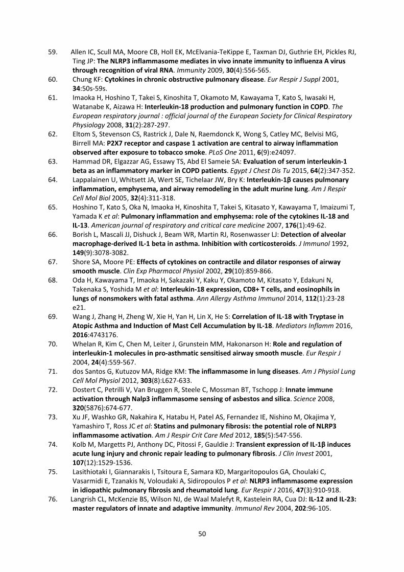
50
59. Allen IC, Scull MA, Moore CB, Holl EK, McElvania-TeKippe E, Taxman DJ, Guthrie EH, Pickles RJ, Ting JP: The NLRP3 inflammasome mediates in vivo innate immunity to influenza A virus through recognition of viral RNA. Immunity 2009, 30(4):556-565.
60. Chung KF: Cytokines in chronic obstructive pulmonary disease. Eur Respir J Suppl 2001, 34:50s-59s.
61. Imaoka H, Hoshino T, Takei S, Kinoshita T, Okamoto M, Kawayama T, Kato S, Iwasaki H, Watanabe K, Aizawa H: Interleukin-18 production and pulmonary function in COPD. The European respiratory journal : official journal of the European Society for Clinical Respiratory Physiology 2008, 31(2):287-297.
62. Eltom S, Stevenson CS, Rastrick J, Dale N, Raemdonck K, Wong S, Catley MC, Belvisi MG, Birrell MA: P2X7 receptor and caspase 1 activation are central to airway inflammation observed after exposure to tobacco smoke. PLoS One 2011, 6(9):e24097.
63. Hammad DR, Elgazzar AG, Essawy TS, Abd El Sameie SA: Evaluation of serum interleukin-1 beta as an inflammatory marker in COPD patients. Egypt J Chest Dis Tu 2015, 64(2):347-352.
64. Lappalainen U, Whitsett JA, Wert SE, Tichelaar JW, Bry K: Interleukin-1β causes pulmonary inflammation, emphysema, and airway remodeling in the adult murine lung. Am J Respir Cell Mol Biol 2005, 32(4):311-318.
65. Hoshino T, Kato S, Oka N, Imaoka H, Kinoshita T, Takei S, Kitasato Y, Kawayama T, Imaizumi T, Yamada K et al: Pulmonary inflammation and emphysema: role of the cytokines IL-18 and IL-13. American journal of respiratory and critical care medicine 2007, 176(1):49-62.
66. Borish L, Mascali JJ, Dishuck J, Beam WR, Martin RJ, Rosenwasser LJ: Detection of alveolar macrophage-derived IL-1 beta in asthma. Inhibition with corticosteroids. J Immunol 1992, 149(9):3078-3082.
67. Shore SA, Moore PE: Effects of cytokines on contractile and dilator responses of airway smooth muscle. Clin Exp Pharmacol Physiol 2002, 29(10):859-866.
68. Oda H, Kawayama T, Imaoka H, Sakazaki Y, Kaku Y, Okamoto M, Kitasato Y, Edakuni N, Takenaka S, Yoshida M et al: Interleukin-18 expression, CD8+ T cells, and eosinophils in lungs of nonsmokers with fatal asthma. Ann Allergy Asthma Immunol 2014, 112(1):23-28 e21.
69. Wang J, Zhang H, Zheng W, Xie H, Yan H, Lin X, He S: Correlation of IL-18 with Tryptase in Atopic Asthma and Induction of Mast Cell Accumulation by IL-18. Mediators Inflamm 2016, 2016:4743176.
70. Whelan R, Kim C, Chen M, Leiter J, Grunstein MM, Hakonarson H: Role and regulation of interleukin-1 molecules in pro-asthmatic sensitised airway smooth muscle. Eur Respir J 2004, 24(4):559-567.
71. dos Santos G, Kutuzov MA, Ridge KM: The inflammasome in lung diseases. Am J Physiol Lung Cell Mol Physiol 2012, 303(8):L627-633.
72. Dostert C, Petrilli V, Van Bruggen R, Steele C, Mossman BT, Tschopp J: Innate immune activation through Nalp3 inflammasome sensing of asbestos and silica. Science 2008, 320(5876):674-677.
73. Xu JF, Washko GR, Nakahira K, Hatabu H, Patel AS, Fernandez IE, Nishino M, Okajima Y, Yamashiro T, Ross JC et al: Statins and pulmonary fibrosis: the potential role of NLRP3 inflammasome activation. Am J Respir Crit Care Med 2012, 185(5):547-556.
74. Kolb M, Margetts PJ, Anthony DC, Pitossi F, Gauldie J: Transient expression of IL-1β induces acute lung injury and chronic repair leading to pulmonary fibrosis. J Clin Invest 2001, 107(12):1529-1536.
75. Lasithiotaki I, Giannarakis I, Tsitoura E, Samara KD, Margaritopoulos GA, Choulaki C, Vasarmidi E, Tzanakis N, Voloudaki A, Sidiropoulos P et al: NLRP3 inflammasome expression in idiopathic pulmonary fibrosis and rheumatoid lung. Eur Respir J 2016, 47(3):910-918.
76. Langrish CL, McKenzie BS, Wilson NJ, de Waal Malefyt R, Kastelein RA, Cua DJ: IL-12 and IL-23: master regulators of innate and adaptive immunity. Immunol Rev 2004, 202:96-105.
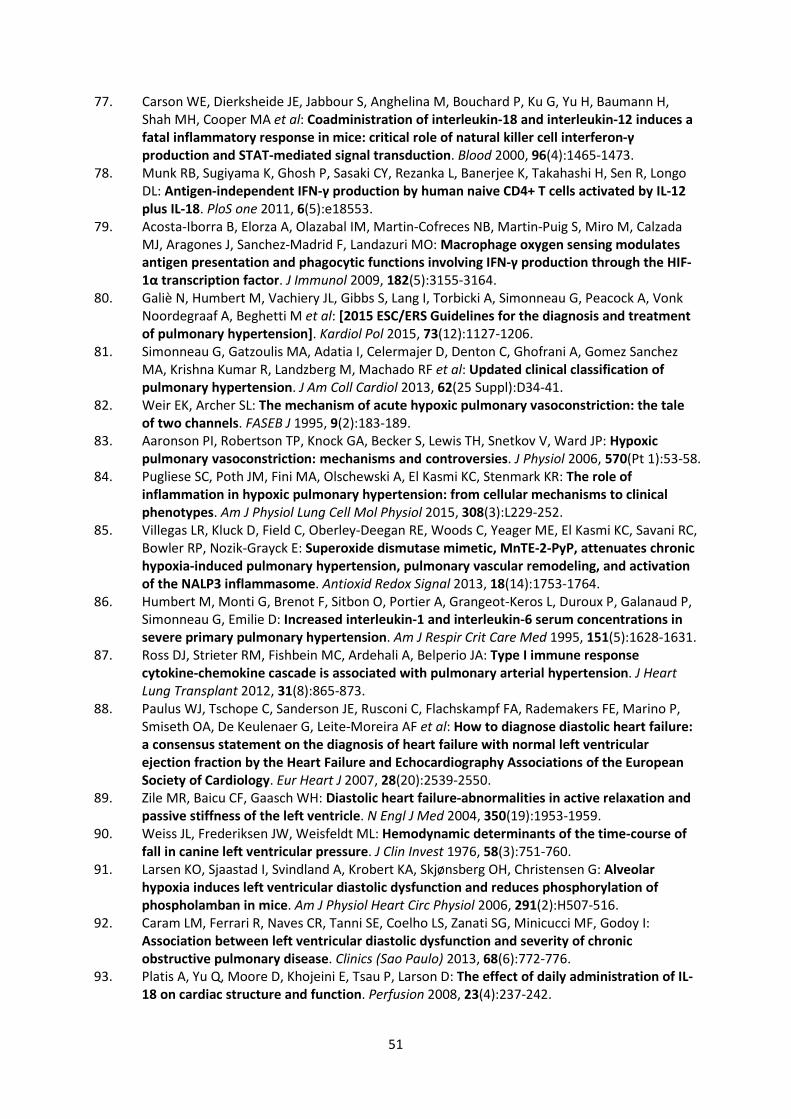
51
77. Carson WE, Dierksheide JE, Jabbour S, Anghelina M, Bouchard P, Ku G, Yu H, Baumann H, Shah MH, Cooper MA et al: Coadministration of interleukin-18 and interleukin-12 induces a fatal inflammatory response in mice: critical role of natural killer cell interferon-γ production and STAT-mediated signal transduction. Blood 2000, 96(4):1465-1473.
78. Munk RB, Sugiyama K, Ghosh P, Sasaki CY, Rezanka L, Banerjee K, Takahashi H, Sen R, Longo DL: Antigen-independent IFN-γ production by human naive CD4+ T cells activated by IL-12 plus IL-18. PloS one 2011, 6(5):e18553.
79. Acosta-Iborra B, Elorza A, Olazabal IM, Martin-Cofreces NB, Martin-Puig S, Miro M, Calzada MJ, Aragones J, Sanchez-Madrid F, Landazuri MO: Macrophage oxygen sensing modulates antigen presentation and phagocytic functions involving IFN-γ production through the HIF-1α transcription factor. J Immunol 2009, 182(5):3155-3164.
80. Galiè N, Humbert M, Vachiery JL, Gibbs S, Lang I, Torbicki A, Simonneau G, Peacock A, Vonk Noordegraaf A, Beghetti M et al: [2015 ESC/ERS Guidelines for the diagnosis and treatment of pulmonary hypertension]. Kardiol Pol 2015, 73(12):1127-1206.
81. Simonneau G, Gatzoulis MA, Adatia I, Celermajer D, Denton C, Ghofrani A, Gomez Sanchez MA, Krishna Kumar R, Landzberg M, Machado RF et al: Updated clinical classification of pulmonary hypertension. J Am Coll Cardiol 2013, 62(25 Suppl):D34-41.
82. Weir EK, Archer SL: The mechanism of acute hypoxic pulmonary vasoconstriction: the tale of two channels. FASEB J 1995, 9(2):183-189.
83. Aaronson PI, Robertson TP, Knock GA, Becker S, Lewis TH, Snetkov V, Ward JP: Hypoxic pulmonary vasoconstriction: mechanisms and controversies. J Physiol 2006, 570(Pt 1):53-58.
84. Pugliese SC, Poth JM, Fini MA, Olschewski A, El Kasmi KC, Stenmark KR: The role of inflammation in hypoxic pulmonary hypertension: from cellular mechanisms to clinical phenotypes. Am J Physiol Lung Cell Mol Physiol 2015, 308(3):L229-252.
85. Villegas LR, Kluck D, Field C, Oberley-Deegan RE, Woods C, Yeager ME, El Kasmi KC, Savani RC, Bowler RP, Nozik-Grayck E: Superoxide dismutase mimetic, MnTE-2-PyP, attenuates chronic hypoxia-induced pulmonary hypertension, pulmonary vascular remodeling, and activation of the NALP3 inflammasome. Antioxid Redox Signal 2013, 18(14):1753-1764.
86. Humbert M, Monti G, Brenot F, Sitbon O, Portier A, Grangeot-Keros L, Duroux P, Galanaud P, Simonneau G, Emilie D: Increased interleukin-1 and interleukin-6 serum concentrations in severe primary pulmonary hypertension. Am J Respir Crit Care Med 1995, 151(5):1628-1631.
87. Ross DJ, Strieter RM, Fishbein MC, Ardehali A, Belperio JA: Type I immune response cytokine-chemokine cascade is associated with pulmonary arterial hypertension. J Heart Lung Transplant 2012, 31(8):865-873.
88. Paulus WJ, Tschope C, Sanderson JE, Rusconi C, Flachskampf FA, Rademakers FE, Marino P, Smiseth OA, De Keulenaer G, Leite-Moreira AF et al: How to diagnose diastolic heart failure: a consensus statement on the diagnosis of heart failure with normal left ventricular ejection fraction by the Heart Failure and Echocardiography Associations of the European Society of Cardiology. Eur Heart J 2007, 28(20):2539-2550.
89. Zile MR, Baicu CF, Gaasch WH: Diastolic heart failure-abnormalities in active relaxation and passive stiffness of the left ventricle. N Engl J Med 2004, 350(19):1953-1959.
90. Weiss JL, Frederiksen JW, Weisfeldt ML: Hemodynamic determinants of the time-course of fall in canine left ventricular pressure. J Clin Invest 1976, 58(3):751-760.
91. Larsen KO, Sjaastad I, Svindland A, Krobert KA, Skjønsberg OH, Christensen G: Alveolar hypoxia induces left ventricular diastolic dysfunction and reduces phosphorylation of phospholamban in mice. Am J Physiol Heart Circ Physiol 2006, 291(2):H507-516.
92. Caram LM, Ferrari R, Naves CR, Tanni SE, Coelho LS, Zanati SG, Minicucci MF, Godoy I: Association between left ventricular diastolic dysfunction and severity of chronic obstructive pulmonary disease. Clinics (Sao Paulo) 2013, 68(6):772-776.
93. Platis A, Yu Q, Moore D, Khojeini E, Tsau P, Larson D: The effect of daily administration of IL-18 on cardiac structure and function. Perfusion 2008, 23(4):237-242.
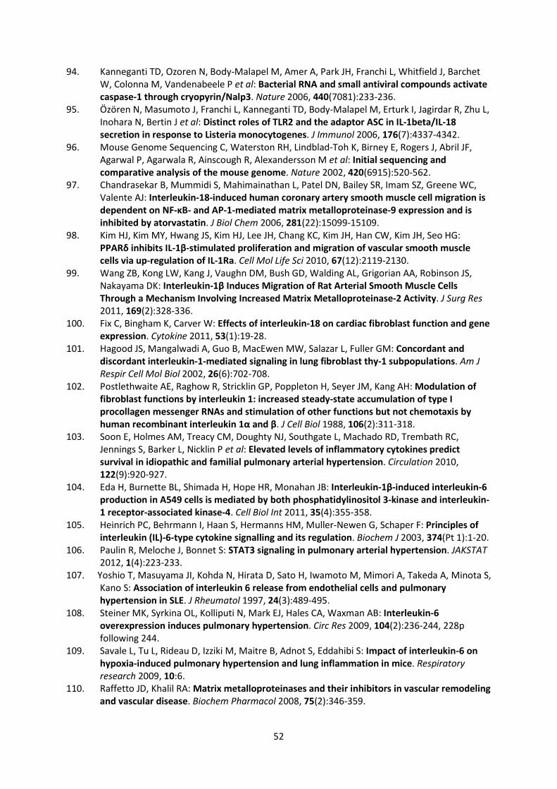
52
94. Kanneganti TD, Ozoren N, Body-Malapel M, Amer A, Park JH, Franchi L, Whitfield J, Barchet W, Colonna M, Vandenabeele P et al: Bacterial RNA and small antiviral compounds activate caspase-1 through cryopyrin/Nalp3. Nature 2006, 440(7081):233-236.
95. Özören N, Masumoto J, Franchi L, Kanneganti TD, Body-Malapel M, Erturk I, Jagirdar R, Zhu L, Inohara N, Bertin J et al: Distinct roles of TLR2 and the adaptor ASC in IL-1beta/IL-18 secretion in response to Listeria monocytogenes. J Immunol 2006, 176(7):4337-4342.
96. Mouse Genome Sequencing C, Waterston RH, Lindblad-Toh K, Birney E, Rogers J, Abril JF, Agarwal P, Agarwala R, Ainscough R, Alexandersson M et al: Initial sequencing and comparative analysis of the mouse genome. Nature 2002, 420(6915):520-562.
97. Chandrasekar B, Mummidi S, Mahimainathan L, Patel DN, Bailey SR, Imam SZ, Greene WC, Valente AJ: Interleukin-18-induced human coronary artery smooth muscle cell migration is dependent on NF-κB- and AP-1-mediated matrix metalloproteinase-9 expression and is inhibited by atorvastatin. J Biol Chem 2006, 281(22):15099-15109.
98. Kim HJ, Kim MY, Hwang JS, Kim HJ, Lee JH, Chang KC, Kim JH, Han CW, Kim JH, Seo HG: PPARδ inhibits IL-1β-stimulated proliferation and migration of vascular smooth muscle cells via up-regulation of IL-1Ra. Cell Mol Life Sci 2010, 67(12):2119-2130.
99. Wang ZB, Kong LW, Kang J, Vaughn DM, Bush GD, Walding AL, Grigorian AA, Robinson JS, Nakayama DK: Interleukin-1β Induces Migration of Rat Arterial Smooth Muscle Cells Through a Mechanism Involving Increased Matrix Metalloproteinase-2 Activity. J Surg Res 2011, 169(2):328-336.
100. Fix C, Bingham K, Carver W: Effects of interleukin-18 on cardiac fibroblast function and gene expression. Cytokine 2011, 53(1):19-28.
101. Hagood JS, Mangalwadi A, Guo B, MacEwen MW, Salazar L, Fuller GM: Concordant and discordant interleukin-1-mediated signaling in lung fibroblast thy-1 subpopulations. Am J Respir Cell Mol Biol 2002, 26(6):702-708.
102. Postlethwaite AE, Raghow R, Stricklin GP, Poppleton H, Seyer JM, Kang AH: Modulation of fibroblast functions by interleukin 1: increased steady-state accumulation of type I procollagen messenger RNAs and stimulation of other functions but not chemotaxis by human recombinant interleukin 1α and β. J Cell Biol 1988, 106(2):311-318.
103. Soon E, Holmes AM, Treacy CM, Doughty NJ, Southgate L, Machado RD, Trembath RC, Jennings S, Barker L, Nicklin P et al: Elevated levels of inflammatory cytokines predict survival in idiopathic and familial pulmonary arterial hypertension. Circulation 2010, 122(9):920-927.
104. Eda H, Burnette BL, Shimada H, Hope HR, Monahan JB: Interleukin-1β-induced interleukin-6 production in A549 cells is mediated by both phosphatidylinositol 3-kinase and interleukin-1 receptor-associated kinase-4. Cell Biol Int 2011, 35(4):355-358.
105. Heinrich PC, Behrmann I, Haan S, Hermanns HM, Muller-Newen G, Schaper F: Principles of interleukin (IL)-6-type cytokine signalling and its regulation. Biochem J 2003, 374(Pt 1):1-20.
106. Paulin R, Meloche J, Bonnet S: STAT3 signaling in pulmonary arterial hypertension. JAKSTAT 2012, 1(4):223-233.
107. Yoshio T, Masuyama JI, Kohda N, Hirata D, Sato H, Iwamoto M, Mimori A, Takeda A, Minota S, Kano S: Association of interleukin 6 release from endothelial cells and pulmonary hypertension in SLE. J Rheumatol 1997, 24(3):489-495.
108. Steiner MK, Syrkina OL, Kolliputi N, Mark EJ, Hales CA, Waxman AB: Interleukin-6 overexpression induces pulmonary hypertension. Circ Res 2009, 104(2):236-244, 228p following 244.
109. Savale L, Tu L, Rideau D, Izziki M, Maitre B, Adnot S, Eddahibi S: Impact of interleukin-6 on hypoxia-induced pulmonary hypertension and lung inflammation in mice. Respiratory research 2009, 10:6.
110. Raffetto JD, Khalil RA: Matrix metalloproteinases and their inhibitors in vascular remodeling and vascular disease. Biochem Pharmacol 2008, 75(2):346-359.
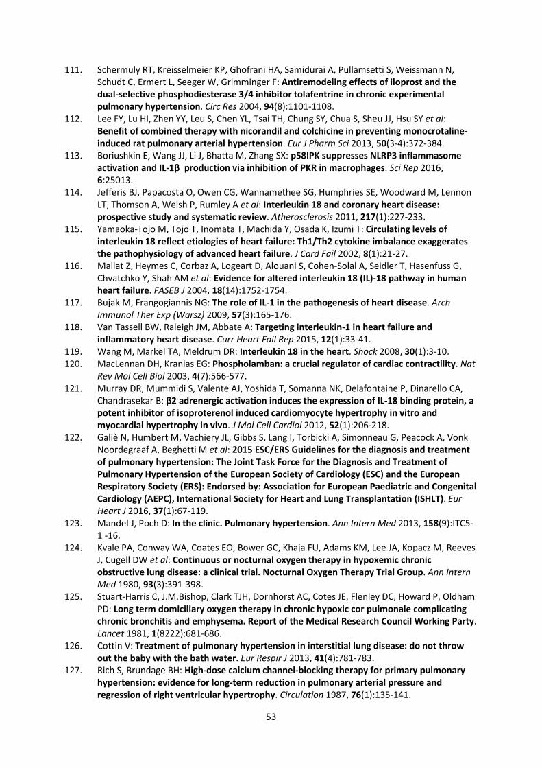
53
111. Schermuly RT, Kreisselmeier KP, Ghofrani HA, Samidurai A, Pullamsetti S, Weissmann N, Schudt C, Ermert L, Seeger W, Grimminger F: Antiremodeling effects of iloprost and the dual-selective phosphodiesterase 3/4 inhibitor tolafentrine in chronic experimental pulmonary hypertension. Circ Res 2004, 94(8):1101-1108.
112. Lee FY, Lu HI, Zhen YY, Leu S, Chen YL, Tsai TH, Chung SY, Chua S, Sheu JJ, Hsu SY et al: Benefit of combined therapy with nicorandil and colchicine in preventing monocrotaline-induced rat pulmonary arterial hypertension. Eur J Pharm Sci 2013, 50(3-4):372-384.
113. Boriushkin E, Wang JJ, Li J, Bhatta M, Zhang SX: p58IPK suppresses NLRP3 inflammasome activation and IL-1β production via inhibition of PKR in macrophages. Sci Rep 2016, 6:25013.
114. Jefferis BJ, Papacosta O, Owen CG, Wannamethee SG, Humphries SE, Woodward M, Lennon LT, Thomson A, Welsh P, Rumley A et al: Interleukin 18 and coronary heart disease: prospective study and systematic review. Atherosclerosis 2011, 217(1):227-233.
115. Yamaoka-Tojo M, Tojo T, Inomata T, Machida Y, Osada K, Izumi T: Circulating levels of interleukin 18 reflect etiologies of heart failure: Th1/Th2 cytokine imbalance exaggerates the pathophysiology of advanced heart failure. J Card Fail 2002, 8(1):21-27.
116. Mallat Z, Heymes C, Corbaz A, Logeart D, Alouani S, Cohen-Solal A, Seidler T, Hasenfuss G, Chvatchko Y, Shah AM et al: Evidence for altered interleukin 18 (IL)-18 pathway in human heart failure. FASEB J 2004, 18(14):1752-1754.
117. Bujak M, Frangogiannis NG: The role of IL-1 in the pathogenesis of heart disease. Arch Immunol Ther Exp (Warsz) 2009, 57(3):165-176.
118. Van Tassell BW, Raleigh JM, Abbate A: Targeting interleukin-1 in heart failure and inflammatory heart disease. Curr Heart Fail Rep 2015, 12(1):33-41.
119. Wang M, Markel TA, Meldrum DR: Interleukin 18 in the heart. Shock 2008, 30(1):3-10. 120. MacLennan DH, Kranias EG: Phospholamban: a crucial regulator of cardiac contractility. Nat
Rev Mol Cell Biol 2003, 4(7):566-577. 121. Murray DR, Mummidi S, Valente AJ, Yoshida T, Somanna NK, Delafontaine P, Dinarello CA,
Chandrasekar B: β2 adrenergic activation induces the expression of IL-18 binding protein, a potent inhibitor of isoproterenol induced cardiomyocyte hypertrophy in vitro and myocardial hypertrophy in vivo. J Mol Cell Cardiol 2012, 52(1):206-218.
122. Galiè N, Humbert M, Vachiery JL, Gibbs S, Lang I, Torbicki A, Simonneau G, Peacock A, Vonk Noordegraaf A, Beghetti M et al: 2015 ESC/ERS Guidelines for the diagnosis and treatment of pulmonary hypertension: The Joint Task Force for the Diagnosis and Treatment of Pulmonary Hypertension of the European Society of Cardiology (ESC) and the European Respiratory Society (ERS): Endorsed by: Association for European Paediatric and Congenital Cardiology (AEPC), International Society for Heart and Lung Transplantation (ISHLT). Eur Heart J 2016, 37(1):67-119.
123. Mandel J, Poch D: In the clinic. Pulmonary hypertension. Ann Intern Med 2013, 158(9):ITC5-1 -16.
124. Kvale PA, Conway WA, Coates EO, Bower GC, Khaja FU, Adams KM, Lee JA, Kopacz M, Reeves J, Cugell DW et al: Continuous or nocturnal oxygen therapy in hypoxemic chronic obstructive lung disease: a clinical trial. Nocturnal Oxygen Therapy Trial Group. Ann Intern Med 1980, 93(3):391-398.
125. Stuart-Harris C, J.M.Bishop, Clark TJH, Dornhorst AC, Cotes JE, Flenley DC, Howard P, Oldham PD: Long term domiciliary oxygen therapy in chronic hypoxic cor pulmonale complicating chronic bronchitis and emphysema. Report of the Medical Research Council Working Party. Lancet 1981, 1(8222):681-686.
126. Cottin V: Treatment of pulmonary hypertension in interstitial lung disease: do not throw out the baby with the bath water. Eur Respir J 2013, 41(4):781-783.
127. Rich S, Brundage BH: High-dose calcium channel-blocking therapy for primary pulmonary hypertension: evidence for long-term reduction in pulmonary arterial pressure and regression of right ventricular hypertrophy. Circulation 1987, 76(1):135-141.
54
128. Hinderliter AL, Willis PWt, Barst RJ, Rich S, Rubin LJ, Badesch DB, Groves BM, McGoon MD, Tapson VF, Bourge RC et al: Effects of long-term infusion of prostacyclin (epoprostenol) on echocardiographic measures of right ventricular structure and function in primary pulmonary hypertension. Primary Pulmonary Hypertension Study Group. Circulation 1997, 95(6):1479-1486.
129. Roeleveld RJ, Vonk-Noordegraaf A, Marcus JT, Bronzwaer JG, Marques KM, Postmus PE, Boonstra A: Effects of epoprostenol on right ventricular hypertrophy and dilatation in pulmonary hypertension. Chest 2004, 125(2):572-579.
130. Lang M, Kojonazarov B, Tian X, Kalymbetov A, Weissmann N, Grimminger F, Kretschmer A, Stasch JP, Seeger W, Ghofrani HA et al: The soluble guanylate cyclase stimulator riociguat ameliorates pulmonary hypertension induced by hypoxia and SU5416 in rats. PLoS One 2012, 7(8):e43433.
131. Kolb TM, Hassoun PM: Right ventricular dysfunction in chronic lung disease. Cardiol Clin 2012, 30(2):243-256.
132. Aziz F, Tk LA, Enweluzo C, Dutta S, Zaeem M: Diastolic heart failure: a concise review. J Clin Med Res 2013, 5(5):327-334.