Embed Size (px)
Citation preview

Cite as: C. A. Thaiss et al., Science 10.1126/science.aar3318 (2018).
RESEARCH ARTICLES
First release: 8 March 2018 www.sciencemag.org (Page numbers not final at time of first release) 1
The obesity pandemic has reached alarming magnitudes, affecting more than 2 billion people worldwide and account-ing for more than 3 million deaths per year (1). A poorly understood feature of the ‘metabolic syndrome’ is its associ-ation with dysfunctions of the intestinal barrier, leading to enhanced permeability and translocation of microbial mole-cules to the intestinal lamina propria and systemic circula-tion (2). This influx of immune-stimulatory microbial ligands into the vasculature, in turn, has been suggested to underlie the chronic inflammatory processes that are fre-quently observed in obesity and its complications (3), while entry of pathogens and pathobionts through an impaired barrier leads to an enhanced risk of infection in obese and diabetic individuals (4, 5), particularly at mucosal sites (6). However, the mechanistic basis for barrier dysfunction ac-companying the metabolic syndrome remains poorly under-stood. Beyond metabolic disease, enhanced intestinal permeability has also been linked with systemic inflamma-tion in a variety of conditions, including cancer (7), neuro-degeneration (8), and aging (9). Thus, there is an urgent scientific need to better define the molecular and cellular orchestrators and disruptors of intestinal barrier function,
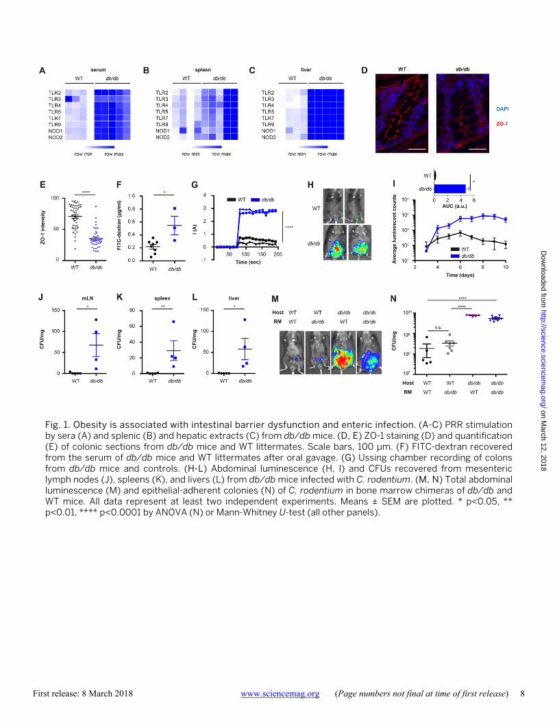
in order to devise strategies to counteract the detrimental systemic consequences of gut barrier alterations. Obesity is associated with, but not required for intesti-nal barrier dysfunction We began our investigation of the drivers of gastrointestinal barrier dysfunction in obesity by hypothesizing that the ad-ipokine leptin, a major orchestrator of mammalian satiety, may act as an obesity-associated regulator of barrier integri-ty. Leptin deficiency and resistance to leptin signaling are strongly associated with morbid obesity in mice and hu-mans, and both leptin deficiency and resistance were previ-ously suggested to contribute to intestinal barrier dysfunction and susceptibility to enteric infection (10–13). We used a mouse model featuring genetic dysfunction of the leptin receptor (LepR), leading to hyperphagia and morbid obesity (db/db, fig. S1A). Indeed, we detected elevated levels of microbial pattern recognition receptor (PRR) ligands at multiple systemic sites in leptin-unresponsive db/db mice (Fig. 1, A-C), indicative of enhanced influx of gut commen-sal-derived products. A similar phenomenon was observed in leptin-deficient mice (ob/ob, fig. S1, B and C). To gain in-
Hyperglycemia drives intestinal barrier dysfunction and risk for enteric infection Christoph A. Thaiss,1 Maayan Levy,1 Inna Grosheva,2 Danping Zheng,1 Eliran Soffer,1 Eran Blacher,1 Sofia Braverman,1 Anouk C. Tengeler,1 Oren Barak,1,3 Maya Elazar,1 Rotem Ben-Zeev,1 Dana Lehavi-Regev,1 Meirav N. Katz,1 Meirav Pevsner-Fischer,1 Arieh Gertler,4 Zamir Halpern,5,6,7 Alon Harmelin,8 Suhail Aamar,9 Patricia Serradas,10 Alexandra Grosfeld,10 Hagit Shapiro,1 Benjamin Geiger,2 Eran Elinav1* 1Department of Immunology, Weizmann Institute of Science, Rehovot, Israel. 2Department of Molecular Cell Biology, Weizmann Institute of Science, Rehovot, Israel. 3Department of Obstetrics and Gynecology, Kaplan Medical Center, Rehovot, affiliated with the Hebrew University and Hadassah School of Medicine, Jerusalem, Israel. 4The Robert H. Smith Faculty of Agriculture, Food and Environment, The Hebrew University, Rehovot, Israel. 5Sackler Faculty of Medicine, Tel Aviv Sourasky Medical Center, Tel Aviv, Israel 6Research Center for Digestive Tract and Liver Diseases, Tel Aviv Sourasky Medical Center, Tel Aviv, Israel. 7Digestive Center, Tel Aviv Sourasky Medical Center, Tel Aviv, Israel. 8Department of Veterinary Resources, Weizmann Institute of Science, Rehovot, Israel. 9Department of Medicine, Hadassah–Hebrew University Hospital, Jerusalem, Israel. 10INSERM UMR-S 1138, Centre de Recherche des Cordeliers, Sorbonne Universités, UPMC Univ Paris 06, Sorbonne Cités, UPD Univ Paris 05, CNRS, IHU ICAN, Paris, France.
*Corresponding author. E-mail: [email protected]
Obesity, diabetes and related manifestations are associated with an enhanced, but poorly understood risk for mucosal infection and systemic inflammation. Here, we show in mouse models of obesity and diabetes that hyperglycemia drives intestinal barrier permeability, through GLUT2-dependent transcriptional reprogramming of intestinal epithelial cells and alteration of tight and adherence junction integrity. Consequently, hyperglycemia-mediated barrier disruption leads to systemic influx of microbial products and enhanced dissemination of enteric infection. Treatment of hyperglycemia, intestinal epithelial-specific GLUT2 deletion, or inhibition of glucose metabolism restores barrier function and bacterial containment. In humans, systemic influx of intestinal microbiome products correlates with individualized glycemic control, indicated by glycated hemoglobin levels. Together, our results mechanistically link hyperglycemia and intestinal barrier function with systemic infectious and inflammatory consequences of obesity and diabetes.
on March 12, 2018
http://science.sciencem
ag.org/D
ownloaded from

First release: 8 March 2018 www.sciencemag.org (Page numbers not final at time of first release) 2
sight into the molecular signatures accompanying barrier dysfunction under aberrant leptin signaling, we performed RNA-sequencing of colonic tissue, obtained from db/db mice and their wild-type (WT) littermates under steady-state conditions. Leptin unresponsiveness was associated with global alterations of transcription (fig. S1D), with several hundred genes featuring differential expression between both groups (fig. S1E). Among the genes whose expression was most strongly abrogated in obese mice were members of the tight and adherence junction structures (fig. S1F), protein complexes that inhibit para-cellular flux of intesti-nal molecules into the lamina propria (14). Consequently, tight junction integrity was compromised in db/db mice (Fig. 1, D and E), leading to enhanced influx of luminal mol-ecules and electrical current measured across the epithelial layer (Fig. 1, F and G).
To determine the consequences of barrier dysfunction in leptin-resistant mice, we used the murine Citrobacter ro-dentium model simulating human enteropathogenic Esche-richia coli infection (15). A bioluminescent variant of C. rodentium allowed us to noninvasively track infection in vivo (16). In WT mice, C. rodentium caused a self-limiting, mainly gut-contained infection (Fig. 1, H-L). In contrast, db/db mice did not clear the pathogen from their intestine (Fig. 1, H and I), in line with previous reports (12). Im-portantly, db/db mice also showed a significantly enhanced bacterial attachment to the intestinal wall (Fig. S1, G and H), and featured C. rodentium colonization at systemic sites (Fig. 1, J-L, and fig. S1I). Similar susceptibility to C. rodenti-um was noted for leptin-deficient ob/ob mice (fig. S1, J-N).
To understand which cell type was responsible for LepR-mediated protection from enteric infection, we generated bone marrow chimeras, in which WT and db/db mice were used as either recipients or donors of bone marrow trans-planted into lethally irradiated mice. Exacerbated infection and systemic spread of C. rodentium was observed whenev-er the bone marrow recipient was LepR-deficient, regardless of the source of bone marrow (Fig. 1, M and N, and fig. S1O), indicating that the non-hematopoietic compartment medi-ated resistance against infection. LepR expression on non-hematopoietic cells has been reported in multiple tissues, including the gut, liver, and most prominently the nervous system (17). Mice lacking LepR in intestinal epithelial cells (Villin-Cre:LepRfl/fl) or hepatocytes (Albumin-Cre:LepRfl/fl) did not show any signs of enhanced susceptibility to C. ro-dentium infection (fig. S2, A-F), while mice with LepR defi-ciency specifically in the nervous system (Nestin-Cre:LepRfl/fl) featured an exacerbated (fig. S2, G-I), yet highly variable (fig. S2, J-O) bacterial growth. To further explore the possibility of neuronal leptin signaling driving barrier dysfunction and risk of infection, we generated mice with a specific deletion of LepR in the paraventricular hypothala-
mus (Sim1-Cre:LepRfl/fl), the ventromedial hypothalamus (SF1-Cre:LepRfl/fl), in cholinergic neurons (ChAT-Cre:LepRfl/fl), and in the arcuate nucleus of the hypothala-mus (POMC-Cre:LepRfl/fl and AgRP-Cre:LepRfl/fl) and infect-ed them with C. rodentium. However, none of these mice showed enhanced susceptibility to pathogenic invasion when compared to littermate controls (fig. S3, A-O). Collec-tively, these results suggested that leptin deficiency per se might not provide a sufficient explanation to barrier dys-function and enhanced risk of enteric infection.
A feature common to all leptin- and LepR-deficient mice exhibiting an impaired barrier function and enhanced C. rodentium dissemination in our studies (db/db, ob/ob and Nestin-Cre:LepRfl/fl) was their tendency to develop obesity. We therefore hypothesized that an obesity-related factor distinct from leptin signaling may predispose these mice to impaired barrier function and exacerbated intestinal infec-tion. Thus, to complement the above genetic models of obe-sity we fed WT mice a high-fat diet (HFD) to induce weight gain (fig. S4A). Similarly to obese leptin- and LepR-deficient mice, HFD-fed obese mice showed elevated steady-state sys-temic PRR ligand influx (Fig. 2A) as well as exacerbated C. rodentium infection and systemic dissemination (Fig. 2, B-E, and fig. S4B). To further test whether obesity is the major driver for barrier dysfunction and impaired C. rodentium containment in LepR-deficient mice, we performed paired-feeding experiments, in which the food access for db/db mice was restricted to the amount consumed by their WT littermates, thereby equalizing body weight between both groups (Fig. 2F). Surprisingly, even after weight reduction to control levels, lean db/db mice were still unable to cope with C. rodentium infection (Fig. 2, G and H), ruling out that obesity per se was directly driving barrier dysfunction and risk for enteric infection in these mice. The lack of a direct causal relationship between obesity and barrier dysfunction was further supported by experiments using a chemical in-hibitor of leptin signaling (18), which rendered WT mice susceptible to exacerbated infection and systemic bacterial spread even prior to the onset of marked obesity (Fig. 2, I-L, and fig. S4, C-F). Together, these data indicated that neither leptin signaling nor obesity per se sufficiently explain the severity of barrier dysfunction and systemic enteric infec-tion in mice with the metabolic syndrome.
In search of a unifying explanation for the above results in multiple mouse models of genetic and acquired obesity and leptin deficiency, we investigated other common fea-tures of the metabolic syndrome that could potentially con-tribute to barrier dysfunction. One such manifestation of the metabolic syndrome, typically accompanying obesity and potentially contributing to barrier dysfunction is glu-cose intolerance and resultant hyperglycemia. Interestingly, all mice featuring marked susceptibility C. rodentium infec-
on March 12, 2018
http://science.sciencem
ag.org/D
ownloaded from

First release: 8 March 2018 www.sciencemag.org (Page numbers not final at time of first release) 3
tion, including obese db/db, pair-fed lean db/db mice, Nes-tin-Cre:LepRfl/fl mice, mice fed a HFD, and mice treated with leptin antagonist showed elevated blood glucose levels (Fig. 2, M-O, and fig. S4, G and H). In contrast, all mouse groups and models that did not develop enhanced C. rodentium susceptibility (Villin-Cre:LepRfl/fl, Albumin-Cre:LepRfl/fl, Sim1-Cre:LepRfl/fl, SF1-Cre:LepRfl/fl, ChAT-Cre:LepRfl/fl, POMC-Cre:LepRfl/fl and AgRP-Cre:LepRfl/fl, as well as those Nestin-Cre:LepRfl/fl mice that did not feature a tendency for severe infection) collectively showed normoglycemic levels (fig. S4, I and J). Together, these results suggested that hy-perglycemia, rather than obesity or alterations in leptin sig-naling, may predispose to barrier dysfunction leading to enhanced enteric infection in the setup of the metabolic syndrome in mice.
Hyperglycemia drives intestinal barrier disruption To test whether elevated glucose levels were causally in-volved in host defense against intestinal infection, we in-duced hyperglycemia in the absence of obesity in a mouse model of type 1 diabetes mellitus through administration of streptozotocin (STZ (19), fig. S5A). Indeed, STZ-treated mice developed severe C. rodentium infection and systemic trans-location, accompanied by enhanced bacterial growth, epi-thelial adherence, and systemic spread (Fig. 3, A-E). STZ treatment also resulted in dysfunction of intestinal epitheli-al adherence junctions under steady-state conditions (Fig. 3, F and G), coupled with systemic dissemination of microbial products (fig. S5, B and C), and enhanced trans-epithelial flux (Fig. 3, H and I).
Oral antibiotic treatment prevented the detection of bac-terial products at systemic sites in STZ-treated mice (Fig. 3, J-L), demonstrating that the intestinal microbiota was the probable source of disseminated microbial molecules. In contrast to the load of bacterial products at distal organs (fig. S5D), the microbial load in the intestinal lumen was unaffected by hyperglycemia (fig. S5E). We next sought to test the possibility that barrier dysfunction in STZ-treated mice was mediated by compositional microbiota alterations. Indeed, 16S rDNA sequencing revealed a taxonomic change in the configuration of the intestinal microbiota of hyper-glycemic mice, which was corrected by insulin treatment and resultant normalization of serum glucose levels (fig. S6, A-D). However, these compositional microbial changes did not seem to play a critical role in glucose-mediated barrier dysfunction, as microbiota transfer from STZ-treated donors and controls to normoglycemic germ-free mice neither in-duced dissemination of bacterial products to systemic sites (fig. S6E) nor increased susceptibility to C. rodentium infec-tion (fig. S6, F-J). These data indicate that while the com-mensal microbiota serves as the reservoir of microbial molecules that translocate to the systemic circulation upon
disruption of the intestinal barrier, compositional microbio-ta alterations arising under hyperglycemic conditions do not directly affect barrier integrity.
To corroborate the specificity of hyperglycemia as a driv-er of susceptibility to intestinal infection, we utilized hyper-glycemic Akita mice (fig. S7A), an STZ-independent model of type I diabetes mellitus that harbors a spontaneous muta-tion in the insulin 2 gene (20). As in STZ-treated mice, we observed in this model elevated C. rodentium growth and pathogenic translocation to systemic tissues (Fig. 3, M and N, and fig. S7, B and C). To further validate the specific im-pact of hyperglycemia as a driver of the barrier dysfunction phenotype, we administered 0.25 U per day of insulin to STZ-treated mice via hyperosmotic pumps for 4 weeks, which restored normoglycemic levels (fig. S7D). Treatment with insulin also prevented the loss of adherence junction integrity (Fig. 4A and fig. S7E), systemic dissemination of microbial products (Fig. 4B), and enhanced C. rodentium growth and pathogenic translocation (Fig. 4, C and D). To-gether, these experiments establish hyperglycemia as a di-rect and specific cause for intestinal barrier dysfunction and susceptibility to enteric infection.
Hyperglycemia reprograms intestinal epithelial cells To determine whether glucose acted directly on intestinal epithelial cells to affect barrier function, we used an in vitro system of cultured intestinal epithelial (Caco-2) cells ex-posed to different concentrations of glucose in the culture medium. We assessed tight junction integrity through au-tomated high-throughput analysis of ZO-1 staining patterns. Indeed, glucose induced barrier alterations in a dose- and time-dependent manner, manifesting visually as increased tortuosity and altered appearance of cell-cell junctions (Fig. 4, E-H). To investigate the mechanisms by which elevated blood glucose levels compromise intestinal epithelial cell function in vivo, we performed RNA-sequencing of purified intestinal epithelial cells from STZ-treated mice and con-trols. Global reprogramming of the epithelial transcriptome was detected in hyperglycemic mice (Fig. 4I), in which more than 1,000 genes were differentially expressed compared to vehicle-treated controls (Fig. 4J). These genes were predom-inantly involved in metabolic pathways, and specifically in N-glycan biosynthesis and pentose-glucuronate interconver-sion (Fig. 4K), two intracellular functions critically involved in the maintenance of epithelial barrier function (21–29). For example, hyperglycemia affected the entire pathway of protein N-glycosylation by provoking marked downregula-tion of central genes (Fig. 4L and fig. S8). In contrast, epi-thelial proliferation or cell death was not affected by STZ treatment (fig. S9, A-D).
In addition to the above epithelial changes, hyperglyce-mia modestly affected the intestinal and splenic immune
on March 12, 2018
http://science.sciencem
ag.org/D
ownloaded from
First release: 8 March 2018 www.sciencemag.org (Page numbers not final at time of first release) 4
system, specifically by causing an increased representation of myeloid cells (fig. S10, A-J), in line with previous reports (30). However, STZ treatment did not provoke an overt in-flammatory state in the intestine (fig. S11, A-E). In particu-lar, cytokines involved in IL-22-mediated barrier function and host defense, which has been implicated in the suscep-tibility of obese mice to infection (12), were unaltered, as was the epithelial transcriptional response to IL-22 (fig. S11F). In fact, hyperglycemia and IL-22 appeared to have additive effects in mediating host defense against C. ro-dentium, since STZ-treated IL-22-deficient mice featured accelerated bacterial growth and mortality when compared to IL-22-deficient controls (fig. S11, G and H). We further compared the involvement of epithelial and immune cells in host defense against another gastrointestinal pathogen, Salmonella Typhimurium. STZ-treated mice orally infected with Salmonella showed enhanced systemic colonization, while intestinal luminal growth was comparable to vehicle-treated controls (fig. S12, A-E). In contrast to this marked susceptibility of STZ-treated mice to oral Salmonella Typhimurium infection, susceptibility of these mice to sys-temic infection was only apparent in the liver (fig. S12, F-H). Interestingly, systemic infection with Salmonella caused enhanced intestinal colonization in STZ-treated mice, poten-tially indicative of retrograde spread of bacteria across a compromised barrier (fig. S12, I and J).
Epithelial reprogramming by hyperglycemia involves glucose metabolism and GLUT2 We next assessed whether epithelial glucose metabolism was involved in the transcriptional reprogramming of STZ-treated mice. Isolated intestinal epithelial cells from hyper-glycemic mice featured elevated levels of metabolites along the glycolytic cascade (fig. S13A). Inhibition of glucose me-tabolism via 2-deoxyglucose (2-DG) rescued glucose-induced barrier aberrations in vitro in a dose-dependent manner (Fig. 5, A-C). In addition, 2-DG administration blocked tran-scriptional reprogramming in STZ-treated mice (Fig. 5D and fig. S13B), including the N-glycan pathway (fig. S13C), pre-vented the systemic dissemination of microbial products (Fig. 5, E and F), and restored host defense against C. ro-dentium (Fig. 5G and fig. S13, D-F). Bacterial growth in the intestinal lumen was unaffected by 2-DG treatment (fig. S13G). To test whether 2-DG could be used to counteract hyperglycemia-mediated loss of barrier integrity beyond the STZ model, we administered 2-DG to C. rodentium-infected db/db mice and assessed its impact on systemic dissemina-tion of the pathogen. Notably, the detectable pathogen load in the mesenteric lymph nodes, spleens and livers of 2-DG-treated db/db mice was strongly reduced under 2-DG treat-ment (Fig. 5H and fig. S13, H and I). Together, these data suggest that glucose-mediated reprogramming of epithelial
cell metabolic function leads to transcriptional alterations, abrogation of the intestinal barrier, and impaired host de-fense against enteric infection.
Glucose transport between the intestinal epithelium and circulation is mediated by the bi-directional glucose trans-porter GLUT2 (31). To determine the role of this transporter in hyperglycemia-mediated epithelial reprogramming, we next used mice selectively lacking GLUT2 in intestinal epi-thelial cells (GLUT2ΔIEC) (32) and induced hyperglycemia in these mice by STZ administration. Indeed, GLUT2ΔIEC mice were resistant to STZ-induced transcriptional reprogram-ming and retained epithelial transcriptomes similar to con-trols (fig. S14, A and B). GLUT2ΔIEC mice also retained intact tight and adherence junction complexes (Fig. 5, I and J, and fig. S14, C and D), reduced transepithelial flux (fig. S14E), and intestinal containment of microbial PRR ligands (Fig. 5K), despite sustained STZ-induced hyperglycemia (fig. S14F). Ablation of GLUT2 also ameliorated the STZ-induced susceptibility to C. rodentium growth and systemic dissemi-nation (Fig. 5 L, and fig. S14, G-I). Collectively, these results indicate that GLUT2 is involved in the hyperglycemia-induced metabolic and transcriptional alterations in intesti-nal epithelial cells, resulting in barrier dysfunction and mi-crobial translocation to the systemic circulation.
Blood glucose levels are associated with microbial product influx in humans Finally, we sought to determine whether glycemic levels similarly correlate with intestinal barrier function in hu-mans. To this end, we recruited 27 healthy individuals (fig. S15, A and B) and performed measurements of multiple se-rum parameters and microbial products in the circulation. Of all variables measured, hemoglobin A1c (HbA1c), indica-tive of an individual’s three-month average plasma glucose concentration, showed the strongest correlation with serum levels of PRR ligands (Fig. 6, A-C, and fig. S15, C-E). In con-trast, high BMI and other hallmarks of metabolic disease did not significantly associate with the influx of microbial products (Fig. 6, A and B, fig. S15F). Total stool bacterial content did not correlate with HbA1c levels (fig. S15G). To-gether, these data suggest that similarly to mice, serum glu-cose levels, rather than obesity, may associate with or potentially even drive intestinal barrier dysfunction in hu-mans.
Discussion Serum glucose is among the most strictly controlled physio-logical variables of organismal homeostasis. Chronically elevated glucose levels, as observed in diabetes mellitus, obesity and associated metabolic disorders, such as non-alcoholic fatty liver disease, result from altered homeostatic set points of the tightly regulated normoglycemic levels (33).
on March 12, 2018
http://science.sciencem
ag.org/D
ownloaded from
First release: 8 March 2018 www.sciencemag.org (Page numbers not final at time of first release) 5
Longstanding hyperglycemia, in turn, leads to a myriad of potentially devastating biochemical and physiological con-sequences, such as the generation of advanced glycation end products, pancreatic glucose toxicity (34, 35), macrovascular and microvascular complications impacting virtually every organ (36), risk of infection (37), and enhanced mortality (38).
In this study, we have identified glucose as an orchestra-tor of intestinal barrier function. Hyperglycemia markedly interfered with homeostatic epithelial integrity, leading to abnormal influx of immune-stimulatory microbial products and a propensity for systemic spread of enteric pathogens. Our results indicate that hyperglycemia causes retrograde transport of glucose into intestinal epithelial cells via GLUT2, followed by alterations in intracellular glucose me-tabolism and transcriptional reprogramming (fig. S16). One of the most strongly affected pathways by hyperglycemia in our study involves the N-glycosylation of proteins in the endoplasmic reticulum and Golgi apparatus, which has been implicated as a key regulator of a multitude of epithelial functions (25). While our study focused on the impact of systemic glucose levels on the intestinal barrier, similar ef-fects might be caused by high-glucose diet, which may affect intestinal epithelial cells in a similar manner, potentially resulting in diet-induced alterations of barrier function. Such potential physiologically important dietary effects on barrier function merit further studies. Furthermore, the im-pact of hyperglycemia on epithelial barrier function might be relevant beyond the gastrointestinal tract and affect oth-er mucosal surfaces, such as the respiratory tract, as was indicated by a recent study of close to 70,000 diabetes pa-tients highlighting a positive correlation between HbA1c values and a variety of mucosal community- and hospital-acquired infections (39).
Collectively, our findings provide a potential molecular explanation for altered barrier function in the context of the metabolic syndrome and the resultant enhanced mucosal infection noted in patients suffering from obesity (5) and diabetes mellitus (40). Furthermore, the link we highlight between hyperglycemia and gut barrier alterations may pro-vide a mechanistic basis for a variety of seemingly unrelated inflammatory manifestations, complications and associa-tions of the metabolic syndrome (collectively termed ‘meta-flammation’ or ‘para-inflammation’ (41, 42)). Examples of these include adipose tissue inflammation driving exacer-bated obesity and glucose intolerance (43), non-alcoholic fatty liver disease progressing to detrimental non-alcoholic steatohepatitis (44), inflammation contributing to athero-sclerosis and associated cardiovascular disease (45) and even recently suggested associations between the metabolic syndrome and neurodegeneration (46). Ultimately, our re-sults may present the starting point for harnessing glucose
metabolism or other regulators of intestinal barrier integrity as potential therapeutic targets in the prevention and ame-lioration of enteric infection and gut-related systemic in-flammation.
REFERENCES AND NOTES 1. N. Stefan, F. Schick, H. U. Häring, Causes, characteristics, and consequences of
metabolically unhealthy normal weight in humans. Cell Metab. 26, 292–300 (2017). doi:10.1016/j.cmet.2017.07.008 Medline
2. D. A. Winer, H. Luck, S. Tsai, S. Winer, The intestinal immune system in obesity and insulin resistance. Cell Metab. 23, 413–426 (2016). doi:10.1016/j.cmet.2016.01.003 Medline
3. P. D. Cani, R. Bibiloni, C. Knauf, A. Waget, A. M. Neyrinck, N. M. Delzenne, R. Burcelin, Changes in gut microbiota control metabolic endotoxemia-induced inflammation in high-fat diet-induced obesity and diabetes in mice. Diabetes 57, 1470–1481 (2008). doi:10.2337/db07-1403 Medline
4. M. E. Falagas, M. Kompoti, Obesity and infection. Lancet Infect. Dis. 6, 438–446 (2006). doi:10.1016/S1473-3099(06)70523-0 Medline
5. K. A. Kaspersen, O. B. Pedersen, M. S. Petersen, H. Hjalgrim, K. Rostgaard, B. K. Møller, C. Juul-Sørensen, S. Kotzé, K. M. Dinh, L. T. Erikstrup, E. Sørensen, L. W. Thørner, K. S. Burgdorf, H. Ullum, C. Erikstrup, Obesity and risk of infection: Results from the Danish Blood Donor Study. Epidemiology 26, 580–589 (2015). doi:10.1097/EDE.0000000000000301 Medline
6. J. Casqueiro, J. Casqueiro, C. Alves, Infections in patients with diabetes mellitus: A review of pathogenesis. Indian J. Endocrinol. Metab. 16 (suppl. S1), 27–36 (2012). doi:10.4103/2230-8210.94253 Medline
7. S. I. Grivennikov, K. Wang, D. Mucida, C. A. Stewart, B. Schnabl, D. Jauch, K. Taniguchi, G.-Y. Yu, C. H. Österreicher, K. E. Hung, C. Datz, Y. Feng, E. R. Fearon, M. Oukka, L. Tessarollo, V. Coppola, F. Yarovinsky, H. Cheroutre, L. Eckmann, G. Trinchieri, M. Karin, Adenoma-linked barrier defects and microbial products drive IL-23/IL-17-mediated tumour growth. Nature 491, 254–258 (2012). doi:10.1038/nature11465 Medline
8. T. R. Sampson, J. W. Debelius, T. Thron, S. Janssen, G. G. Shastri, Z. E. Ilhan, C. Challis, C. E. Schretter, S. Rocha, V. Gradinaru, M.-F. Chesselet, A. Keshavarzian, K. M. Shannon, R. Krajmalnik-Brown, P. Wittung-Stafshede, R. Knight, S. K. Mazmanian, Gut microbiota regulate motor deficits and neuroinflammation in a model of Parkinson’s disease. Cell 167, 1469–1480.e12 (2016). doi:10.1016/j.cell.2016.11.018 Medline
9. N. Thevaranjan, A. Puchta, C. Schulz, A. Naidoo, J. C. Szamosi, C. P. Verschoor, D. Loukov, L. P. Schenck, J. Jury, K. P. Foley, J. D. Schertzer, M. J. Larché, D. J. Davidson, E. F. Verdú, M. G. Surette, D. M. E. Bowdish, Age-associated microbial dysbiosis promotes intestinal permeability, systemic inflammation, and macrophage dysfunction. Cell Host Microbe 21, 455–466.e4 (2017). doi:10.1016/j.chom.2017.03.002 Medline
10. R. Ahmad, B. Rah, D. Bastola, P. Dhawan, A. B. Singh, Obesity-induces organ and tissue specific tight junction restructuring and barrier deregulation by claudin switching. Sci. Rep. 7, 5125 (2017). doi:10.1038/s41598-017-04989-8 Medline
11. N. M. Mackey-Lawrence, W. A. Petri Jr., Leptin and mucosal immunity. Mucosal Immunol. 5, 472–479 (2012). doi:10.1038/mi.2012.40 Medline
12. X. Wang, N. Ota, P. Manzanillo, L. Kates, J. Zavala-Solorio, C. Eidenschenk, J. Zhang, J. Lesch, W. P. Lee, J. Ross, L. Diehl, N. van Bruggen, G. Kolumam, W. Ouyang, Interleukin-22 alleviates metabolic disorders and restores mucosal immunity in diabetes. Nature 514, 237–241 (2014). doi:10.1038/nature13564 Medline
13. R. Madan, X. Guo, C. Naylor, E. L. Buonomo, D. Mackay, Z. Noor, P. Concannon, K. W. Scully, P. Pramoonjago, G. L. Kolling, C. A. Warren, P. Duggal, W. A. Petri Jr., Role of leptin-mediated colonic inflammation in defense against Clostridium difficile colitis. Infect. Immun. 82, 341–349 (2014). doi:10.1128/IAI.00972-13 Medline
14. C. Guillot, T. Lecuit, Mechanics of epithelial tissue homeostasis and morphogenesis. Science 340, 1185–1189 (2013). doi:10.1126/science.1235249 Medline
15. J. W. Collins, K. M. Keeney, V. F. Crepin, V. A. K. Rathinam, K. A. Fitzgerald, B. B. Finlay, G. Frankel, Citrobacter rodentium: Infection, inflammation and the microbiota. Nat. Rev. Microbiol. 12, 612–623 (2014). doi:10.1038/nrmicro3315
on March 12, 2018
http://science.sciencem
ag.org/D
ownloaded from

First release: 8 March 2018 www.sciencemag.org (Page numbers not final at time of first release) 6
Medline 16. M. Wlodarska, C. A. Thaiss, R. Nowarski, J. Henao-Mejia, J.-P. Zhang, E. M. Brown,
G. Frankel, M. Levy, M. N. Katz, W. M. Philbrick, E. Elinav, B. B. Finlay, R. A. Flavell, NLRP6 inflammasome orchestrates the colonic host-microbial interface by regulating goblet cell mucus secretion. Cell 156, 1045–1059 (2014). doi:10.1016/j.cell.2014.01.026 Medline
17. M. G. Myers Jr., H. Münzberg, G. M. Leinninger, R. L. Leshan, The geometry of leptin action in the brain: More complicated than a simple ARC. Cell Metab. 9, 117–123 (2009). doi:10.1016/j.cmet.2008.12.001 Medline
18. E. Elinav, L. Niv-Spector, M. Katz, T. O. Price, M. Ali, M. Yacobovitz, G. Solomon, S. Reicher, J. L. Lynch, Z. Halpern, W. A. Banks, A. Gertler, Pegylated leptin antagonist is a potent orexigenic agent: Preparation and mechanism of activity. Endocrinology 150, 3083–3091 (2009). doi:10.1210/en.2008-1706 Medline
19. M. S. Islam, D. T. Loots, Experimental rodent models of type 2 diabetes: A review. Methods Find. Exp. Clin. Pharmacol. 31, 249–261 (2009). doi:10.1358/mf.2009.31.4.1362513 Medline
20. J. Wang, T. Takeuchi, S. Tanaka, S.-K. Kubo, T. Kayo, D. Lu, K. Takata, A. Koizumi, T. Izumi, A mutation in the insulin 2 gene induces diabetes with severe pancreatic beta-cell dysfunction in the Mody mouse. J. Clin. Invest. 103, 27–37 (1999). doi:10.1172/JCI4431 Medline
21. D. W. Scott, C. E. Tolbert, D. M. Graham, E. Wittchen, J. E. Bear, K. Burridge, N-glycosylation controls the function of junctional adhesion molecule-A. Mol. Biol. Cell 26, 3205–3214 (2015). doi:10.1091/mbc.E14-12-1604 Medline
22. M. Nita-Lazar, I. Rebustini, J. Walker, M. A. Kukuruzinska, Hypoglycosylated E-cadherin promotes the assembly of tight junctions through the recruitment of PP2A to adherens junctions. Exp. Cell Res. 316, 1871–1884 (2010). doi:10.1016/j.yexcr.2010.02.008 Medline
23. B. T. Jamal, M. Nita-Lazar, Z. Gao, B. Amin, J. Walker, M. A. Kukuruzinska, N-glycosylation status of E-cadherin controls cytoskeletal dynamics through the organization of distinct β-catenin- and γ-catenin-containing AJs. Cell Health Cytoskelet. 2009, 67–80 (2009). Medline
24. Y. Goto, T. Obata, J. Kunisawa, S. Sato, I. I. Ivanov, A. Lamichhane, N. Takeyama, M. Kamioka, M. Sakamoto, T. Matsuki, H. Setoyama, A. Imaoka, S. Uematsu, S. Akira, S. E. Domino, P. Kulig, B. Becher, J.-C. Renauld, C. Sasakawa, Y. Umesaki, Y. Benno, H. Kiyono, Innate lymphoid cells regulate intestinal epithelial cell glycosylation. Science 345, 1254009 (2014). doi:10.1126/science.1254009 Medline
25. Y. Goto, S. Uematsu, H. Kiyono, Epithelial glycosylation in gut homeostasis and inflammation. Nat. Immunol. 17, 1244–1251 (2016). doi:10.1038/ni.3587 Medline
26. T. A. Pham, S. Clare, D. Goulding, J. M. Arasteh, M. D. Stares, H. P. Browne, J. A. Keane, A. J. Page, N. Kumasaka, L. Kane, L. Mottram, K. Harcourt, C. Hale, M. J. Arends, D. J. Gaffney, G. Dougan, T. D. Lawley; Sanger Mouse Genetics Project, Epithelial IL-22RA1-mediated fucosylation promotes intestinal colonization resistance to an opportunistic pathogen. Cell Host Microbe 16, 504–516 (2014). doi:10.1016/j.chom.2014.08.017 Medline
27. J. M. Pickard, C. F. Maurice, M. A. Kinnebrew, M. C. Abt, D. Schenten, T. V. Golovkina, S. R. Bogatyrev, R. F. Ismagilov, E. G. Pamer, P. J. Turnbaugh, A. V. Chervonsky, Rapid fucosylation of intestinal epithelium sustains host-commensal symbiosis in sickness. Nature 514, 638–641 (2014). doi:10.1038/nature13823 Medline
28. K. S. Bergstrom, V. Kissoon-Singh, D. L. Gibson, C. Ma, M. Montero, H. P. Sham, N. Ryz, T. Huang, A. Velcich, B. B. Finlay, K. Chadee, B. A. Vallance, Muc2 protects against lethal infectious colitis by disassociating pathogenic and commensal bacteria from the colonic mucosa. PLOS Pathog. 6, e1000902 (2010). doi:10.1371/journal.ppat.1000902 Medline
29. M. Van der Sluis, B. A. E. De Koning, A. C. J. M. De Bruijn, A. Velcich, J. P. P. Meijerink, J. B. Van Goudoever, H. A. Büller, J. Dekker, I. Van Seuningen, I. B. Renes, A. W. C. Einerhand, Muc2-deficient mice spontaneously develop colitis, indicating that MUC2 is critical for colonic protection. Gastroenterology 131, 117–129 (2006). doi:10.1053/j.gastro.2006.04.020 Medline
30. S. Niu, Z. Bian, A. Tremblay, Y. Luo, K. Kidder, A. Mansour, K. Zen, Y. Liu, Broad infiltration of macrophages leads to a proinflammatory state in streptozotocin-induced hyperglycemic mice. J. Immunol. 197, 3293–3301 (2016). doi:10.4049/jimmunol.1502494 Medline
31. B. Thorens, GLUT2, glucose sensing and glucose homeostasis. Diabetologia 58,
221–232 (2015). doi:10.1007/s00125-014-3451-1 Medline 32. C. C. Schmitt, T. Aranias, T. Viel, D. Chateau, M. Le Gall, A.-J. Waligora-Dupriet, C.
Melchior, O. Rouxel, N. Kapel, G. Gourcerol, B. Tavitian, A. Lehuen, E. Brot-Laroche, A. Leturque, P. Serradas, A. Grosfeld, Intestinal invalidation of the glucose transporter GLUT2 delays tissue distribution of glucose and reveals an unexpected role in gut homeostasis. Mol. Metab. 6, 61–72 (2016). doi:10.1016/j.molmet.2016.10.008 Medline
33. M. E. Kotas, R. Medzhitov, Homeostasis, inflammation, and disease susceptibility. Cell 160, 816–827 (2015). doi:10.1016/j.cell.2015.02.010 Medline
34. A. Bangert, M. Andrassy, A.-M. Müller, M. Bockstahler, A. Fischer, C. H. Volz, C. Leib, S. Göser, S. Korkmaz-Icöz, S. Zittrich, A. Jungmann, F. Lasitschka, G. Pfitzer, O. J. Müller, H. A. Katus, Z. Kaya, Critical role of RAGE and HMGB1 in inflammatory heart disease. Proc. Natl. Acad. Sci. U.S.A. 113, E155–E164 (2016). doi:10.1073/pnas.1522288113 Medline
35. Y. Tanaka, P. O. Tran, J. Harmon, R. P. Robertson, A role for glutathione peroxidase in protecting pancreatic beta cells against oxidative stress in a model of glucose toxicity. Proc. Natl. Acad. Sci. U.S.A. 99, 12363–12368 (2002). doi:10.1073/pnas.192445199 Medline
36. R. Madonna, R. De Caterina, Cellular and molecular mechanisms of vascular injury in diabetes—part I: Pathways of vascular disease in diabetes. Vascul. Pharmacol. 54, 68–74 (2011). doi:10.1016/j.vph.2011.03.005 Medline
37. S. O. Butler, I. F. Btaiche, C. Alaniz, Relationship between hyperglycemia and infection in critically ill patients. Pharmacotherapy 25, 963–976 (2005). doi:10.1592/phco.2005.25.7.963 Medline
38. S. A. Leite, S. B. Locatelli, S. P. Niece, A. R. F. Oliveira, D. Tockus, T. Tosin, Impact of hyperglycemia on morbidity and mortality, length of hospitalization and rates of re-hospitalization in a general hospital setting in Brazil. Diabetol. Metab. Syndr. 2, 49 (2010). doi:10.1186/1758-5996-2-49 Medline
39. A. Mor, O. M. Dekkers, J. S. Nielsen, H. Beck-Nielsen, H. T. Sørensen, R. W. Thomsen, Impact of glycemic control on risk of infections in patients with type 2 diabetes: A population-based cohort study. Am. J. Epidemiol. 186, 227–236 (2017). doi:10.1093/aje/kwx049 Medline
40. L. M. Muller, K. J. Gorter, E. Hak, W. L. Goudzwaard, F. G. Schellevis, A. I. Hoepelman, G. E. Rutten, Increased risk of common infections in patients with type 1 and type 2 diabetes mellitus. Clin. Infect Dis. 41, 281–288 (2005). doi:10.1086/431587 Medline
41. R. Medzhitov, Origin and physiological roles of inflammation. Nature 454, 428–435 (2008). doi:10.1038/nature07201 Medline
42. G. S. Hotamisligil, Inflammation, metaflammation and immunometabolic disorders. Nature 542, 177–185 (2017). doi:10.1038/nature21363 Medline
43. D. Okin, R. Medzhitov, The effect of sustained inflammation on hepatic mevalonate pathway results in hyperglycemia. Cell 165, 343–356 (2016). doi:10.1016/j.cell.2016.02.023 Medline
44. A. Nakamura, M. Yoneda, K. Fujita, K. Tajima, K. Kikuchi, A. Nakajima, S. Maeda, Y. Terauchi, Impact of glucose tolerance on the severity of non-alcoholic steatohepatitis. J. Diabetes Investig. 2, 483–489 (2011). doi:10.1111/j.2040-1124.2011.00134.x Medline
45. F. Pistrosch, A. Natali, M. Hanefeld, Is hyperglycemia a cardiovascular risk factor? Diabetes Care 34 (suppl. 2), S128–S131 (2011). doi:10.2337/dc11-s207 Medline
46. M. Barbagallo, L. J. Dominguez, Type 2 diabetes mellitus and Alzheimer’s disease. World J. Diabetes 5, 889–893 (2014). doi:10.4239/wjd.v5.i6.889 Medline
47. A. Wang, S. C. Huen, H. H. Luan, S. Yu, C. Zhang, J.-D. Gallezot, C. J. Booth, R. Medzhitov, Opposing effects of fasting metabolism on tissue tolerance in bacterial and viral inflammation. Cell 166, 1512–1525.e12 (2016). doi:10.1016/j.cell.2016.07.026 Medline
48. C. A. Thaiss, S. Itav, D. Rothschild, M. Meijer, M. Levy, C. Moresi, L. Dohnalová, S. Braverman, S. Rozin, S. Malitsky, M. Dori-Bachash, Y. Kuperman, I. Biton, A. Gertler, A. Harmelin, H. Shapiro, Z. Halpern, A. Aharoni, E. Segal, E. Elinav, Persistent microbiome alterations modulate the rate of post-dieting weight regain. Nature 540, 544–551 (2016). doi:10.1038/nature20796 Medline
49. M. Barthel, S. Hapfelmeier, L. Quintanilla-Martínez, M. Kremer, M. Rohde, M. Hogardt, K. Pfeffer, H. Rüssmann, W.-D. Hardt, Pretreatment of mice with streptomycin provides a Salmonella enterica serovar Typhimurium colitis model
on March 12, 2018
http://science.sciencem
ag.org/D
ownloaded from
First release: 8 March 2018 www.sciencemag.org (Page numbers not final at time of first release) 7
that allows analysis of both pathogen and host. Infect. Immun. 71, 2839–2858 (2003). doi:10.1128/IAI.71.5.2839-2858.2003 Medline
50. P. K. Anand, R. K. S. Malireddi, J. R. Lukens, P. Vogel, J. Bertin, M. Lamkanfi, T.-D. Kanneganti, NLRP6 negatively regulates innate immunity and host defence against bacterial pathogens. Nature 488, 389–393 (2012). doi:10.1038/nature11250 Medline
51. D. A. Jaitin, E. Kenigsberg, H. Keren-Shaul, N. Elefant, F. Paul, I. Zaretsky, A. Mildner, N. Cohen, S. Jung, A. Tanay, I. Amit, Massively parallel single-cell RNA-seq for marker-free decomposition of tissues into cell types. Science 343, 776–779 (2014). doi:10.1126/science.1247651 Medline
52. C. Trapnell, L. Pachter, S. L. Salzberg, TopHat: Discovering splice junctions with RNA-Seq. Bioinformatics 25, 1105–1111 (2009). doi:10.1093/bioinformatics/btp120 Medline
53. S. Heinz, C. Benner, N. Spann, E. Bertolino, Y. C. Lin, P. Laslo, J. X. Cheng, C. Murre, H. Singh, C. K. Glass, Simple combinations of lineage-determining transcription factors prime cis-regulatory elements required for macrophage and B cell identities. Mol. Cell 38, 576–589 (2010). doi:10.1016/j.molcel.2010.05.004 Medline
54. G. Dennis Jr., B. T. Sherman, D. A. Hosack, J. Yang, W. Gao, H. C. Lane, R. A. Lempicki, DAVID: Database for Annotation, Visualization, and Integrated Discovery. Genome Biol. 4, P3 (2003). doi:10.1186/gb-2003-4-5-p3 Medline
55. J. G. Caporaso, J. Kuczynski, J. Stombaugh, K. Bittinger, F. D. Bushman, E. K. Costello, N. Fierer, A. G. Peña, J. K. Goodrich, J. I. Gordon, G. A. Huttley, S. T. Kelley, D. Knights, J. E. Koenig, R. E. Ley, C. A. Lozupone, D. McDonald, B. D. Muegge, M. Pirrung, J. Reeder, J. R. Sevinsky, P. J. Turnbaugh, W. A. Walters, J. Widmann, T. Yatsunenko, J. Zaneveld, R. Knight, QIIME allows analysis of high-throughput community sequencing data. Nat. Methods 7, 335–336 (2010). doi:10.1038/nmeth.f.303 Medline
56. C. A. Thaiss, M. Levy, T. Korem, L. Dohnalová, H. Shapiro, D. A. Jaitin, E. David, D. R. Winter, M. Gury-BenAri, E. Tatirovsky, T. Tuganbaev, S. Federici, N. Zmora, D. Zeevi, M. Dori-Bachash, M. Pevsner-Fischer, E. Kartvelishvily, A. Brandis, A. Harmelin, O. Shibolet, Z. Halpern, K. Honda, I. Amit, E. Segal, E. Elinav, Microbiota diurnal rhythmicity programs host transcriptome oscillations. Cell 167, 1495–1510.e12 (2016). doi:10.1016/j.cell.2016.11.003 Medline
ACKNOWLEDGMENTS
We thank the members of the Elinav lab for discussions, Jeffrey Friedman for kindly providing mice, and Ayelet Erez and Shiran Limanovich for helpful advice. Funding: C.A.T. received a Boehringer Ingelheim Fonds PhD Fellowship. B.G. holds the Erwin Neter professorial Chair in cell and tumor biology. B. G. and E.E. are supported by the Leona M. and Harry B. Helmsley Charitable Trust; E.E. is supported by Y. and R. Ungar; the Adelis Foundation; the Gurwin Family Fund for Scientific Research; the Crown Endowment Fund for Immunological Research; the estate of J. Gitlitz; the estate of L. Hershkovich; the Benoziyo Endowment Fund for the Advancement of Science; J. L. and V. Schwartz; A. and G. Markovitz; A. and C. Adelson; the French National Center for Scientific Research (CNRS); D. L. Schwarz; the V. R. Schwartz Research Fellow Chair; L. Steinberg; J. N. Halpern; A. Edelheit; and by grants funded by the European Research Council; a Marie Curie Integration grant; the German-Israeli Foundation for Scientific Research and Development; the Israel Science Foundation; the Helmholtz Foundation; and the European Foundation for the Study of Diabetes. P.S. and A.G. acknowledge ANR-ALIA 007-01 Nutra2-sense for the generation of the intestinal epithelial-specific GLUT2 KO mice. E.E. holds the Sir Marc & Lady Tania Feldmann professorial Chair in immunology, is a senior fellow of the Canadian Institute For Advanced Research (CIFAR), and an international researcher, the Bill & Melinda Gates Foundation and Howard Hughes Medical Institute (HHMI). Author contributions: C.A.T. conceived the study, designed, performed, analyzed and interpreted experiments, and wrote the manuscript. M.L., I.G., D.Z., E.S., E.B., S.B., A.C.T., M.N.K. and M.P.-F. performed and analyzed experiments. O.B., M.E., R.B.-Z. and D.L-R., Z.H., and S.A. carried out the human study. A.G., A.H., P.S., A.G., H.G. and B.G. provided critical tools, insights and advice. E.E. conceived the study, supervised and mentored its participants, interpreted results and wrote the manuscript. The authors declare that they have no competing interests. Data and materials availability: All data and code to understand and assess the conclusions of this research are available in the main text, supplementary
materials and via the following repositories: European Nucleotide Archive (ENA) accession no. PRJEB24760. This work is licensed under a Creative Commons Attribution 4.0 International (CC BY 4.0) license, which permits unrestricted use, distribution, and reproduction in any medium, provided the original work is properly cited. To view a copy of this license, visit http://creativecommons.org/licenses/by/4.0/. This license does not apply to figures/photos/artwork or other content included in the article that is credited to a third party; obtain authorization from the rights holder before using such material.
SUPPLEMENTARY MATERIALS www.sciencemag.org/cgi/content/full/science.aar3318/DC1 Materials and Methods Figs. S1 to S16 References (47–56) 26 October 2017; accepted 6 February 2018 Published online 8 March 2018 10.1126/aar3318
on March 12, 2018
http://science.sciencem
ag.org/D
ownloaded from
First release: 8 March 2018 www.sciencemag.org (Page numbers not final at time of first release) 8
Fig. 1. Obesity is associated with intestinal barrier dysfunction and enteric infection. (A-C) PRR stimulation by sera (A) and splenic (B) and hepatic extracts (C) from db/db mice. (D, E) ZO-1 staining (D) and quantification (E) of colonic sections from db/db mice and WT littermates. Scale bars, 100 μm. (F) FITC-dextran recovered from the serum of db/db mice and WT littermates after oral gavage. (G) Ussing chamber recording of colons from db/db mice and controls. (H-L) Abdominal luminescence (H, I) and CFUs recovered from mesenteric lymph nodes (J), spleens (K), and livers (L) from db/db mice infected with C. rodentium. (M, N) Total abdominal luminescence (M) and epithelial-adherent colonies (N) of C. rodentium in bone marrow chimeras of db/db and WT mice. All data represent at least two independent experiments. Means ± SEM are plotted. * p<0.05, ** p<0.01, **** p<0.0001 by ANOVA (N) or Mann-Whitney U-test (all other panels).
on March 12, 2018
http://science.sciencem
ag.org/D
ownloaded from

First release: 8 March 2018 www.sciencemag.org (Page numbers not final at time of first release) 9
Fig. 2. Obesity does not suffice to explain susceptibility to enteric infection. (A) PRR stimulation by splenic extracts from mice fed a high-fat diet (HFD). (B-E) Abdominal luminescence (B) and CFUs recovered from colonic tissue (C), spleens (D) and livers (E) of HFD-fed mice infected with luminescent C. rodentium. (F-H) Body weight (F), C. rodentium luminescence (G), and C. rodentium-induced mortality (H) in paired-fed db/db mice and controls. (I-L) Total abdominal luminescence signals (I) and live CFUs recovered from colonic tissue (J), mesenteric lymph nodes (K) and livers (L) from leptin antagonist (LeptAnt)-treated mice infected with bioluminescent C. rodentium. (M-O) Blood glucose levels in paired-fed db/db mice (M), HFD-fed mice (N) and LeptAnt-treated mice (O). All data represent at least two independent experiments. Means ± SEM are plotted. * p<0.05, ** p<0.01, *** p<0.001, **** p<0.0001 by ANOVA (F, M) or Mann-Whitney U-test (all other panels).
on March 12, 2018
http://science.sciencem
ag.org/D
ownloaded from

First release: 8 March 2018 www.sciencemag.org (Page numbers not final at time of first release) 10
Fig. 3. Hyperglycemia causes susceptibility to enteric infection. (A-E) Abdominal luminescence (A, B) and CFUs recovered from colonic tissue (C), spleens (D) and livers (E) from STZ-treated mice infected with bioluminescent C. rodentium. (F, G) E-cadherin staining (F) and quantification (G) of colons from STZ-treated mice and controls. Scale bars, 25 μm. (H) Ussing chamber recordings from colons of STZ-treated mice and controls. (I) FITC-dextran recovered from the serum of STZ-treated mice after oral gavage. (J) Detection of 16S rDNA in livers of STZ- and Abx-treated mice. (K, L) PRR stimulation by sera (K) and hepatic extracts (L) from STZ-treated mice and controls, with or without antibiotic (Abx) treatment. (M, N) Abdominal luminescence (M) and hepatic CFUs (N) from C. rodentium-infected Akita mice. All data represent at least two independent experiments. Means ± SEM are plotted. * p<0.05, *** p<0.001, **** p<0.0001 by ANOVA (J) or Mann-Whitney U-test (all other panels).
on March 12, 2018
http://science.sciencem
ag.org/D
ownloaded from

First release: 8 March 2018 www.sciencemag.org (Page numbers not final at time of first release) 11
Fig. 4. Hyperglycemia alters intestinal epithelial cell function. (A, B) Colonic E-cadherin intensity (A) and PRR ligand stimulation by sera (B) from STZ-treated mice and controls, with or without insulin administration. Scale bars, 25 μm. (C, D) Abdominal luminescence (C) and CFUs recovered from the spleen (D) of STZ- and insulin-treated mice after C. rodentium infection. (E-H) Quantification of barrier tortuosity (E, G) and representative ZO-1 staining (F, H) of Caco-2 cells treated with different concentrations and exposure times of glucose. Scale bars, 10 μm. (I-K) PCA (I), heatmap (J) and KEGG pathway annotation (K) of differentially expressed genes in the epithelium of STZ-treated mice and controls. (L) Differentially expressed genes contributing to N-glycan biosynthesis. All data represent at least two independent experiments. Means ± SEM are plotted. n.s. not significant, ** p<0.01, **** p<0.001 by ANOVA.
on March 12, 2018
http://science.sciencem
ag.org/D
ownloaded from

First release: 8 March 2018 www.sciencemag.org (Page numbers not final at time of first release) 12
Fig. 5. Epithelial reprogramming by hyperglycemia involves glucose metabolism and GLUT2. (A-C) Quantification of barrier tortuosity (A, C) and representative ZO-1 staining (B) of Caco-2 cells treated with the indicated concentrations of glucose and 2-deoxyglucose (2-DG). Scale bars, 10 μm. (D) Similarity matrix of the epithelial transcriptomes of STZ-treated mice, with or without 2-DG administration. (E, F) PRR stimulation by hepatic extracts (E) and sera (F) from STZ-treated mice, with or without 2-DG administration. (G, H) Splenic CFUs from C. rodentium-infected and 2-DG-treated STZ (G) and db/db mice (H). (I-K) Colonic ZO-1 (I) and E-cadherin intensity (J) and PRR stimulation by hepatic extracts (K) from STZ-treated GLUT2ΔIEC mice and controls. (L) CFUs recovered from spleens of STZ-treated GLUT2ΔIEC mice and controls infected with C. rodentium. All data represent at least two independent experiments. Means ± SEM are plotted. n.s. not significant, * p<0.05, ** p<0.01, *** p<0.001, **** p<0.0001 by ANOVA.
on March 12, 2018
http://science.sciencem
ag.org/D
ownloaded from

First release: 8 March 2018 www.sciencemag.org (Page numbers not final at time of first release) 13
Fig. 6. Hyperglycemia is associated with influx of microbial products in humans. (A, B) Correlation matrix (A) and average correlations with systemic PRR ligands (B) of the indicated parameters in the serum of 27 healthy volunteers. (C) Correlation of HbA1c with serum levels of TLR4 ligands.
on March 12, 2018
http://science.sciencem
ag.org/D
ownloaded from

Hyperglycemia drives intestinal barrier dysfunction and risk for enteric infection
ElinavZamir Halpern, Alon Harmelin, Suhail Aamar, Patricia Serradas, Alexandra Grosfeld, Hagit Shapiro, Benjamin Geiger and EranTengeler, Oren Barak, Maya Elazar, Rotem Ben-Zeev, Dana Lehavi-Regev, Meirav N. Katz, Meirav Pevsner-Fischer, Arieh Gertler, Christoph A. Thaiss, Maayan Levy, Inna Grosheva, Danping Zheng, Eliran Soffer, Eran Blacher, Sofia Braverman, Anouk C.
published online March 8, 2018
ARTICLE TOOLS http://science.sciencemag.org/content/early/2018/03/07/science.aar3318
MATERIALSSUPPLEMENTARY http://science.sciencemag.org/content/suppl/2018/03/07/science.aar3318.DC1
CONTENTRELATED
http://science.sciencemag.org/content/sci/359/6380/1156.fullhttp://science.sciencemag.org/content/sci/359/6380/1161.fullhttp://science.sciencemag.org/content/sci/359/6380/1097.full
REFERENCES
http://science.sciencemag.org/content/early/2018/03/07/science.aar3318#BIBLThis article cites 56 articles, 11 of which you can access for free
PERMISSIONS http://www.sciencemag.org/help/reprints-and-permissions
Terms of ServiceUse of this article is subject to the
registered trademark of AAAS. is aScienceAmerican Association for the Advancement of Science. No claim to original U.S. Government Works. The title
Science, 1200 New York Avenue NW, Washington, DC 20005. 2017 © The Authors, some rights reserved; exclusive licensee (print ISSN 0036-8075; online ISSN 1095-9203) is published by the American Association for the Advancement ofScience
on March 12, 2018
http://science.sciencem
ag.org/D
ownloaded from


![Th17:ANewParticipantinGutDysfunctionin MiceInfectedwith ...downloads.hindawi.com/journals/mi/2009/517052.pdf · pathogenesis of intestinal dysfunction [3–5]. The immune changes](https://img.pdfslide.us/doc/110x75/60689d9dd998b7002c1a31a7/th17anewparticipantingutdysfunctionin-miceinfectedwith-pathogenesis-of-intestinal.jpg)