Embed Size (px)
Citation preview
Hypaxial Muscle: Controversial
Classification and Controversial Data?
Karl R. Wotton, Frank R. Schubert, and Susanne Dietrich
Abstract Hypaxial muscle is the anatomical term commonly used when referring
to all the ventrally located musculature in the body of vertebrates, including
muscles of the body wall and the limbs. Yet these muscles had very humble
beginnings when vertebrates evolved from their chordate ancestors, and complex
anatomical changes and changes in underlying gene regulatory networks occurred.
This review summarises the current knowledge and controversies regarding the
development and evolution of hypaxial muscles.
1 Introduction
Vertebrates evolved from chordate ancestors that lived in water (reviewed in (Clack
2002; Freitas et al. 2014). Their mode of movement was a side-to-side undulation of
body and tail, which can still be seen in extant chordates, but also in aquatic and
semi-aquatic vertebrates (exception: aquatic mammals; see below). This side-to-
side undulation is facilitated by reiterated (segmented) blocks of muscle—the
myotomes (Goodrich 1958). The myotomes work against a central skeletal element,
initially the notochord, later the vertebral column. The myotomes are innervated by
somatic motor neurons which are connected with activating and inhibitory inter-
neurons such that muscle contracts in an alternating fashion on either side of the
K.R. Wotton
EMBL/CRG Systems Biology Research Unit, Centre for Genomic Regulation (CRG),
Dr. Aiguader 88, 08003 Barcelona, Spain
Universitat Pompeu Fabra (UPF), Barcelona, Spain
F.R. Schubert
Institute for Biomedical and Biomolecular Science (IBBS), School of Biology, University of
Portsmouth, King Henry Building, King Henry I Street, Portsmouth PO1 2DY, UK
S. Dietrich (*)
Institute for Biomedical and Biomolecular Science (IBBS), School of Pharmacy and
Biomedical Sciences, University of Portsmouth, St. Michael’s Building, White Swan Road,
Portsmouth PO1 2DT, UK
e-mail: [email protected]
© Springer-Verlag Berlin Heidelberg 2015
B. Brand-Saberi (ed.), Vertebrate Myogenesis, Results and Problems in Cell
Differentiation 56, DOI 10.1007/978-3-662-44608-9_2
25
body, and a wave of contractions runs from rostral to caudal. This creates a force
against water as a viscous medium and propels the body forward (Kiehn 2011).
The myotomes were initially set up as dorsoventrally continuous half-rings
(Goodrich 1958; Fetcho 1987). Yet in the ancestor of jawed vertebrates, muscle
became split into distinct, separately innervated dorsal and ventral muscle blocks,
which allowed full three-dimensional movements for the first time (Goodrich 1958;
Fetcho 1987; Fig. 1). Moreover, when the lateral mesoderm evolved to form two
distinct leaves, an inner splanchnopleura and an outer somatopleura, muscle pene-
trated the outer layer, thus leading to a muscularised body wall (Onimaru et al. 2011).
Finally paired fins evolved. In most cartilaginous vertebrates (chondrichthyans) and
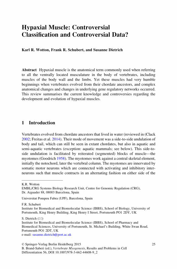
Fig. 1 Vertebrate phylogenetic tree, indicating the key changes in the organisation of their body
musculature that underpinned changes in movement pattern. The basic chordate movement
pattern is swimming via side-to-side undulations of the body and tail, relying on segmented
muscle blocks—the myotomes—on either side of the body. The first steps towards three-
dimensional mobility were taken in the shared ancestors of agnathans and gnathostomes, when
Engrailed was recruited into the somite to facilitate the separate innervation of the dorsal/
epaxial and ventral/hypaxial domain of the myotome. Likewise, in this ancestor the most rostral
segments became incorporated into the head and muscle was deviated from a role in locomotion
to role in pharyngeal support, respiration and food uptake. Specifically, the ventral/hypaxial
muscle precursors were recruited to provide elaborate hypobranchial muscles. In the lineage
leading to jawed vertebrates, epaxial–hypaxial muscle became fully segregated. Moreover, the
lateral mesoderm developed two distinct leafs, facilitating the establishment of a muscularised
body wall and the evolution of paired fins. Initially, muscle penetrated the outer, somatopleural
aspect of the lateral mesoderm as a somitic outgrowth. Yet in the lineage leading to
osteichthyans, a molecular program that allowed the de-epithelialisation and emigration of
muscle precursors evolved. This program was first used to generate the muscles of the pectoral
fins, but in the lineage leading to sarcopterygians, it was co-opted into the pelvic fins/hind limbs.
It is thought that the resolution of segmental boundaries allowed the redistribution of muscle
cells and, together with the more mobile insertion of the sarcopterygian fin/limb in the shoulder
girdle, it facilitated the evolution of load-bearing limbs
26 K.R. Wotton et al.
in ray-finned (actinopterygian) bony1 vertebrates (osteichthyans), paired fins are
mainly used for steering. Yet in lobe-finned animals (sarcopterygian osteichthyans),
these fins eventually changed into load-bearing limbs that allowed the tetrapods to
conquer land and to take to the air (Clack 2002). These amazing changes in body
plans and locomotion took some 500 million years and allowed vertebrates to
populate every ecological niche on Earth: land, air, fresh and marine waters. With
that respect, vertebrates are one of the most successful animal group ever.
The anatomical changes that allowed the change of vertebrate movement pat-
terns predominantly affected the lay-out of the ventral muscular system, tradition-
ally referred to as “hypaxial”, and this review will retrace their development and
evolution. It also will discuss the “primaxial–abaxial” subdivision of muscle that is
often portrayed as contrasting concept. Finally, the review will provide an overview
of a specialised type of hypaxial muscle precursors that evolved in the osteichthyan
lineage and that is thought to have aided the evolution of load-bearing limbs—the
migratory hypaxial muscle precursors.
2 Developmental Anatomy of Dorso-Ventral Muscles
and the Classical Epaxial–Hypaxial Concept
In all vertebrates, the skeletal musculature of the body and fins/limbs originates
from the segmented paraxial (¼next to the axial notochord) mesoderm termed
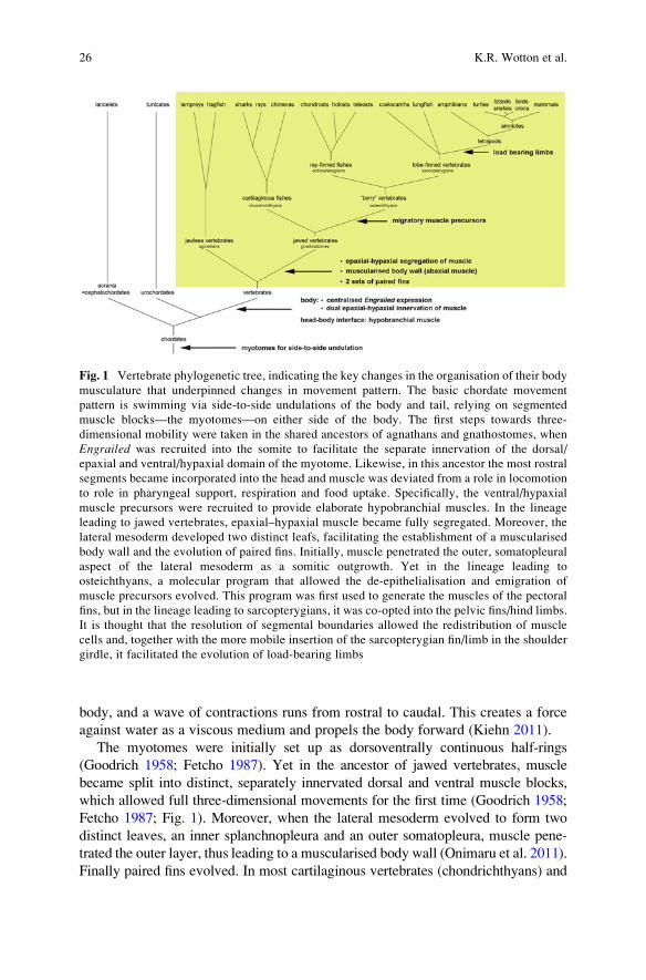
somites, and muscle is laid down in waves (reviewed in: Bryson-Richardson and
Currie 2008; Buckingham 2006; Fig. 2). The first muscle (primary myotomes) is
immediately contractile. This is essential since anamniote vertebrates (as well as
their chordate relatives) develop via free-feeding, motile larvae (Goodrich 1958).
Yet, differentiated muscle fibres are postmitotic, thus limiting muscle growth to an
increase in fibre size by hypertrophy. However, vertebrates have set aside muscle
stem cells that drive hyperplastic muscle growth and muscle repair upon injury
(reviewed in: Bryson-Richardson and Currie 2008; Buckingham 2006). These stem
cells initially reside in a structure superficial to the myotome, the dermomyotome.
However, eventually these cells populate the muscles, thereby establishing a
resident pool of stem cells. In the adult, these cells are located underneath the
basal lamina of muscle fibres and are referred to as satellite cells. In
actinopterygians such as teleosts, muscle stem cells drive the continuous muscle
growth typical for these animals. In amniotes, the satellite cells become quiescent
and are only activated and proliferative upon injury.
In jawed vertebrates, the myotomes become segregated into dorsal and ventral
components. This is achieved by the intercalation of a physical boundary, the
1 The phrase “bony” vertebrate as a more colloquial term for osteichthyans is somewhat mislead-
ing since mineralised bone was already present in stem group gnathostomes and was secondarily
lost in sharks and rays, (Zhu et al. 2013).
Hypaxial Muscle: Controversial Classification and Controversial Data? 27
Fig. 2 Vertebrate muscle is
generated in waves.
Schematic cross sections of
amniote flank somites,
dorsal to the top, modelled
after the chicken. In line
with the rostrocaudal
progression of somite
formation and
differentiation, the
developmentally youngest
somites are shown at the
bottom, the most mature at
the top. (a) Soon after the
epithelial somite formed, its
dorsal territory becomes
specified as dermomyotome
(light green), its ventralterritory as sclerotome
(light pink), and cells in the
medial wall of the somite
are specified as the first
myogenic cells (turquoise).(b) The myogenic cells, also
referred to as muscle
pioneers, spread (redarrow) and form a scaffold
between the now
morphologically defined
dermomyotome (green) andsclerotome (pink). (c) More
cells are being added to the
scaffold from the
dorsomedial, ventrolateral,
rostral and caudal edges of
the dermomyotome (redarrows), leading to a
morphologically well-
defined, contractile
myotome (turquoise). (d)Eventually, the
dermomyotome
de-epithelialises, releasing
myogenic stem cells into the
myotome (red arrows).These cells drive the foetal
and perinatal growth of
muscle and provide the
adult muscle stem cells
(satellite cells)
28 K.R. Wotton et al.
thoracolumbar fascia or horizontal myoseptum (Goodrich 1958; Gray 1995). In
teleost fish, the dorsoventral subdivision of muscle occurs at an early time point and
is organised by specialised, engrailed expressing slow muscle cells. These cells are
somewhat misleadingly termed muscle pioneers since they express muscle struc-
tural genes at an early time point (Devoto et al. 1996; Currie and Ingham 1996).
However, their key role is to serve as a first target for the axons of the three large,
primary somatic motorneurons and organise the projection of one of them to the
dorsal muscle block, one to the ventral muscle block, and one to innervate the slow-
twitch muscle at the dorsoventral boundary (Beattie and Eisen 1997), reviewed by
(Lewis and Eisen 2003). When engrailed function is disrupted, severe innervation
defects occur (Ahmed et al. manuscript in preparation; Fig. 3).
In teleosts, eventually the smaller, secondary motorneurons outnumber the
primary motorneurons and take over to drive muscle contraction (Fetcho 1987;
Lewis and Eisen 2003). However, these neurons use the pre-existing scaffold for
their axonal pathfinding. In amniotes in contrast, it is generally held that only
secondary-type motorneurons are being used (Fetcho 1987). They are organised
into two pools in the ventral spinal cord, with the medially located pool (medial
motor column) destined to innervate the dorsal muscles, and the laterally located
neurons (hypaxial motor column) innervating the ventral muscles (reviewed in:
Tsuchida et al. 1994). Yet in all vertebrates, the physical segregation of muscle is
matched by their separate innervation, such that the dorsal and ventral muscles can
contract independently. According to their distinct innervation, muscles have
classically been distinguished as epaxial (innervated by the medial motor column
via the dorsal ramus of the spinal nerve) or hypaxial (innervated by the hypaxial
motor column via the ventral ramus of the spinal nerve; Fig. 4a, Ai).
Interestingly, in amniotes, the dorsoventrally distinct innervation of body muscle
occurs before the myotome is physically segregated (Tosney and Landmesser 1985;
Tosney 1987; reviewed in: Fetcho 1987). Yet this innervation pattern also relies on
Engrailed (En1; Ahmed et al., manuscript in preparation). En1 is initially expressedin the central dermomyotome where it sets up a molecular and compartment
boundary (Cheng et al. 2004). Expression of En1 is brought into the myotome
when the muscle stem cells arrive from the de-epithelialising dermomyotome
(Ahmed et al. 2006). En1 then supports the outgrowth of the dorsally projecting
axons and suppresses the outgrowth of the ventrolaterally projecting axons (Ahmed
et al., manuscript in preparation; Fig. 3). Thus, although En1 function has shifted intime, it is remarkable that in jawed vertebrates, it is associated with epaxial–
hypaxial boundary formation and innervation. Moreover, in all vertebrates inves-
tigated Engrailed expression is controlled by the Shh signalling molecule released
from the notochord, suggesting the conservation of key parts of the underlying
regulatory network (Cheng et al. 2004; Hammond et al. 2009; Maurya et al. 2011).
Jawless vertebrates such as the lamprey already have a dorsal and ventral
innervation point of their myotome even though a horizontal myoseptum is absent
(Fetcho 1987). Moreover, markers have been identified that distinguish the dorsal
and the lateral edge of the somite, suggesting that the first steps towards an epaxial–
hypaxial segregation of muscle had been taken before the agnathan-gnathostome
Hypaxial Muscle: Controversial Classification and Controversial Data? 29
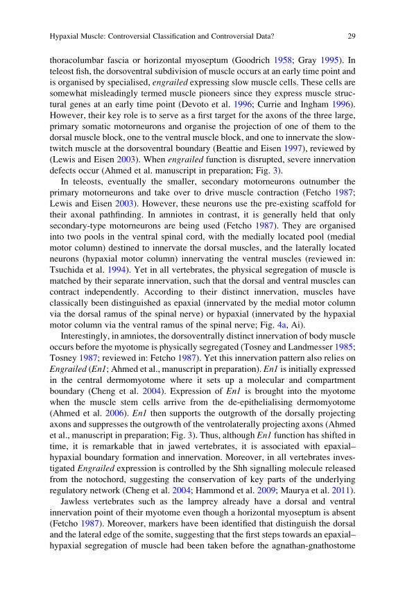
Fig. 3 Conserved role of Engrailed in organising the epaxial–hypaxial innervation of muscle.
(a–c) Role of En1 in the chicken embryo. (a) Cross sections of chicken flank somites at embryonic
day E4.5 of development, dorsal to the top, medial to the left. Engrailed 1 (En1) expression is
shown in blue, intermediate neurofilaments of nerve axons are revealed with the RMO antibody in
30 K.R. Wotton et al.
divide (Kusakabe et al. 2011). Interestingly, also in the lamprey, one of its
engrailed genes is expressed in the centre of the myotome, molecularly separating
its dorsal and ventral aspect (Hammond et al. 2009; Matsuura et al. 2008). This
suggests that already before the agnathan-gnathostome divide, eng/En had been
⁄�
Fig. 3 (continued) brown. Note that En1 expressing cells are leaving the central dermomyotome
and populate the central myotome underneath. The developing ventral ramus of the spinal nerve
navigates around the En1 expression domain and targets the hypaxial myotome; first contact with
the myotome is made when axons of the cutaneous branch of the ventral ramus travel along the
ventral boundary of the En1 domain and project towards the dermis (small arrows). The dorsal
ramus lags developmentally behind; its axons target the En1 domain (arrowhead). (b–c) Gain-of-function experiment; phenotype displayed on cross sections. Flank somites were electroporated at
E2.5 with (b) a GFP expressing control construct or (c) a bi-cistronic GFP and En1 expressing
experimental construct. 24 h later, expression of the construct was revealed by in situ hybridisation
in blue and the position of the axons by RMO staining in brown. Note that in the En1misexpressing somites, the ventral ramus of the spinal nerve defasciculated and failed to form
the cutaneous branch of this ramus (open arrowhead). (d–f) Role of En1 in the mouse embryo. (d)
Cross sections of mouse flank somites at E11.5 of development, dorsal to the top, medial to the left;En1 expression in blue, RMO staining in brown. Note that marker gene expression and axonal
projections in the mouse closely match that of the chicken. (e–f) Loss-of-function experiment,
axons were revealed by RMO antibody staining on cross sections (green staining). Note that in
wildtype litter mates, the dorsal ramus of the spinal nerve is well developed and innervates the
epaxial myotome (e). In En1 deficient embryos, that dorsal ramus falls short of its target (f, openarrowhead). (g–k) Role of engrailed genes in the zebrafish. (g) Cross section of a 36hpf zebrafish
eng2a:GFP embryo. engrailed expression as revealed by the GFP transgene expression is shown in
red; axons are stained with the znp1 antibody (green) and cell nuclei with Dapi (blue). Initially, theprimary motor neurons all project to the eng expressing muscle pioneers (+, mp) which organise
the formation of the horizontal myoseptum. Subsequently, the primary and accompanying sec-
ondary motor neurons located next to the posterior somite half extend their axons ventrally to
contribute to the ventral ramus of the spinal nerve and to innervate the hypaxial myotome. Motor
neurons neighbouring the anterior somite half send their axons laterally along the developing
horizontal myoseptum to form the fish-specific medial ramus and to target the superficial slow
muscles. Motor neurons in the middle of each segment withdraw their connection to the muscle
pioneers and project dorsally to form the dorsal ramus and to innervate the epaxial myotome. (h–k)
Lateral views of 36hpf experimental zebrafish embryos, anterior to the left. The position of the
developing horizontal myoseptum is indicated by a stippled line. (h–i) Gain-of-function experi-
ment: Transgenic α actin-Gal4 embryos were used to drive expression of (H) a UAS-tRFP control
construct or (i) a bi-cistronic construct encoding tRFP as well as zebrafish engrailed 2a. Cells
expressing the constructs fluoresce in red; axons are revealed with the znp1 antibody in green.Note that engrailed misexpression leads to severe misguidance of motor axons (i, arrowheads).(j–k) Loss-of-function experiment: eng2a:GFP embryos (transgene expression shown in red, znp1-
stained axons in green as in (g) were treated with (j) a control morpholino or (k) a morpholino
cocktail targeting the splice sites of engrailed1a, 1b and 2a which are all expressed in muscle
pioneers. Note that this knock down of eng function blocked axonal outgrowth, and axons stalled ortook up erratic paths in search for their targets (k, open arrowheads). The data shown here are the
work of Mohi U. Ahmed, Ashish K. Maurya, Louise Cheng, Erika C. Jorge, Frank R. Schubert,
Pascal Maire, M. Albert Basson, Philip W. Ingham and Susanne Dietrich. d dermis precursors, dmdermomyotome, dml dorsomedial lip of the dermomyotome, dr dorsal ramus of the spinal nerve, drgdorsal root ganglion, m myotome, mp muscle pioneers, mr medial ramus of the spinal nerve, sclsclerotome, vll ventrolateral lip of the dermomyotome, vr ventral ramus of the spinal nerve. The
asterisk in (a,d) marks the axons of motor neurons projecting out of the neural tube
Hypaxial Muscle: Controversial Classification and Controversial Data? 31
recruited into the developing musculature where it paved the way for the dorso-
ventral segregation and innervation of muscle and the evolution of full three-
dimensional mobility. On the other hand, when mammals returned to the sea,
they adapted to movement in water with fully segregated epaxial–hypaxial muscle
in place. It can be speculated that this divide was the basis to evolve upwards-
downwards undulations of the body and tail as seen best in dolphins and whales.
3 The Primaxial–Abaxial Concept and the Lateral Somitic
Frontier
Skeletal muscle fibres are organised into anatomically defined muscles via several
layers of connective tissue (Gray 1995). Moreover, muscle can only fulfil its role
when anchored on skeletal elements via tendons or aponeuroses. Thus, functional
muscle has an intricate relationship with the various types of connective tissue.
Indeed, even though initially muscle and connective tissue develop independently,
eventually both tissues rely on each other for function and survival (Murphy
et al. 2011). Interestingly, in muscle-less limbs, connective tissue organises itself
in the correct anatomical pattern, anticipating the position of muscle (Kardon
et al. 2003). This indicates that the connective tissue directs the muscle cells to
their defined places. Yet at different locations in the body, connective tissue is made
from different cell types, and hence, muscle has to adjust to different partners. This
has led to the primaxial–abaxial concept of muscle development (Fig. 4b,Bi).
Epaxial muscles, but also some hypaxial muscles—for example the
sub-vertebral muscle of the neck and the muscles associates with the ribs—receive
their connective tissue from the somites. Thus, the cells always remain in a
paraxial—or primaxial—environment. On the other hand, hypaxial muscle pre-
cursors for muscles associated with the sternum, the body wall or the limbs enter a
new environment, namely the dorsal leaf of the lateral mesoderm (somatopleura),
and all the connective tissue is derived from this environment (Durland et al. 2008).
When heterotopically grafted, muscle precursors entering the lateral plate environ-
ment switch Hox gene expression to match the position values found on site
(Nowicki and Burke 2000). Thus, these hypaxial cells cross a boundary, termed
“lateral somitic frontier”. They will settle far from their original position and are
patterned by their new environment, and hence have been termed “abaxial”
(reviewed in: Shearman and Burke 2009). In Amphioxus and in the lamprey, the
lateral mesoderm of the body does not split into splanchnic and somatic lateral
mesoderm, and muscle does not enter this environment (Onimaru et al. 2011;
Tulenko et al. 2013). Thus, abaxial body muscle, i.e. a muscularised body wall
and derivates thereof (see below) may be a novelty that emerged in the lineage
which eventually led to the jawed vertebrates (Fig. 1). How connective tissue cells
communicate their positional values to muscle cells is not clear. It has been shown,
however, that homeodomain transcription factors can act as short range signalling
32 K.R. Wotton et al.
molecules, both in vertebrates and in insects (reviewed in: Brunet et al. 2007;
Layalle et al. 2011). Thus, it is tempting to speculate that a similar process may
allow the alignment of Hox gene expression patterns. However, single cells versus
group cell grafting performed in a cranial environment suggested that, if the grafted
cells have enough neighbours of their own kind, they retain their original Hox code
(Trainor and Krumlauf 2000). Thus, more work is needed to determine cell–cell
communication at the lateral somitic frontier. Yet, it should be emphasized that the
classical epaxial–hypaxial concept and the primaxial–abaxial concept are not
necessarily exclusive; they simply refer to different aspects of ventrolateral muscle
development (Fig. 4).
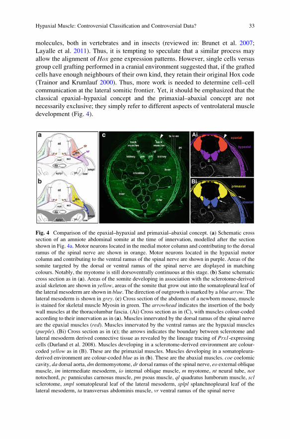
Fig. 4 Comparison of the epaxial–hypaxial and primaxial–abaxial concept. (a) Schematic cross
section of an amniote abdominal somite at the time of innervation, modelled after the section
shown in Fig. 4a. Motor neurons located in the medial motor column and contributing to the dorsal
ramus of the spinal nerve are shown in orange. Motor neurons located in the hypaxial motor
column and contributing to the ventral ramus of the spinal nerve are shown in purple. Areas of the
somite targeted by the dorsal or ventral ramus of the spinal nerve are displayed in matching
colours. Notably, the myotome is still dorsoventrally continuous at this stage. (b) Same schematic
cross section as in (a). Areas of the somite developing in association with the sclerotome-derived
axial skeleton are shown in yellow, areas of the somite that grow out into the somatopleural leaf of
the lateral mesoderm are shown in blue. The direction of outgrowth is marked by a blue arrow. Thelateral mesoderm is shown in grey. (c) Cross section of the abdomen of a newborn mouse, muscle
is stained for skeletal muscle Myosin in green. The arrowhead indicates the insertion of the body
wall muscles at the thoracolumbar fascia. (Ai) Cross section as in (C), with muscles colour-coded
according to their innervation as in (a). Muscles innervated by the dorsal ramus of the spinal nerve
are the epaxial muscles (red). Muscles innervated by the ventral ramus are the hypaxial muscles
(purple). (Bi) Cross section as in (c); the arrows indicates the boundary between sclerotome and
lateral mesoderm derived connective tissue as revealed by the lineage tracing of Prx1-expressingcells (Durland et al. 2008). Muscles developing in a sclerotome-derived environment are colour-
coded yellow as in (B). These are the primaxial muscles. Muscles developing in a somatopleura-
derived environment are colour-coded blue as in (b). These are the abaxial muscles. coe coelomic
cavity, da dorsal aorta, dm dermomyotome, dr dorsal ramus of the spinal nerve, eo external obliquemuscle, im intermediate mesoderm, io internal oblique muscle, m myotome, nt neural tube, notnotochord, pc panniculus carnosus muscle, pm psoas muscle, ql quadratus lumborum muscle, sclsclerotome, smpl somatopleural leaf of the lateral mesoderm, splpl splanchnopleural leaf of thelateral mesoderm, ta transversus abdominis muscle, vr ventral ramus of the spinal nerve
Hypaxial Muscle: Controversial Classification and Controversial Data? 33
4 Migratory Muscle Precursors for the Paired Fins
and Limbs: An Osteichthyan Innovation
In tetrapods, even though the lateral swaying of the body is still an important part in
the movement of amphibians and reptiles, locomotion clearly relies on load-bearing
limbs and their associated musculature. Innervated by the lateral motor column
a specialist group of hypaxial motor neurons only found at limb levels reviewed in
Murakami and Tanaka 2011 and developing in the lateral mesoderm that provides
the limb connective tissue and the limb skeleton, limb muscles are both hypaxial
and abaxial. Embryological studies in the chicken established that in amniotes, limb
muscles develop from cells that detach from the lateral lip of the dermomyotome
and actively migrate into the limb, where they become organised into dorsal and
ventral muscles masses to give rise to the extensor and flexor muscles groups,
respectively (Chevallier et al. 1977; Christ et al. 1977; Hayashi and Ozawa 1995;
Fig. 5a). Painstaking histological and lineage tracing experiments in various fish
species showed that muscle precursors undertaking long-range migration
muscularise the pectoral fins of teleosts (ray-finned “bony” vertebrate), while in
cartilaginous vertebrates, somites form extensions that reach into the fin anlage in a
similar fashion as they grow out into the body wall to form abdominal muscles
(Neyt et al. 2000). The pelvic fins of lungfish (a lobe-finned relative of tetrapods),
teleosts and paddlefish (both ray-finned osteichthyans) receive muscle precursors
from somitic extensions that, when close to their target site, deepithelialise to
release individual cells. The pelvic fin muscles of sharks and chimeras are made
in the same way as their pectoral musculature, namely from somitic extensions
(Cole et al. 2011). This has led to the view that hypaxial/abaxial muscle formation
via somitic extensions is the evolutionarily older mechanism of muscle delivery,
while migratory muscle precursors evolved later in the lineage leading to
osteichthyans. They were first established for the pectoral fin/forelimb and subse-
quently for the pelvic fin/hind limb. In line with this view, a molecular program has
been deciphered that specifically operates at teleost pectoral fin levels/tetrapod limb
levels, and acts on top of the generic program for hypaxial myogenesis (Fig. 5b).
The generic program for hypaxial myogenesis is best researched in amniotes.
Here, the lateral somite domain is specified by Bmp4 released from the lateral
mesoderm (Pourquie et al. 1996). Together with Wnt signals from the surface
ectoderm, Bmp upregulates the expression of the pre-myogenic gene Pax3(Dietrich et al. 1998; Fan et al. 1997; Tajbakhsh et al. 1998). Six transcription
factors, when activated by their Eya partners, contribute to the upregulation of
Pax3, and Pax3 enhances its own expression. Together, Six and Pax3 transcription
factors facilitate the generation of hypaxial skeletal muscle cells (Tremblay
et al. 1998; Borycki et al. 1999; Grifone et al. 2005; Grifone et al. 2007). This
occurs after Bmp4 cooperated with Notch signalling to facilitate the release of
smooth muscle and endothelial precursors (Ben-Yair and Kalcheim 2008).
At limb levels, muscle precursors destined to emigrate express the
homeodomain transcriptional repressor Lbx1, and in animals as diverse as teleosts
34 K.R. Wotton et al.
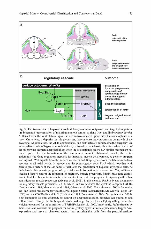
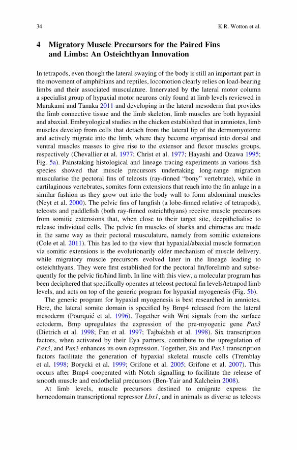
Fig. 5 The two modes of hypaxial muscle delivery—somitic outgrowth and targeted migration.
(a) Schematic representation of maturing amniote somites at flank (top) and limb (bottom levels).At flank levels, the ventrolateral lip of the dermomyotome (vll) penetrates the somatopleura as a
sheet. On its way, it deposits muscle precursors, thereby ensuring concomitant outgrowth of the
myotome. At limb levels, the vll de-epithelialises, and cells actively migrate into the periphery. An
intermediate mode of hypaxial muscle delivery is found in the teleost pelvic fins, where the vll of
the outgrowing segment deepithelialises when the destination is reached. A similar mechanism has
been reported for the formation of the ventralmost amniote abdominal muscle, the rectus
abdominis. (b) Gene regulatory network for hypaxial muscle development. A generic program
starting with Wnt signals from the surface ectoderm and Bmp signals from the lateral mesoderm
operates at all axial levels. It upregulates the premyogenic gene Pax3 which, together with
premyogenic factors of the Six family, facilitates the generation of hypaxial myogenic cells. At
limb levels, this generic program of hypaxial muscle formation is in operation. Yet, additional
localised factors control the formation of migratory muscle precursors. Firstly, Hox gene expres-sion in limb levels somites instructs these somites to activate the program of migratory rather than
non-migratory muscle precursors (Alvares et al. 2003). In this context, Pax3 activates the marker
for migratory muscle precursors, Lbx1, which in turn activates the cytokine receptor CXCR4
(Dietrich et al. 1999; Mennerich et al. 1998; Odemis et al. 2005; Vasyutina et al. 2005). Secondly,
the limb lateral mesoderm provides the cMet ligand Scatter Factor/Hepatocyte Growth Factor (SF/
HGF) and the CXCR4 ligand Sdf1 (Bladt et al. 1995; Prunotto et al. 2004; Vasyutina et al. 2005).
Both signalling systems cooperate to control lip deepithelialisation, targeted cell migration and
cell survival. Thirdly, the limb apical ectodermal ridge (aer) releases Fgf signalling molecules
which are required for the expression of SF/HGF (Scaal et al. 1999). Importantly, Fgf molecules by
themselves can override the program for non-migratory hypaxial muscle precursors, trigger Lbx1expression and serve as chemoattractants, thus ensuring that cells from the paraxial territory
Hypaxial Muscle: Controversial Classification and Controversial Data? 35
(actinopterygians), lungfish and tetrapods (sarcopterygians), Lbx1 (teleosts: lbx1a,b) is the bona fide marker for migratory muscle precursors (Cole et al. 2011;
Dietrich 1999; Dietrich et al. 1999; Jagla et al. 1995; Martin and Harland 2006;
Ochi and Westerfield 2009; Thisse et al. 2004); Figs. 5b and 6a, b, d and 7).
Absence of Lbx1 or misexpression of a dominant negative Lbx1 construct prevents
cell emigration into the paired fins/limbs, and only a subset of forelimb flexor
muscle at the ventral junction to the limbs develops under the influence of local
cues (Schafer and Braun 1999; Gross et al. 2000; Brohmann et al. 2000; Ochi and
Westerfield 2009; Martin and Harland 2006; Lours-Calet et al. 2014); Fig. 6f,Fi).
Given this important role of Lbx1, the question of migratory muscle precursor
formation has frequently been seen as a problem of localised Lbx1 induction.
The mouse mutant Splotch is a well known model for muscle-less limbs (Franz
et al. 1993; Bober et al. 1994; Tremblay et al. 1998), and in this animal, Lbx1expression is lost (Dietrich et al. 1999; Mennerich et al. 1998). Splotchmice carry a
mutation for the paired box transcription factor Pax3, yet Pax3 is expressed in the
early somite, in the dermomyotome of more mature somites and is upregulated in
the dorsomedial and ventrolateral dermomyotomal lips of all somites along the
rostrocaudal body axis. Thus, while Pax3 is necessary for Lbx1 expression, it is notsufficient to position expression in limb-level somites. Interestingly, experiments in
the zebrafish indicated that here, the duplicated pax3b gene had its expression
restricted to pectoral fin somites and is required for lbx expression (lbx2 in this
case; Minchin et al. 2013). Thus, while displaying a variation on the theme, it
suggests that the relationship of Pax3 and Lbx is evolutionarily ancient.
It is well established that heterotopic transplantation of limbs or limb induction
via implantation of Fgf beads in the flank of chicken embryos lead to the develop-
ment of a muscularised and fully innervated ectopic limb (Chevallier et al. 1977;
Christ et al. 1977; Hayashi and Ozawa 1995; Cohn et al. 1995). Moreover, the
ectopic limb, its apical ectodermal ridge (aer) or the Fgf4/8 signalling molecule
produced by the aer, all induce somitic Lbx1 expression and the emigration of
muscle precursors (Alvares et al. 2003). Furthermore, regulated by FGF from the
aer, the limb mesenchyme produces the signalling molecule Scatter factor/Hepato-
cyte growth factor (Scaal et al. 1999). Its receptor cMet is found in all ventrolateral
dermomyotomal lips, but the local activation of cMet leads to local lip deepithe-
lialisation only (Bladt et al. 1995; Prunotto et al. 2004). Similarly, the cytokine Sdf1
which assists SF/HGF is expressed in the limb mesenchyme, and its CXCR4
receptor in the somitic dermomyotome (Odemis et al. 2005; Vasyutina
et al. 2005). Together, this has led to the view that the limb overrides any
pre-existing programme and is the key inducer of migratory muscle precursors.
Fig. 5 (continued) are recruited into the limb (Alvares et al. 2003). coe coelomic cavity, da dorsalaorta, dm dermomyotome, im intermediate mesoderm, m myotome, MMP migratory muscle
precursors, nt neural tube, not notochord, scl sclerotome, smpl somatopleural leaf of the lateral
mesoderm, splpl splanchnopleural leaf of the lateral mesoderm
36 K.R. Wotton et al.
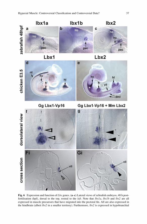
Fig. 6 Expression and function of Lbx genes. (a–c) Lateral views of zebrafish embryos, 48 h post-
fertilisation (hpf), dorsal to the top, rostral to the left. Note that lbx1a, lbx1b and lbx2 are all
expressed in muscle precursors that have migrated into the pectoral fin. All are also expressed in
the hindbrain (albeit lbx2 in a smaller territory). Furthermore, lbx2 is expressed in hypobranchial
Hypaxial Muscle: Controversial Classification and Controversial Data? 37
However, evidence has accumulated that suggests the role of the limb has been
overrated, and a more complex gene regulatory network has emerged (Fig. 5b).
A detailed characterisation of ectopic limbs revealed that the limb is not able to
fully reprogram flank tissues since it does not force the spinal cord to generate a
lateral motor column. Instead, the ectopic limb deviates axons originating from the
flank hypaxial motor column to innervate the ectopic limb muscle rather than its
normal target, the body wall musculature (Turney et al. 2003). Secondly, in cMet
and SF/HGF mutants, while muscle precursors de-epithelialisation and emigration
fails, Lbx1 is well expressed (Dietrich et al. 1999), and in migratory muscle
precursors, expression of CXCR4 is downstream of Lbx1 (Vasyutina et al. 2005).
This indicates that the specification of migratory muscle precursors is independent
from cell de-epithelisation. Thirdly and most importantly, Lbx1 expressing muscle
precursors develop when competent, limb-level somites are exposed to any type of
lateral mesoderm, including lateral mesoderm from the flank (Alvares et al. 2003).
When Hox genes were misexpressed to switch the axial identity of flank somites to
that of limb somites, these somites faithfully expressed Lbx1 (Alvares et al. 2003;
Fig. 5b). This indicates that intrinsic, Hox-dependent cues predispose somites
towards either generating non-migratory or migratory muscle precursors.
5 Migratory Muscle Precursors for the Paired Fins
and Limbs: A Vertebrate Innovation?
In osteichthyans, Lbx1 genes are exclusively expressed in cells detaching and
migrating away from the somites (Cole et al. 2011; Dietrich 1999; Dietrich
et al. 1999; Jagla et al. 1995; Martin and Harland 2006; Ochi and Westerfield
2009; Thisse et al. 2004); this article; Fig. 7). Possibly the most extreme case is
Fig. 6 (continued) muscle precursors coalescing in the hypoglossal cord (hc) and temporarily in
the dorsal and ventral tips of the myotome (not shown). (d, e) Lateral views of chicken embryos at
embryonic day E3.5 days of development, rostral to the top-right. (d) Lbx1, in addition to its
expression in the neural tube, is expressed in the migratory muscle precursors populating the limbs
and the hypoglossal cord. Lbx2 is not expressed in neural tissues, but shows a widespread
expression in myogenic cells including muscle precursors in all ventrolateral dermomyotomal
lips (migratory and non-migratory), muscle precursors in all dorsomedial lips and the myogenic
neck lateral mesoderm (asterisk). (f, g) Dorsolateral views and (Fi,Gi) corresponding cross
sections of electroporated chicken somites at forelimb levels. (f, Fi) Misexpression of a dominant
negative chicken Lbx1 construct (Gg Lbx1-Vp16; blue staining) interferes with the emigration of
muscle precursors into the forelimb (open arrowheads). (g, Gi) Co-expression of the dominant
negative construct (blue staining) together with mouse (Mm) Lbx2 rescues muscle precursor
emigration even though Mm Lbx2 is divergent and not expressed in myogenic cells. The data
shown here are the work of Karl Wotton and Susanne Dietrich. dml dorsomedial lip of the
dermomyotome, fl fore limb, hb hind brain, hc hypoglossal cord, hl hind limb, nt neural tube,pec pectoral fin, vll ventrolateral lip of the dermomyotome
38 K.R. Wotton et al.
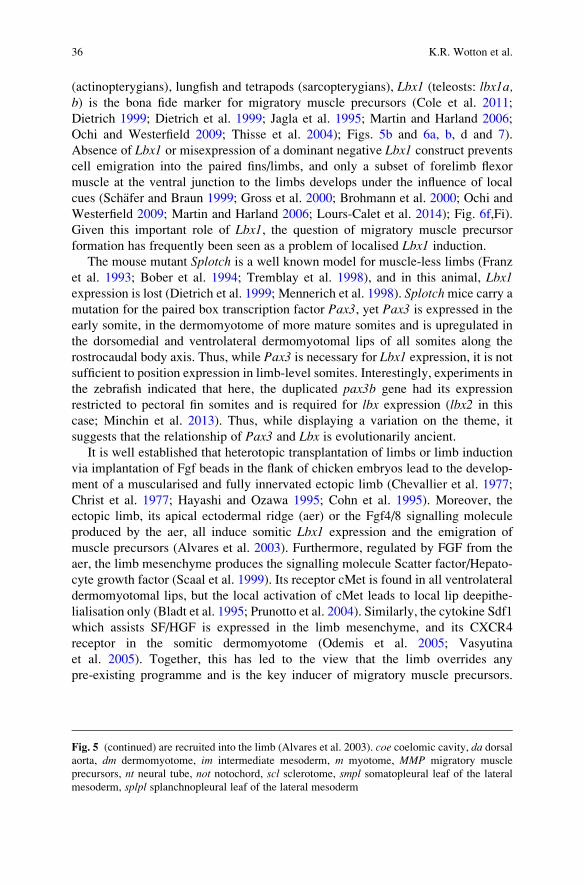
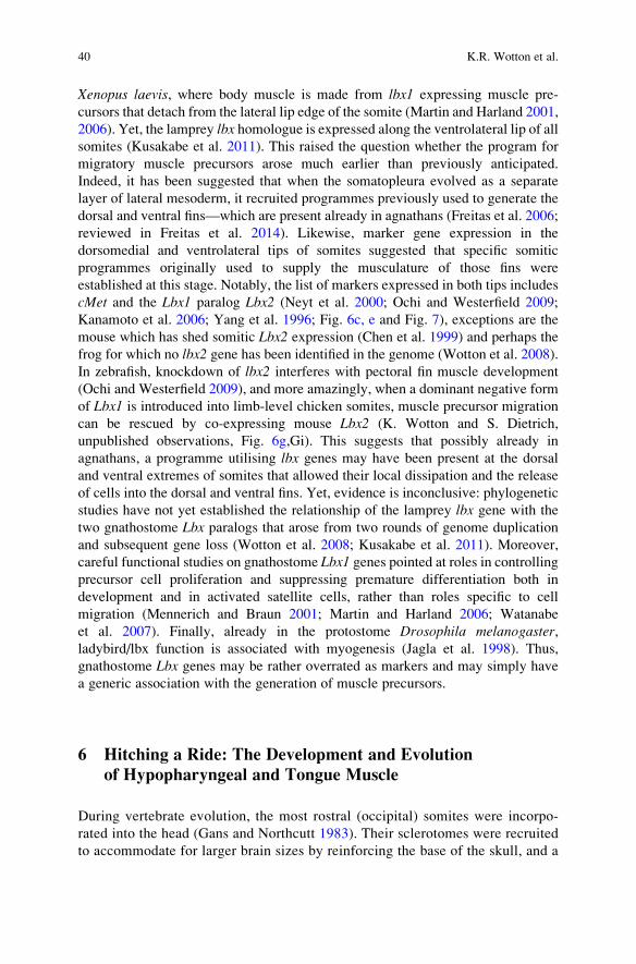
lbx/Lbx1-type genes Lbx2-type genesSpecies Gene Expression in: Reference Gene Expression in: Reference
all dml
all vll
vll producing MMP only
all dml
all vll
vll producing MMP only
Lamprey lbx-a*
� (Kusakabe et al. 2011)
Zebrafish lbx1a � (Ochi and Westerfield 2009); this ar�cle
Zebrafish lbx1b � (Thisse et al. 2004); this ar�cle
lbx2 � � (Neyt et al. 2000; Ochi and Westerfield 2009); this ar�cle
Xenopus lbx1 �**
�**
(Mar�n and Harland 2006)
No lbx2 gene in theXenopus genome assembly
(Wo�on et al. 2008)
Chicken Lbx1 � (Dietrich et al. 1998)
Lbx2 � � (Kanamoto et al. 2006); this ar�cle
Mouse Lbx1 � (Jagla et al. 1995; Dietrich et al. 1999)
Lbx2 Not expressed in somites (Chen et al. 1999)
* Phylogene�c analyses did not fully resolve whether the lamprey lbx-A gene is an ortholog of gnathostome Lbx1, or whether the gene arose before the two rounds (teleosts: three rounds) of gnathostome genome duplica�on and hence, would be a homologue of both, gnathostome Lbx1 and Lbx2 (Kusakabe et al. 2011; Wo�on et al. 2008).
** Frog body wall muscles seem to form from migratory cells (Mar�n and Harland 2006).
Fig. 7 Presence and myogenic expression of vertebrate Lbx genesExtant gnathostomes show evidence of 2 rounds of whole genome duplication during evolution,
with additional lineages, including that of the teleosts, undergoing a third. Yet, owing to the early
loss of duplicates, gnathostomes genomes only have a Lbx1 gene (teleosts: lbx1a and 1b) and a
Lbx2 gene; frogs may have lost their lbx2. Of these, Lbx1 genes are almost invariably associated
with migratory muscle precursors. Lbx2 genes have a more widespread expression, labelling the
dorsomedial as well as ventrolateral lips. The exceptions are mammals that have lost somitic Lbx2expression.
It is currently controversial whether the aforementioned genome duplications occurred before or
after the gnathostome-agnathan split, whether an independent genome duplication occurred in
agnathans, or whether in the agnathan lineage numerous individual genes were duplicated. Thus,
the phylogenetic relationship of the only lamprey lbx gene identified so far, lbx-A, is unclear. Yet itis remarkable that this gene is expressed in all vll’s along the body.
dml dorsomedial lips of the dermomyotome, MMP migratory muscle precursors, vll ventrolaterallips of the dermomyotome
Hypaxial Muscle: Controversial Classification and Controversial Data? 39
Xenopus laevis, where body muscle is made from lbx1 expressing muscle pre-
cursors that detach from the lateral lip edge of the somite (Martin and Harland 2001,
2006). Yet, the lamprey lbx homologue is expressed along the ventrolateral lip of all
somites (Kusakabe et al. 2011). This raised the question whether the program for
migratory muscle precursors arose much earlier than previously anticipated.
Indeed, it has been suggested that when the somatopleura evolved as a separate
layer of lateral mesoderm, it recruited programmes previously used to generate the
dorsal and ventral fins—which are present already in agnathans (Freitas et al. 2006;
reviewed in Freitas et al. 2014). Likewise, marker gene expression in the
dorsomedial and ventrolateral tips of somites suggested that specific somitic
programmes originally used to supply the musculature of those fins were
established at this stage. Notably, the list of markers expressed in both tips includes
cMet and the Lbx1 paralog Lbx2 (Neyt et al. 2000; Ochi and Westerfield 2009;
Kanamoto et al. 2006; Yang et al. 1996; Fig. 6c, e and Fig. 7), exceptions are the
mouse which has shed somitic Lbx2 expression (Chen et al. 1999) and perhaps the
frog for which no lbx2 gene has been identified in the genome (Wotton et al. 2008).
In zebrafish, knockdown of lbx2 interferes with pectoral fin muscle development
(Ochi and Westerfield 2009), and more amazingly, when a dominant negative form
of Lbx1 is introduced into limb-level chicken somites, muscle precursor migration
can be rescued by co-expressing mouse Lbx2 (K. Wotton and S. Dietrich,
unpublished observations, Fig. 6g,Gi). This suggests that possibly already in
agnathans, a programme utilising lbx genes may have been present at the dorsal
and ventral extremes of somites that allowed their local dissipation and the release
of cells into the dorsal and ventral fins. Yet, evidence is inconclusive: phylogenetic
studies have not yet established the relationship of the lamprey lbx gene with the
two gnathostome Lbx paralogs that arose from two rounds of genome duplication
and subsequent gene loss (Wotton et al. 2008; Kusakabe et al. 2011). Moreover,
careful functional studies on gnathostome Lbx1 genes pointed at roles in controllingprecursor cell proliferation and suppressing premature differentiation both in
development and in activated satellite cells, rather than roles specific to cell
migration (Mennerich and Braun 2001; Martin and Harland 2006; Watanabe
et al. 2007). Finally, already in the protostome Drosophila melanogaster,ladybird/lbx function is associated with myogenesis (Jagla et al. 1998). Thus,
gnathostome Lbx genes may be rather overrated as markers and may simply have
a generic association with the generation of muscle precursors.
6 Hitching a Ride: The Development and Evolution
of Hypopharyngeal and Tongue Muscle
During vertebrate evolution, the most rostral (occipital) somites were incorpo-
rated into the head (Gans and Northcutt 1983). Their sclerotomes were recruited
to accommodate for larger brain sizes by reinforcing the base of the skull, and a
40 K.R. Wotton et al.
proportion of muscle precursors were deviated from making muscle for locomo-
tion. Instead, these cells were recruited to contribute to the caudal pharyngeal
arches and to provide an elaborate hypopharyngeal muscular system, all crucial in
ventilating the gills and in food uptake (Goodrich 1958), recently reviewed in
(Sambasivan et al. 2011). In lung-breathing tetrapods, the pharyngeal arches are
not required for respiration any more, but the role in particular of the
hypopharyngeal and, as an important component, the tongue muscles, remained
crucial. Interestingly, in osteichthyans the hypopharyngeal/tongue muscle pre-
cursors undertake long-range migration, and they all express Lbx1 (Dietrich
et al. 1999; Lours-Calet et al. 2014; Martin and Harland 2006). Yet, in mouse
mutants for the upstream regulator Pax3, in cMet and SF/HGF mutants as well as
in Lbx1mutants, hypobranchial muscle formation is reduced, not abolished (Bladt
et al. 1995; Prunotto et al. 2004; Schafer and Braun 1999; Gross et al. 2000;
Brohmann et al. 2000; Lours-Calet et al. 2014). Similarly, misexpression of a
dominant negative Lbx1 construct in occipital somites in the chicken only delayed
the formation of hypopharyngeal muscle (Lours-Calet et al. 2014). Likewise,
when somites normally producing non-migratory muscle precursors only, or
head mesoderm that is unable to read signals for somitic myogenesis, were grafted
in place of the somites normally providing hypobranchial muscle, the grafts
contributed cells to this musculature (Mackenzie et al. 1998; Lours-Calet
et al. 2014). Finally, the hypopharyngeal musculature of the lamprey is thought
to derive from somitic extensions, not migratory cells (Goodrich 1958). Together,
this suggests that there is an alternative, evolutionarily ancestral mechanism of
hypopharyngeal muscle precursor transport that does not require active migration.
Intriguingly, molecular markers for all occipital tissues and cells types extend
their expression along the floor of the pharynx along the same path that is taken by
the muscle precursors, and markers for the lateral mesoderm and overlying
ectoderm precede those of other tissues (Lours-Calet et al. 2014). Lineage tracing
experiments revealed that this extension of marker gene expression is due to cells
moving along this circumpharyngeal path (Lours-Calet et al. 2014; Fig. 8).
Specifically, the lateral mesoderm originating from the level of the first somite
leads the procession, with the lateral mesoderm originating from the level of
somite 2 following and embedding the hypopharyngeal muscle precursors; more
caudal lateral mesoderm becomes displaced caudally. Studies on cells moving as
a tissue sheet have shown that the moving sheet is able to drag non-migratory cells
along (reviewed in (Montell 2008). Thus, it can be speculated that the newly
discovered occipital cell movements, deep in the evolution of vertebrates, pro-
vided the original transport system for the initially non-migratory hypopharyngeal
muscle cells.
Hypaxial Muscle: Controversial Classification and Controversial Data? 41
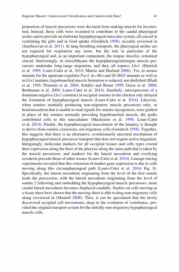
Fig. 8 An alternative mode of hypaxial cell transport at the head–neck interface. When genes
required for the emigration of limb muscle precursors are mutated, limb muscles fail to form. Yet
in most cases, hypobranchial muscle formation is merely delayed. This figure shows DiO-labelings
(green) of the lateral mesoderm and DiI labelling (red) of a somite destined to produce
42 K.R. Wotton et al.
7 Outlook
Hypaxial muscles have been remodelled quite extensively during the evolution of
vertebrates, and many aspects of their development have been deciphered. How-
ever, a number of questions, mainly surrounding migratory muscle precursors,
remain: is it indeed possible that these cells evolved earlier than the emergence of
osteichthyans, and is the underlying molecular programme an adaptation of
programmes used for the dorsal and ventral fin muscles? And how does the
formation of migratory muscle precursors for the fins/limbs relate to the release
of individual cells to form the body wall muscle in frog (Martin and Harland 2001,
2006), the contribution of migratory cells to the rostral body wall in teleosts
(Windner et al. 2011) or the de-epithelialisation of the somitic lips when the
outgrowing somite reaches the ventral midline in amniotes (Christ et al. 1983)? Is
it possible that, even though the cells destined to contribute to the body wall are
mesenchymal, they migrate as sheet rather than individual cells? And what are the
underlying molecular mechanisms? These questions may seem to have mainly
academic merit, yet answers may provide knowledge and understanding for the
therapy of birth defects such as gastroschisis or for the reconstruction or regener-
ation of muscle and limbs. Recently, it has been suggested that a mutation of the
human LBX1 gene may be responsible for the myopathy and severe vertebral
column malformation in a patient (Fernandez-Jaen et al. 2014), reinforcing how
basic research informs Medicine.
Acknowledgements We are grateful to Mohi U. Ahmed, Ashish K. Maurya, Louise Cheng, Erika
C. Jorge, Pascal Maire, M. Albert Basson and Philip W. Ingham for agreeing to include currently
unpublished data in this review article. The work was supported by the Human Frontier Science
Program, Grant No R6Y0056/2004-C201, the European Network of Excellence Myores, Grant No
EU LSHG-CT-2004-511978 MYORES, the Association Francaise contre les Myopathies, Grant
No CL/NM/2005.2088/Following number 11378, Groupe A07/06/05 and CA 01/07/05, DRM05/
BUIOCE/REFAP/MUSAI/ANGLE.
⁄�
Fig. 8 (continued) hypobranchial muscle precursors (HMP) in a 36 hours chicken embryo (a,c,e).
The embryos were analysed 24 h later when hypobranchial muscle precursors start to emigrate
(b,d,f). Notably, all occipital lateral mesoderm moves ventrolaterally. However, eventually the
streams of cells deviate. Cells originating from a position next to the most rostral somite (somite 1)
take a rostral path along the floor of the pharynx which anticipates the course of the HMP (a,b).
Lateral mesoderm from the level of somite 2 contributes both to the rostrally and ventrally—
caudally directed stream. HMP become embedded in the rostrally projecting stream (c,d). Cells
from the level of the third somite project exclusively caudally and are out of the way when HMP
start to emigrate (e, f). This suggests extensive cells movements at the head–trunk interface. It
furthermore suggests that there is a rostrally directed stream that is suited to carry non-migratory
cells along. This stream is conserved and may represent the evolutionarily ancestral way of muscle
precursor transport. lm lateral mesoderm, ma mandibular arch, ov otic vesicle, s somite
Hypaxial Muscle: Controversial Classification and Controversial Data? 43
References
Ahmed MU, Cheng L, Dietrich S (2006) Establishment of the epaxial-hypaxial boundary in the
avian myotome. Dev Dyn 235(7):1884–1894
Alvares LE, Schubert FR, Thorpe C, Mootoosamy RC, Cheng L, Parkyn G, Lumsden A, Dietrich S
(2003) Intrinsic, Hox-dependent cues determine the fate of skeletal muscle precursors. Dev
Cell 5(3):379–390
Beattie CE, Eisen JS (1997) Notochord alters the permissiveness of myotome for pathfinding by an
identified motoneuron in embryonic zebrafish. Development 124(3):713–720
Ben-Yair R, Kalcheim C (2008) Notch and bone morphogenetic protein differentially act on
dermomyotome cells to generate endothelium, smooth, and striated muscle. J Cell Biol 180
(3):607–618. doi:10.1083/jcb.200707206
Bladt F, Riethmacher D, Isenmann S, Aguzzi A, Birchmeier C (1995) Essential role for the c-met
receptor in the migration of myogenic precursor cells into the limb bud. Nature 376
(6543):768–771. doi:10.1038/376768a0
Bober E, Franz T, Arnold H-H, Gruss P, Tremblay P (1994) Pax-3 is required for the development
of limb muscles: a possible role for the migration of dermomyotomal muscle progenitor cells.
Development 120:603–612
Borycki A-G, Li J, Jin F, Emerson CP (1999) Pax3 functions in cell survival and in Pax7
regulation. Development 126:1665–1674
Brohmann H, Jagla K, Birchmeier C (2000) The role of Lbx1 in migration of muscle precursor
cells. Development 127(2):437–445
Brunet I, Di Nardo AA, Sonnier L, Beurdeley M, Prochiantz A (2007) The topological role of
homeoproteins in the developing central nervous system. Trends Neurosci 30(6):260–267
Bryson-Richardson RJ, Currie PD (2008) The genetics of vertebrate myogenesis. Nat Rev Genet 9
(8):632–646
Buckingham M (2006) Myogenic progenitor cells and skeletal myogenesis in vertebrates. Curr
Opin Genet Dev 16(5):525–532
Chen F, Liu KC, Epstein JA (1999) Lbx2, a novel murine homeobox gene related to the Drosophila
ladybird genes is expressed in the developing urogenital system, eye and brain. Mech Dev 84
(1–2):181–184
Cheng L, Alvares LE, Ahmed MU, El-Hanfy AS, Dietrich S (2004) The epaxial-hypaxial
subdivision of the avian somite. Dev Biol 274(2):348–369
Chevallier A, Kieny M, Mauger A (1977) Limb-somite relationship: origin of the limb muscula-
ture. J Embryol Exp Morphol 41:245–258
Christ B, Jacob HJ, Jacob M (1977) Experimental analysis of the origin of the wing musculature in
avian embryos. Anat Embryol 150:171–186
Christ B, Jacob M, Jacob HJ (1983) On the origin and development of the ventrolateral abdominal
muscles in the avian embryo. An experimental and ultrastructural study. Anat Embryol
166:87–101
Clack JA (2002) Gaining ground. The origin and evolution of tetrapods. Indiana University Press,
Bloomington, IN
Cohn MJ, Izpisua-Belmonte JC, Abud H, Heath JK, Tickle C (1995) Fibroblast growth factors
induce additional limb development from the flank of chick embryos. Cell 80(5):739–746
Cole NJ, Hall TE, Don EK, Berger S, Boisvert CA, Neyt C, Ericsson R, Joss J, Gurevich DB,
Currie PD (2011) Development and evolution of the muscles of the pelvic fin. PLoS Biol 9
(10):1001168. doi:10.1371/journal.pbio.1001168
Currie PD, Ingham PW (1996) Induction of a specific muscle cell type by a hedgehog-like protein
in zebrafish. Nature 382(6590):452–455
Devoto SH, Melancon E, Eisen JS, Westerfield M (1996) Identification of separate slow and fast
muscle precursor cells in vivo, prior to somite formation. Development 122(11):3371–3380
44 K.R. Wotton et al.
Dietrich S (1999) Regulation of hypaxial muscle development. Cell Tissue Res 296(1):175–182
Dietrich S, Abou-Rebyeh F, Brohmann H, Bladt F, Sonnenberg-Riethmacher E, Yamaai T,
Lumsden A, Brand-Saberi B, Birchmeier C (1999) The role of SF/HGF and c-Met in the
development of skeletal muscle. Development 126(8):1621–1629
Dietrich S, Schubert FR, Healy C, Sharpe PT, Lumsden A (1998) Specification of the hypaxial
musculature. Development 125(12):2235–2249
Durland JL, Sferlazzo M, Logan M, Burke AC (2008) Visualizing the lateral somitic frontier in the
Prx1Cre transgenic mouse. J Anat 212(5):590–602. doi:10.1111/j.1469-7580.2008.00879.x
Fan CM, Lee CS, Tessier-Lavigne M (1997) A role for WNT proteins in induction of
dermomyotome. Dev Biol 191(1):160–165
Fernandez-Jaen A, Suela J, Fernandez-Mayoralas DM, Fernandez-Perrone AL, Wotton KR,
Dietrich S, Castellanos MD, Cigudosa JC, Calleja-Perez B, Lopez-Martin S (2014)
Microduplication 10q24.31 in a Spanish girl with scoliosis and myopathy: the critical role of
LBX. Am J Med Genet A 168:2074–8
Fetcho JR (1987) A review of the organization and evolution of motoneurons innervating the axial
musculature of vertebrates. Brain Res 434(3):243–280
Franz T, Kothary R, Surani MAH, Halata Z, Grim M (1993) The Splotch mutation interferes with
muscle development in the limbs. Anat Embryol 187:153–160
Freitas R, Gomez-Skarmeta JL, Rodrigues PN (2014) New frontiers in the evolution of fin
development. J Exp Zool (Mol Dev Evol) 9999B:1–13
Freitas R, Zhang G, Cohn MJ (2006) Evidence that mechanisms of fin development evolved in the
midline of early vertebrates. Nature 442(7106):1033–1037
Gans C, Northcutt RG (1983) Neural crest and the origin of vertebrates: a new head. Science
220:268–274
Goodrich ES (1958) Studies on the structure and development of vertebrates, 2nd edn. Dover,
New York
Gray H (1995) Gray’s anatomy, 38th edn. Churchill Livingstone, Edinburgh, London
Grifone R, Demignon J, Giordani J, Niro C, Souil E, Bertin F, Laclef C, Xu PX, Maire P (2007)
Eya1 and Eya2 proteins are required for hypaxial somitic myogenesis in the mouse embryo.
Dev Biol 302(2):602–616
Grifone R, Demignon J, Houbron C, Souil E, Niro C, Seller MJ, Hamard G, Maire P (2005) Six1
and Six4 homeoproteins are required for Pax3 and Mrf expression during myogenesis in the
mouse embryo. Development 132(9):2235–2249
Gross MK, Moran-Rivard L, Velasquez T, Nakatsu MN, Jagla K, Goulding M (2000) Lbx1 is
required for muscle precursor migration along a lateral pathway into the limb. Development
127(2):413–424
Hammond KL, Baxendale S, McCauley DW, Ingham PW, Whitfield TT (2009) Expression of
patched, prdm1 and engrailed in the lamprey somite reveals conserved responses to Hedgehog
signaling. Evol Dev 11(1):27–40
Hayashi K, Ozawa E (1995) Myogenic cell migration from somites is induced by tissue contact
with medial region of the presumptive limb mesoderm in chick embryos. Development
121:661–669
Jagla K, Dolle P, Mattei M-G, Jagla T, Schuhbaur B, Dretzen G, Bellard F, Bellard M (1995)
Mouse Lbx1 and human LBX1 define a novel mammalian homeobox gene family related to the
Drosophila ladybird genes. Mech Dev 53:345–356
Jagla T, Bellard F, Lutz Y, Dretzen G, Bellard M, Jagla K (1998) ladybird determines cell fate
decisions during diversification of Drosophila somatic muscles. Development 125(18):3699–
3708
Kanamoto T, Terada K, Yoshikawa H, Furukawa T (2006) Cloning and expression pattern of lbx3,
a novel chick homeobox gene. Gene Expr Patterns 6(3):241–246
Kardon G, Harfe BD, Tabin CJ (2003) A Tcf4-positive mesodermal population provides a
prepattern for vertebrate limb muscle patterning. Dev Cell 5(6):937–944
Hypaxial Muscle: Controversial Classification and Controversial Data? 45

Kiehn O (2011) Development and functional organization of spinal locomotor circuits. Curr Opin
Neurobiol 21(1):100–109. doi:10.1016/j.conb.2010.09.004
Kusakabe R, Kuraku S, Kuratani S (2011) Expression and interaction of muscle-related genes in the
lamprey imply the evolutionary scenario for vertebrate skeletal muscle, in association with the
acquisition of the neck and fins. Dev Biol 350(1):217–227. doi:10.1016/j.ydbio.2010.10.029
Layalle S, Volovitch M, Mugat B, Bonneaud N, Parmentier ML, Prochiantz A, Joliot A, Maschat F
(2011) Engrailed homeoprotein acts as a signaling molecule in the developing fly. Develop-
ment 138(11):2315–2323. doi:10.1242/dev.057059
Lewis KE, Eisen JS (2003) From cells to circuits: development of the zebrafish spinal cord. Prog
Neurobiol 69(6):419–449
Lours-Calet C, Alvares LE, El-Hanfy AS, Gandesha S, Walters EH, Sobreira DR, Wotton KR,
Jorge EC, Lawson JA, Kelsey Lewis A, Tada M, Sharpe C, Kardon G, Dietrich S (2014)
Evolutionarily conserved morphogenetic movements at the vertebrate head-trunk interface
coordinate the transport and assembly of hypopharyngeal structures. Dev Biol 390(2):231–246.
doi:10.1016/j.ydbio.2014.03.003
Mackenzie S, Walsh FS, Graham A (1998) Migration of hypoglossal myoblast precursors. Dev
Dyn 213(4):349–358
Martin BL, Harland RM (2001) Hypaxial muscle migration during primary myogenesis in
Xenopus laevis. Dev Biol 239(2):270–280. doi:10.1006/dbio.2001.0434
Martin BL, Harland RM (2006) A novel role for lbx1 in Xenopus hypaxial myogenesis. Devel-
opment 133(2):195–208
Matsuura M, Nishihara H, Onimaru K, Kokubo N, Kuraku S, Kusakabe R, Okada N, Kuratani S,
Tanaka M (2008) Identification of four Engrailed genes in the Japanese lamprey, Lethenteron
japonicum. Dev Dyn 237(6):1581–1589
Maurya AK, Tan H, Souren M, Wang X, Wittbrodt J, Ingham PW (2011) Integration of Hedgehog
and BMP signalling by the engrailed2a gene in the zebrafish myotome. Development 138
(4):755–765. doi:10.1242/dev.062521
Mennerich D, Braun T (2001) Activation of myogenesis by the homeobox gene Lbx1 requires cell
proliferation. EMBO J 20(24):7174–7183
Mennerich D, Schafer K, Braun T (1998) Pax-3 is necessary but not sufficient for lbx1 expression
in myogenic precursor cells of the limb. Mech Dev 73(2):147–158
Minchin JE, Williams VC, Hinits Y, Low S, Tandon P, Fan CM, Rawls JF, Hughes SM (2013)
Oesophageal and sternohyal muscle fibres are novel Pax3-dependent migratory somite deriv-
atives essential for ingestion. Development 140(14):2972–2984. doi:10.1242/dev.090050
Montell DJ (2008) Morphogenetic cell movements: diversity from modular mechanical properties.
Science 322(5907):1502–1505
Murakami Y, Tanaka M (2011) Evolution of motor innervation to vertebrate fins and limbs. Dev
Biol 355(1):164–172. doi:10.1016/j.ydbio.2011.04.009
Murphy MM, Lawson JA, Mathew SJ, Hutcheson DA, Kardon G (2011) Satellite cells, connective
tissue fibroblasts and their interactions are crucial for muscle regeneration. Development 138
(17):3625–3637. doi:10.1242/dev.064162
Neyt C, Jagla K, Thisse C, Thisse B, Haines L, Currie PD (2000) Evolutionary origins of
vertebrate appendicular muscle. Nature 408(6808):82–86
Nowicki JL, Burke AC (2000) Hox genes and morphological identity: axial versus lateral
patterning in the vertebrate mesoderm. Development 127(19):4265–4275
Ochi H, Westerfield M (2009) Lbx2 regulates formation of myofibrils. BMC Dev Biol 9:13.
doi:10.1186/1471-213X-9-13
Odemis V, Lamp E, Pezeshki G, Moepps B, Schilling K, Gierschik P, Littman DR, Engele J (2005)
Mice deficient in the chemokine receptor CXCR4 exhibit impaired limb innervation and
myogenesis. Mol Cell Neurosci 30(4):494–505
46 K.R. Wotton et al.
Onimaru K, Shoguchi E, Kuratani S, Tanaka M (2011) Development and evolution of the lateral
plate mesoderm: comparative analysis of amphioxus and lamprey with implications for the
acquisition of paired fins. Dev Biol 359(1):124–136. doi:10.1016/j.ydbio.2011.08.003
Pourquie O, Fan CM, Coltey M, Hirsinger E, Watanabe Y, Breant C, Francis-West P, Brickell P,
Tessier-Lavigne M, Le Douarin NM (1996) Lateral and axial signals involved in avian somite
patterning: a role for BMP4. Cell 84(3):461–471
Prunotto C, Crepaldi T, Forni PE, Ieraci A, Kelly RG, Tajbakhsh S, Buckingham M, Ponzetto C
(2004) Analysis of Mlc-lacZMet mutants highlights the essential function of Met for migratory
precursors of hypaxial muscles and reveals a role for Met in the development of hyoid arch-
derived facial muscles. Dev Dyn 231(3):582–591
Sambasivan R, Kuratani S, Tajbakhsh S (2011) An eye on the head: the development and evolution
of craniofacial muscles. Development 138(12):2401–2415. doi:10.1242/dev.040972
Scaal M, Bonafede A, Dathe V, Sachs M, Cann G, Christ B, Brand-Saberi B (1999) SF/HGF is a
mediator between limb patterning and muscle development. Development 126(21):4885–4893
Schafer K, Braun T (1999) Early specification of limb muscle precursor cells by the homeobox
gene Lbx1h. Nat Genet 23(2):213–216
Shearman RM, Burke AC (2009) The lateral somitic frontier in ontogeny and phylogeny. J Exp
Zool B Mol Dev Evol 312(6):603–612. doi:10.1002/jez.b.21246
Tajbakhsh S, Borello U, Vivarelli E, Kelly R, Papkoff J, Duprez D, Buckingham M, Cossu G
(1998) Differential activation of Myf5 and MyoD by different Wnts in explants of mouse
paraxial mesoderm and the later activation of myogenesis in the absence of Myf5. Develop-
ment 125(21):4155–4162
Thisse B, Heyer V, Lux A, Alunni V, Degrave A, Seiliez I, Kirchner J, Parkhill JP, Thisse C (2004)
Spatial and temporal expression of the zebrafish genome by large-scale in situ hybridization
screening. Methods Cell Biol 77:505–519
Tosney KW (1987) Proximal tissues and patterned neurite outgrowth at the lumbosacral level of
the chick embryo: deletion of the dermamyotome. Dev Biol 122(2):540–558
Tosney KW, Landmesser LT (1985) Growth cone morphology and trajectory in the lumbosacral
region of the chick embryo. J Neurosci 5(9):2345–2358
Trainor P, Krumlauf R (2000) Plasticity in mouse neural crest cells reveals a new patterning role
for cranial mesoderm. Nat Cell Biol 2(2):96–102
Tremblay P, Dietrich S, Meriskay M, Schubert FR, Li Z, Paulin D (1998) A crucial role for Pax3 inthe development of the hypaxial musculature and the long-range migration of muscle pre-
cursors. Dev Biol 203:49–61
Tsuchida T, Ensini M, Morton SB, Baldassare M, Edlund T, Jessell TM, Pfaff SL (1994)
Topographic organization of embryonic motor neurons defined by expression of LIM homeo-
box genes. Cell 79(6):957–970
Tulenko FJ, McCauley DW, Mackenzie EL, Mazan S, Kuratani S, Sugahara F, Kusakabe R, Burke
AC (2013) Body wall development in lamprey and a new perspective on the origin of vertebrate
paired fins. Proc Natl Acad Sci U S A 110(29):11899–11904. doi:10.1073/pnas.1304210110
Turney BW, Rowan-Hull AM, Brown JM (2003) The innervation of FGF-induced additional
limbs in the chick embryo. J Anat 202(1):83–92
Vasyutina E, Stebler J, Brand-Saberi B, Schulz S, Raz E, Birchmeier C (2005) CXCR4 and Gab1
cooperate to control the development of migrating muscle progenitor cells. Genes Dev 19
(18):2187–2198
Watanabe S, Kondo S, Hayasaka M, Hanaoka K (2007) Functional analysis of homeodomain-
containing transcription factor Lbx1 in satellite cells of mouse skeletal muscle. J Cell Sci 120
(Pt 23):4178–4187. doi:10.1242/jcs.011668
Windner SE, Steinbacher P, Obermayer A, Kasiba B, Zweimueller-Mayer J, Stoiber W (2011)
Distinct modes of vertebrate hypaxial muscle formation contribute to the teleost body wall
musculature. Dev Genes Evol 221(3):167–178. doi:10.1007/s00427-011-0369-1
Hypaxial Muscle: Controversial Classification and Controversial Data? 47
Wotton KR, Weierud FK, Dietrich S, Lewis KE (2008) Comparative genomics of Lbx loci reveals
conservation of identical Lbx ohnologs in bony vertebrates. BMC Evol Biol 8:171–186
Yang X-M, Vogan K, Gros P, Park M (1996) Expression of the met receptor tyrosine kinase in
muscle progenitor cells in somites and limbs is absent in Splotch mice. Development 122
(7):2163–2171
Zhu M, Yu X, Ahlberg PE, Choo B, Lu J, Qiao T, Qu Q, Zhao W, Jia L, Blom H, Zhu Y (2013) A
Silurian placoderm with osteichthyan-like marginal jaw bones. Nature 502(7470):188–193.
doi:10.1038/nature12617
48 K.R. Wotton et al.

http://www.springer.com/978-3-662-44607-2