Embed Size (px)
Citation preview
PROPERTIES OF CELL-FREE HYDROGENASES OFESCHERICHIA COLI AND RHODOSPIRILLUM
RUBRUM'HOWARD GEST
Department of Microbiology, Western Reserve University School of Medicine,Cleveland, Ohio
Received for publication July 24, 1951
The mechanism of conversion of substrate hydrogen to H2 in microbial fer-mentations is still obscure. One of the enzymes presumably involved in thefinal phase of H2 formation is hydrogenase, which catalyzes the reaction H2 =-2H+ + 2e (Stephenson and Stickland, 1931). The present investigation wasstimulated by our interest in the presumed participation of hydrogenase inphotochemical production of H2 by photosynthetic bacteria and the apparentrelationship of this enzyme to fixation of N2 in these organisms (Gest, Kamen,and Bregoff, 1950). Several properties of hydrogenase in cell-free preparationsof Rhodospirillum rubrum, a photosynthetic N2-fixing organism, have beenstudied and are compared with the corresponding enzyme of the heterotrophicbacterium, Escherichia coli, which has thus far not been shown to fix N2 (Lind-strom, Lewis, and Pinsky, 1951).
METHODS AND RESULJTS
Preparation of cell-free hydrogenases and partial purification of the enzyme.A. Rhodospirillum hydrogenase. Rhodospirillum rubrum (SI) was grown an-aerobically in the light in the G3X medium described by Kohlmiller and Gest(1951). In some instances, this medium was supplemented with 0.1 per centacid-hydrolyzed casein; this addition did not noticeably affect the propertiesof the organisms with respect to the present studies. Cell-free extracts contain-ing active hydrogenase, as measured by H2 uptake in the presence of methyleneblue, were obtained by three procedures: (a) grinding with glass as describedby Kalnitsky, Utter, and Werkman (1945), (b) grinding with "alumina A303"(Aluminum Company of America) according to the directions of McIlwain(1948), and (c) sonic disintegration of cells suspended in water or 0.5 per centKCl. After centrifugation to remove abrasives and cell debris, clear deep-redextracts were obtained. Freezing and thawing of the extracts led to formationof a considerable amount of red precipitate (particularly in the more concen-trated sonic extracts), which was removed by high speed centrifugation. Thesupernates, still intensely colored, were stored under H2 at 5 C and used for theexperiments described hereafter. Preliminary experiments with these prepara-tions indicated that Rhodospirillum hydrogenase behaves very much like thehydrogenase of E. coli with respect to further purification.
1 This investigation was performed under a contract (no. N6-ori-208, T.O.1) administeredby the Office of Naval Research for the Atomic Energy Commission.
111
Dow
nloa
ded
from
http
s://j
ourn
als.
asm
.org
/jour
nal/j
b on
17
Nov
embe
r 20
21 b
y 13
3.11
4.68
.211
.
HOWARD GEST
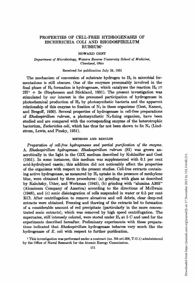
B. Escherichia coli hydrogenase. Escherichia coli, strain B, was grown for8 to 24 hours in deep stationary culture at 37 C in the following medium: Difcopeptone, 5 g; Difco beef extract, 3 g; glucose, 10 g; NaCl, 5 g; distilled water,1 liter; pH adjusted to 7.5.Acetone powders (prepared in the usual manner) of the cells contained active
hydrogenase which could be extracted as indicated in table 1. From the resultsgiven, it appears that alumina grinding is the most effective means of extractingthe enzyme.Although acetone powders (and lyophilized cells) offer advantages as source
material for preparation of the enzyme, our experience indicates that the useof fresh cells is preferable; the crude extracts prepared from fresh cells usuallyhave considerably higher specific activity. The highest specific activity prepara-
TABLE 1Extraction of Escherichia coli hydrogenase from acetone powder
EXTRACTING AGENT MG PROTEIN/ML OF ct
A. Water.................................. 0.88 78B. Phosphate buffer (0.05 M; pH 7) ............ 2.29 63C. KCl (1 per cent)......................... 0.64 186D. Alumina grinding ............. 1.91 291
In A, B, and C, 50 mg of acetone powder were suspended in 5 ml of extracting agentfor 30 minutes at 30 C. In D, 100 mg of powder plus 0.9 ml of water were ground withalumina and the paste extracted with 10 ml of 0.05 M phosphate pH 7. In all instances,insoluble debris, etc., was removed by centrifugation at 20,000 Xg for 20 minutes.
* The proteins were precipitated with trichloroacetic acid and the precipitate analyzedfor protein content by the procedure described by Sutherland et al. (1949) using crystallineserum albumin as the standard.
t Q'c = ,ul H2/hr/mg protein. The rates given are based on the uptake observed duringthe first 20 minutes, using 0.7 ml of extract + 0.3 ml 0.5 M phosphate buffer pH 6.7 + 0.2ml neutralized methylene blue (8 um) in 10 ml Warburg vessels.
tion obtained thus far was made by the following procedure: 12 g (wet weight)of 8-hr old cells were washed once with 25 ml of water and then ground by handwith 30 g of "alumina A303" in a large mortar. The paste was extracted with60 ml of water for 20 min and alumina, etc., removed by centrifugation at highspeed. A considerable amount of nucleoprotein was precipitated from the browncolored extract by addition of one-twentieth volume of 1 M MnCl2. After re-moval of the precipitate, the hydrogenase was precipitated by 50 per centsaturation with (NH4)2S04. This protein fraction was dissolved in water andcentrifuged to remove residual insoluble particles. For most of the experimentsdescribed later, preparations at this stage of purification were used; in the bestcases, the specific activity at this point was approximately 30,000 JAI H2 per hrper mg protein N at 37 C with methylene blue as the acceptor.
It has been observed that further purification can be achieved by a subsequentadsorption step using reagent grade MnO2 (Merck) as follows: the solution is
112 [VOL. 63
Dow
nloa
ded
from
http
s://j
ourn
als.
asm
.org
/jour
nal/j
b on
17
Nov
embe
r 20
21 b
y 13
3.11
4.68
.211
.
PROPERTIES OF CELL-FREE HYDROGENASES
adjusted to 0.05 M phosphate pH 5.5 to 5.9 and solid MnO2 added (MnO2/
protein _ 20140). After 10 minutes, the MnO2 is centrifuged out, washed with
water, and hydrogenase eluted from the adsorbent with 0.2 M phosphate bufferpH 7.3. Proper application of this adsorption step (i.e., fractional adsorptionand elution) should facilitate further purification of the enzyme.
In the procedures previously outlined, all steps are conducted in the cold asrapidly as possible because of the lability of the enzyme with respect to oxida-tion. When storage periods greater than 2 or 3 hours were necessary, the prepara-tions were kept under H2 at 5 C.
Hydrogenase assay. Enzyme activity in the cell-free preparations was de-termined by measuring H2 consumption at 30 C or 37 C with methylene blueas the acceptor. Methylene blue chloride National Aniline was used; the dyesolution was neutralized to a pH between 6 and 7. It should be noted that atconcentrations required for adequate manometric assay, methylene blue maycause protein precipitation, particularly in crude extracts.Hydrogenase shows maximal activity over a rather broad pH range (Joklik,
1950a); in the present experiments, 0.125 M (final conc) phosphate buffer of pH6.7 was generally used. Phosphate is not required for activity of the enzyme,and the concentration of this buffer does not affect the rate of H2 uptake ap-preciably (0.06 to 0.25 M; Rhodospirillum hydrogenase). It is of interest thatH2 uptake has not been observed in the absence of methylene blue, even incrude extracts.Using crude preparations, with relatively high protein content, the rate of
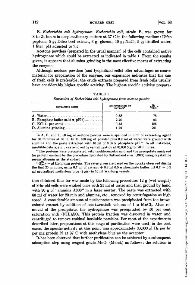
H2 consumption is ordinarily observed to be linear until most of the methyleneblue is reduced to the leucoform. More purified preparations, on the other hand,may show marked instability during the activity assay. This point is illustratedin figure 1 for a case (Escherichia hydrogenase) in which the protein concentra-tion was approximately 33 ,g per ml in the reaction mixture. In phosphatebuffer, the activity falls off rapidly. Better assay conditions are obtained whenversene buffer is used in place of phosphate. Addition of crystalline serum al-bumin to phosphate buffer is most effective as indicated by curve A. The partialprotection by versene buffer suggests that the progressive inactivation in phos-phate is partly due to metal ions (possibly introduced as impurities in the methyl-ene blue).
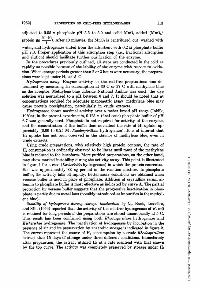
Stability of hydrogenase during storage: inactivation by 02. Back, Lascelles,and Still (1946) reported that the activity of the cell-free hydrogenase of E. coliis retained for long periods if the preparations are stored anaerobically at 5 C.This result has been confirmed using both Rhodospirillum hydrogenase andEscherichia hydrogenase. The inactivation of hydrogenase by incubation in thepresence of air and its preservation by anaerobic storage is indicated in figure 2.The curves represent the course of H2 consumption by a crude Rhodospirillumextract after 15 days of storage under three different conditions. Immediatelyafter preparation, the extract utilized H2 at a rate identical with that shownby the top curve. The activity was completely preserved by storage under H2
19521 113
Dow
nloa
ded
from
http
s://j
ourn
als.
asm
.org
/jour
nal/j
b on
17
Nov
embe
r 20
21 b
y 13
3.11
4.68
.211
.
HOWARD GEST
A
-C
A- 0.125 M PHOSPHATEBUFFER + 1.5mg AL-BUMIN/.I
B- 0.125 M VERSENEBUFFER
C- 0.125M PHOSPHATEBUFFER
TIME, MINUTES100
Figure 1. Stability of E8cherichia coli hydrogenase during activity assay under threedifferent conditions. At zero time, 0.2 ml of methylene blue (approximately 8 pM) wa tippedinto 1.0 ml of buffer-enzyme mixture containing 40 pg of enzyme protein. The pH in A andC was 6.65; in B, 6.3. Temperature, 37 C.
l lI I I
500__
4,00 - 5tored under H2 at 5 C.
E300 -
200 -
Storedc froze
Stored in air at 5 C
20 40 60 80 100 120 140 160Time in minutes
Figure 2. Activity of cell-free Rhodospirillum rubrum hydrogenase after storage underthree different conditions. The curves represent the hydrogenase activity (at 37 C and pH7) observed on the 15th day of storage under the conditions noted.
200
C 160w
I-0
o 120'
w
= 80z00
' 40- /
20
114 [VOL. 63
Dow
nloa
ded
from
http
s://j
ourn
als.
asm
.org
/jour
nal/j
b on
17
Nov
embe
r 20
21 b
y 13
3.11
4.68
.211
.
PROPERTIES OF CELL-FREE HYDROGENASES
at 5 C. As is evident from the lower curves, considerable activity is lost if thepreparation is maintained at 5 C in contact with air or in the frozen state. Inview of the stability of the enzyme under H2 at 5 C, all preparations of Rhodo-spirillum hydrogenase and Escherichia hydrogenase were stored in this manner.
It has been recently suggested by Joklik (1950a) that the inactivation ofEscherichia hydrogenase by oxygen is due to oxidation of essential sulfhydrylgroups. This view was based on the observations that the enzyme was inhibited
200
(f)160xla
0) 120
2 80mCl)z00
A-ENZYME STOREDUDERH2
B4ERATESENZYMEC-AERATEd' ENZYME
PREINCUBATEDWITH 0.I MCYSTEINE
20 40TIME, MINUTES
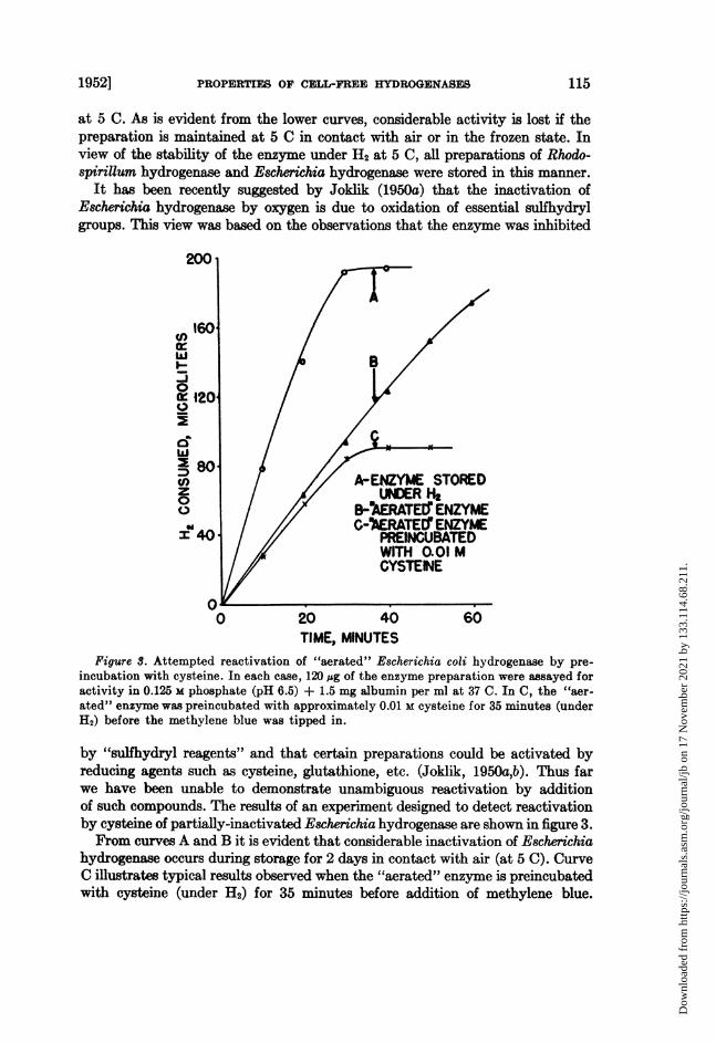
Figure S. Attempted reactivation of "aerated" Escherichia coli hydrogenase by pre-incubation with cysteine. In each case, 120 sg of the enzyme preparation were assayed foractivity in 0.125 M phosphate (pH 6.5) + 1.5 mg albumin per ml at 37 C. In C, the "aer-ated" enzyme was preincubated with approximately 0.01 M cysteine for 35 minutes (underH2) before the methylene blue was tipped in.
by "sulfhydryl reagents" and that certain preparations could be activated byreducing agents such as cysteine, glutathione, etc. (Joklik, 1950a,b). Thus farwe have been unable to demonstrate unambiguous reactivation by additionof such compounds. The results of an experiment designed to detect reactivationby cysteine of partially-inactivated Escherichia hydrogenase are shown in figure 3.From curves A and B it is evident that considerable inactivation of Escherichia
hydrogenase occurs during storage for 2 days in contact with air (at 5 C). CurveC illustrates typical results observed when the "aerated" enzyme is preincubatedwith cysteine (under H2) for 35 minutes before addition of methylene blue.
1952] 115
Dow
nloa
ded
from
http
s://j
ourn
als.
asm
.org
/jour
nal/j
b on
17
Nov
embe
r 20
21 b
y 13
3.11
4.68
.211
.
HOWARD GEST
There is no acceleration of H2 uptake, and it is to be noted that the total amountof H2 consumed is markedly smaller than that observed in the absence of cysteine.Addition of cysteine after methylene blue has been added to the reaction mixturedoes not cause reactivation, and the total H2 uptake is again found to be de-creased as compared with the controls. Diminution of the total H2 consumptionis also observed when other reducing agents such as glutathione, ascorbic acid,FeSO4, or sodium hydrosulfite are added (also found with RhodospiriUum hydro-genase). In the presence of reducing substances, the cessation of H2 uptake iscoincident with decolorization of the methylene blue which indicates that poison-ing of the enzyme is not the cause of the decreased total uptake. The diminishedtotal uptake observed in these instances is undoubtedly due to nonenzymaticreduction of part of the methylene blue by the added reducing agents.The failure to obtain a clearcut reactivation of "aerated" hydrogenase by
cysteine, etc. suggests that the effect of 02 cannot be attributed solely to oxida-tion of essential sulfhydryl groups on the enzyme. In this connection it is ofinterest to note that 10-3 M p-chloromercuribenzoate, which is considered to beone of the most specific inhibitors of -SH enzymes, does not appreciably in-hibit Rhodospirillum hydrogenase in crude sonic extracts or purified Escherichiahydrogenase.
Partial reactivation of "aerated" Rhodospirillum hydrogenase has been ob-tained by storing preparations under H2 at 5 C for several days. A similar re-activation of Escherichia hydrogenase, presumably oxidized during preparationof the enzyme, was reported by Back et al. (1946).
Attempts to demonstrate a cofactor in hydrogenase activity. The observationsdescribed hereafter indicate that an easily dissociable cofactor is not involvedin hydrogenase activity (with methylene blue as acceptor):
(1) Anaerobic dialysis of Rhodospirillum hydrogenase against oxygen-freewater (saturated with H2) for periods as long as 18 hours does not cause ap-preciable loss of activity. Preparations dialyzed for this length of time fre-quently show an unexplained slight lag in H2 uptake; this lag is usually of theorder of 10 minutes. Addition of boiled juices or triphosphopyridine nucleotideto extracts dialyzed for 5 hours did not accelerate H2 consumption.
(2) Attempts to resolve Escherichia hydrogenase by precipitation of the apo-enzyme with HCl-(NH4)2SO4 according to the procedure of Warburg andChristian (1938) were not successful. Some inactivation occurred as a result ofthis treatment, and it could not be reversed by addition of supplements suchas boiled juices and yeast extract.
(3) Exposure of Escherichia hydrogenase to the action of trypsin or alkalinephosphatase (which have been reported to be effective for resolution of certainother enzymes) under appropriate conditions did not cause any diminutionin hydrogenase activity as compared with controls incubated in the absence ofthese enzymes.
(4) Metal complexing agents such as 1,10 phenanthroline, a, a'-dipyridyl,and diethyldithiocarbamate in concentrations up to 0.01 M do not inhibit Escheri-chia hydrogenase or Rhodospirillum hydrogenase appreciably even after pro-
16 [VOL. 63
Dow
nloa
ded
from
http
s://j
ourn
als.
asm
.org
/jour
nal/j
b on
17
Nov
embe
r 20
21 b
y 13
3.11
4.68
.211
.
PROPERTIES OF CELL-FREE HYDROGENASES
longed contact. This indicates that heavy metal cations do not participate inhydrogenase activity.
(5) Attempts to demonstrate reduction of diphosphopyridine nucleotide byEscherichia hydrogenase in the presence of H2 in the Beckman spectrophotom-eter have given negative results. Diphosphopyridine nucleotide in substratequantities did not serve as an acceptor for H2 in the presence of Rhodospirillumhydrogenase; similar results for Escherichia hydrogenase were reported byJoklik (1950a). We have also attempted to couple hydrogenase with (a) thediphosphopyridine nucleotide enzyme, formic dehydrogenase (prepared frompeas)-no H2 was evolved from reaction mixtures containing formate, diphospho-pyridine nucleotide, and the two enzymes under an atmosphere of helium, andwith (b) the triphosphopyridine nucleotide Zwischenferment enzyme system-with similar results.H2 utilization with acceptors other than methylene blue. It was previously reported
(Gest, 1950, 1951) that intact cells of R. rubrum rapidly oxidize H2 in the darkwith K3Fe(CN)6 as the acceptor. This reaction can be readily demonstrated bysuspending cells in a solution of 0.2 M phosphate pH 6.8 which is 0.1 M withrespect to KgFe(CN)6; KOH must be placed in the center well to absorb therelatively large amount of CO2 produced from endogenous substrates whenK3Fe(CN). is present. The ability of the latter compound to act as an acceptorappears to explain satisfactorily the observation that photoevolution of H2from malate or pyruvate does not occur when KsFe(CN)6 is added (Gest, 1950).2
Intact cells of E. coli also can use K3Fe(CN)6 as an acceptor for oxidation ofH2. In contrast to the intact cells, cell-free Escherichia hydrogenase and Rhodo-spirillum hydrogenase have not been found to consistently oxidize H2 in thepresence of KsFe(CN)6. A slight H2 uptake is usually noted just after K3Fe(CN)6is added, but the total amount consumed is far below the theoretically expectedquantity. When present at a concentration of 0.01 M, KaFe(CN)s inhibits theconsumption of H2 with methylene blue (0.001 M K3Fe(CN)o may cause a slightlag but does not affect the subsequent rate as compared with the controls).If reducing agents such as cysteine (0.02 M) are added to the inhibited system,immediate consumption of 12 is observed, but the total quantity utilized isagain well below the expected amount. Interpretation of these inhibition ex-periments is dfficult because of the occurrence of nonenzymatic interaction be-tween KgFe(CN)6 and methylene blue; nonenzymatic reduction of ferricyanideto ferrocyanide by the added reducing agent can account for the "reactivation"observed. In view of the sensitivity of Escherichia hydrogenase and Rhodo-spirillum hydrogenase in the cell-free state to oxidizing agents, one would expectthat KXFe(CN)0 could not be a satisfactory acceptor. Cell-free Azotobacter hydro-genase appears to differ from Escherichia hydrogenase (and Rhodospirillumhydrogenase) in that K3Fe(CN)6 can act as an acceptor for the former enzyme
2 We have found that intense illumination has no effect on hydrogenase activity of intactcells or extracts of R. rubrum using KsFe(CN). or methylene blue as the respective accep-tors; this result supports the notion that the activation of H2 as a hydrogen donor in photo-synthetic reactions and photoevolution of H2 is not directly light-dependent.
19521 117
Dow
nloa
ded
from
http
s://j
ourn
als.
asm
.org
/jour
nal/j
b on
17
Nov
embe
r 20
21 b
y 13
3.11
4.68
.211
.
8HOWARD GEST
(Hyndman and Wilson, 1951). The explanation for this difference is not apparentat the present time.The ability of various other substances to act as acceptors for Rhodospirifum
hydrogenase has been tested. Negative results were observed with fumarate,pyruvate, diphosphopyridine nucleotide (see before), SO-, NO2, and riboflavin.Intact cells of Rhodospirillum also do not reduce fumarate, malate, or diphospho-pyridine nucleotide with H2. Joklik (1950a) has reported that partially purifiedEscherichia hydrogenase reduces only dyes oh-he type of methylene blue.
Spectroscopic observations. Concentrated "solutions" of the 50 per cent (NH4)2SO4 fraction from E. coli have been visually examined for absorption bandscharacteristic of porphyrin proteins with a Hilger angular spectroscope. A weakband can be observed in such preparations at approximately 560 m,u after in-cubation with H2 or reduction by sodium hydrosulfite. Incubation under heliumdoes not cause appearance of the band. The band decreases in intensity in thepresence of air and can be restored again by incubation with H2. The same phe-nomena can also be demonstrated in thick suspensions of intact cells. Althoughit is possible that the prosthetic group of hydrogenase itself is a porphyrin, theseobservations can also be interpreted on the basis that other known porphyrinenzymes may act as carriers in the utilization of H2.
DISCUSSION
Inhibitor studies with 02, CO, and KCN using intact bacteria have led tothe suggestion that hydrogenase is an iron porphyrin protein which is activeonly in the reduced state. The results reported with CO and KCN are stillsomewhat contradictory. The "Knailgas" reaction, H2 + 'O2 -+ H20, in Azoto-bacter (Wilson and Wilson, 1943) and the reduction of methylene blue by E. coli(Lascelles and Still, 1946a) are inhibited by CO, but the inhibitions could notbe reversed by light. Catalysis of the exchange reaction between water and H2by the hydrogenase of Proteus vulgaris is also inhibited by CO, and in this in-stance a partial reversal by light has been reported, thus indicating participationof an iron porphyrin enzyme (Hoberman and Rittenberg, 1943). One of theenzymes involved in H2 evolution from sugars by Clostridium butyricum, pre-sumably hydrogenase, is sensitive to CO, and the inhibition has been shown tobe reversed by high light intensities; the "absorption spectrum" of the hydrogen-evolving enzyme in intact cells as determined by photochemical means.however, suggests an iron enzyme which does not appear to be a typical ironporphyrin (Kempner and Kubowitz, 1933). Experiments with the cell-free hy-drogenase of E. coli by Joklik (1950b) showed that the reduction of methyleneblue in the partially purified system is also CO sensitive, but attempts toreverse the inhibition by high light intensities were unsuccessful.
Similar ambiguities have been found with regard to cyanide inhibition. The"Knallgas" reaction and the reduction of methylene blue and certain other ac-ceptors in various intact organisms are markedly inhibited by cyanide (Wilsonand Wilson, 1943; Lascelles and Still, 1946a,b). In contrast to these cases the"Knallgas" reaction in Lactobacillus delbrueckii, which apparently metabolizes
118 [VOL. 63
Dow
nloa
ded
from
http
s://j
ourn
als.
asm
.org
/jour
nal/j
b on
17
Nov
embe
r 20
21 b
y 13
3.11
4.68
.211
.
PROPERTIES OF CELL-FREE HYDROGENASES
mainly by means of flavin enzymes, is not sensitive to cyanide (Yamagata andNakamura, 1938). Recent studies with the cell-free hydrogenase of E. coli haveshown that the reduction of methylene blue in this system is also not markedlysuppressed by cyanide (Joklik, 1950b).Many of the foregoing observations can be adequately rationalized if it is
assumed that the oxidation of H2 by bacteria involves a complex system of thetype proposed by Yamagata and Nakamura (1938), viz.
H2 -I I Intermediary carriers - AcceptorT T
Hydrogenase Specific(+ other enzymes?) "acceptor" enzymes
Thus depending on the nature of the acceptor, and consequently on the specificenzymes concerned with the acceptor, the utilization of H2 in a particular casemay or may not be inhibited by agents such as CO and KCN. Inhibition wouldbe expected when the "acceptor-enzymes" are typical iron porphyrins; forexample, in E. coli when the acceptor is 02 or nitrate.3 The participation of por-phyrin carriers in certain types of hydrogenase activity in E. coli therefore ap-pears likely. This view is supported to some extent by the spectroscopic ob-servations previously described and by the dramatic effect of iron nutrition onthe level of hydrogenase activity in intact cells (Waring and Werkman, 1944).The fact that hydrogenase activity in intact cells is inhibited by incubation
with oxygen and can be reactivated by reducing agents has also been used insupport of the hypothesis that the enzyme itself possesses an iron porphyrinprosthetic group. It is evident that these effects may well be indirect; in orderto determine the nature of the hydrogenase prosthetic group conclusively,further purification of the enzyme is required. The suggestion that oxygen in-hibition is due to oxidation of essential sulflhydryl groups (Joklik, 1950a) of theenzyme is not supported by the present studies. In this connection, it shouldbe noted that Joklik found "reactivation" of the cell-free enzyme by reducingagents to be greater under H2 than under N2, and he concludes that "it has notyet been definitely determined whether H2 is necessary for the action of -SHreagents or not."
In considering the apparent contradictions between effects of inhibitors onintact cells and cell-free systems, it is evident that corresponding results wouldnot necessarily be expected. Observation of differences in this respect obviouslydoes not justify the conclusion that the inherent properties of hydrogenase aresignificantly altered by extraction and purification procedures as has been sug-gested by Joklik (1950b).The present results indicate that the cell-free hydrogenases (defined on the
basis of H2 utilization with methylene blue) of E. coli and R. rubrum are verysimilar in all respects. Both of these organisms, as grown in the present instance,produce H2 as a major metabolic product. There is little question that hydro-
' The nitrate reductase of E. coli appears to be an iron porphyrin enzyme of the cyto-chrome b type (Sato and Egami, 1949).
19521 119
Dow
nloa
ded
from
http
s://j
ourn
als.
asm
.org
/jour
nal/j
b on
17
Nov
embe
r 20
21 b
y 13
3.11
4.68
.211
.
HOWARD GEST
genase is essential in the enzyme complex responsible for H2 formation in theseorganisms. Thus far there is no information available on the nature of the carriersinvolved, and it is conceivable that they may be different from those participat-ing in reduction of various terminal acceptors by H2. Although direct or indirectreduction of known dissociable coenzymes as a result of H2 oxidation has notyet been demonstrated, it seems reasonable to presume that such reactions occursince numerous organisms are capable of fulfilling all their necessary metabolicrequirements with H2 as the primary hydrogen donor.
ACKNOWLEDGMENTS
The author wishes to express his appreciation to Drs. L. 0. Krampitz, J. 0Lampen, and S. J. Cooperstein for their interest and advice during this investiga-tion. The technical assistance of Mrs. M. A. Lasoski and Mrs. S. J. Corn isgratefully acknowledged.
SUMMARY
Cell-free hydrogenases have been prepared from Escherichia coli and Rhodo-spirillum rubrum by a variety of methods and the enzyme partially purified.A survey of properties indicates that the enzymes from both sources are es-sentially similar. Attempts to demonstrate participation of a dissociable cofactorin the reaction: H2 + methylene blue -* leucomethylene blue, have given nega-tive results. Evidence suggesting participation of iron porphyrin enzymes inhydrogenase activity has been obtained by visual spectroscopic observationson concentrated solutions of the enzyme from E. coli and on intact cells; theseobservations have disclosed an absorption band (at about 560 mu) which isaccentuated by incubation with H2 and discharged by aeration. Some of thefactors involved in assay of hydrogenase and in the lability of the enzyme withrespect to oxygen have been studied; oxidation of essential sulfhydryl groupson the enzyme does not appear to explain the inactivation by air. The presentresults are discussed in relation to the mechanisms of various metabolic re-actions involving H2.
REFERENCESBACK, K. J. C., LASCELLES, J., AND STILL, J. L. 1946 Hydrogenase. Australian J. Sci.
Research, 9, 25.GEST, H. 1950 Anaerobic oxidation of malate and hydrogen in the dark by Rhodospiril-
lum rubrum. Bact. Proc., 1960, 136-137.GEST, H. 1951 Enzymatic oxidation of molecular hydrogen by bacterial extracts. Fed-
eration Proc., 10, 188.GEST, H., KAMEN, M. D., AND BREGOFF, H. M. 1950 Studies on the metabolism of photo-
synthetic bacteria. V. Photoproduction of hydrogen and nitrogen fixation by Rhodo-spirillum rubrum. J. Biol. Chem., 182, 153-170.
HOBERMAN, H. D., AND RITTENBERG, D. 1943 Biological catalysis of the exchange reac-tion between water and hydrogen. J. Biol. Chem., 147, 211-227.
HYNDMAN, L. A., AND WILSON, P. W. 1951 Studies on hydrogenase of Azotobacter vine-landii. Bact. Proc. 1961, 126.
JOKLIK, W. K. 1950a The hydrogenase of E. coli in the cell-free state. I. Concentration,properties and activation. Australian J. Exptl. Biol. Med. Sci., 28, 321-329.
120 [voL. 63
Dow
nloa
ded
from
http
s://j
ourn
als.
asm
.org
/jour
nal/j
b on
17
Nov
embe
r 20
21 b
y 13
3.11
4.68
.211
.
PROPERTIES OF CELL-FREE HYDROGENASES
JORLIK, W. K. 1950b The hydrogenase of E. coli in the cell-free state. II. The effectof certain inhibitors on hydrogenase. Australian J. Exptl. Biol. Med. Sci., 28, 331-338.
KALNITSKY, G., UTTER, M. F., AND WERKMAN, C. H. 1945 Active enzyme preparationsfrom bacteria. J. Bact., 49, 595-602.
KEMPNER, W., AND KUBOWITZ, F. 1933 Wirkung des Lichtes auf die Kohlenoxydhem-mung der Buttersaure garung. Biochem. Z., 265, 245-252.
KOHLMILLER, E. F., AND GEST, H. 1951 A comparative study of the light and darkfermentations of organic acids by Rhodospirillum rubrum. J. Bact., 61, 269-282.
LASCELLES, J., AND STILL, J. L. 1946a Utilization of molecular hydrogen by bacteria.Australian J. Exptl. Biol. Med. Sci., 24, 37-48.
LASCELLES, J., AND STILL, J. L. 1946b The reduction of nitrate, nitrite and hydroxyl-amine by E. coli. Australian J. Exptl. Biol. Med. Sci., 24, 159-167.
LINDSTROM, E. S., LEwIS, S. M., AND PINS'KY, M. J. 1951 Nitrogen fixation and hydro-genase in various bacterial species. J. Bact., 61, 481-487.
McILWAIN, H. 1948 Preparation of cell-free bacterial extracts with powdered alumina.J. Gen. Microbiol., 2, 288-291.
SATO, R., AND EGAMI, F. 1949 Studies on nitrate reductase III. Bull. Chem. Soc. Japan,22, 137-143.
STEPHENSON, M., AND STICKLAND, L. H. 1931 Hydrogenase: a bacterial enzyme activat-ing molecular hydrogen. I. The properties of the enzyme. Biochem. J., 25, 205-214.
SUTHERLAND, E. W., CORI, C. F., HAYNES, R., AND OLSEN, N. S 1949 Purification ofthe hyperglycemic-glycogenolytic factor from insulin and from gastric mucosa. J.Biol. Chem., 180, 825-837.
WARBURG, O., AND CHRXSTIAN, W. 1938 Isolierung der prosthetischen gruppe der d-Aminosbiureoxydase. Biochem. Z., 298, 150-168.
WARING, W. S., AND WERKMAN, C. H. 1944 Iron deficiency in bacterial metabolism.Arch. Biochem., 4, 75-87.
WILSON, J. B., AND WILSON, P. W. 1943 Action of inhibitors on hydrogenase in Azoto-bacter. J. Gen. Physiol., 26, 277-286.
YAMAGATA, S., AND NAKAMURA, H. 1938 tlber die Hydrogenase, nebst einer Bemerkunguber den Mechanismus der bakteriellen Knallgasreaktion. Acta Phytochim., 10,297-311.
1952] 121
Dow
nloa
ded
from
http
s://j
ourn
als.
asm
.org
/jour
nal/j
b on
17
Nov
embe
r 20
21 b
y 13
3.11
4.68
.211
.


![How oxygen attacks [FeFe] hydrogenases from photosynthetic ... · How oxygen attacks [FeFe] hydrogenases from photosynthetic organisms Sven T. Strippa, Gabrielle Goldetb, Caterina](https://img.pdfslide.us/doc/110x75/603ba0c1ea10106e07149762/how-oxygen-attacks-fefe-hydrogenases-from-photosynthetic-how-oxygen-attacks.jpg)

![Supplementary Information Discovery of Novel [FeFe ... · Supplementary Information Discovery of Novel [FeFe]-Hydrogenases for Biocatalytic H2- Production Henrik Land,a Pierre Ceccaldi,a](https://img.pdfslide.us/doc/110x75/600f4cccf0018f3e2604f859/supplementary-information-discovery-of-novel-fefe-supplementary-information.jpg)


![RESEARCH ARTICLE Open Access Zymographic …RESEARCH ARTICLE Open Access Zymographic differentiation of [NiFe]-Hydrogenases 1, 2 and 3 of Escherichia coli K-12 Constanze Pinske1, Monique](https://img.pdfslide.us/doc/110x75/60fc2ba80443e671fd673977/research-article-open-access-zymographic-research-article-open-access-zymographic.jpg)


![Zymographic differentiation of [NiFe]-Hydrogenases 1, 2 and 3 of](https://img.pdfslide.us/doc/110x75/61fb0e812e268c58cd59a4f6/zymographic-differentiation-of-nife-hydrogenases-1-2-and-3-of.jpg)