Embed Size (px)
Citation preview

TThhee FFiifftteeeenntthh AAnnnnuuaall MMeeeettiinngg
TThhee EEggyyppttiiaann SSoocciieettyy ooff NNuucclleeaarr MMeeddiicciinnee SSppeecciiaalliissttss
Hurghada
Egypt
4 – 6 March, 2015

General Information Organizing Committee President: Consultant..Dr. Adel Khidr Head of the Society Prof. Dr. Hosna Moustafa Secretary of the Congress: Consultant. Dr. Khalid Taalab Program Directors: Prof. Gaber Ziada Prof. Walid Omar Board of Directors ESNMS
President: Prof. Dr. Hosna Moustafa (President) Members: Consultant. Dr. Adel Khidr (Vice President) Prof. Dr. Walid Omar (General Secretary) Consultant. Dr. Khaled Taalab Prof. Dr. Abdel-Hamied El-Gazzar Eng. Mohamed Abdou (Treasurer) Lecturer. Mai Amr
Invited Speakers:
Dr. Al- Rowaily, M. (Saudi Arabia) Mr / Claude Lalou (France) Prof. Dr. Osman, M. (U.S.A) Ass. Prof. Rabiea, H. (Saudi Arabia) Prof.Dr.Taalab, Kh. (Egypt) Prof. Dr. Ziada, G. (Kwait)

Local Organizing Committee: Prof. Dr. Moustafa, H. Consultant. Taalab, Kh. Prof. Dr. Omar, W. Eng. Abdou, M. Ass. Lecturer. Amr, M. Phy. El-Magrapy, Sh. Congress Location: Desert Rose – Hurghada, Egypt Congress Language: Official language of the congress is English. No simultaneous translation will be provided. Projection: Computer projection is available and Computer data should be handed over to the congress office one-hour before the session.
Climate: The weather during March in Hurghada region is generally sunny by day and cool by night.
Visas: Citizens of most countries require entry visa for Egypt.The Egyptian Embassy and/or Consulate in your country can inform you if a visa is necessary. Travel to Hurghada: Hurghada could be easily reached by plane, as well as cars or buses.

Cancellations: Only written cancellations will be accepted. - Up to 5 st February 2015, all payments for registration fees are refunded with less 10% for accommodation service charge. - Up to 20th February 2015, no refund for registration fees and all other payments are refunded less 25% for accommodation cancellation charges. - No refund will be made after 26th February 2015.
Awards of Society:
(1) Professor/ Abdel-Razzak award
For young doctors in Nuclear Medicine field less than 35 years.
They should have oral presentation during the Annual Meeting 2015.
The Doctor will receive a certificate and 1500 L.E.
(2) Professor/ Abdel-Dayem award
For doctors less than 50 years during the Annual Meeting 2015.
They should submit (3-5) international and national articles in Nuclear
Medicine field published in the last 3 Years before 1/2/2015; for
evaluation (3 Copies).
The Doctor will receive a certificate and 3000 L.E.

Important Guidelines For chair persons: - Please be in your session place at least 10 minutes before its start. - Speakers should strictly observe timing of presentation and discussion. - Discussants should clearly state their name. - Participants should not speak without permission. For speakers: - Turn in your data one hour prior to the start of the session. - Collect your data from the preview room immediately after the session. - You should be in session room at least 10 minutes before its onset. - Time allowed for presentation is 10 minutes. - Follow chair persons instructions. - Discussion is strictly at time indicated. Computer Center: - Located in a room outside the congress hall. NO SMOKING IN MEETING ROOM
Congress office: Prior to and after the meeting Consultant Dr. Adel, Khidr.
Cellular : 002 01222282131 E-mail: [email protected]. During the meeting Desert Rose – Hurghada, Egypt

Social program: Wednesday 04/03 16:00 - 17:00 Registration.
Friday 06/03 14:45 - 15:15 Closing ceremony
Excursions: web site: www.esnms.net. Mail box: [email protected]. Mail of Secretary: [email protected]. * Abstract Book is included in congress bag.
* Eight issue of ESNMS Magazine will be available for members of society. * A copy of preview meetings CD is included in congress bag. * Certificate of attendance is included in congress bag.

Wednesday MMaarrcchh 44
First Day Wednesday 4 Th March 2015
Registration 16:00 – 17:00

Presidential address 17:00 – 17:15 Dear colleagues and members of nuclear medicine society
Dear colleagues, guests and family members! It is truly an honor and a privilege for me to speak at the opening of the 15th. Conference of the ESNMS; this meeting is a culmination of efforts of many people who worked for almost one year to bring us together in very hard conditions of our homeland.
The Purpose of the Conference, Is awareness by the recent updates in Nuclear Medicine; in diagnosis and therapy.
First and foremost, I would like to take this opportunity to thank everyone who accepted our invitation to come to Egypt and share us our Congress;
We are delighted that: Prof. Osman; Prof. Ziada; Prof. Omar; Prof. Taalab; Ass.Prof. Farghaly; Dr. Abu-Zaid; Dr. Al Rowaily; and Dr. Houseni sharing with us this Conference.
We are thankful and board members and Organize committee for helping in preparation of this meeting
We are greatly honored for all members to be with us in this event.
I would like to cordially thank our sponsors from Gamma Trade; Ghalioungie ; General Electric; IEC; Siemens; Maggie Medical; Gulf ; and Emerald companies; for without their support this Conference would not have been possible.
It is great that all of us got here today in one piece that makes us happy.
Head of congress
Prof .Dr. Adel Khidr

Session I 17:15 – 19:00 Read with the expert / Advances In Oncologic
FDG application & Free papers Chair Persons: Consultant, Dr. Khidr, A. (Egypt)
Prof, Dr. Ziada, G. (Kwait) 1 17:15 – 18:00 Read and Explain clinical physics with the expert
Prof. Dr . Ziada, G. (Kuwait)
2 18:00 – 18:30 Advances In Oncologic PET/CT IMAGING Prof. Dr. Rabiea, H. (Saudi Arabia)
Free Papers 18:30 - 18:40 S/I -1
F-18 FDG PET-CT versus RAI-131 MIBG in pediatric Neuroblastoma
patient’s Comparative study
Amr, M. Omar, W. Kotb, M and Moustafa, H.
Nuclear Medicine departments in NCI and NEMROCK Center, Cairo University, Egypt
18:40 - 18:50 S/I -2 Estimation of Staff to Patient Absorbed Dose Ratios for Different
Radiopharmaceuticals in Nuclear Medicine in Egypt
Samy, S. Guirguis, O. and Saad, I.
Oncology and Nuclear Medicine Department in Kasr Al-Ainy & Department of Biophysics,Cairo
University, Egypt.
18:50 - 19:00 Discussion

Thursday MMaarrcchh 55
Targeted Radionuclides Therapy / Radiation Protection & Free papers

Second Day Thursday 5 th March 2015 Session II 10:00 – 12:45
Targeted Radionuclides Therapies Radiation Protection & Free papers
Chair Persons: Prof. Dr. Omar, W. (Egypt) Dr. Abu-zeid, M. (Saudi Arabia) 3 10:00- 10:30 General Electric Presentation
Discovery IQ, a new generation of PET/CT Mr / Claude Lalou (France)
4 10:30 – 11:00 New Generation Targeted Radionuclides Therapies.
Dr. Al-Rowaily, M. (Saudi Arabia)
5 11:00- 11:45 Radiation dose to and from patients in Nuclear Medicine: challenges and solutions
Prof.Dr. Osman, M. (USA)
Free Papers
11:45- 11:55 S/II -3
Correlation Between Different 18F FDG PET/CT Quantitative Parameters
And Response To Therapy In Patients with Non-small Cell Lung Cancer Nagui, H. Gaber, Y. Abdel-meguid, R. Osaama, A, El-refaei, Sh.
Oncology and Nuclear Medicine Department, Radiology Departments, Cairo University, Egypt.
11:55- 12:05 S/II -4
Prognostic Value Of Different F-18 FDG PET/CT Quantitative Analytical
Methodologies In Pediatric Hodgkin’s Lymphoma Serry, O . Kandeel, A. El-Sayed, A. Omar, W.
Oncology and Nuclear Medicine Department in Kasr Al-Ainy, National Cancer Institute, Egypt.
12:05 - 12:15 Discussion
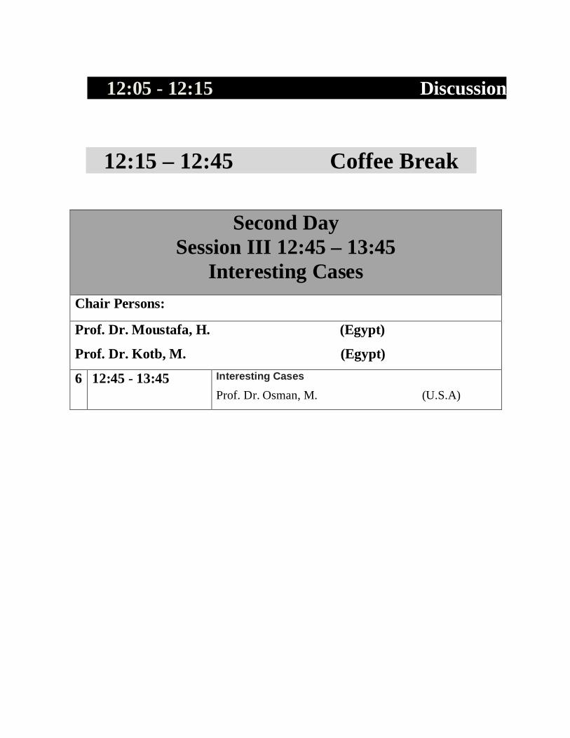
12:15 – 12:45 Coffee Break
Second Day Session III 12:45 – 13:45
Interesting Cases Chair Persons:
Prof. Dr. Moustafa, H. (Egypt)
Prof. Dr. Kotb, M. (Egypt)
6 12:45 - 13:45 Interesting Cases
Prof. Dr. Osman, M. (U.S.A)

Friday MMaarrcchh 66
Read with Expert / New Pharmaceuticals Molecular Imaging& Free Papers

Third Day Friday 6 th March 2015
Session IV 10:00 – 11:45
68Ga generator / SPECT/CT application& Free Papers
Chair Persons: Consultant. Dr. Taalab, Kh. (Egypt) Prof.Dr. Osman, M (U.S.A) 7 10:00- 11:00 Overview on 68Ga generator, chemistry, labelling and
challenges
Dr. Al-Rowaily, M. (Saudi Arabia)
11:00- 11:10 S/ IV -5 The value of Tc-99m Sesta MIBI washout rate in detection of ischemia compared
with standard myocardial perfusion imaging Omar, M. Abu-Gabel, M. and Moustafa, H.
Nuclear medicine department, NEMROCK center, Egypt.
11:10- 11:20 S/ IV -6 Thyroid remnant ablation of differentiated thyroid carcinoma: a comparison of
ablation success with high and low doses of radioiodine (I-131) El-rasad, Sh. Abdel-meguid, R. Abdel-haffez, Y . El-refaei, Sh.
Oncology and Nuclear Medicine Department in Kasr Al-Ainy, Cairo University, Egypt.
11:20- 11:30 Discussion

11:30 – 13:00
Friday Prayer & Coffee Break &Photo Picture
Session V 13:00 – 14:15 Molecular Imaging & Free Papers
Chair Persons:
Prof. Dr. Mostafa, H. ( Egypt) Consultant. Dr. Abdel-Samie, M. (Egypt) 8 13:00- 14:00 SPET/CT Interesting cases
Prof.Dr. Taalab, Kh. (Egypt) 14:00- 14:10 S/V -7 Pitfalls and Artifacts in Pediatric PET/CT Nawwar, A. Abou – Gabal, M. Tawakol, A. Moustafa, H. Omar, W.
Oncology and Nuclear Medicine Department in Kasr Al-Ainy, Cairo University, Egypt.
14:10- 14:20 S/V -8 The Role of 18F-FDG-PET Imaging for the Detection of Hepatocellular
Carcinoma in cirrhotic Patients Ali, E1. Abou – Gabal, M. Alhuseny, M. Moustafa, H.
Department of nuclear medicine, Sohag Oncology Center, Monofia liver Oncology institute
and Oncology and Nuclear Medicine Department in Kasr Al-Ainy, Cairo University, Egypt.

14:20- 14:30 S/V -9 Comparison between different techniques in purification of 18O enriched water
after cyclotron irradiation Shahat - M. F. El Kholany-, A. S. El S. Masoud M.
Faculty of Science, Alexandria University, Nuclear Medicine Department International
Medical Center and Faculty of Science, in Shams University, Egypt.
14:30- 14:40 S/V -10 Stability of Liver SUV between Initial and Interim 18F-FDG-PET in Paediatric
Hodgkin Lymphoma Patients
Hussien, A. Omar, W.
Oncology and Nuclear Medicine Department, Sohag University and NCI, Cairo University, Egypt.
14:40- 15:00 Discussion 15:00- 15:30 Closing Ceremony + Awards
S/I -1 F-18 FDG PET-CT Versus RAI-131 MIBG in
Pediatric Neuroblastoma Patients: Comparative Study
Amr, M1.Omar, W1. Kotb, M1 and Moustafa, H2
Nuclear Medicine departments in NCI 1 and Oncology and Nuclear Medicine Department,
Kasr Al-Aini Hospital 2 ,Cairo University, Egypt
Purpose: To compare diagnostic performance of F-18 FDG PET/CT & I-131 MIBG at neuroblastoma lesions. Materials and method: cross sectional study with 63 pathologically proved NB patients with dominating high risk category (~65.1 %) who underwent paired F-18 FDG PET/CT and MIBG scans (with maximum 2 weeks interval) using standard techniques for purpose of initial , post-therapy or follow up assessment. . clinico-pathological , radiological and follow up data were also collected. Results: A site based analysis was performed with a total of 194 positive neuroblastoma regions were identified in the whole study group (proved histo-pathologically or via FU). Lesions were divided into: - I) NB soft tissue lesions (n= 87): (41 primary sites, 26 regional nodes & 20 distant lesions. II) NB bone lesions (n=107).In lesions, a wide range of sensitivity variation was noted with I131 MIBG, where higher sensitivity was seen at local sites (83% for primary & 69 % for regional LNs) compared to 45 % for distant soft tissue metastases with global soft tissue lesions detection sensitivity of 70.1%. On the other hand, No significant site related altered sensitivity was seen with FDG PET/CT where sensitivity ranges from 93 to 100 % at different sites. This gap of sensitivity between both modalities was statistically significant (P-value 0.05).High specificity (100 %) the neuroblastoma specific tracer (MIBG) yet with no significant statistical difference from FDG PET/CT as the latter specificity (ranging from 96.1 to 100%) was not far away from that of MIBG (P-value > 0.05).The rest of parameters including accuracy, NPV & PPV didn't show statistical significance when comparing both modalities yet a trend was noted with total accuracy (P-value = 0.06) and NPV (P-value= 0.07) between both modalities. On lesion based analysis of neuroblastoma bone metastases, though non-optimum sensitivity was noted with both modalities yet statistically significant higher sensitivity was seen with FDG PET/CT (73.1%) as compared to that of I-131 MIBG (55.2 %) (p- Value=0.03). Higher (non-statistically significant) specificity of 100% was seen with MIBG compared to 92.5% for FDG PET/CT (p-value=0.95)
Conclusion:
I -131 MIBG shows high specificity in both soft tissue and osseous neuroblastoma lesions,
meanwhile FDG PET-CT with its technical superiority revealed higher sensitivity and
comparable specificity to I -131 MIBG and can be successfully added into the diagnostic
workup of NB.

S/I -2
Estimation of Staff to Patient Absorbed Dose Ratios for Different Radiopharmaceuticals in
Nuclear Medicine in Egypt
Samy, S1 .Guirguis, O. 2 and Saad, I1.
Oncology and Nuclear Medicine Department, Kasr Al-Aini Hospital 1, Faculty of Medicine
and Department of Biophysics, Faculty of Science 2, Cairo University, Egypt.
There are many potential sources of radiation exposure in any nuclear medicine unit. Staff to
patient absorbed dose ratio during each nuclear medicine scan as well as the factors
governing these ratios was studied. Materials and Methods: 80 patients were referred to
Nuclear Medicine Departments in Cairo University hospitals to undergo different nuclear
medicine scans. Patients were injected with a predetermined dose of the radiopharmaceutical
and the equivalent dose for the patient is measured by the digital dosimeter. Also, the
equivalent dose rate for the technician is measured during each scan. Then the equivalent
dose ratio per scan was calculated. The data were classified into 10 groups according to 3
main differences: (1) scan type (2) time per scan and (3) the patient injected dose. Results:
For data classification according to scan type, a statistically significant difference (P<0.05)
between the ratio for bone and thyroid scans. While a non-significant difference (P>0.05)
between the ratios for cardiac and renal scans. A statistically significant difference between
the ratios for scans having the same time period; this is attributed to the difference in imaging
room design and shielding conditions among in which the scans performed. For data
classification according to patient injected dose (5-15 mCi) showed a statistically significant
difference as compared to a dose of (15-20 mCi).
Conclusion: There are different factors that affect the ratio of dose equivalent
between patient and the staff, which can be used to emphasis the ALARA principle.

S/II -3
Correlation Between Different 18F FDG PET/CTQuantitative Parameters and Response to
Therapy In Patients with Non-small Cell LungCancer
Nagui, H 1. Gaber, Y2. Abdel-meguid, R1. Osaama, A 3, El-refaei,
Sh 1.
Oncology and Nuclear Medicine Department, Kasr Al-Aini Hospital, Cairo University 1.
Radiotherapy and Nuclear Medicine Department, South Egypt Cancer Institute, Assiut
University 2. Radiology Departments, Kasr Al-Aini Hospital, Cairo University3, Egypt.
Patients and Methods: This retrospective study included thirty patients with newly
diagnosed advanced NSCLC who were referred for whole body 18F FDG-PET/CT as a
baseline staging method before therapy and later referred again to monitor the response to the
therapy taken. For each patient, maximum, mean and peak SUVs, metabolic tumor volume
(MTV) and total lesion glycolysis (TLG) of the primary tumor were determined at the pre-
treatment scan. The tumor volume was measured using a semi-automatic contouring
software. The selected volumes were based on the PERCIST threshold level (drawn on the
right lobe of the liver).Two weeks after the end of treatment, the metabolic response of the
primary tumor were evaluated using the EORTC response criteria.
The correlation between each parameter and the response was done. Results: ROC analysis
identified SUV max value of 8.7, MTV value of 12.18 and TLG value of 283.9 as the best
predictive cut-off values for the presence of response. These values gave modest sensitivity
of 67%, 42% and 67% and specificity of 56%, 78% and 62% respectively. Though not high,
the accuracy of TLG (61%) was highest in predicting the tumor response to therapy, and the
accuracy of MTV (58%), which may indicate that volume-based parameters are more
accurate than SUV max (49%) in identifying future responders from non-responders prior to
treatment.
Conclusion:
Baseline TLG has better predictive value than SUV max for the response to chemotherapy in
advanced NSCLC.

S/II -4 Prognostic Value Of Different F-18 FDG PET/CT
Quantitative Analytical Methodologies In Pediatric Hodgkin’s Lymphoma
Serry, O1. Kandeel, A1. El-Sayed, A1. Omar, W2
Oncology and Nuclear Medicine Department , Kasr Al-Ainy Hospital 1 and Nuclear Medicine Department, National Cancer Institute 2, Cairo University, Egypt
Introduction and aim of work: Assessment of the individualized SUVs, PET-derived total
metabolic tumor volume (TMTV) and the product of both parameters, termed total lesion
glycolysis (TLG) in both initial and interim PET if it carries a better PPV in early assessment
of response to therapy in pediatric Hodgkin’s lymphoma (PHL) patients. Patients and
Methods: Retrospective analysis of PET/CT results was performed on 60 patients (42 males
and 18 females; mean age 8.7±4.2 years). To assess the prognostic value of initial and
interim 18F-FDG PET/CT, different semi-quantitative parameters such as SUVmax,
SUVmean, Total lesion glycolysis (TLG) and TMTV of all lesions using SUV max & mean
including SUV2.5 and 40% of SUV max as cut-off values were calculated. Follow up for 24
months from initial treatment with calculation of Disease Specific Survival (DSS). According to the recommendations of Deauville criteria interim PET (PET2) results were
identified into three groups; PET2-negative (PET2-ve), PET2-positive (PET2+ve), and
PET2-minimal residual uptake (PET2-MRU), the cut-off between PET2+ve and PET2-MRU
was 3-4 in the 5-point scale. Results: Out of the 60 interim-PET scans, 50 scans were
considered as PET2-ve (83.3%), 5 scans as PET2+ve (8.3%) and 5 scans as PET2-MRU
(8.3%). The risk of the disease and the visual scoring assessment were significantly
correlated with patient's outcome (whether Negative or Residual/Relapse) (p <0.0001). Different results were obtained; the most important were TLGmax2.5 (cut-off 2.5),
TLGmean2.5 (cut-off 2) and TMTV2.5 (cut-off 0.75 ccm) in interim PET showed the highest
sensitivity, specificity, PPV and NPV (58.5%, 97.9%, 87.5% and 90.3% respectively for the
3 parameters).
Conclusion: TLGmax2.5, TLGmean2.5 and TMTV2.5 are the most relevant parameters for
predicting the outcome in patients with PHL, and can add a significant prognostic insight to
interim PET response assessment, thus may guide clinicians in their choice of therapeutic
strategy.

S/IV -5
The Value of Tc-99m SestaMIBI Washout Rate in Detection of Ischemia compared with standard
Myocardial Perfusion Imaging Omar, M. Abu-Gabel, M. and Moustafa, H.
Oncology and Nuclear Medicine Department, Kasr Al-Aini Hospital, Cairo University,
Egypt.
Objective: we aimed to estimate the rate of MIBI washout of myocardium in patients with
clinical ischemia as compared to the degree of reversibility between stress and rest studies.
Patients and methods: this prospective study included 50 patients [34 males (60%) & 16
females (30 %)] with mean age 55.3 ± 10.1 years. All patients underwent ECG-gated SPECT
Tc-99m SestaMIBI myocardial perfusion imaging. Two days protocol (rest/stress) was used,
the rest study was performed at 90 min and delayed images at 4 hours post-injection. While
in stress phase images were performed after 30 min. The polar map of perfusion images
acquired at stress and rest images at 90 min to detect reversibility while polar map of 90 min
was compared with delayed perfusion images at 4 h to calculate washout rate. Results: there
was higher WR in the ischemic myocardial region of LAD (21.18±7.2) compared to the
normal one (9.96±2.49), (p < 0.001). Also, in the region of RCA WR was 19.17±3.86 in
ischemic wall versus 9.59±1.69 in normal walls (p<0.02) and (LCX) WR was 17.02 ± 2.6 in
ischemic wall versus 9.63 ± 1.76 in normal walls (p<0.04). Additionally, the linear
correlation of regional WR of each vascular territory as compared with the corresponding
degree of reversibility was statistically significant for LAD (0.77), LCx (0.86) and RCA
(0.64). Conclusion: There is higher WR of MIBI in ischemic walls in all vascular territories with
correlation with its degree of reversibility that may potentiate the results of stress study.

S/IV -6
Thyroid Remnant Ablation of Differentiated Thyroid Carcinoma: a comparison of Ablation
success with High and Low Doses of Radioiodine (I-131)
El-rasad, Sh. Abd-elmeguid, R. Abd-elhaffez, Y . Elrefaei, Sh.
Oncology and Nuclear Medicine Department, Kasr Al-Aini Hospital, Cairo University,
Egypt.
Aim of study: To assess efficiency of low dose I131in thyroid remnant ablation of patients
with differentiated thyroid cancer after surgical treatment. Material and Methods:. 128
patients with differentiated thyroid cancer,(age 20-75 years) tumor stage T1 to T3, with
disease confined to the thyroid or cervical lymph nodes were treated with I131 after total
thyroidectomy and pathologic lymph node resection, if present. A randomized double-armed
prospective trial comparing low-dose and high-dose radioiodine ablation. Results were
available for 88 cases. 39 patients received low dose [1110MBq (30mCi)] and 49 patients
received high dose [2960-3700 MBq (80-100mCi)]. Six months after the administration of
radioiodine, measurements of Tg, anti-Tg antibodies together with neck ultrasound exam and
I131whole-body scan were performed. The success rate of ablation is determined by negative
whole body I131 scan, negative neck ultrasonography and serum thyroglobulin level less than
2 ng/mL.Results:Successful ablation reported in 23 out of 39 cases (58.9 %) in the group
receiving low-dose radioiodine [1110MBq] versus 37 out of 49 cases (75.5 %) in the group
receiving the high dose [2960-3700 MBq]. (P value= 0.098).Six months later (1 year after
the ablative dose) a second follow up was performed for the cases who had successive
ablation from both groups. In the low dose groupit was available for 12 out of 23 patients
(52%), all of them didn't show disease recurrence, versus 17 cases out of 37 from the high
dose group, 16 of them didn't had recurrence (43.2%), while in one case there was a recurrent
disease at the thyroid bed.
Conclusion: There is no significant difference in successful ablation with low and high dose
of 131-iodine. This is work in progress.

S/V -7
Pitfalls and Artifacts in Pediatric PET/CT Nawwar, A1. Abou - Gabal, M1. Omar, W2.Tawakol, A1. Moustafa,
H1.
Oncology and Nuclear Medicine Department, Kasr Al-Aini Hospital 1and NCI 2,
Cairo University, Egypt
Introduction: provides vital information as localization and accuracy and therefore its role
must be highlighted and emphasized upon. Aim: The purpose of this study was to determine
the prevalence, location and appearance of the non- tumoural F18 FDG focal uptakes (potential
pitfalls) and various artifacts in pediatric patients undergoing PET/CT scans. Materials and
Methods: The study was carried out on 100 pediatric patients of both genders and various
indications, primarily for staging of various primary malignancies. All PET/CT scans were
obtained at the CCHE over the past 2 years and were prospectively reviewed. Detailed clinical
history was obtained followed by scanning using the standardized protocol for the hospital.
Results: Out of the one hundred patients the highest artifact was misregistration in 90%
(respiratory motion), patient movement in 16%, there was mal-positioning in10%, diaper
contamination in 4%, and injection was out in 3%. Pitfalls included focal urinary tract uptake in
44%, muscular uptake in 29%, CT related artifacts in 20%, focal cardiac uptake in 17%, and
brown adipose tissue in 13%. Physiologic tonsillar uptake in 20%, non- specific lymph node
uptake in 16%, nasopharyngeal uptake in 21%, laryngeal uptake in 14%, salivary uptake in
10%. Inflammatory and benign lesions with pulmonary uptakes in 14%, thyroid uptake in 10%,
changes with colonic uptake in 6% and gastric uptake in 3%, sites of subcutaneous injection in
3%. Post therapy changes with thymus uptake in 27%, diffuse bone marrow uptake in 46%.
Conclusion: The prevalence of pitfalls and artifacts in pediatric PET/CT scans are somewhat
high, especially misregistration due to respiratory motion in patients of such age group due to
failure to hold their breath (respiratory motion). It is vitally important to know the incidence of
such pitfalls and artifacts in order to avoid misinterpretation of scans and result in better
treatment and follow up of patients.

S/V -8
The Role of 18F-FDG-PET Imaging for the
Detection of Hepatocellular Carcinoma in Cirrhotic
Patients
Ali, E1. Abou – Gabal, M3. Alhuseny, M 2. Moustafa, H3.
Department of nuclear medicine, Sohag Oncology Center 1, Monofia liver institute 2,
Oncology and Nuclear Medicine Departement ,Kasr Al-Ainy Cairo University 3, Egypt. Introduction: PET (18F-FDG) has been reported to have inadequate sensitivity of 50-
55% in hepatocellular carcinoma with higher sensitivity in high grade. Material and
Methods: 77 patients with liver cirrhosis and suspicious liver lesions underwent a whole-
body PET/CT scan for detection of HCC and extra hepatic metastases at Alpha Scan center
in the period between November 2011 and December 2014. All patients underwent PET/CT
imaging. 18 F-FDG uptake was assessed in patients with different liver lesions associated
with liver cirrhosis and its prognostic significance was investigated. Data collected included
gender, age, PET/CT and Tri-phasic CT imaging findings, tumor number and histological
data. Results: 54 patients with liver cirrhosis had positive PET/CT scans for poorly
differentiated HCC, with sensitivity 75%, Specificity 21.7%. The association between
histological grade and PET/CT findings did not reach statiscal significant difference
(Kappa0.2). Also, 53 patients of them had positive results in Tri-phasic CT (69 %). While 18
patients had negative FDG scan in moderately differentiated HCC and all of them were
positive on tri-phasic CT. Moreover, the extra hepatic metastases were significantly higher
than of primary lesions using PET/CT with positive finding in PET/CT (87.9%) (Kappa0.8).
Conclusion: PET/CT imaging could be a good tool for assessment the primary HCC in high
histological grade with accurate detection of extra hepatic metastases.

S/V -9
Comparison Between Different Techniques in Purification of 18O Enriched Water after Cyclotron
Irradiation
Masoud, MS 1. El-Kholany, AS 2 and El- Shahat, MF 3.
Faculty of Science, Chemistry Department, Alexandria University 1. Nuclear Medicine
Department in the International Medical Center 2 and Chemistry Department, Faculty of
Science, Ain Shams University 3 Egypt.
In the synthesis of 18F-FDG by nucleophilic substitution method, 18O-H2O is usually used as
target water. The high cost of virgin 18O-H2O enriched water pointed to recycle process after
the first irradiation for the production of radiopharmaceuticals. The irradiated 18O-H2O was
contaminated by both organic substances (ethanol, acetonitrile, etc.) and inorganic ions (Cd2+,
Na+, K+, Cl-, etc). In this study different techniques (ozonolysis, UV, distillation and resin)
were used to minimize the concentration of both organic substances and inorganic ions and
evaluate the effectiveness of resin as method for the purification. Material and Methods:
Two different techniques (distillation and resin) were used to decrease the concentration of
inorganic ions in the irradiated water. This work was done at the IMC Cairo, Egypt. Results:
showed that column risen is more efficient than distillation. Also, two different techniques
were compared to eliminate the organic residues from the irradiated water ozonolysis and UV
irradiation. The results showed that ozonolysis is more efficient and take much less time than
UV technique.
Conclusion: Column risen more is efficient than distillation in elimination of inorganic
residues but the concentration of the organic residues raised in case of column risen. So
ozonolysis and distillation gave the best results in minimizing both organic substances and
inorganic ions respectively.

S/V -10
Stability of Liver SUV Between Initial and Interim
18F-FDG-PET in Paediatric Hodgkin Lymphoma
Patients
Hussien, A1. Omar, W 2.
Oncology and Nuclear Medicine Department, Sohag University 1. Oncology and Nuclear
Medicine Department, National cancer institute2, Cairo University, Egypt.
Aim of the study: To assess stability of liver SUV in paediatric Hodgkin Lymphoma patients
are comparing SUVs of PET1 and PET2 testing the effect of first 2 cycles of chemotherapy.
Methods: 137 pHL patients were studied (33 female and 104 male) before chemotherapy
(PET1) and after 2 cycles of chemotherapy (PET2). Mean values for blood glucose level,
injected dose, and uptake period didn’t differ between both PET studies. Spherical VOIs were
placed on the dome of the liver to calculate mean and maximum SUVs. Patients were grouped
according to sex and age stage. Student T test, Pearson correlation, and Bland-Altman plots
were used to test SUVs investigating if they are significantly different between both studies,
correlate to each other and if there is agreement between them. Results: No significant
difference was found neither in SUV max nor in SUV mean of liver between both PET scans.
There is no significant difference between females and males for tested SUVs. Meanwhile,
significant difference was found in tested SUV values between childhood and adolescent age
stages. Significant positive correlation was found in SUV max and SUV mean values between
PET1 and PET2. Better agreement by Bland-Altman plot was encountered for SUV mean;
where the mean difference between the two measurements was 0,1±0,4 SUV (range: 1,3 –
1,0).
Conclusion: Maximal and mean SUVs measured in normal liver of paediatric HL patients
were stable over time. Nevertheless, the agreement between both measurements was not
clinically suitable to be used interchangeably between studies of the same patient, only if both
liver uptake measurements are identical or carry little difference.