Embed Size (px)
Citation preview
ISSN: 2312-5365 print LJMR.com.ly ISSN: 2413-6069 online
Vol. 9, No. 2: Year 201539
Humoral responses toward cercarial secretions of Schistosoma Mansoni: a
relationship with age, sex and prevalence of infection
Fawzia Shawesh¹, Jan A. Bradley², Altayeb Elazomi¹,³, Hatem Khpiza¹, Mike Doenhoff²
¹Faculty of Medical Technology, Zawia University, ³National Medical Research Centre, Zawia, Libya and ²School of
Biomedical Sciences, The University of Nottingham, Nottingham, UK
Abstract: Exposure to mercurial secretions induces specific antibody responses, which can be
useful to evaluate exposure to S. ma`nsoni infection. This paper describes work designed to measure
the anti-CTF response (IgG1, IgG4 and IgE) in individuals from a schistosomiasis endemic area of
Piida village, Uganda. The predominant anti-CTF antibodies in sera were IgG1 and IgG4. IgG4
specifically recognized antigens at approximately 30 kDa, 46 kDa and 58 kDa molecules. In
addition, IgE antibody weakly recognized some molecules of CTF at approximately 22 kDa, 58 kDa
and 80 kDa. The effects and the interactions of age, sex, prevalence and intensity of infection on
specific antibody levels were also assessed. This study demonstrated that there were different
responses in sex dependent age groups. In addition, the anti-CTF IgG1, and IgG4 responses were
significantly higher in the age groups of 10-14 and 20-24 years. There was however, no remarkable
effect of age on IgE anti-CTF. In addition, there were significant positive correlations between egg-
count and the anti-CTF antibody isotypes responses. This study also investigated the relationship
between anti-CTF and antibody responses to other S. mansoni antigens, including adult (AWA), egg
(SEA) and whole cercarial homogenate. Most of these antibodies were strongly correlated with each
other. These results suggest that the anti-CTF antibody response appears a reliable indicator of
exposure to S. mansoni in endemic areas, and might also be exploited for schistosomiasis
epidemiological studies.
Key words: Schistosoma Mansoni, Schistosomiasis, immune responses, ELISA, SDS-PAGE.
Introduction
Schistosomiasis infection with any of the five
species that infect man causes a range of
immune-related events at the site of infection
in general, and in particular, stimulate
antibody production (1). Animal models were
often used to study immune responses, but
they had many strict limitations. Therefore,
recent studies had focused on in vitro
investigations using sera from people living in
endemic areas (2-5). In the endemic areas, the
population was exposed to the Schistosoma
infection from a very early age (6).
Researchers pointed out the importance to
investigate factors affecting the immuno-
epidemiology in these areas such as age, sex
and the intensity of infection (3, 5, 7, 8). There
was evidence of earlier changes in the
equilibrium of antibodies in more intensely
infected populations (9, 10). The protective
immunity appeared to increase slowly and the
susceptibility to decrease in older children or
adults, in spite of evidence that some people
were repeatedly infected from a young age
(10). It was pointed out that some people were
more susceptible to re-infection, while others
appeared resistant after treatment for
schistosomiasis but the reasons behind these
observations were not known (11). The
heterogeneous nature of the human exposure
to contaminated water was perhaps one
ISSN: 2312-5365 print LJMR.com.ly ISSN: 2413-6069 online
Vol. 9, No. 2: Year 201540
reason; therefore to be able to discriminate
between lack of cercarial exposure and
acquired resistance would be helpful (5).
Several studies reported that the IgG1and
IgG4 were the predominant anti-S. mansoni
isotypes induced in the sera of infected
humans (12, 13). Seroepidemiologic studies in
Kenya (14) and in Brazil (15) indicated that
the early and high levels of production of IgG4
against adult and egg antigens of S. mansoni
may block the activity of IgE (16).
Aaccumulating evidence also indicated that
the levels of IgE against worm and egg antigen
tended to increase with age (3, 5, 17). IgG4
and IgE antibodies had been characterized as
markers for developing protection to infection,
as well as a risk for immuno-pathology (18).
People’s contact with water containing
cercariae had been extensively studied and had
been shown to correlate negatively with age
(10, 19).
In S. mansoni endemic areas, the intensity of
infection peaked between 6-20 years of age
and declined rapidly after this age suggesting
that the adaptive immune response increased
(5, 10). The difference in the intensity of S.
mansoni infection between the genders from
the same community was highlighted, females
being with lower intensity of infection than
males (3, 20). This was related to different sex
behavioural and/or different social culture
factors (18, 20, 21). However, a study in mice
illustrated that the gender difference might be
due to the difference in susceptibility to
infection, and due to decreased immunity to
infection amongst males (23). Webster et al.
(7) suggested that the difference in the
infection rate between the sexes could be
dependent on hormone changes around
puberty. The skin penetration process was
facilitated by the cercarial transformation fluid
secretions (CTF) containing molecules that
induce cellular and humoral immune
responses (24, 25). Although, antibody
responses had been studied intensively to S.
mansoni adult and egg antigens (4, 5),
schistosomula tegument extract (26) and
cercarial homogenate antigens (17), the
antibody response to CTF was not investigated
in detail. Few studies reported that anti-CTF
antibody was considerably more specific than
anti-SEA antibody for antibody detection
diagnostic test in endemic area (27-29). The
main objective of the present study was to
measure the antibody response to CTF
antigens in humans residing in high endemic
area of S. mansoni in order to answer the main
question: Does anti-CTF antibody responses
predict exposure to S. mansoni infection?.
Materials and methods
Preparation of cercarial transformation fluid
(CTF) from S. mansoni: The CTF that was
used for the present experiments was provided
by Prof. Mike Doenhoff, University of
Nottingham. The material was prepared as
follows: B. glabrata snails with patient S.
mansoni infections were placed in distilled
water in glass beakers and incubated under a
60 watt tungsten light to induce the snails to
shed cercariae into the distilled water. The
cercariae were concentrated over a glass fiber
filter into a smaller volume of water
(approximately 10 ml) and placed in ice to
cause sedimentation by gravity. The
supernatant was discarded and the cercarial
pellet was resus-pended in an appropriate
volume of PBS, approximately 5 ml PBS per
ml of gravity-packed cercariae and the larvae
were mechanically stimulated to release the
components of their acetabular glands and
break off their tails by drawing the suspension
through a 20 G needle approximately 15 times.
Larval bodies and tails were incubated at 37
ºC in a 10 cm diameter plastic Petri dish for 2
ISSN: 2312-5365 print LJMR.com.ly ISSN: 2413-6069 online
Vol. 9, No. 2: Year 201541
hours after which the suspension was
centrifuged at 2000 g for 10 min. CTF was
collected and stored at - 80 ºC.
Human sera and population
The human sera were kindly donated by Prof
Dunne form Cambridge University. Two
hundred ninety nine sera, of persons aged 5 to
60 years old and described by Kebatereine and
others (30), were kindly provided. The sera
were randomly collected from people infected
with S. mansoni in North-western Uganda. No
schistosomiasis treatment had been offered to
the population before the sera collection.
Parasitological status was assessed by
analyzing three stool samples collected on
three successive days. For each individual,
eggs were counted using the microscope,
while personal data was also recorded.
Immunoassay ELISA
Nine of the 384 well flat-bottom microlon 600
high binding plates were coated with CTF
diluted in a coating buffer at a final
concentration of 5.0 µg/ml. Following the
blocking of nonspecific binding sites of the
plates with a blocking buffer for 1 hour at
room temperature, human infected and
uninfected sera were diluted 1: 20 for
detecting parasite specific IgE and 1:200 for
detecting IgG1 and IgG4 with dilution buffer
and incubated overnight at 4ºC. The following
day, the plates were probed with monoclonal
biotinylated mouse anti-human IgG1, mouse
anti-human IgG4 and mouse anti-human IgE
at 0.5 µg/ml and were diluted with an
incubation buffer and incubated for 2 hours at
room temperature with gentle shaking. Poly-
HRP strepatavidin complex was added to each
plate at a dilution of 1:3000 and incubated for
2 hours at room temperature. After each step
described above, the plates were washed with
washing buffer 5 times for 15 min. The
reaction was then visualized by adding
substrate solution for 10-30 minutes. The
reaction was stopped by addition of a stopping
buffer; the absorbance was read at a test
wavelength 450 nm, and reference wavelength
630 nm. A total of 299 individuals’ sera
samples were used for measuring antibody
responses towards CTF.
Western immunoblotting
Cercarial transformation fluid at final
concentration of 2.0 mg/ml was analyzed by
Sodium dodecyl sulphate- polyacrylamide gel
electrophoresis (SDS-PAGE), following the
method proposed by previously by Laemmli
and others. Thus, the CTF molecules of the
four sections of the gel were transferred onto a
nitrocellulose membrane (NCM).
Immunoblotting followed the method
developed by Towbin and others. Sera from
healthy European volunteers were used as
negative controls. Seventeen human sera from
the S. mansoni endemic area in Uganda were
randomly selected for the test.
Statistical analysis
The statistical analyses were performed using
SPSS version 19. The data included sex, age
categories and egg-count as independent
variables, and immunological parameters
(antibody response) as dependent variables.
The intensity of infection was analyzed using
the Hierarchical Loglinear function.
Quantitative analysis of egg-count was done
by age and sex based on general linear models
(GLIM), and the residuals were indicated if
the egg-count was not normally distributed
(31-33). The statistical analysis of the effect of
sex and age in the prevalence of infection were
performed by a full factorial model. The
population was divided into 7 age classes (1 =
5-9 years, 2 = 10-14 years, 3 = 15-19 years, 4
= 20-24 years, 5= 25-29 years, 6=30-39 years,
and 7= 40-60 years). The intensity of infection
ISSN: 2312-5365 print LJMR.com.ly ISSN: 2413-6069 online
Vol. 9, No. 2: Year 201542
was analyzed in the seven age groups and in
two age groups, the age group 20-24 years old
and all other age groups as the second age
group. A non-parametric model (Kruskal-
Wallis test) was used to analyze two or more
groups. The Mann-Whitney U test was used to
determine variation within two groups. It was
further analyzed by categorizing the intensity
of infection into 7 groups according to the egg
load in the faecal samples. These were: group
1 = 0 egg-count, group 2 = 1-200 egg-count,
group 3 = 201-400, group 4 = 401-800, group
5 = 801-1200, group 6 = 1201-2000, and
group 7 = 2001-8000.
In order to characterize the relationship
between age and IgG1, IgG4 and IgE
antibodies specific to CTF from S. mansoni
infected sera, the individuals were divided into
five age groups. The effect of sex, age
categories and egg-count on antibody
responses were analyzed using GLIM,
multivariate and univariate approaches. The
distribution normality of 12 immunological
parameters was tested. These parameters were
reduced by the Principal Component Analysis
into four groups (34). The antibody response
to CTF was component 1, response to AWA
was component 2 and response to SEA was
component 3. The intensity of parasite
infections with these principal components
was examined for potential effects of age, sex
and egg-count, using a non-parametric model
(Kruskal-Wallis test). The four principal
components were employed as dependent
variables and the age (7 levels) and the sex (2
levels) were used as main factors. Pearson’s
correlation coefficients were used to evaluate
the relationships between IgG1, IgG4 and IgE
antibody responses to CTF, and between the
four antigens of S. mansoni. Significance was
indicated at the 5% level.
Reuslts
The study group is described in
, including the parasitological characteristics
as measured by the antibody responses to CTF
of S. mansoni. The prevalence of S. mansoni
infection, in the study population, was very
high, 89%.
Table 1: Description of the study group
Number of
Individuals Age Sex Prevalence Intensity
a299 a5/60 a134/165 b47% /55%
a266 b89%
a8226-0 c(1060.24)
aNumbers and ranges of study group, bPercentage of infected people (different genders) and
prevalence of infections. c Mean of egg-count (eggs per gram of faeces) of individuals.
ISSN: 2312-5365 print LJMR.com.ly ISSN: 2413-6069 online
Vol. 9, No. 2: Year 201543
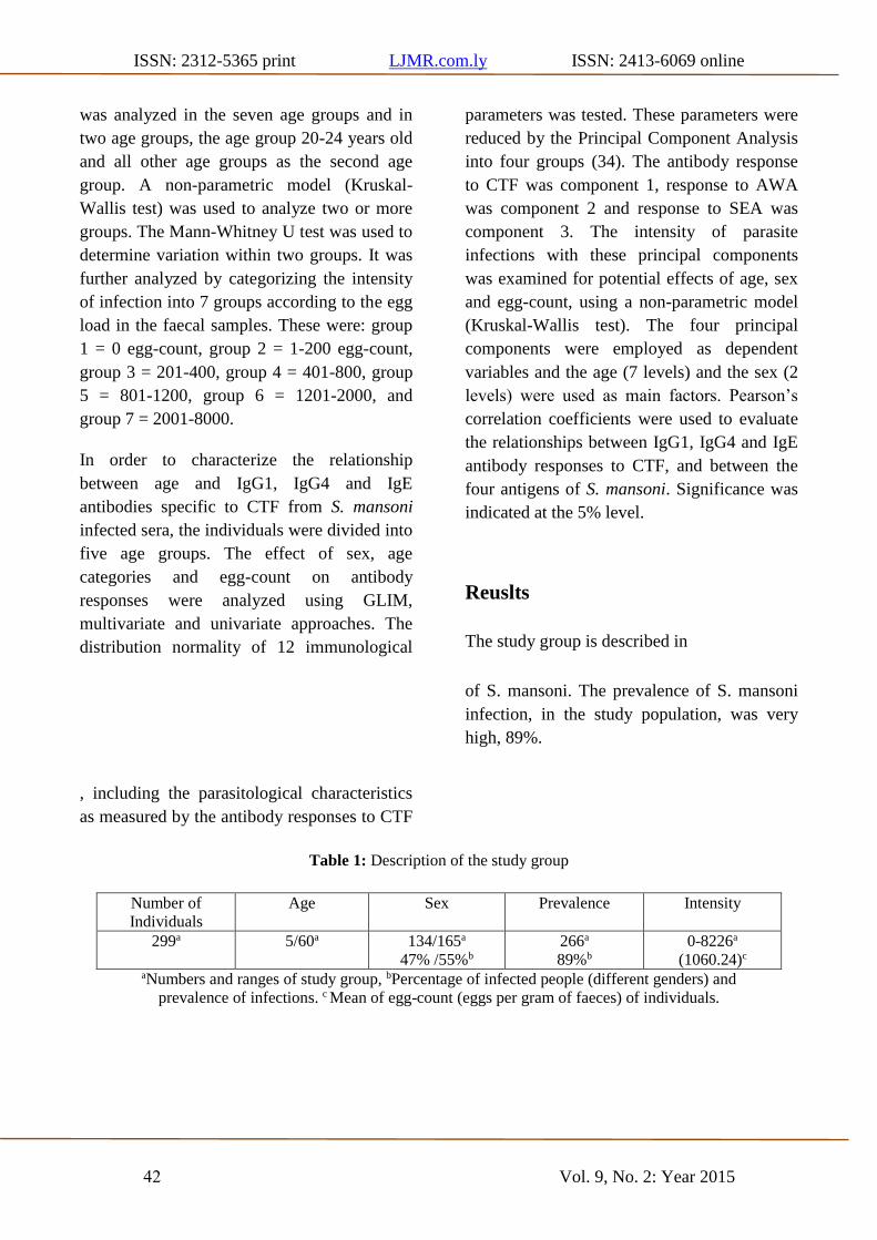
Age and gender-profiles of intensity and
prevalence of infection
Although age was significantly related to
the intensity of infection, the parameters of
infection of S. mansoni: the intensity and
the prevalence, did not show any
significant differences between the genders
(P > 0.05). However, females had a
slightly higher intensity of infection than
males in the (5-9) and (20-24) age groups,
and it was higher in the males in the (10-
14), (15-19), (20-24) and (25-29) age
groups (Figure 1). In both genders, the
intensity of infection was high around the
age group of 20-24. The analysis of the
intensity of infection in the two age groups
(20-24 and all other age groups) confirmed
that the parasite egg yield dramatically
increased in the 20-24 years old and that
during all other age groups the egg-count
decreased or stable (Kruskal-Wallis test,
X21 = 6.194, P = 0.013). The prevalence of
infection by age and sex (Figure 2) showed
a similar trend to that of intensity of
infection.
Figure 1: Intensity of infection by age group and sex
A g e g r o u p (y e a r s )
Me
an
of
eg
g c
ou
nt
(ep
g)
5-9
10-1
4
15-1
9
20-2
4
25-2
9
30-3
9
40-6
0
0
5 0 0
1 0 0 0
1 5 0 0
2 0 0 0
2 5 0 0
A g e g r o u p ( y e a r s )
Pr
ev
ale
nc
e o
fS
. m
an
son
i (%
)
5 -9
1 0 -14
1 5 -19
2 0 -24
2 5 -29
3 0 -39
4 0 -60
2 0
4 0
6 0
8 0
1 0 0
Red symbols represent the means ± SD of females, while the blue symbols represent means ± SD of males in
each age group (5-9, 10-14, 15-19, 20-24, 25-29, 30-39 and 40-60. The standard error bars show the range of
egg-count data, excluding extreme values
Figure 2: Relationships between age-prevalence of S. mansoni and sex in North-western Uganda
ISSN: 2312-5365 print LJMR.com.ly ISSN: 2413-6069 online
Vol. 9, No. 2: Year 201544
Blue symbols represent males, while the red symbols represent females in 7 age groups
(5-9, 10-14, 15-19, 20-24, 25-29, 30-39 and 40-60)
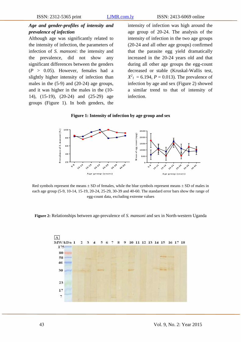
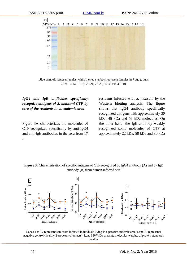
IgG4 and IgE antibodies specifically
recognize antigens of S. mansoni CTF by
sera of the residents in an endemic area
Figure 3A characterizes the molecules of
CTF recognized specifically by anti-IgG4
and anti-IgE antibodies in the sera from 17
residents infected with S. mansoni by the
Western blotting analysis. The figure
shows that IgG4 antibody specifically
recognized antigens with approximately 30
kDa, 46 kDa and 58 kDa molecules. On
the other hand, the IgE antibody weakly
recognized some molecules of CTF at
approximately 22 kDa, 58 kDa and 80 kDa
.
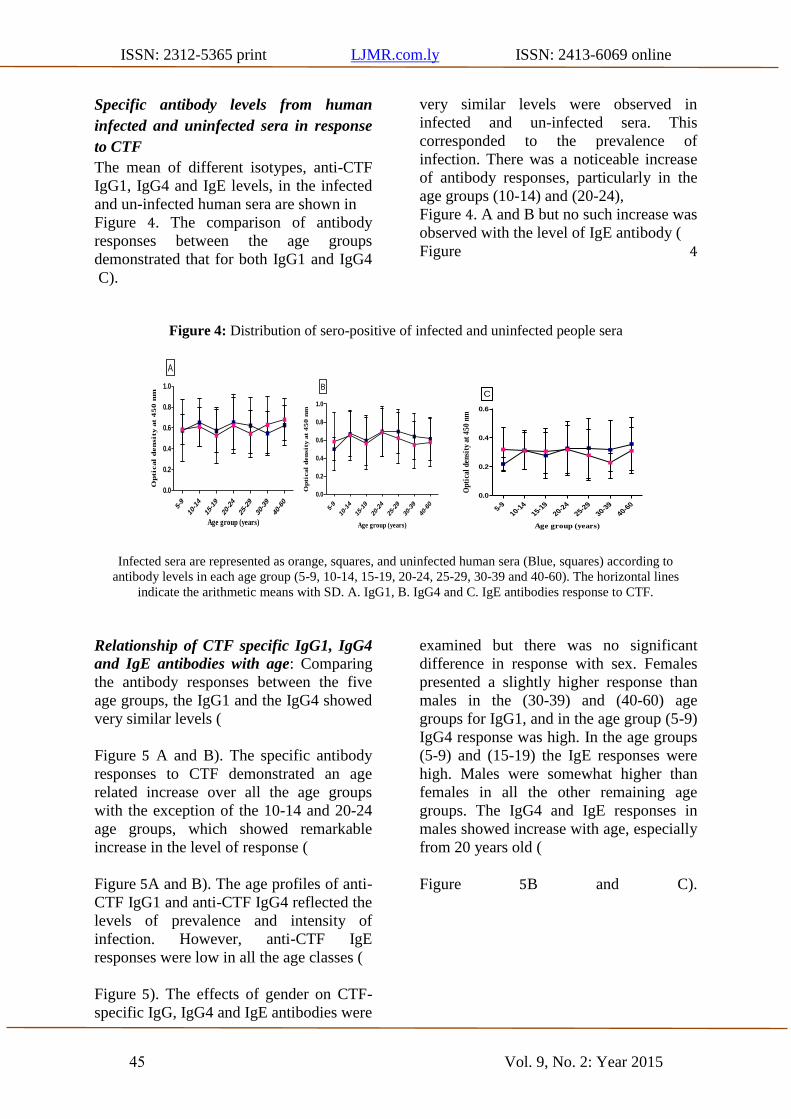
Figure 3: Characterisation of specific antigens of CTF recognised by IgG4 antibody (A) and by IgE
antibody (B) from human infected sera
Age group (years)
Op
tical
de
nsit
y a
t 4
50
nm
5-9
10-1
4
15-1
9
20-2
4
25-2
9
30-3
9
40-6
0
0.0
0.5
1.0
1.5
A
Age group (years)
Op
tical
den
sit
y a
t 4
50
nm
5-9
10-1
4
15-1
9
20-2
4
25-2
9
30-3
9
40-6
0
0.0
0.5
1.0
1.5
B
Age group (years)
Op
tica
l d
ensi
ty a
t 4
50
nm
5-9
10-14
15-19
20-24
25-29
30-39
40-60
0.0
0.2
0.4
0.6
C
Lanes 1 to 17 represent sera from infected individuals living in a parasite endemic area. Lane 18 represents
negative control (healthy European volunteers). Lane MW/kDa presents molecular weights of protein standards
in kDa
ISSN: 2312-5365 print LJMR.com.ly ISSN: 2413-6069 online
Vol. 9, No. 2: Year 201545
Specific antibody levels from human
infected and uninfected sera in response
to CTF
The mean of different isotypes, anti-CTF
IgG1, IgG4 and IgE levels, in the infected
and un-infected human sera are shown in
Figure 4. The comparison of antibody
responses between the age groups
demonstrated that for both IgG1 and IgG4
very similar levels were observed in
infected and un-infected sera. This
corresponded to the prevalence of
infection. There was a noticeable increase
of antibody responses, particularly in the
age groups (10-14) and (20-24),
Figure 4. A and B but no such increase was
observed with the level of IgE antibody (
Figure 4
C).
Figure 4: Distribution of sero-positive of infected and uninfected people sera
Age group (years)
Op
tical d
en
sity a
t 4
50
nm
5-9
10-14
15-19
20-24
25-29
30-39
40-60
0.0
0.2
0.4
0.6
0.8
1.0
A
Age group (years)
Op
tical
den
sit
y a
t 4
50
nm
5-9
10-1
4
15-1
9
20-2
4
25-2
9
30-3
9
40-6
0
0.0
0.2
0.4
0.6
0.8
1.0
B
Age group (years)
Op
tica
l d
ensi
ty a
t 4
50
nm
5-9
10-14
15-19
20-24
25-29
30-39
40-60
0.0
0.2
0.4
0.6
C
Infected sera are represented as orange, squares, and uninfected human sera (Blue, squares) according to
antibody levels in each age group (5-9, 10-14, 15-19, 20-24, 25-29, 30-39 and 40-60). The horizontal lines
indicate the arithmetic means with SD. A. IgG1, B. IgG4 and C. IgE antibodies response to CTF.
Relationship of CTF specific IgG1, IgG4
and IgE antibodies with age: Comparing
the antibody responses between the five
age groups, the IgG1 and the IgG4 showed
very similar levels (
Figure 5 A and B). The specific antibody
responses to CTF demonstrated an age
related increase over all the age groups
with the exception of the 10-14 and 20-24
age groups, which showed remarkable
increase in the level of response (
Figure 5A and B). The age profiles of anti-
CTF IgG1 and anti-CTF IgG4 reflected the
levels of prevalence and intensity of
infection. However, anti-CTF IgE
responses were low in all the age classes (
Figure 5). The effects of gender on CTF-
specific IgG, IgG4 and IgE antibodies were
examined but there was no significant
difference in response with sex. Females
presented a slightly higher response than
males in the (30-39) and (40-60) age
groups for IgG1, and in the age group (5-9)
IgG4 response was high. In the age groups
(5-9) and (15-19) the IgE responses were
high. Males were somewhat higher than
females in all the other remaining age
groups. The IgG4 and IgE responses in
males showed increase with age, especially
from 20 years old (
Figure 5B and C).
ISSN: 2312-5365 print LJMR.com.ly ISSN: 2413-6069 online
Vol. 9, No. 2: Year 201546
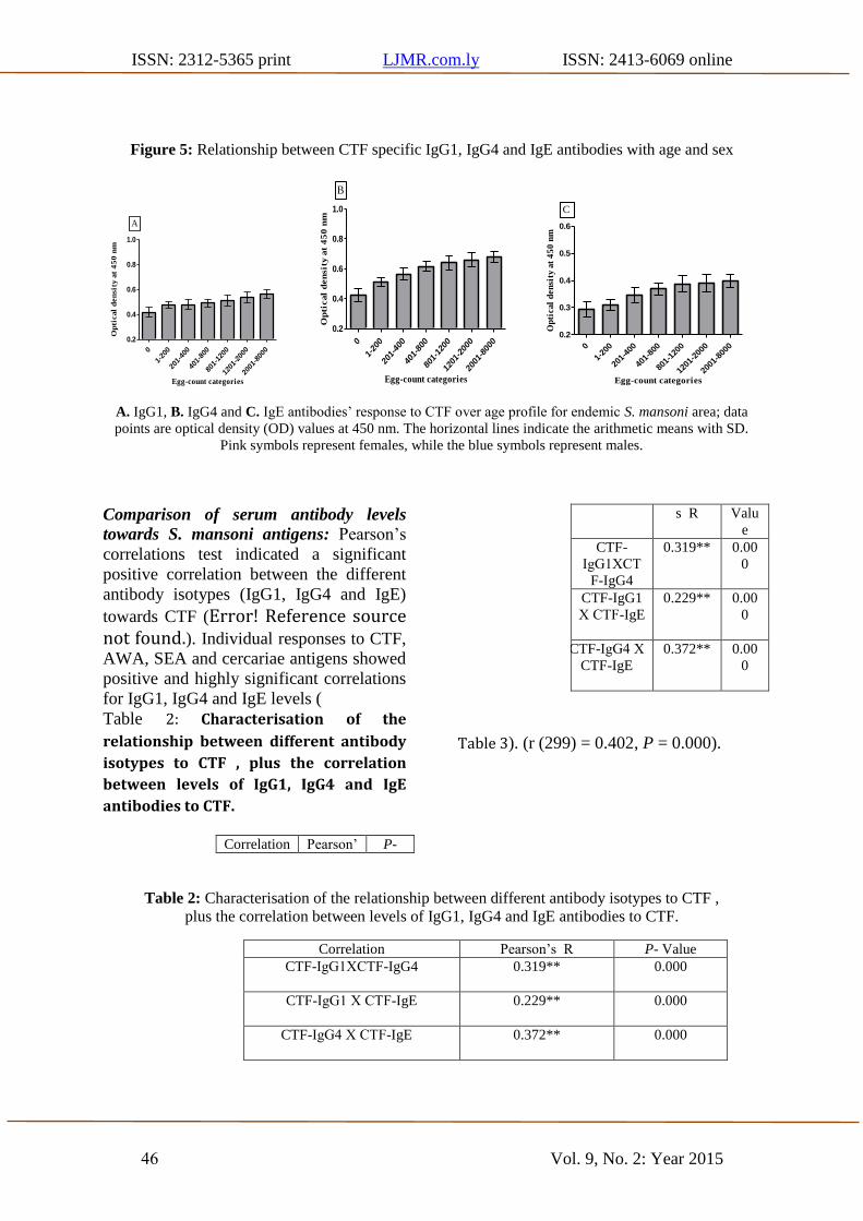
Figure 5: Relationship between CTF specific IgG1, IgG4 and IgE antibodies with age and sex
Egg-count categories
Op
tical
den
sit
y a
t 4
50
nm
0
1-20
0
201-
400
401-
800
801-
1200
1201
-200
0
2001
-800
0
0.2
0.4
0.6
0.8
1.0
A
Egg-count categories
Op
tical
den
sit
y a
t 4
50
nm
0
1-20
0
201-
400
401-
800
801-
1200
1201
-200
0
2001
-800
0
0.2
0.4
0.6
0.8
1.0
B
Egg-count categories
Op
tical
den
sity
at
45
0 n
m
0
1-20
0
201-
400
401-
800
801-
1200
1201
-200
0
2001
-800
0
0.2
0.3
0.4
0.5
0.6
C
A. IgG1, B. IgG4 and C. IgE antibodies’ response to CTF over age profile for endemic S. mansoni area; data
points are optical density (OD) values at 450 nm. The horizontal lines indicate the arithmetic means with SD.
Pink symbols represent females, while the blue symbols represent males.
Comparison of serum antibody levels
towards S. mansoni antigens: Pearson’s
correlations test indicated a significant
positive correlation between the different
antibody isotypes (IgG1, IgG4 and IgE)
towards CTF (Error! Reference source not found.). Individual responses to CTF,
AWA, SEA and cercariae antigens showed
positive and highly significant correlations
for IgG1, IgG4 and IgE levels (
Table 2: Characterisation of the
relationship between different antibody
isotypes to CTF , plus the correlation
between levels of IgG1, IgG4 and IgE
antibodies to CTF.
Correlation Pearson’ P-
s R Valu
e
CTF-
IgG1ХCT
F-IgG4
0.319** 0.00
0
CTF-IgG1
Х CTF-IgE
0.229**
0.00
0
CTF-IgG4 Х
CTF-IgE
0.372** 0.00
0
Table 3). (r (299) = 0.402, P = 0.000).
Table 2: Characterisation of the relationship between different antibody isotypes to CTF ,
plus the correlation between levels of IgG1, IgG4 and IgE antibodies to CTF.
Correlation Pearson’s R P- Value
CTF-IgG1ХCTF-IgG4 0.319** 0.000
CTF-IgG1 Х CTF-IgE
0.229**
0.000
CTF-IgG4 Х CTF-IgE 0.372** 0.000
ISSN: 2312-5365 print LJMR.com.ly ISSN: 2413-6069 online
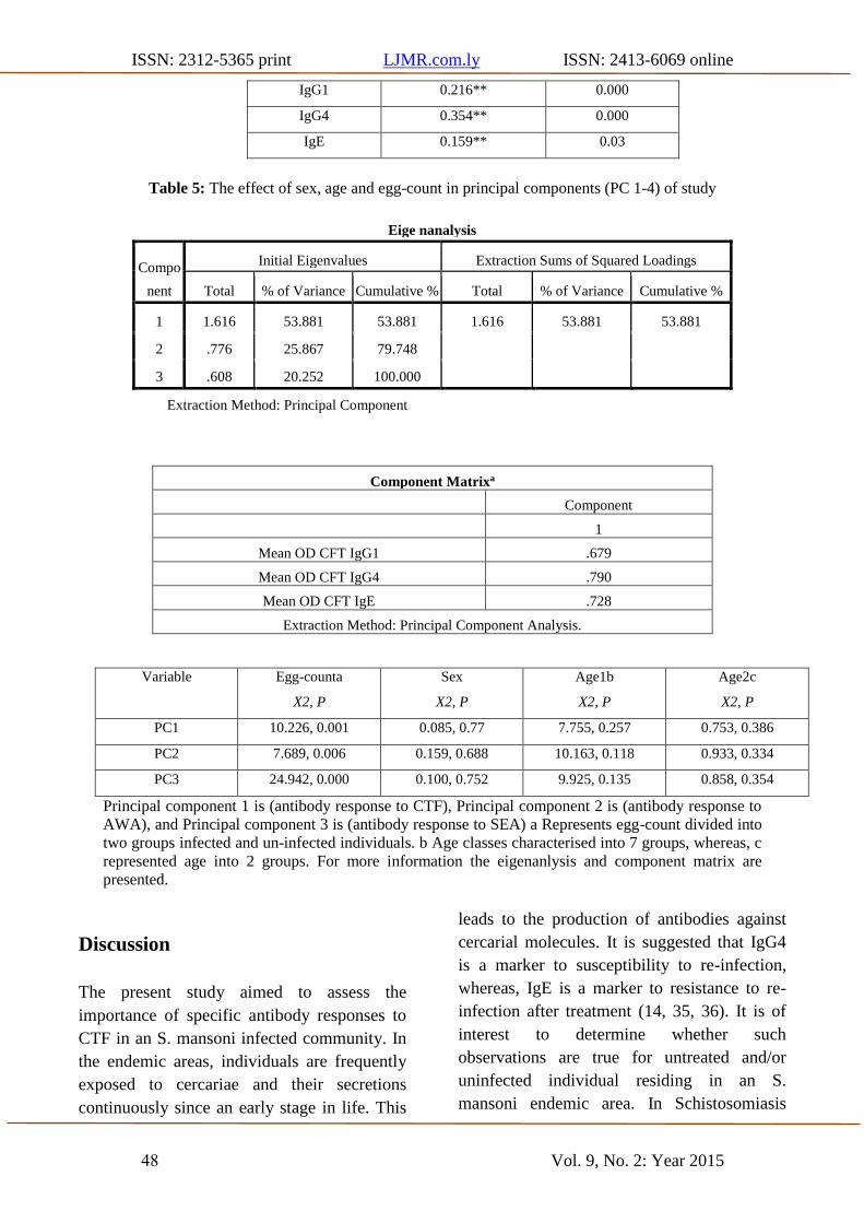
Vol. 9, No. 2: Year 201547
Table 3: The relationship between different antibody isotypes to some S. mansoni antigens
Correlation
Anti-body
isotype
Pearson’s R P- Value
CTF Х AWA
IgG1 0.441 0.000**
IgG4 0.239 0.000**
IgE 0.173 0.003**
CTF Х SEA
IgG1 0.542 0.000**
IgG4 0.114 0.050
IgE 0.227 0.000**
CTF Х Cercariae IgG1 0.390 0.000**
IgG4 0.144 0.013
Antibody levels specific for CTF, AWA, SEA and cercariae were measured by ELISA and
correlated with one another using Pearson’s rank correlation model
S. mansoni antigens specific IgG1, IgG4
and IgE antibodies and egg-count
There were significant positive correlations
between the egg-count and the antibody
response specific to CTF (Table 4). The
increase in the egg-count related to the
increase in the antibody response to CTF.
The comparison of the antibodies in the 7
egg-count categories and their responses to
CTF indicated that all antibody isotypes’
responses were higher in individuals with
egg-count from 1201-2000 epg (Error!
Reference source not found.). The
IgG1 levels were significantly higher in the
egg-count category 2001-8000 when
compared to group 1 (epg = 0) and group 2
(1-200 epg) (Mann-whitney U-test, z = -
2.587, P = 0.01 and P = 0.03
respectively), Error! Reference source
not found. A. A significant positive
correlation was found between IgG4
antibody responses to CTF and
approximately all egg-count categories in
individuals with 2001-8000 epg when
compared with groups 1 and 2 (r (299,
0.66) = 0.521, P = 0.000), with group 3 (P
= 0.005) and with group 4 (P = 0.015).
Group 1 significantly differed with group 4
(P = 0.012), with group 5 (P = 0.006) and
with group 6 (P = 0.000), Error!
Reference source not found. B.
Additionally, anti-CTF IgE response was
significantly different between group 1 and
the highest egg-count group (Mann-
whitney U-test, z = -2.485, P = 0.012),
Error! Reference source not found.C).
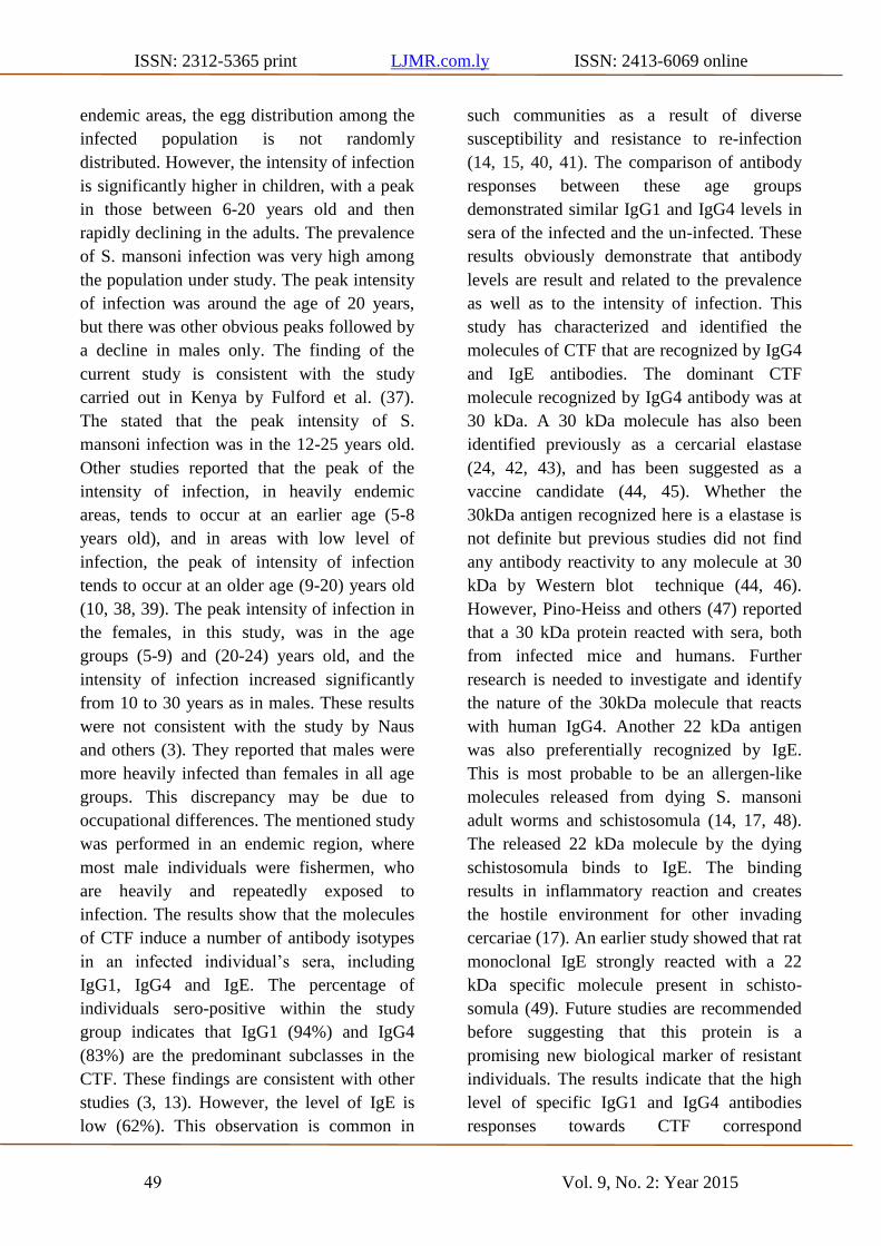
Consequently, principal components (PC1-
3) were examined in relation to sex, age
groups and egg-count (infected and
uninfected). For this, the non-parametric
test (Kruskal-Wallis test) was employed;
egg-count did indeed significantly change
between all the principal components (
Table 5). However, in relation to gender,
no significant differences were found with
all principal components (reflective of
antibody responses). Similarly, there was
no significant influence in terms of age
classes (7 groups and 2 groups) in terms of
all principal components.
Table 4 : Correlation between egg-count with IgG1, IgG4 and IgE antibody response to CTF
Correlation Pearson’s R P value
ISSN: 2312-5365 print LJMR.com.ly ISSN: 2413-6069 online
Vol. 9, No. 2: Year 201548
IgG1 0.216** 0.000
IgG4 0.354** 0.000
IgE 0.159** 0.03
Table 5: The effect of sex, age and egg-count in principal components (PC 1-4) of study
Component Matrixa
Component
1
Mean OD CFT IgG1 .679
Mean OD CFT IgG4 .790
Mean OD CFT IgE .728
Extraction Method: Principal Component Analysis.
Variable Egg-counta
X2, P
Sex
X2, P
Age1b
X2, P
Age2c
X2, P
PC1 10.226, 0.001 0.085, 0.77 7.755, 0.257 0.753, 0.386
PC2 7.689, 0.006 0.159, 0.688 10.163, 0.118 0.933, 0.334
PC3 24.942, 0.000 0.100, 0.752 9.925, 0.135 0.858, 0.354
Principal component 1 is (antibody response to CTF), Principal component 2 is (antibody response to
AWA), and Principal component 3 is (antibody response to SEA) a Represents egg-count divided into
two groups infected and un-infected individuals. b Age classes characterised into 7 groups, whereas, c
represented age into 2 groups. For more information the eigenanlysis and component matrix are
presented.
Discussion
The present study aimed to assess the
importance of specific antibody responses to
CTF in an S. mansoni infected community. In
the endemic areas, individuals are frequently
exposed to cercariae and their secretions
continuously since an early stage in life. This
leads to the production of antibodies against
cercarial molecules. It is suggested that IgG4
is a marker to susceptibility to re-infection,
whereas, IgE is a marker to resistance to re-
infection after treatment (14, 35, 36). It is of
interest to determine whether such
observations are true for untreated and/or
uninfected individual residing in an S.
mansoni endemic area. In Schistosomiasis
Eige nanalysis
Compo
nent
Initial Eigenvalues Extraction Sums of Squared Loadings
Total % of Variance Cumulative % Total % of Variance Cumulative %
1 1.616 53.881 53.881 1.616 53.881 53.881
2 .776 25.867 79.748
3 .608 20.252 100.000
Extraction Method: Principal Component
ISSN: 2312-5365 print LJMR.com.ly ISSN: 2413-6069 online
Vol. 9, No. 2: Year 201549
endemic areas, the egg distribution among the
infected population is not randomly
distributed. However, the intensity of infection
is significantly higher in children, with a peak
in those between 6-20 years old and then
rapidly declining in the adults. The prevalence
of S. mansoni infection was very high among
the population under study. The peak intensity
of infection was around the age of 20 years,
but there was other obvious peaks followed by
a decline in males only. The finding of the
current study is consistent with the study
carried out in Kenya by Fulford et al. (37).
The stated that the peak intensity of S.
mansoni infection was in the 12-25 years old.
Other studies reported that the peak of the
intensity of infection, in heavily endemic
areas, tends to occur at an earlier age (5-8
years old), and in areas with low level of
infection, the peak of intensity of infection
tends to occur at an older age (9-20) years old
(10, 38, 39). The peak intensity of infection in
the females, in this study, was in the age
groups (5-9) and (20-24) years old, and the
intensity of infection increased significantly
from 10 to 30 years as in males. These results
were not consistent with the study by Naus
and others (3). They reported that males were
more heavily infected than females in all age
groups. This discrepancy may be due to
occupational differences. The mentioned study
was performed in an endemic region, where
most male individuals were fishermen, who
are heavily and repeatedly exposed to
infection. The results show that the molecules
of CTF induce a number of antibody isotypes
in an infected individual’s sera, including
IgG1, IgG4 and IgE. The percentage of
individuals sero-positive within the study
group indicates that IgG1 (94%) and IgG4
(83%) are the predominant subclasses in the
CTF. These findings are consistent with other
studies (3, 13). However, the level of IgE is
low (62%). This observation is common in
such communities as a result of diverse
susceptibility and resistance to re-infection
(14, 15, 40, 41). The comparison of antibody
responses between these age groups
demonstrated similar IgG1 and IgG4 levels in
sera of the infected and the un-infected. These
results obviously demonstrate that antibody
levels are result and related to the prevalence
as well as to the intensity of infection. This
study has characterized and identified the
molecules of CTF that are recognized by IgG4
and IgE antibodies. The dominant CTF
molecule recognized by IgG4 antibody was at
30 kDa. A 30 kDa molecule has also been
identified previously as a cercarial elastase
(24, 42, 43), and has been suggested as a
vaccine candidate (44, 45). Whether the
30kDa antigen recognized here is a elastase is
not definite but previous studies did not find
any antibody reactivity to any molecule at 30
kDa by Western blot technique (44, 46).
However, Pino-Heiss and others (47) reported
that a 30 kDa protein reacted with sera, both
from infected mice and humans. Further
research is needed to investigate and identify
the nature of the 30kDa molecule that reacts
with human IgG4. Another 22 kDa antigen
was also preferentially recognized by IgE.
This is most probable to be an allergen-like
molecules released from dying S. mansoni
adult worms and schistosomula (14, 17, 48).
The released 22 kDa molecule by the dying
schistosomula binds to IgE. The binding
results in inflammatory reaction and creates
the hostile environment for other invading
cercariae (17). An earlier study showed that rat
monoclonal IgE strongly reacted with a 22
kDa specific molecule present in schisto-
somula (49). Future studies are recommended
before suggesting that this protein is a
promising new biological marker of resistant
individuals. The results indicate that the high
level of specific IgG1 and IgG4 antibodies
responses towards CTF correspond
ISSN: 2312-5365 print LJMR.com.ly ISSN: 2413-6069 online
Vol. 9, No. 2: Year 201550
particularly with the peaks of the prevalence
of infection with age groups with the highest
percentage of infection (10-14 years and 20-24
years). Previous studies reported that antibody
response against AWA and SEA were
associated strongly with age and with the
intensity of infection (40). This peak of
antibodies in these age groups (10-14 and 20-
24) is most probably associated with the
variation in the individual personal and
behavioral period of exposure to infection.
The findings suggested a significant positive
correlation between egg-count and antibody
responses. This supports the reports by
previous researchers (3, 7). The analysis by
two age groups, the group of 20-24 years old
(group 1) and all other age groups (group 2)
showed that group 1 is characterized by a
significantly higher prevalence than group 2
and that the antibody levels of the individuals
of this group are also very high, especially
IgG1and IgG4. There is a strong correlation
between IgG4 and the different S. mansoni
antigens (CTF, AWA, SEA and cercariae
homogenate) as well as a positive correlation
between IgG4 and IgF responses to CTF. The
strong correlation between the IgG4 and the
different S. mansoni anti-genes is most
probably due to that these antigens expressed
identical common epitopes, which directly
bind to IgG4. Hussain and others (50)
suggested that IgE and IgG4 antibodies might
bind to the same epitopes. Thus, the effector
function of IgE is blocked by IgG4 as they are
both directed to the same epitopes (36).
However, Li et al. (1999) (51) demonstrated
that the two antibodies are independently
regulated by different mechanisms. The IgG4
response is significantly different in the
different age groups, depending upon the
prevalence and the intensity of the infection,
whereas no such observation is demonstrated
with IgE response to CTF. Also there are no
gender differences between IgG1, IgG4 and
IgE responses. However, the levels of IgG4
and IgE increase from 20 years old in the
males, while an increase in the levels of IgG1
level is observed from 30 years in females.
The IgG4 level is considerably higher in
females than males in age 5-9 year old, an
observation reported by several workers (3, 7).
Two possible explanations were suggested.
The first is that males are exposed to cercarial
antigens more than females, because of
behavioural differences (21). The second is
that the difference could be due to different
hormonal factors between sexes (7). It will be
useful to conduct further studies with a large
number of adults over 20 years to assess the
cumulative exposure to cercarial secretions,
combined with a water contact survey. It is
interesting to observe that only egg-count is
significantly related to all S. mansoni antigen,
CTF, AWA, SEA, the principal components.
This suggests that the antibody response is a
reliable indicator of infection with S. mansoni
in endemic areas and it might be also exploited
for schistosomiasis epidemiological studies.
The antibody responses are significantly
correlated with the prevalence of infection.
The results indicate that IgG4 and IgE
responses are associated with sex, age as well
as with the prevalence of infection and that
anti-IgG4 and anti-IgG1 against CTF increase
significantly with the egg abundance. Sera
from uninfected individuals, according to
negative egg-count results (zero), had sero-
positive results to CTF antigen with detectable
levels of IgG1, IgG4 and IgE. This conclusion
is consistent with those of other studies and
suggests that such a reaction reflects the
limited of microscopic sensitivity of egg
combined with a very high sensitivity and
specificity of ELISA to detect antibody (52,
53). The sensitivity of antibody detection is a
more effective method than parasitology (54).
ISSN: 2312-5365 print LJMR.com.ly ISSN: 2413-6069 online
Vol. 9, No. 2: Year 201551
In conclusion, the results suggest that anti-
CTF antibody responses predict exposure to S.
mansoni but further in depth studies are
needed.
ISSN: 2312-5365 print LJMR.com.ly ISSN: 2413-6069 online
Vol. 9, No. 2: Year 201552
References
1. Standley CJ, Mugisha L, Dobson AP and Stothard JR (2012) Zoonotic schistosomiasis in
non-human primates: past, present and future activities at the human-wildlife interface in
Africa. J Helminthol. 86: 131-140.
2. Silveira AM, Bethony J, Gazzinelli A, Kloos H, Fraga LA, Alvares MC, Prata A, Guerra HL,
Loverde PT, Correa-Oliveira R and Gazzinelli G (2002) High levels of IgG4 to Schistosoma
mansoni egg antigens in individuals with periportal fibrosis. Am J Trop Med Hyg. 66: 542-
549.
3. Naus CW, Booth M, Jones FM, Kemijumbi J, Vennervald BJ, Kariuki CH, Ouma JH,
Kabatereine NB and Dunne DW (2003) The relationship between age, sex, egg-count and
specific antibody responses against Schistosoma mansoni antigens in a Ugandan fishing
community. Trop. Med Int Health: TM & IH. 8: 561-568.
4. Walter K, Fulford AJ, McBeath R, Joseph S, Jones FM, Kariuki HC, Mwatha JK, Kimani G,
Kabatereine NB, Vennervald BJ, Ouma JH and Dunne DW (2006) Increased human IgE
induced by killing Schistosoma mansoni in vivo is associated with pretreatment Th2 cytokine
responsiveness to worm antigens. J Immunol. 177: 5490-5498.
5. Pinot de Moira A, Fulford AJ, Kabatereine NB, Ouma JH, Booth M and Dunne DW (2010)
Analysis of complex patterns of human exposure and immunity to Schistosomiasis mansoni:
the influence of age, sex, ethnicity and IgE. PLoS Negl Trop Dis. 4.
6. Vereecken K, Naus, CW, Polman K, Scott JT, Diop M, Gryseels B and Kestens L (2007)
Associations between specific antibody responses and resistance to reinfection in a
Senegalese population recently exposed to Schistosoma mansoni. Trop Med Int Health: TM
& IH. 12: 431-444.
7. Webster M, Libranda-Ramirez BD, Aligui GD, Olveda RM, Ouma JH, Kariuki HC, Kimani
G, Olds GR, Fulford AJ, Butterworth AE and Dunne DW (1997) The influence of sex and
age on antibody isotype responses to Schistosoma mansoni and Schistosoma japonicum in
human populations in Kenya and the Philippines. Parasitol. 114(Pt 4): 383-393.
8. Naus CW, Kimani G, Ouma JH, Fulford AJ, Webster M, van Dam GJ, Deelder AM,
Butterworth AE and Dunne DW (1999) Development of antibody isotype responses to
Schistosoma mansoni in an immunologically naive immigrant population: influence of
infection duration, infection intensity, and host age. Infect Immunol. 67: 3444-3451.
9. Mutapi F, Ndhlovu PD, Hagan P and Woolhouse ME (1997) A comparison of humoral
responses to Schistosoma haematobium in areas with low and high levels of infection.
Parasite Immunol. 19: 255-263.
10. Mitchell KM, Mutapi F, Savill NJ and Woolhouse ME (2011) Explaining observed infection
and antibody age-profiles in populations with urogenital schistosomiasis. PLoS Comput Biol.
7: e1002237.
11. Wang LD, Chen HG, Guo JG, Zeng XJ, Hong XL, Xiong JJ, Wu XH, Wang XH, Wang LY,
Xia G, Hao Y, Chin DP and Zhou XN (2009) A strategy to control transmission of
Schistosoma japonicum in China. N Engl J Med. 360: 121-128.
12. Naus CW, van Remoortere A, Ouma JH, Kimani G, Dunne DW, Kamerling JP, Deelder AM
and Hokke CH (2003) Specific antibody responses to three schistosome-related carbohydrate
structures in recently exposed immigrants and established residents in an area of Schistosoma
mansoni endemicity. Infect Immunol. 71, 5676-5681. 13. Caldas IR, Campi-Azevedo AC, Oliveira LF, Silveira AM, Oliveira RC and Gazzinelli G
(2008) Human schistosomiasis mansoni: immune responses during acute and chronic phases
of the infection. Acta Trop. 108: 109-117.
ISSN: 2312-5365 print LJMR.com.ly ISSN: 2413-6069 online
Vol. 9, No. 2: Year 201553
14. Dunne DW, Butterworth AE, Fulford AJ, Kariuki HC, Langley JG, Ouma JH, Capron A,
Pierce RJ and Sturrock RF (1992) Immunity after treatment of human schistosomiasis:
association between IgE antibodies to adult worm antigens and resistance to reinfection. Eur J
Immunol. 22: 1483-1494.
15. Jiz M, Friedman JF, Leenstra T, Jarilla B, Pablo A, Langdon G, Pond-Tor S, Wu HW,
Manalo D, Olveda R, Acosta L and Kurtis JD (2009) Immunoglobulin E (IgE) responses to
paramyosin predict resistance to reinfection with Schistosoma japonicum and are attenuated
by IgG4. Infect Immunol. 77: 2051-2058.
16. Aalberse R, Stapel S, Schuurman J and Rispens T (2009) Immunoglobulin G4: an odd
antibody. Clin Exp Allergy. 469-477.
17. Fitzsimmons CM, McBeath R, Joseph S, Jones FM, Walter K, Hoffmann KF, Kariuki HC,
Mwatha JK, Kimani G, Kabatereine NB, Vennervald BJ, Ouma JH and Dunne DW (2007)
Factors affecting human IgE and IgG responses to allergen-like Schistosoma mansoni
antigens: Molecular structure and patterns of in vivo exposure. Int Arch Allergy Immunol.
142, 40-50.
18. Imai N, Rujeni N, Nausch N, Bourke CD, Appleby LJ, Cowan G, Gwisai R, Midzi N,
Cavanagh D, Mduluza T, Taylor D and Mutapi F (2011) Exposure, infection, systemic
cytokine levels and antibody responses in young children concurrently exposed to
schistosomiasis and malaria. Parasitol. 138: 1519-1533.
19. Dalton PR and Pole D (1978) Water-contact patterns in relation to Schistosoma haematobium
infection. Bull WHO. 56: 417-426.
20. de Lima e Costa MF, Rocha RS, Coura Filho P and Katz N (1993) A 13-year follow-up of
treatment and snail control in an area endemic for Schistosoma mansoni in Brazil: incidence
of infection and reinfection. Bull WHO. 71: 197-205.
21. Kabatereine NB, Brooker S, Tukahebwa EM, Kazibwe F and Onapa AW (2004)
Epidemiology and geography of Schistosoma mansoni in Uganda: implications for planning
control. Trop Med Int Health: TM & IH 9: 372-380.
22. Matthys B, Tschannen AB, Tian-Bi NT, Comoe H, Diabate S, Traore M, Vounatsou P Raso
G, Gosoniu L, Tanner M, Cisse G, N'Goran EK and Utzinger J (2007) Risk factors for
Schistosoma mansoni and hookworm in urban farming communities in western Cote d'Ivoire.
Trop Med Int Health: TM & IH 12: 709-723.
23. Eloi-Santos S, Olsen NJ, Correa-Oliveira R and Colley DG (1992) Schistosoma mansoni:
mortality, pathophysiology, and susceptibility differences in male and female mice. Exp
Parasitol. 75: 168-175.
24. Knudsen GM, Medzihradszky KF, Lim KC, Hansell E and McKerrow JH (2005) Proteomic
analysis of Schistosoma mansoni cercarial secretions. Mol Cell Proteomics. 4: 1862-1875.
25. Curwen RS, Ashton PD, Sundaralingam S and Wilson RA (2006) Identification of novel
proteases and immunomodulators in the secretions of schistosome cercariae that facilitate
host entry. Mol Cell Proteomics. 5: 835-844. 26. Sepulveda J Tremblay JM, DeGnore JP, Skelly PJ and Shoemaker CB (2010) Schistosoma
mansoni host-exposed surface antigens characterized by sera and recombinant antibodies
from schistosomiasis-resistant rats. Int J Parasitol. 40: 1407-1417.
27. Chand MA, Chiodini PL and Doenhoff MJ (2010) Development of a new assay for the
diagnosis of schistosomiasis, using cercarial antigens. Trans R Soc Trop Med Hyg. 104: 255-
258. 28. El Aswad Bel D, Doenhoff MJ, El Hadidi AS, Schwaeble WJ and Lynch NJ (2011) Use of
recombinant calreticulin and cercarial transformation fluid (CTF) in the serodiagnosis of
Schistosoma mansoni. Immunobiol. 216: 379-385.
29. Smith H, Doenhoff M, Aitken C, Bailey W, Ji M, Dawson E, Gilis H, Spence G, Alexander C
and van Gool T (2012) Comparison of Schistosoma mansoni soluble cercarial antigens and
ISSN: 2312-5365 print LJMR.com.ly ISSN: 2413-6069 online
Vol. 9, No. 2: Year 201554
soluble egg antigens for serodiagnosing schistosome infections. PLoS Negl Trop Dis. 6:
e1815.
30. Kabatereine NB, Kemijumbi J, Ouma JH, Sturrock RF, Butterworth AE, Madsen H, Ornbjerg
N, Dunne DW and Vennnervald BJ (2003) Efficacy and side effects of praziquantel treatment
in a highly endemic Schistosoma mansoni focus at Lake Albert, Uganda. Trans R Soc Trop
Med Hyg. 97: 599-603.
31. Behnke JM, Pritchard DI, Wakelin D, Park JR, McNicholas AM and Gilbert FS (1994) Effect
of ivermectin on infection with gastro-intestinal nematodes in Sierra Leone. J Helminthol. 68:
187-195.
32. Wilson K and Grenfell BT (1997) Generalized linear modelling for parasitologists. Parasitol
Today 13: 33-38.
33. Behnke JM, Lewis JW, Zain SN and Gilbert FS (1999) Helminth infections in Apodemus
sylvaticus in southern England: interactive effects of host age, sex and year on the prevalence
and abundance of infections. J Helminthol. 73: 31-44.
34. Sokal RR and Rohlf FJ (1981) In: Biometry. Freeman & Co, New York.
35. Hagan P, Blumenthal UJ, Dunn D, Simpson AJ, Wilkins HA (1991) Human IgE, IgG4 and
resistance to reinfection with Schistosoma haematobium. Nature. 349: 243-245.
36. Demeure CE, Rihet P, Abel L, Ouattara M, Bourgois A and Dessein AJ (1993) Resistance to
Schistosoma mansoni in humans: influence of the IgE/IgG4 balance and IgG2 in immunity to
reinfection after chemotherapy. J Infect Dis. 168: 1000-1008.
37. Fulford AJ, Butterworth AE, Sturrock RF and Ouma JH (1992) On the use of age-intensity
data to detect immunity to parasitic infections, with special reference to Schistosoma mansoni
in Kenya. Parasitol. 105 (Pt 2): 219-227.
38. Woolhouse ME (1998) Patterns in parasite epidemiology: the peak shift. Parasitol Today. 14:
428-434.
39. Duerr HP, Dietz K and Eichner M (2003) On the interpretation of age-intensity profiles and
dispersion patterns in parasitological surveys. Parasitol. 126: 87-101.
40. Satti MZ, Lind P, Vennervald BJ, Sulaiman SM, Daffalla AA and Ghalib HW (1996)
Specific immunoglobulin measurements related to exposure and resistance to Schistosoma
mansoni infection in Sudanese canal cleaners. Clin Exp Immunol. 106: 45-54.
41. Naus CW, van Dam GJ, Kremsner PG, Krijger FW and Deelder AM (1998) Human IgE, IgG
subclass, and IgM responses to worm and egg antigens in schistosomiasis haematobium: a
12-month study of reinfection in Cameroonian children. Clinical infectious diseases : an
official publication of the Infectious Diseases Society of America. 26: 1142-1147.
42. Landsperger WJ, Stirewalt MA and Dresden MH (1982) Purification and properties of a
proteolytic enzyme from the cercariae of the human trematode parasite Schistosoma mansoni.
Biochem J. 201: 137-144.
43. Hansell E, Braschi S, Medzihradszky KF, Sajid M, Debnath M, Ingram J, Lim KC and
McKerrow JH (2008) Proteomic analysis of skin invasion by blood fluke larvae. PLoS Negl
Trop Dis 2, e262.
44. Cardoso FC, Pacifico RN, Mortara RA and Oliveira SC (2006) Human antibody responses of
patients living in endemic areas for schistosomiasis to the tegumental protein Sm29 identified
through genomic studies. Clin Exp Immunol. 144: 382-391.
45. Ingram JR, Rafi SB, Eroy-Reveles AA, Ray M, Lambeth L, Hsieh I, Ruelas D, Lim KC,
Sakanari J, Craik CS, Jacobson MP and McKerrow JH (2012) Investigation of the proteolytic
functions of an expanded cercarial elastase gene family in Schistosoma mansoni. PLoS Negl
Trop Dis. 6: e1589.
46. Bahgat M, Francklow K, Doenhoff MJ, Li YL, Ramzy RM, Kirsten C and Ruppel A (2001)
Infection induces antibodies against the cercarial secretions, but not against the cercarial
ISSN: 2312-5365 print LJMR.com.ly ISSN: 2413-6069 online
Vol. 9, No. 2: Year 201555
elastases of Schistosoma mansoni, Schistosoma haematobium, Schistosoma japonicum and
Trichobilharzia ocellata. Parasite Immunol. 23: 557-565.
47. Pino-Heiss S, Petitt M, Beckstead JH and McKerrow JH (1986) Preparation of mouse
monoclonal antibodies and evidence for a host immune response to the preacetabular gland
proteinase of Schistosoma mansoni cercariae. Am J Trop Med Hyg. 35, 536-543.
48. Dunne DW, Webster M, Smith P, Langley JG, Richardson BA, Fulford AJ, Butterworth AE,
Sturrock RF, Kariuki HC and Ouma JH(1997) The isolation of a 22 kDa band after SDS-
PAGE of Schistosoma mansoni adult worms and its use to demonstrate that IgE responses
against the antigen(s) it contains are associated with human resistance to reinfection. Parasite
Immunol. 19: 79-89.
49. Verwaerde C, Joseph M, Capron M, Pierce RJ, Damonneville M, Velge F, Auriault C and
Capron A (1987) Functional properties of a rat monoclonal IgE antibody specific for
Schistosoma mansoni. J Immunol. 138: 4441-4446.
50. Hussain R, Poindexter RW, Ottesen EA and Reimer CB (1986) Use of monoclonal antibodies
to quantify subclasses of human IgG. II. Enzyme immunoassay to define antigen specific
(anti-filarial) IgG subclass antibodies. J Immunol Methods. 94: 73-80.
51. Li YS, Ross AG, Sleigh AC, Li Y, Waine GJ, Williams GJ, Tanner M and McManus DP
(1999) Antibody isotype responses, infection and re-infection for Schistosoma japonicum in a
marshland area of China. Acta Trop. 73: 79-92.
52. de Vlas SJ and Gryseels B (1992) Underestimation of Schistosoma mansoni prevalences.
Parasitol Today. 8: 274-277.
53. Utzinger J, Booth M, N'Goran EK, Muller I, Tanner M and Lengele C (2001) Relative
contribution of day-to-day and intra-specimen variation in faecal egg counts of Schistosoma
mansoni before and after treatment with praziquantel. Parasitol. 122: 537-544.
54. Doenhoff MJ, Chiodini PL and Hamilton JV (2004) Specific and sensitive diagnosis of
schistosome infection: can it be done with antibodies? Trends Parasitol. 20: 35-39.