Embed Size (px)
Citation preview

Human Vision

The Visual System
• The visual system consists of two parts.
– The eyes act as image receptors.
– The brain acts as an image processing andinterpretation unit.
• Understanding how we see requires that we understandboth components.
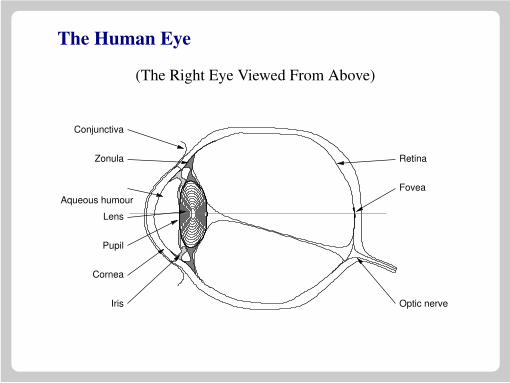
The Human Eye
(The Right Eye Viewed From Above)
Optic nerve
Fovea
Retina
Iris
Cornea
Pupil
Lens
Aqueous humour
Zonula
Conjunctiva

Constricted and Dilated Pupils
Constricted Dilated

Retinal Structure
• Receptor cells are at the back of the retina.
• Light passes “through the wiring”.
• Horizontal cells do some initial signal processing.
Light
horizontal cell
bipolar cell cone
rod
choroidganglion cell

Photo Receptor Cells
• The eye contains two different classes of light sensitivecells (or photo receptors).
• Rod cells provide sensitive low-light vision.
• Cone cells provide normal vision.

Rod Cells
• Rod cells provide low-light vision.
• There is only one type of rod cell.
• Rod cells provide no colour discrimination.

Cone Cells
• Cone cells operate at medium and high light levels.
• There are three different kinds of cone cells, eachsensitive to different light wavelengths.
• The sensitivity to different wavelengths is what providesus with colour vision.

Rod And Cone Distribution
• The distribution of rod and cone cells is not uniformacross the retina.
• The cones are concentrated at the centre rear of theretina and the rods are more evenly distributed awayfrom that area.

−80 −60 −40 −20 0 20 40 60 80
0
50
100
150
200
The Distribution of Rod and Cone Cells
Den
sity
in th
ousa
nds
per
squa
re m
m
Angular distance from the fovea (in degrees)
Rods
Cones

The Macula And Fovea
• The centre-rear of the retina has a small yellow spotknown as the macula.
• At the centre of the macula is a region called the foveawhich has a large concentration of cone cells, and norod cells at all.
• In normal light, the fovea is the part of the eye whichyields the highest resolution.
• The fovea occupies about 1.5◦ of our visual field(slightly larger than the moon).
• The highest resolution of all is given by the foveacentralis which gives occupies about one tenth of this.

High Resolution Vision
The picture on the right shows the resolution with we wouldexpect to see the face of the child if we were to stare fixedly atthe child’s right eye.

Neural Connections and the Blind Spot
• A network of nerves gathers signals from thephoto-sensitive cells of the retina and conducts them tothe optic nerve, which is attached to the sclera at therear of the eye.
• The optic nerve conveys the information from the retinato the visual processing centres of the brain.
• There are no photo receptors where the optic nervereaches the retina.
• This creates a “blind spot” in the visual field of eacheye.

Finding Your Blind Spot
• Draw a cross and a dot about 8cm apart as shown below.
• Close your right eye.
• Look directly at the cross
• Move the page backward and forward until you see thedot disappear.

Retinal Circuitry
• In the retina, horizontal cells connect photo receptorstogether.
• These connections mean that some simple imageprocessing can take place in the eye.
• Some of the most important operations are based onlateral inhibition.

Lateral Inhibition
• A network exhibits lateral inhibition if a positiveoutcome in one element of the network, induces anegative outcome in its neighbours.
• Lateral inhibition can be used to produce a simple formof edge enhancement.
• It can also be used to help provide hue and tonediscrimination.
• The phenomenon of Mach Banding shows the effect oflateral inhibition in the vision system.

A “Lateral Inhibition” Filter
• Consider the image filter takes an image and creates anew one by replacing each pixel by 3 times the value ofthat pixel minus the twice the average of its eightneighbours.
1
4
−1 −1 −1−1 12 −1−1 −1 −1
• The effect of the filter is to exaggerate (or enhance) any
edges present in an image.
• This is directly analogous to the lateral inhibition foundin the neural circuitry of the eye and brain.
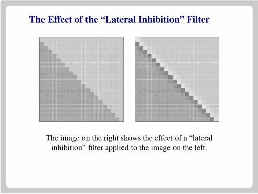
The Effect of the “Lateral Inhibition” Filter
The image on the right shows the effect of a “lateralinhibition” filter applied to the image on the left.

Mach Bands
Notice the apparent dark band to the left and apparent lightband to the right of the grey ramp. This can be explained bythe effect of lateral inhibition.

The Chevreul Illusion
The bands in this image are each a fixed shade of grey, butappear to be lighter on their right side than on their left one.

Neural Processing
• After the visual signal leaves the eye, further processingtakes place in stages down the visual pathway.
• Many of the steps in the processing of the visual signalare incompletely understood, but some progress hasbeen made in understanding the process.
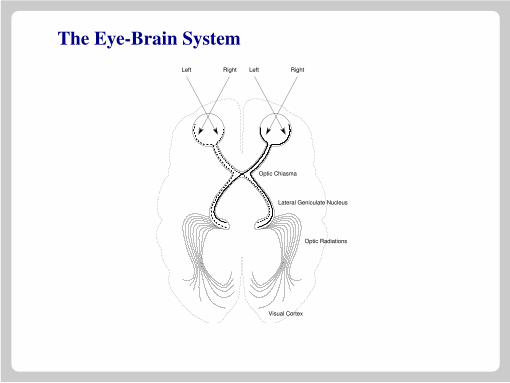
The Eye-Brain SystemRightLeftRightLeft
Optic Chiasma
Optic Radiations
Lateral Geniculate Nucleus
Visual Cortex

Transmission To The Cortex
• The visual signal from the retina is transmitted down theoptic nerve.
• There are roughly 1,000,000 nerve fibres in the opticnerve and some 125,000,000 receptors in the retina. Thesignal is thus not transmitted in a one-fibre per receptorfashion.
• The greatest convergence occurs for the rod cells — thesignals from 1000 or more rods may be carried by thesame nerve fibre.
• There is evidence that each receptor in the central foveais connected to two nerve cells.

The Optic Chiasm
• The optic nerves from the two eyes converge and crossat structure call the optic chiasm.
• Signals from each eye are mixed at this point. Suchcombination is clearly necessary for filling in theblind-spot and for comparison leading to the extractionof depth information.

The Lateral Geniculate Nucleus
• The signals a transmitted back through the brain to thelateral geniculate nucleus. The LGN acts as a kind ofrelay station which dispatches the signal to the visualcortex.
• The true function of the LGN is more complex than justacting as a relay, because it also receives a good deal ofinput from the cortex in addition to the optic nerve.

The Visual Cortex
• The visual cortex contains a number of neural structureswhich extract basic information from the visual signal.
• There are groups of neurons which are dedicated todetecting lines with particular orientation.
• Other groups take this information and use it to detectshapes.
• The signal is processed at higher and higher levels andeventually passed forward to the higher brain centres.

High Resolution Vision
• For a full visual examination of objects, we must moveour eyes so that all parts of the object’s image fall for atime on the fovea.
• We do this by moving our eyes from place to place overthe object in a jerky fashion.

Eye Movements
• Fixation : A period of time when the eye is focused on asingle point.
• Saccade : An eye motion from one fixation point toanother.
• Normal vision alternates between saccades andfixations, with each lasting just 100ths of a second.
Eye Movement Studies
• An image is presented to a subject.
• The subject may (or may not) be given a specific task tocarry out.
• A record is made of where the subjects eyes are directedas they study the image.

Studies by I. L. Yarbus
• Experimental work carried out in Russia during the1940s and 1950s.
• A suction “cap” was fitted directly onto a subject’seyeball.
• A small mirror on the cap reflected a light beam on tophoto-sensitive paper.

How We Look At Faces
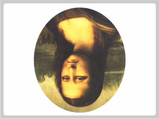


An Unexpected Visitor by I. E. Repin.

Examine the picture.

Decide how wealthy the family is.

Estimate the ages of the people in the room.

Decide what the family were doing before the visitor arrived.

Remember the position of the objects and people in the room.

Estimate how long the visitor has been away.
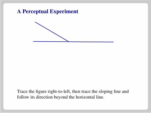
A Perceptual Experiment
Trace the figure right-to-left, then trace the sloping line andfollow its direction beyond the horizontal line.
A Perceptual Experiment
Trace the figure right-to-left, then trace the sloping line andfollow its direction beyond the horizontal line.

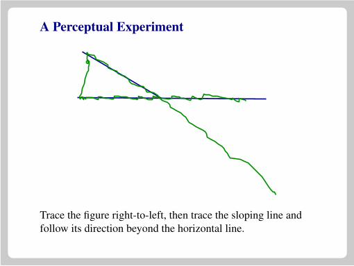
A Perceptual Experiment
Trace the figure right-to-left, then trace the sloping line andfollow its direction beyond the horizontal line.

Seeing in Three Dimensions
• Images projected through the pupil on to the retina aretwo dimensional.
• When we look at the world, we perceive it as threedimensional.
• How do we achieve this sense of depth?

The Eye as a Pinhole Camera
• Because light passes through a narrow hole (the pupil)before begin projected on the retina, the eye acts as apinhole camera.
• Left/Right and Up/Down are inverted.

Perspective
d
h
h’
d’
h′
d′ =h
dh′ =
d′
dh

Perspective Example

Projections
Orthographic Perspective

Perspective as a Depth Cue
• Perspective gives us information about depth for objectsat near to medium distances.
• When there is any hint of perspective, our visualsystems try hard to extract depth information from thescene.
• We can be fooled by the appearance of perspective.

The Ames Room

The Ames Room Explanation

Stereoscopic Vision
• Each of our eyes occupies a different position in space.
• When we look at an object, each eye sees a slightlydifferent image.
• The brain can use the difference between the images toinfer depth information.

A Cube As Seen By The Left And Right Eyes

A Stereopticon and Virtual Reality Helmet

Light and Shade
• The pattern which light makes when it falls on an objectcan give us strong depth information.
• Light and shadow can reveal very fine detail on thesurface of illuminated objects.
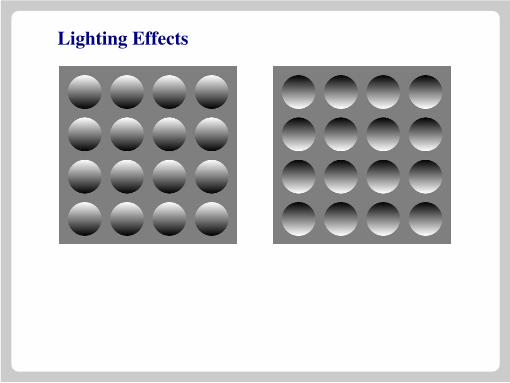
Lighting Effects

A Shaded Relief Topographic Map

Occlusion
Objects which are close, hide objects further away.

Haze and Fog


![Illumination and Vision [Kompatibilit?tsmodus]site.iugaza.edu.ps/aschokry/files/2014/01/Illumination-and-Vision.pdf · to the eyeglass prescription required to correct vision. Instead,](https://img.pdfslide.us/doc/110x75/5fd252f9be8aa43ca370c0a6/illumination-and-vision-kompatibilittsmodussite-to-the-eyeglass-prescription.jpg)