Embed Size (px)
Citation preview
8/22/2019 Human Tendon Stem Cells Better Maintain Their Stemness in Hypoxic Culture Conditions
http://slidepdf.com/reader/full/human-tendon-stem-cells-better-maintain-their-stemness-in-hypoxic-culture-conditions 1/10
Human Tendon Stem Cells Better Maintain TheirStemness in Hypoxic Culture Conditions
Jianying Zhang, James H.-C. Wang*
MechanoBiology Laboratory, Departments of Orthopaedic Surgery, Bioengineering, Mechanical Engineering and Materials Science, and Physical Medicine and
Rehabilitation, University of Pittsburgh, Pittsburgh, Pennsylvania, United States of America
Abstract
Tissues and organs in vivo are under a hypoxic condition; that is, the oxygen tension is typically much lower than in ambientair. However, the effects of such a hypoxic condition on tendon stem cells, a recently identified tendon cell, remainincompletely defined. In cell culture experiments, we subjected human tendon stem cells (hTSCs) to a hypoxic conditionwith 5% O2, while subjecting control cells to a normaxic condition with 20% O2. We found that hTSCs at 5% O2 hadsignificantly greater cell proliferation than those at 20% O2. Moreover, the expression of two stem cell marker genes, Nanogand Oct-4, was upregulated in the cells cultured in 5% O2. Finally, in cultures under 5% O2, more hTSCs expressed the stemcell markers nucleostemin, Oct-4, Nanog and SSEA-4. In an in vivo experiment, we found that when both cell groups wereimplanted with tendon-derived matrix, more tendon-like structures formed in the 5% O2 treated hTSCs than in 20% O2
treated hTSCs. Additionally, when both cell groups were implanted with Matrigel, the 5% O 2 treated hTSCs showed moreextensive formation of fatty, cartilage-like and bone-like tissues than the 20% O2 treated cells. Together, the findings of thisstudy show that oxygen tension is a niche factor that regulates the stemness of hTSCs, and that less oxygen is better formaintaining hTSCs in culture and expanding them for cell therapy of tendon injuries.
Citation: Zhang J, Wang JH-C (2013) Human Tendon Stem Cells Better Maintain Their Stemness in Hypoxic Culture Conditions. PLoS ONE 8(4): e61424.doi:10.1371/journal.pone.0061424
Editor: Sudha Agarwal, Ohio State University, United States of America
Received January 24, 2013; Accepted March 13, 2013; Published April 16, 2013
Copyright: ß 2013 Wang, Zhang. This is an open-access article distributed under the terms of the Creative Commons Attribution License, which permitsunrestricted use, distribution, and reproduction in any medium, provided the original author and source are credited.
Funding: This project was funded by National Institutes of Health grants AR049921 and AR06139. The funders had no role in study design, data collection andanalysis, decision to publish, or preparation of the manuscript.
Competing Interests: The authors have declared that no competing interests exist.
* E-mail: [email protected]
Introduction
Tendons connect muscles to bones to enable joint movement.
As a result, they are subjected to large mechanical loads and hence
are frequently injured. Full recovery of injured tendons requires a
long, complex healing process, particularly in the case of complete
tendon rupture when tendon retraction occurs. Moreover, healed
tendons consist of scar tissue that has lower mechanical strength
than normal tendon tissue. This mechanical weakness not only
impairs normal tendon function and joint kinematics, but also
predisposes patients to further tendon injury [1].
Restoring normal structure and function to injured tendons is
challenging and a number of ways are being discovered to
promote tendon regeneration after injury. Tissue engineering is
one such approach that uses cells, scaffolds and growth factors to
effectively repair or regenerate injured tendons more effectively.
Cell therapy in particular, is important in tissue engineering to
repair injured tendons or other tissues. For example, bone marrow
mesenchymal stem cells (BMSCs) in conjugation with collagen
gels, have been used to repair injured tendons [2] although these
have resulted in ectopic bone formation in rabbit tendon injury
models [3]. In addition, embryonic stem cells (ESCs) have also
been used to repair injured tendons. However, ESCs implantation
could result in teratoma formation, which occurs due to difficulty
in controlling ESCs differentiation in vivo when compared to adult
stem cells such as BMSCs. These and other studies clearly indicate
that stem cells from non-tendinous tissues may not be optimal to
restore the normal structure and function of injured tendons using
cell therapy.Implantation of autologous tenocytes, which are resident tendon
cells responsible for the maintenance and repair of tendons has
resulted only in a slight improvement in tendon quality [4]. A new
type of recently discovered tendon cells called tendon stem cells
(TSCs) have a great potential to repair injured tendons and have
been identified in humans, rabbits, rats and mice [5–7]. Like adult
stem cells, TSCs have the capacity for self-renewal, which enables
them to make more stem cells by cell division and also possess
multi-differentiation potential, which enables them to become
specialized cell types. Under normal conditions, TSCs differentiate
into tenocytes [6]. However, when implanted with engineered
tendon matrix (ETM), TSCs form tendon-like tissues in nude rats
[8]. Therefore, TSCs may be an ideal cell source for tissue
engineering approaches that could effectively repair injuredtendons.
To obtain sufficient numbers of cells for cell therapy of injured
tendons, TSCs must be expanded in culture. However, under
regular culture conditions that use 95% air and 5% CO2, TSCs
tend to differentiate and consequently lose their stemness quickly.
In vivo, tendons, which are collagen-rich structures with only a few
blood vessels, have low oxygen levels when compared to vascular-
rich organs and tissues such as the lungs, heart, liver and kidneys
where the oxygen levels range from 10 to 13% [9]. However, the
effects of low oxygen concentrations on TSCs have not been
completely defined yet. In this study, we tested the hypothesis that
PLOS ONE | www.plosone.org 1 April 2013 | Volume 8 | Issue 4 | e61424

8/22/2019 Human Tendon Stem Cells Better Maintain Their Stemness in Hypoxic Culture Conditions
http://slidepdf.com/reader/full/human-tendon-stem-cells-better-maintain-their-stemness-in-hypoxic-culture-conditions 2/10
under hypoxic conditions TSCs better maintain their stemness.
Indeed, our findings show that low oxygen tension enhances the
stemness of TSCs, which was characterized by quicker cell
proliferation, higher expression of stem cell markers in vitro and
more extensive formation of tendon-like and non-tendon-like
tissues in vivo.
Materials and Methods
Ethics StatementNormal human knee tissues were obtained within 24 hours of
death of donors from the Gift of Hope Organ and Tissue Donor
Network (Elmhurst, IL) with approval from the local ethics
committee (Gift of Hope Organ and Tissue Donor Network).
Written consent from the families was obtained and approved by
the Gift of Hope Organ and Tissue donor Network. Tissue
specimens were obtained for investigation only. The protocol to
use human tendon tissues for subsequent cell culture and animal
studies was approved by the University of Pittsburgh IRB.
This project did not involve human subjects, and the authors
conducting research did not obtain data through intervention or
interaction with individuals or obtain identifiable private infor-
mation.
In addition, protocol for the use of rats for in vivo experimen-tation was approved by the University of Pittsburgh IACUC. All
animal surgery was performed under general anesthesia and efforts
were made to minimize suffering.
Control of Hypoxic and Normoxic Culture ConditionsWe used a dedicated tri-gas incubator (Thermo Scientific
Heracell 150i, Thermo Scientific, and Pittsburgh, PA) to achieve
hypoxic conditions (5% O2 ) in cell culture experiments. Concen-
tration of oxygen in the incubator was precisely controlled by two
gas controllers and an oxygen sensor. Nitrogen and carbon dioxide
gases were supplied using a nitrogen gas controller (Thermo
Scientific) connected to two nitrogen tanks and a carbon dioxide
gas controller connected to two carbon dioxide tanks. The set up
was such that the supply of gas automatically switched from the
first to the second tank when the first tank was empty. To avoid air
flow into the incubator during brief openings of the door the
incubator was separated into three isolated chambers with each
chamber closed by double doors. With these control devices in
place, oxygen concentration in the incubator was maintained at a
constant level of 5% during all cell culture experiments.
To maintain normoxic culture conditions (20% O2 ) a regular
tissue culture incubator (Thermo Scientific) was used. About 20%
O2 concentration was achieved inside the incubator by feeding
95% air and 5% carbon dioxide from tanks.
Human TSC CultureHuman TSC (hTSCs) were obtained from the patellar tendons
of six young adult donors aged 26 to 49 years following our
previously published method [6].
Cell Proliferation ExperimenthTSCs were seeded into 6-well culture plates at a density of
40,000 cells/well in 3 ml DMEM growth medium with 20%
FBS and maintained in the tri-gas incubator to achieve a 5%
O2 culture condition or the regular incubator to provide a 20%
O2 culture condition. Replacement medium for the cells
cultured in the tri-gas incubator was prepared by pre-
conditioning the medium in the tri-gas incubator for at least
30 min before use. The medium was changed every two days
under both hypoxic and normaxic culture conditions. Colony
formation by hTSCs cultured in the two oxygen conditions was
tested by staining with methyl violet. Cell proliferation was
determined by counting cells on days 1, 2, 6 and 12 after
seeding, as previously described [10].
Stem Cell Marker ExpressionTo characterize the stemness of hTSCs in hypoxic and
normaxic culture conditions, we determined differential expression
of stem cell markers in hTSCs in both culture conditions. Cellswere seeded into 12-well plates at a density of 20,000 cells/well
with 1.5 ml medium and cultured either with 5 or 20% O2 for 3–5
days. Expression of four stem cell markers including nucleostemin
(NS), octamer-binding transcription factor 4 (Oct-4), Nanog and
stage-specific embryonic antigen-4 (SSEA-4) was measured using
immunocytochemistry. Briefly, hTSCs were fixed in 4% parafor-
maldehyde in PBS for 20 min at room temperature. For Oct-4,
Nanog and nucleostemin staining fixed cells were treated with
0.5% Triton-X-100 in PBS for 15 min and washed with 2%
mouse or goat serum-PBS for 30 min. The cells were then
incubated with either mouse anti-human Oct-4 (1:500), rabbit
anti-human Nanog (1:500) or goat anti-human nucleostemin
(1:500) overnight at 4̊C. After washing in PBS three times, the cells
were again incubated with either Cy-3-conjugated goat anti-mouseIgG antibodies (1:1000), Cy3-conjugated goat anti-rabbit IgG
(1:500) or Cy-3-conjugated donkey anti-goat IgG antibodies
(1:500) for 2 hrs at room temperature to detect Oct-4, Nanog
and nucleostemin respectively. To stain for SSEA-4, fixed cells
were blocked with 2% mouse serum for 1 hr and incubated with
mouse anti-human SSEA-4 antibody (1:500) for 2 hrs at room
temperature. After subsequent washing with PBS, TSCs were
treated with Cy3-conjugated goat anti-mouse IgG antibody
(1:1000) for 1 hr at room temperature. Stained cells were then
examined using fluorescence microscopy. All antibodies were
obtained from Chemicon International (Temecula, CA), BD
Biosciences (Franklin Lakes, NJ), Neuromics (Edina, MN) or Santa
Cruz Biotechnology Inc. (Santa Cruz, CA).
Multi-differentiation PotentialsThe differentiation capacity of hTSCs in hypoxic and normaxic
culture conditions was examined in vitro by testing their abilities to
undergo adipogenesis, chondrogenesis and osteogenesis. Cells at
passage 1 were seeded into 6-well plates at a density of 24 6 104
cells/well in basic growth medium (DMEM plus 10% FBS) and
cultured in either 5 or 20% O2. To measure adipogenic potential,
hTSCs were cultured in adipogenic induction medium (Millipore,
Billerica, MA) that consists of basic growth medium supplemented
with dexamethasone (1 mM), insulin (10 mg/ml), indomethacin
(100 mM) and isobutylmethylxanthine (0.5 mM). To determine
chondrogenic potential, hTSCs were cultured in basic growth
medium supplemented with proline (40 mg/ml), dexamethasone
(39 ng/ml), TGF-b3 (10 ng/ml), ascorbic 2-phosphate (50 mg/ml),
sodium pyruvate (100 mg/ml) and insulin-transferrin-seleniousacid mix (50 mg/ml) (BD Bioscience, Bedford, MA). Finally,
osteogenic potential of hTSCs in both hypoxic and normaxic
culture conditions was studied by culturing cells in osteogenic
induction medium (Millipore, Billerica, MA) consisting of basic
growth medium supplemented with dexamethasone (0.1 mM),
ascorbic 2-phosphate (0.2 mM) and glycerol 2-phosphate
(10 mM). hTSCs were grown in above three media for 21 days
followed by Oil red O assay for adipogenesis, Safranin O assay for
chondrogenesis and Alizarin red S assay for osteogenesis as
described previously [6].
Hypoxic Effects on Human Tendon Stem Cells
PLOS ONE | www.plosone.org 2 April 2013 | Volume 8 | Issue 4 | e61424
8/22/2019 Human Tendon Stem Cells Better Maintain Their Stemness in Hypoxic Culture Conditions
http://slidepdf.com/reader/full/human-tendon-stem-cells-better-maintain-their-stemness-in-hypoxic-culture-conditions 3/10
Semi-quantification of the Extent of hTSC DifferentiationFor the semi-quantification of cell differentiation, twelve images
of each well were randomly taken under a microscope (Nikon
eclipse, TE2000-U). Then areas with positive staining were
manually identified from each picture and computed by SPOTTM
imaging software (Diagnostic Instruments, Inc., Sterling Heights,
MI). Proportion of positive staining was calculated by dividing the
positively stained area by the total area viewed under the
microscope. These values were obtained for all twelve images of a well and their average was used to represent the percentage of
positive staining, which is the extent of cell differentiation in the
respective induction media described under multi-differentiation
potentials.
Quantitative Real-time PCR (qRT-PCR)To measure the stemness of hTSCs under hypoxic (5% O2 ) and
normaxic culture conditions (20% O2 ) we performed qRT-PCR
analysis. Total RNA was extracted from hTSCs using an RNeasy
Mini Kit with an on-column DNase I digest (Qiagen). First-strand
cDNA was synthesized by reverse transcribing 1 mg total RNA
with SuperScript II (Invitrogen) in a 20 ml reaction volume. The
conditions for cDNA synthesis included: 65uC for 5 min followed
by cooling at 4u
C for 1 min, then 42u
C for 50 min and finally72uC for 15 min. qRT-PCR was carried out using 2 ml cDNA
(approximately 100 ng RNA) in a 25 ml PCR reaction volume
using QIAGEN QuantiTect SYBR Green PCR Kit (Qiagen) in a
Chromo 4 Detector (MJ Research). To determine stemness of
TSCs gene-specific primers of human Oct-4, Nanog, and
tenocyte-related genes, including collagen type I and tenascin C
were used. Glyceraldehyde-3-phosphate dehydrogenase (GAPDH)
was used as an internal control. Forward and reverse primers for
all genes were designed based on previously published sequences
[11–13] and were synthesized by Invitrogen (Carlsbad, CA).
Relative gene expression levels in hTSCs under hypoxic and
normaxic culture conditions were determined using the formula
DDCT=(CTtarget2CTGAPDH )Hypoxia2(CTtarget2CTGAPDH )Nor-
moxia, where CT represents cycle threshold of each RNA sample.
At least three replicates were performed for each gene and eachexperimental condition.
Preparation of hTSCs for in vivo ImplantationhTSCs for implantation were prepared by plating cells from
passage 2 into two 24-well plates at a seeding density of 66104/
well and were allowed to grow in 5 or 20% O2 culture conditions.
After one week, cells under both culture conditions were collected
and each was mixed separately with 0.5 ml 5% ETM made from
rabbit patellar tendon samples according to our previously
published method [8] or 0.5 ml Matrigel (Cat. # 354234, BD
Biosciences, Bedford, MA). The cell-ETM or cell-Matrigel
composites were then reseeded into a 24-well plate and cultured
overnight with 5 or 20% O2 to maintain hypoxic or normaxic
conditions respectively.
In vivo Implantation ExperimentFour 10 weeks old female nude rats weighing between 200–
250 g were used for hTSC implantation experiments. Protocol for
the use of rats for in vivo experimentation was approved by the
University of Pittsburgh IACUC. Before implantation, all rats
were given general anesthesia by intramuscular injection of a
mixture of ketamine hydrochloride (75 mg/kg body weight) and
xylazine hydrochloride (5 mg/kg body weight). Two rats each
were implanted with hTSCs cultured in 5 or 20% O2 conditions.
A total of six distinct wounds were made on the back of each rat
and each wound was filled with a piece of cell-ETM or cell-
Matrigel composite. Three weeks after implantation, the wound
sites were opened and tissues in the area were harvested. The
tissue samples were then immersed in frozen section medium (Neg
50; Richard-Allan Scientific; Kalamazoo, MI) in pre-labeled base
molds and were quickly frozen in 2-methylbutane chilled with
liquid nitrogen. Frozen tissue blocks were then placed on dry ice
and stored in -80uC until further use for histological and
immunohistochemical analyses. At least three replicates wereperformed for each experimental condition.
Detection of hTSC Differentiation in vivo
Frozen tissue blocks were cut into 8 mm thick sections, fixed in
4% paraformaldehyde for 15 min and stained with mouse anti-
human collagen type I (1:100, Millipore, Cat. #MAB1340;
Temecula, CA), mouse anti-human adiponectin (1:300, Millipore;
Cat. #MAB3604; Temecula, CA), mouse anti-human collagen
type II (1:100, Millipore, Cat. #MAB1330, Temecula, CA) and
mouse anti-human osteocalcin (1:200, Abcam, Cat #13418,
Cambridge, MA) at room temperature for 2 hrs. Cy-3 conjugated
goat anti-mouse IgG (1:500, Jackson ImmunoResearch Labora-
tories, Inc., Cat. #115-165-146, West Grove, PA) was used as the
secondary antibody to detect collagen type I, collagen type II and
osteocalcin at room temperature for 2 hrs. FITC-conjugated goatanti-mouse IgM (1:500, Santa Cruz Biotechnology, Cat. #sc-
2082, Santa Cruz, CA) was used as the secondary antibody to
detect adiponectin. The tissue sections were also treated with
Hoechst 33342 (Sigma, Cat. #B2261, St. Louis, MO) to stain
nuclei.
Statistical AnalysisOne-way analysis of variance (ANOVA) followed by either
Fisher’s predicted least-square difference (PLSD) for multiple
comparisons or two tailed, paired or unpaired student t -test were
performed wherever applicable. Differences between two groups
(hypoxic vs. normaxic conditions) were considered significant
when P-value was below 0.05.
Results
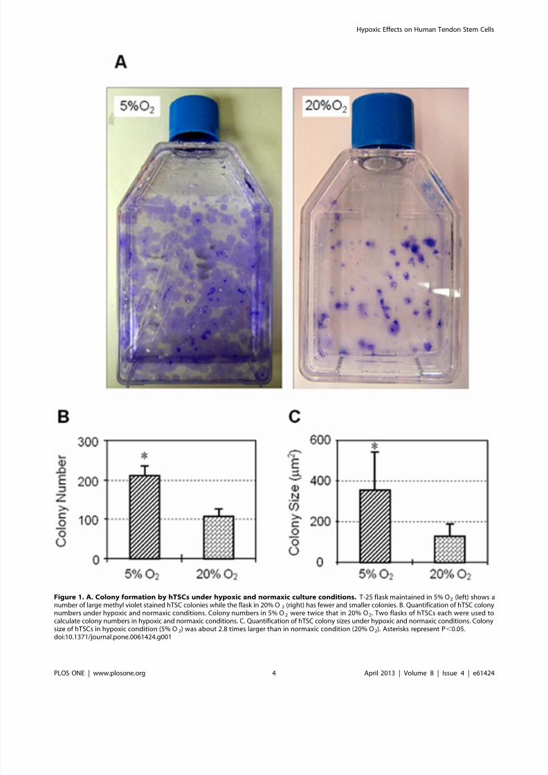
To determine the effects of hypoxia, similar numbers of hTSCs
were seeded and cultured in both hypoxic and normaxic
conditions. We found that hTSCs cultured in 5% O2 formed
more colonies that were also larger than cells cultured in 20% O2
(Figure 1A, 1B). Colonies formed in 5% O2 were twice as many as
those in 20% O2 (Figure 1B) and the colony size in 5% O2 was on
an average about 2.8 times larger than those in 20% O2
(Figure 1C). In addition, proliferation of hTSCs in both culture
conditions increased in the days following seeding and the number
of cells in 5% O2 was higher than in 20% O2 from day 1 through
days 2, 6 and 12 (Figure 2).
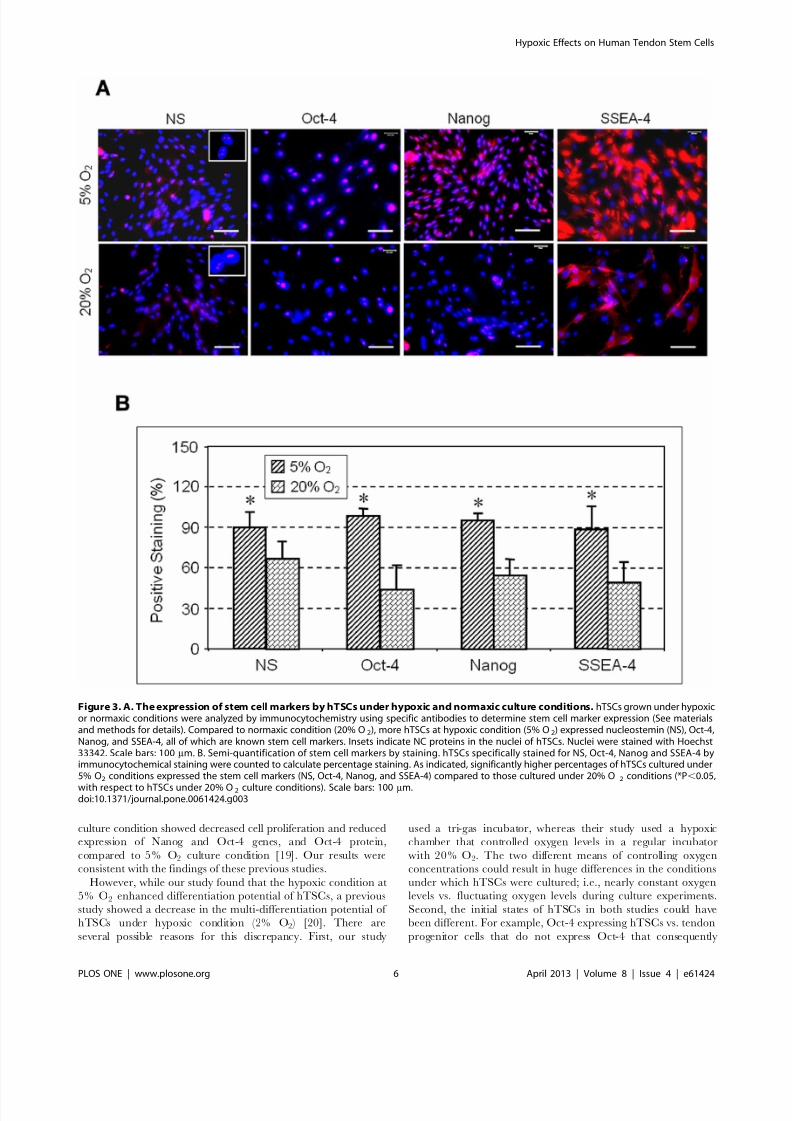
Expression of stem cell markers NS, Oct-4, Nanog and SSEA-4
determined by immunocytochemistry was also higher in coloniescultured in 5% O2 compared to hTSCs grown in 20% O2
(Figure 3A). Semi-quantification of the immuno-stained cells
further showed that more than 90% of hTSCs cultured in 5% O2
were NS positive compared to 66% in hTSCs cultured in 20% O2.
Oct-4 expression was also higher (98%) in hTSCs cultured at 5%
O2 when compare to cells cultured in 20% O2 (44%). Similarly,
the expression of Nanog and SSEA-4 in hTSCs cultured in 5% O2
was 95 and 88% respectively when compared to the lower
percentages (55 and 49%) observed in hTSCs cultured in 20% O2
(Figure 3B). Consistent with these results RT-PCR analysis also
showed higher expression levels of both Oct-4 and Nanog genes in
Hypoxic Effects on Human Tendon Stem Cells
PLOS ONE | www.plosone.org 3 April 2013 | Volume 8 | Issue 4 | e61424
8/22/2019 Human Tendon Stem Cells Better Maintain Their Stemness in Hypoxic Culture Conditions
http://slidepdf.com/reader/full/human-tendon-stem-cells-better-maintain-their-stemness-in-hypoxic-culture-conditions 4/10
Figure 1. A. Colony formation by hTSCs under hypoxic and normaxic culture conditions. T-25 flask maintained in 5% O2 (left) shows anumber of large methyl violet stained hTSC colonies while the flask in 20% O 2 (right) has fewer and smaller colonies. B. Quantification of hTSC colonynumbers under hypoxic and normaxic conditions. Colony numbers in 5% O2 were twice that in 20% O2. Two flasks of hTSCs each were used tocalculate colony numbers in hypoxic and normaxic conditions. C. Quantification of hTSC colony sizes under hypoxic and normaxic conditions. Colonysize of hTSCs in hypoxic condition (5% O2) was about 2.8 times larger than in normaxic condition (20% O2). Asterisks represent P,0.05.doi:10.1371/journal.pone.0061424.g001
Hypoxic Effects on Human Tendon Stem Cells
PLOS ONE | www.plosone.org 4 April 2013 | Volume 8 | Issue 4 | e61424

8/22/2019 Human Tendon Stem Cells Better Maintain Their Stemness in Hypoxic Culture Conditions
http://slidepdf.com/reader/full/human-tendon-stem-cells-better-maintain-their-stemness-in-hypoxic-culture-conditions 5/10
5% O2 compared to 20% O2 (Figure 4). Cells cultured in both
conditions showed no significant difference in the expression of the
tenocyte-related gene, collagen type I, but the level of tenascin C
expression was more than 2-fold higher in 5% O2 compared to
20% O2 (Figure 5A). Moreover, the expressions of non-tenocyte-related genes Sox-9 and Runx-2 were significantly lower in 5% O2
than in 20% O2 culture conditions while expression of PPARc was
marginally lower (Figure 5B).
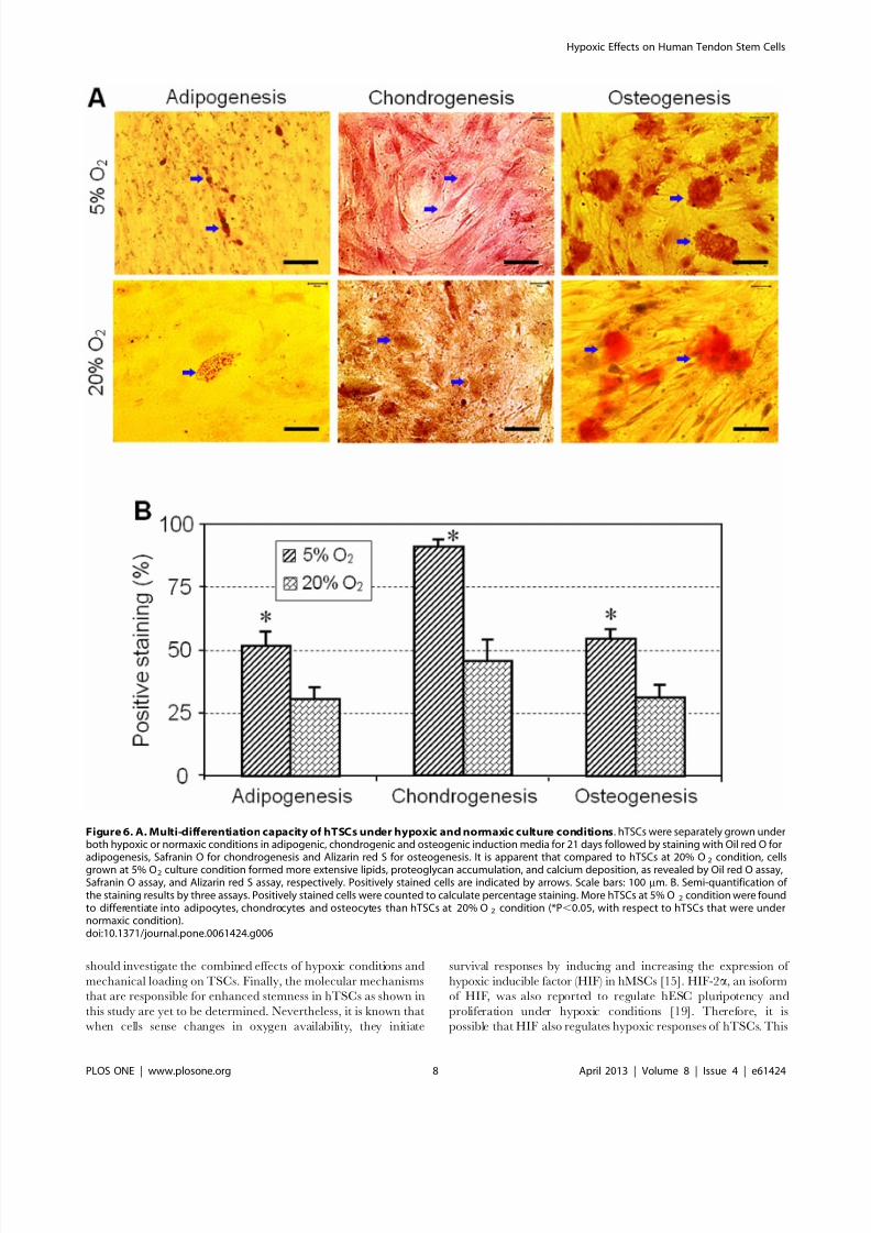
We next examined the multi-differentiation capacity of TSCs
under hypoxic and normaxic culture conditions. After 21 days in
culture, the degree of adipogenesis, chondrogenesis and osteogen-
esis of hTSCs was more extensive in 5% O2 condition compared
to 20% O2 (Figure 6A). Semi-quantitative analysis was also
consistent with these results with the percentages of hTSCs that
differentiated into adipocytes, osteocytes and chondrocytes about
51, 90 and 54% respectively in 5% O2 conditions when compared
to lesser percentages of 31, 46 and 31% respectively in 20% O2
(Figure 6B).
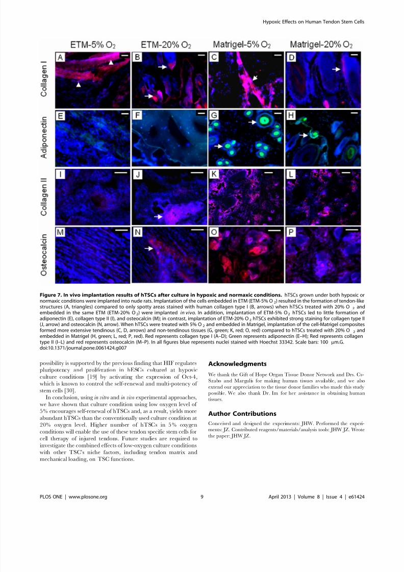
To further characterize hTSCs after exposure to hypoxic andnormaxic conditions, we implanted the cells grown under the two
conditions into nude rats subcutaneously. Three weeks after
implantation hTSCs cultured in 5% O2 and embedded in ETM
resulted in extensive formation of bands that corresponded to
tendon-like structures, as evidenced by strong staining for human
collagen type I (hCT-I). In contrast, hTSCs cultured in 20% O 2
before embedding in ETM and transplantation, showed only
discreet areas which were stained positive for hCT-I. Staining for
adiponectin (a marker for adipogenesis), collagen type II (a marker
for chondrogenesis) and osteocalcin (a marker for osteogenesis)
showed minimal formation of fatty, cartilage, and bony tissues in
5% O2 treated hTSCs and embedded in ETM (ETM-5% O2 )
whereas the cells treated with 20% O2 and embedded in ETM
(ETM-20% O2 ) formed well- developed cartilage and bony tissues.
Similarly, when 5 and 20% O2 treated hTSCs were embedded in
Matrigel and implanted, we observed formation of all four types of
tissues (tendinous, fatty, cartilage-like and bony tissues), which was
more extensive in 5% O2 treated hTSCs compared to 20% O2
treated hTSCs (Figure 7). Semi-quantitative analysis further
showed that compared to normaxic condition, hypoxic condition
resulted in higher amounts of both tendinous protein (collagen
type I) and non-tendinous proteins (adiponectin, collagen type II
and osteocalcin) (data not shown).
Discussion
Tendon injury is common in both occupational and athletic
settings. Currently, there are no effective means to restore normal
structure and function to injured tendons. TSCs, which were only
recently identified, are tendon-specific adult stem cells that are
thought to play a critical role in the repair of injured tendons.
Therefore, TSCs may be an optimal cell source for effective tissue
engineering of injured tendons. However, the major obstacle using
such cell therapy is that once TSCs are isolated from tendons and
grown in a conventional in vitro environment, they tend to
differentiate quickly. Considering that TSCs in vivo are under
hypoxic conditions due to poor vascularity in tendon substances,
we designed this study to investigate the effects of hypoxic
conditions on hTSCs. By performing cell culture experiments, we
have shown that the stemness of hTSCs cultured in 5% O2 was
better than hTSCs at 20% oxygen levels. Specifically, under the
hypoxic culture condition (5% O2 ), growth of hTSCs was faster,
higher number of cells expressed stem cell markers (NS, Oct-4,
Nanog and SSEA-4) and the expression level of stem cell marker
genes (Oct-4 and Nanog) was also significantly higher. In addition,
while tenocyte-related gene expression levels were similar under
both hypoxic and normaxic conditions (5% O2 vs. 20% O2 ), non-
tenocyte related gene expression at hypoxic condition wassignificantly lower than in normaxic condition. Moreover, hTSCs
cultured under the hypoxic condition (5% O2 ) exhibited more
potent multi-differentiation capacity in terms of adipogenesis,
chondrogenesis and osteogenesis. By performing an in vivo
implantation experiment, we were also able to show that when
implanted together with ETM, hTSCs in hypoxic condition
produced more extensive tendon-like tissues than in normaxic
condition. The hypoxic condition also resulted in more tendinous
and non-tendinous tissues than normaxic condition when both
were implanted with Matrigel. The in vivo results further showed
that hypoxic condition enhanced multi-differentiation potential of
hTSCs.
The findings of this study show that oxygen is an important
niche factor for the maintenance of stemness by hTSCs. These
findings also indicate that for effective tissue engineering of
injured tendons, TSCs should be cultured in a hypoxic
environment. This hypoxic condition can promote TSCs’ self-
renewal, thus allowing sufficient numbers of TSCs to be
obtained for tissue engineering, which may repair injured
tendons more effectively. The high self-renewal rate of hTSCs
under a hypoxic condition is consistent with the concept that
adult stem cells like hTSCs ensure maintenance of their pool for
tissue repair or regeneration when the tissue is injured [14].
Finally, this study also indicates that caution should be exercised
before using the so-called hyperbaric oxygen therapy to treat
injured tendons, at levels as high as .20% O2 that could
potentially deplete the TSCs pool quickly by promoting their
differentiation into specialized cell types and consequently could
hinder the repair of tendons after re-injury.Many studies have investigated the effects of various hypoxic
conditions on cells. For example, compared to normaxic condition
(20% O2 ), hypoxic condition (1.5% to 5% O2 ) increases
proliferation of human mesenchymal stem cells (hMSCs) [15]. In
addition, hMSCs grown in 2% O2 exhibited enhanced colony-
forming capabilities and had a higher expression of Oct-4 [16,17].
Hypoxic conditions also produced greater numbers of stem cell
colonies that proliferated more rapidly in culture. Rat MSCs
cultured in 5% O2 produced more bone than cells cultured in 20%
O2 when the cells were loaded into porous ceramic cubes and
implanted into animals [18]. In addition, hESCs in 20% O2
Figure 2. Proliferation of hTSCs cultured under hypoxic andnormaxic culture conditions. hTSCs were grown in DMEM growthmedium with FBS under hypoxic or normaxic conditions and colonyformation was determined by counting cells stained with methyl violet.While the cells grew at both culture conditions, at all time points (days1, 2, 6, and 12), hTSCs at 5% O2 grew significantly quicker than at 20%O2.doi:10.1371/journal.pone.0061424.g002
Hypoxic Effects on Human Tendon Stem Cells
PLOS ONE | www.plosone.org 5 April 2013 | Volume 8 | Issue 4 | e61424
8/22/2019 Human Tendon Stem Cells Better Maintain Their Stemness in Hypoxic Culture Conditions
http://slidepdf.com/reader/full/human-tendon-stem-cells-better-maintain-their-stemness-in-hypoxic-culture-conditions 6/10
culture condition showed decreased cell proliferation and reducedexpression of Nanog and Oct-4 genes, and Oct-4 protein,
compared to 5% O2 culture condition [19]. Our results were
consistent with the findings of these previous studies.
However, while our study found that the hypoxic condition at
5% O2 enhanced differentiation potential of hTSCs, a previous
study showed a decrease in the multi-differentiation potential of
hTSCs under hypoxic condition (2% O2 ) [20]. There are
several possible reasons for this discrepancy. First, our study
used a tri-gas incubator, whereas their study used a hypoxic
chamber that controlled oxygen levels in a regular incubator
with 20% O2. The two different means of controlling oxygen
concentrations could result in huge differences in the conditions
under which hTSCs were cultured; i.e., nearly constant oxygen
levels vs. fluctuating oxygen levels during culture experiments.
Second, the initial states of hTSCs in both studies could have
been different. For example, Oct-4 expressing hTSCs vs. tendon
progenitor cells that do not express Oct-4 that consequently
Figure 3. A. The expression of stem cell markers by hTSCs under hypoxic and normaxic culture conditions. hTSCs grown under hypoxicor normaxic conditions were analyzed by immunocytochemistry using specific antibodies to determine stem cell marker expression (See materialsand methods for details). Compared to normaxic condition (20% O2), more hTSCs at hypoxic condition (5% O2) expressed nucleostemin (NS), Oct-4,Nanog, and SSEA-4, all of which are known stem cell markers. Insets indicate NC proteins in the nuclei of hTSCs. Nuclei were stained with Hoechst33342. Scale bars: 100 mm. B. Semi-quantification of stem cell markers by staining. hTSCs specifically stained for NS, Oct-4, Nanog and SSEA-4 byimmunocytochemical staining were counted to calculate percentage staining. As indicated, significantly higher percentages of hTSCs cultured under5% O2 conditions expressed the stem cell markers (NS, Oct-4, Nanog, and SSEA-4) compared to those cultured under 20% O 2 conditions (*P,0.05,
with respect to hTSCs under 20% O2 culture conditions). Scale bars: 100 mm.doi:10.1371/journal.pone.0061424.g003
Hypoxic Effects on Human Tendon Stem Cells
PLOS ONE | www.plosone.org 6 April 2013 | Volume 8 | Issue 4 | e61424

8/22/2019 Human Tendon Stem Cells Better Maintain Their Stemness in Hypoxic Culture Conditions
http://slidepdf.com/reader/full/human-tendon-stem-cells-better-maintain-their-stemness-in-hypoxic-culture-conditions 7/10
resulted in differences in cellular responses to similar hypoxic
conditions. Finally, there could be differences in experimental
conditions used in hTSCs culture including the density of cells,
depth of medium, pre-conditioning of medium and cellular
respiration, all of which could alter the oxygen tension at the
surface of cultured cells, consequently leading to differential
responses of hTSCs to hypoxic conditions.
Because there are no specific stem cell markers for hTSCs, we
used general stem cell markers (NS, Oct-4, Nanog, and SSEA-4) to
characterize their stemness under both hypoxic and normaxic
conditions. Nucleostemin (NS), that controls cell cycle progression,
is exclusively expressed in stem cells, and is therefore not expressed
in committed and terminally differentiated cells [21]. Nanog, a
unique homeobox transcription factor, was reported to be
expressed in pluripotent stem cells, and its expression was
associated with stem cell differentiation [22]. Typically expressedin embryonic stem cells (ESCs) during development, Oct-4 is a
transcription factor that is known to mediate pluripotency in ESCs
[23]. Oct-4 is also essential for maintaining pluripotent stem cells,
and is not expressed in differentiated cells [24]. Finally, SSEA-4 is
a transcription factor specific to undifferentiated pluripotent
human or mouse stem cells [25–27]. Thus, the higher expression
levels of these stem cell markers in hypoxic condition (5% O2 )
observed in this study indicate that more hTSCs were kept in an
undifferentiated state and self-renewed when they were cultured at
hypoxic condition (5% O2 ) than at normaxic condition (20% O2 ).
It is generally accepted that 3 to 5% oxygen levels are present in
tissues, although the actual O2 concentration in situ depends on
vascularization of the tissue and its metabolic activity [15]. To our
best knowledge, the physiological oxygen tension of the humanpatellar tendon remains unknown. In the articular cartilage,
however, oxygen tension is known to be less than 10% at the
surface and less than 1% in the deepest layer [28]. Considering
that tendons are largely avascular, it is likely that their oxygen
tension is higher than 1% but lower than 10%. This is the reason
we chose a 5% O2 level in this study. Use of 5% O2 level also
makes it possible to control oxygen levels in an incubator more
precisely, as too low levels of oxygen, which creates a high gradient
of oxygen against the environment, is technically demanding in
terms of precisely controlling constant oxygen levels to culture
cells.
There are a few limitations associated with this study. First, we
grew hTSCs in plastic dishes, which itself is ‘‘foreign’’ to hTSCs
and therefore may cause cell differentiation in culture. Ourprevious study showed that TSCs grown on tendon matrix coated
plastic surfaces can encourage self-renewal of TSCs. Therefore, it
seems reasonable to speculate that culture of hTSCs in tendon
matrix under a hypoxic condition will result in an even higher
stemness of hTSCs, especially in long term cell culture. Second,
tendons in vivo are constantly subjected to mechanical loading
because of their role in the transmission of muscular forces to
bones. Mechanical loading, however, was not included in our cell
culture experiment although, our previous study showed that
mechanical loading itself can regulate TSC functions including
proliferation and differentiation [29]. Therefore, future studies
Figure 4. Stem cell gene analysis by qRT-PCR. Total RNA extractedfrom hTSCs grown under hypoxic or normaxic conditions was used tosynthesize cDNA, which was used as a template in qRT-PCR usingprimers specific to Oct-4 and Nanog. GAPDH was used as an internalcontrol. Y- axis represents relative gene expression when compared toGAPDH expression levels. Ct values were normalized against hTSCscultured under 20% O2. Both stem cell marker genes (Oct-4 and Nanog)cultured at 5% O2 culture conditions were expressed at significantlyhigher g005levels than those cultured at 20% O2 culture conditions.doi:10.1371/journal.pone.0061424.g004
Figure 5. A. Tenocyte related gene expression by hTSCs underhypoxic and normaxic culture conditions. Total RNA extractedfrom hTSCs grown under hypoxic or normaxic conditions was used tosynthesize cDNA, which was used as a template in qRT-PCR usingprimers specific to Collagen-1 and Tenascin C. GAPDH was used as aninternal control. Y- axis represents relative gene expression whencompared to GAPDH expression levels. Ct values were normalizedagainst hTSCs cultured under 20% O2. At both oxygen conditions (5%and 20% O2), there was no significant difference in the expression of collagen type I, but the expression of tenascin C in the hypoxic groupwas significantly higher than in the normaxic group (*P,0.05). B. Non-
tenocyte related gene expression by hTSCs under the above twooxygen conditions. Total RNA extracted from hTSCs grown underhypoxic or normaxic conditions was used to synthesize cDNA, whichwas used as a template in qRT-PCR using primers specific to PPARc,Sox-9 and Runx-2. GAPDH was used as an internal control. Y- axisrepresents relative gene expression when compared to GAPDHexpression. Ct values were normalized against hTSCs cultured under20% O2. The cellular expression of PPARc, a marker for adipogenesis,was not significantly different in 5 and 20% O2 conditions. However,Sox-9 and Runx-2 (markers for chondrogenesis and osteogenesis,respectively) were expressed at significantly lower levels when hTSCswere cultured at 5% O2 condition in comparison to 20% O2 (*P,0.05,respective to hTSCs that were under normaxic conditions).doi:10.1371/journal.pone.0061424.g005
Hypoxic Effects on Human Tendon Stem Cells
PLOS ONE | www.plosone.org 7 April 2013 | Volume 8 | Issue 4 | e61424
8/22/2019 Human Tendon Stem Cells Better Maintain Their Stemness in Hypoxic Culture Conditions
http://slidepdf.com/reader/full/human-tendon-stem-cells-better-maintain-their-stemness-in-hypoxic-culture-conditions 8/10
should investigate the combined effects of hypoxic conditions and
mechanical loading on TSCs. Finally, the molecular mechanisms
that are responsible for enhanced stemness in hTSCs as shown in
this study are yet to be determined. Nevertheless, it is known that
when cells sense changes in oxygen availability, they initiate
survival responses by inducing and increasing the expression of
hypoxic inducible factor (HIF) in hMSCs [15]. HIF-2a, an isoform
of HIF, was also reported to regulate hESC pluripotency and
proliferation under hypoxic conditions [19]. Therefore, it is
possible that HIF also regulates hypoxic responses of hTSCs. This
Figure 6. A. Multi-differentiation capacity of hTSCs under hypoxic and normaxic culture conditions. hTSCs were separately grown underboth hypoxic or normaxic conditions in adipogenic, chondrogenic and osteogenic induction media for 21 days followed by staining with Oil red O foradipogenesis, Safranin O for chondrogenesis and Alizarin red S for osteogenesis. It is apparent that compared to hTSCs at 20% O2 condition, cellsgrown at 5% O2 culture condition formed more extensive lipids, proteoglycan accumulation, and calcium deposition, as revealed by Oil red O assay,Safranin O assay, and Alizarin red S assay, respectively. Positively stained cells are indicated by arrows. Scale bars: 100 mm. B. Semi-quantification of the staining results by three assays. Positively stained cells were counted to calculate percentage staining. More hTSCs at 5% O 2 condition were foundto differentiate into adipocytes, chondrocytes and osteocytes than hTSCs at 20% O2 condition (*P,0.05, with respect to hTSCs that were undernormaxic condition).doi:10.1371/journal.pone.0061424.g006
Hypoxic Effects on Human Tendon Stem Cells
PLOS ONE | www.plosone.org 8 April 2013 | Volume 8 | Issue 4 | e61424
8/22/2019 Human Tendon Stem Cells Better Maintain Their Stemness in Hypoxic Culture Conditions
http://slidepdf.com/reader/full/human-tendon-stem-cells-better-maintain-their-stemness-in-hypoxic-culture-conditions 9/10
possibility is supported by the previous finding that HIF regulatespluripotency and proliferation in hESCs cultured at hypoxic
culture conditions [19] by activating the expression of Oct-4,
which is known to control the self-renewal and multi-potency of
stem cells [30].
In conclusion, using in vitro and in vivo experimental approaches,
we have shown that culture condition using low oxygen level of
5% encourages self-renewal of hTSCs and, as a result, yields more
abundant hTSCs than the conventionally used culture condition at
20% oxygen level. Higher number of hTSCs in 5% oxygen
conditions will enable the use of these tendon specific stem cells for
cell therapy of injured tendons. Future studies are required to
investigate the combined effects of low-oxygen culture conditions
with other TSC’s niche factors, including tendon matrix and
mechanical loading, on TSC functions.
Acknowledgments
We thank the Gift of Hope Organ Tissue Donor Network and Drs. Cs-
Szabo and Margulis for making human tissues available, and we also
extend our appreciation to the tissue donor families who made this study
possible. We also thank Dr. Im for her assistance in obtaining human
tissues.
Author Contributions
Conceived and designed the experiments: JHW. Performed the experi-
ments: JZ. Contributed reagents/materials/analysis tools: JHW JZ. Wrote
the paper: JHW JZ.
Figure 7. In vivo implantation results of hTSCs after culture in hypoxic and normaxic conditions. hTSCs grown under both hypoxic or
normaxic conditions were implanted into nude rats. Implantation of the cells embedded in ETM (ETM-5% O2) resulted in the formation of tendon-likestructures (A, triangles) compared to only spotty areas stained with human collagen type I (B, arrows) when hTSCs treated with 20% O 2 andembedded in the same ETM (ETM-20% O2) were implanted in vivo. In addition, implantation of ETM-5% O2 hTSCs led to little formation of adiponectin (E), collagen type II (I), and osteocalcin (M); in contrast, implantation of ETM-20% O2 hTSCs exhibited strong staining for collagen type II(J, arrow) and osteocalcin (N, arrow). When hTSCs were treated with 5% O2 and embedded in Matrigel, implantation of the cell-Matrigel compositesformed more extensive tendinous (C, D, arrows) and non-tendinous tissues (G, green; K, red; O, red) compared to hTSCs treated with 20% O 2 andembedded in Matrigel (H, green; L, red; P, red). Red represents collagen type I (A–D); Green represents adiponectin (E–H); Red represents collagentype II (I–L) and red represents osteocalcin (M–P). In all figures blue represents nuclei stained with Hoechst 33342. Scale bars: 100 mm.G.doi:10.1371/journal.pone.0061424.g007
Hypoxic Effects on Human Tendon Stem Cells
PLOS ONE | www.plosone.org 9 April 2013 | Volume 8 | Issue 4 | e61424
8/22/2019 Human Tendon Stem Cells Better Maintain Their Stemness in Hypoxic Culture Conditions
http://slidepdf.com/reader/full/human-tendon-stem-cells-better-maintain-their-stemness-in-hypoxic-culture-conditions 10/10
References
1. Butler DL, Juncosa-Melvin N, Boivin GP, Galloway MT, Shearn JT, et al.
(2008) Functional tissue engineering for tendon repair: A multidisciplinary
strategy using mesenchymal stem cells, bioscaffolds, and mechanical stimulation.
J Orthop Res 26: 1–9.
2. Awad HA, Boivin GP, Dressler MR, Smith FN, Young RG, et al. (2003) Repair
of patellar tendon injuries using a cell-collagen composite. J Orthop Res 21:
420–431.
3. Harris MT, Butler DL, Boivin GP, Florer JB, Schantz EJ, et al. (2004)
Mesenchymal stem cells used for rabbit tendon repair can form ectopic bone and
express alkaline phosphatase activity in constructs. J Orthop Res 22: 998–1003.
4. Cao Y, Liu Y, Liu W, Shan Q, Buonocore SD, et al. (2002) Bridging tendon
defects using autologous tenocyte engineered tendon in a hen model. Plast
Reconstr Surg 110: 1280–1289.
5. Bi Y, Ehirchiou D, Kilts TM, Inkson CA, Embree MC, et al. (2007)
Identification of tendon stem/progenitor cells and the role of the extracellular
matrix in their niche. Nat Med 13: 1219–1227.
6. Zhang J, Wang JH (2010) Characterization of differential properties of rabbit
tendon stem cells and tenocytes. BMC Musculoskelet Disord 11: 10.
7. Rui YF, Lui PP, Li G, Fu SC, Lee YW, et al. (2010) Isolation and
characterization of multipotent rat tendon-derived stem cells. Tissue Eng
Part A 16: 1549–1558.
8. Zhang J, Li B, Wang JH (2011) The role of engineered tendon matrix in the
stemness of tendon stem cells in vitro and the promotion of tendon-like tissue
formation in vivo. Biomaterials 32: 6972–6981.
9. D’Ippolito G, Diabira S, Howard GA, Roos BA, Schiller PC (2006) Low oxygen
tension inhibits osteogenic differentiation and enhances stemness of human
MIAMI cells. Bone 39: 513–522.10. Zhang J, Wang JH (2010) Production of PGE(2) increases in tendons subjected
to repetitive mechanical loading and induces differentiation of tendon stem cells
into non-tenocytes. J Orthop Res 28: 198–203.
11. Huangfu D, Osafune K, Maehr R, Guo W, Eijkelenboom A, et al. (2008)
Induction of pluripotent stem cells from primary human fibroblasts with only
Oct4 and Sox2. Nat Biotechnol 26: 1269–1275.
12. Liu H, Fan H, Wang Y, Toh SL, Goh JC (2008) The interaction between a
combined knitted silk scaffold and microporous silk sponge with human
mesenchymal stem cells for ligament tissue engineering. Biomaterials 29: 662–
674.
13. Risbud MV, Guttapalli A, Tsai TT, Lee JY, Danielson KG, et al. (2007)
Evidence for skeletal progenitor cells in the degenerate human intervertebral
disc. Spine (Phila Pa 1976) 32: 2537–2544.
14. Ivanovic Z (2009) Hypoxia or in situ normoxia: The stem cell paradigm. J Cell
Physiol 219: 271–275.
15. Lavrentieva A, Majore I, Kasper C, Hass R (2010) Effects of hypoxic cultureconditions on umbilical cord-derived human mesenchymal stem cells. CellCommun Signal 8: 18.
16. Grayson WL, Zhao F, Izadpanah R, Bunnell B, Ma T (2006) Effects of hypoxiaon human mesenchymal stem cell expansion and plasticity in 3D constructs.
J Cell Physiol 207: 331–339.17. Grayson WL, Zhao F, Bunnell B, Ma T (2007) Hypoxia enhances proliferation
and tissue formation of human mesenchymal stem cells. Biochem Biophys Res
Commun 358: 948–953.18. Lennon DP, Edmison JM, Caplan AI (2001) Cultivation of rat marrow-derived
mesenchymal stem cells in reduced oxygen tension: effects on in vitro andin vivo osteochondrogenesis. J Cell Physiol 187: 345–355.
19. Forristal CE, Wright KL, Hanley NA, Oreffo RO, Houghton FD (2010)Hypoxia inducible factors regulate pluripotency and proliferation in humanembryonic stem cells cultured at reduced oxygen tensions. Reproduction 139:85–97.
20. Lee WY, Lui PP, Rui YF (2012) Hypoxia-mediated efficient expansion of humantendon-derived stem cells in vitro. Tissue Eng Part A 18: 484–498.
21. Tsai RY, McKay RD (2002) A nucleolar mechanism controlling cellproliferation in stem cells and cancer cells. Genes Dev 16: 2991–3003.
22. Pan G, Thomson JA (2007) Nanog and transcriptional networks in embryonicstem cell pluripotency. Cell Res 17: 42–49.
23. Greco SJ, Liu K, Rameshwar P (2007) Functional similarities among genesregulated by OCT4 in human mesenchymal and embryonic stem cells. StemCells 25: 3143–3154.
24. Pesce M, Scholer HR (2001) Oct-4: gatekeeper in the beginnings of mammaliandevelopment. Stem Cells 19: 271–278.
25. Gang EJ, Bosnakovski D, Figueiredo CA, Visser JW, Perlingeiro RC (2007)SSEA-4 identifies mesenchymal stem cells from bone marrow. Blood 109: 1743– 1751.
26. Henderson JK, Draper JS, Baillie HS, Fishel S, Thomson JA, et al. (2002)Preimplantation human embryos and embryonic stem cells show comparableexpression of stage-specific embryonic antigens. Stem Cells 20: 329–337.
27. Cui L, Johkura K, Yue F, Ogiwara N, Okouchi Y, et al. (2004) Spatialdistribution and initial changes of SSEA-1 and other cell adhesion-relatedmolecules on mouse embryonic stem cells before and during differentiation.
J Histochem Cytochem 52: 1447–1457.28. Grimshaw MJ, Mason RM (2000) Bovine articular chondrocyte function in vitro
depends upon oxygen tension. Osteoarthritis Cartilage 8: 386–392.29. Zhang J, Wang JH (2010) Mechanobiological response of tendon stem cells:
implications of tendon homeostasis and pathogenesis of tendinopathy. J OrthopRes 28: 639–643.
30. Keith B, Simon MC (2007) Hypoxia-inducible factors, stem cells, and cancer.Cell 129: 465–472.
Hypoxic Effects on Human Tendon Stem Cells
PLOS ONE | www.plosone.org 10 April 2013 | Volume 8 | Issue 4 | e61424