Embed Size (px)
Citation preview
Human Reproduction
Male & Female Systems & Menstration
Make a Sperm & Ovum
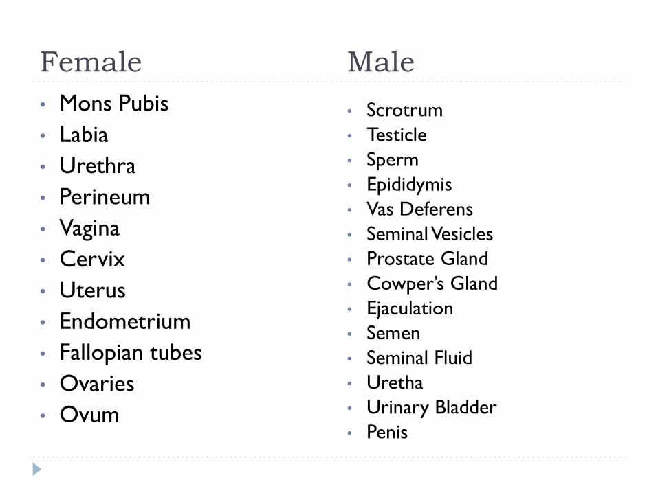
Female
• Mons Pubis
• Labia
• Urethra
• Perineum
• Vagina
• Cervix
• Uterus
• Endometrium
• Fallopian tubes
• Ovaries
• Ovum
Male
• Scrotrum
• Testicle
• Sperm
• Epididymis
• Vas Deferens
• Seminal Vesicles
• Prostate Gland
• Cowper’s Gland
• Ejaculation
• Semen
• Seminal Fluid
• Uretha
• Urinary Bladder
• Penis
A five year old girl asked the question that every parent
dreads, ―Mommy, how are babies made?‖ The mom did
her best to explain, but the daughter still looked
confused.
―But what about kittens?‖ she asked.
―Well, it is exactly the same way.‖ I said.
―Wow!‖ she said excitedly, ―My Daddy can
do anything!‖
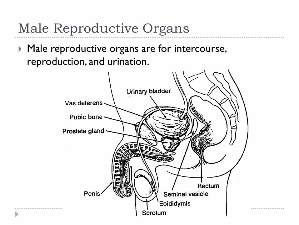
Male Reproductive Organs
Male reproductive organs are for intercourse,
reproduction, and urination.
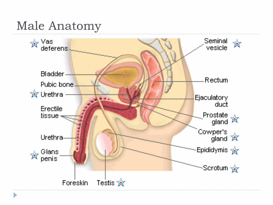
Male Anatomy
Male Timeline
• Infancy
– Erections begin
• Ages 11-14
– Secondary sex characteristics appear
• Ages 13-16
– Sperm produced in adult amounts (puberty)
• Late teens
– Peak sexual urges for boys
• Throughout life
– If good health is present, there is the sex urge and ability to
father children

Scrotum (j)
A sac-like pouch located behind the penis that holds each
testes and helps regulate temperature for sperm
production.

Testicles or Testes (i)
• The two testes are small organs that lie in the scrotum and produce sperm and the male hormone testosterone.
• The testicles are the male sex gland.
• The testicles are outside the body because the male sperm that is manufactured in the testes need cooler-than-body temperature for normal growth and development.
• They are the counterpart to the female ovary.
• Loss of one does not impair the function of the other.
• Four to five billion sperm cells are produced each month.
Testosterone
• The male reproductive hormone made by the testicles which causes the changes of puberty.
• This hormone causes secondary sex characteristics, production of sperm and sexual urge.
• It is produced in the testicles and enters the bloodstream at a fairly constant rate.

Sperm• The microscopic cells produced by
the male's testicles which can fertilize the female's ovum.
• They are tiny, living cells 100 times smaller than a pencil dot. (the smallest cell in a mans body
• Enough sperm would fit on the head of a pin to re-populate the earth if each sperm fertilized an egg.
• It is destroyed by warm body temperature, acidic environment.
• It can survive in a women’s body for 5-8 days.
• Any sperm not ejaculated are passed in the urine.
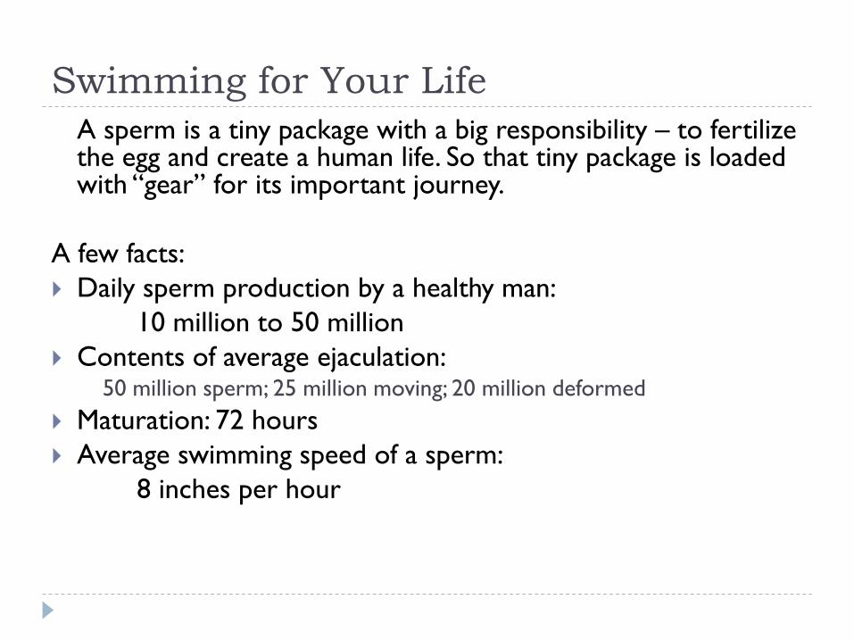
Swimming for Your Life
A sperm is a tiny package with a big responsibility – to fertilize the egg and create a human life. So that tiny package is loaded with ―gear‖ for its important journey.
A few facts:
Daily sperm production by a healthy man:
10 million to 50 million
Contents of average ejaculation:50 million sperm; 25 million moving; 20 million deformed
Maturation: 72 hours
Average swimming speed of a sperm:
8 inches per hour
Epididymis (h)
• The structure that forms a mass over the back and
upper part of each testes.
• Sperm are stored there for as long as six weeks while
they ripen to maturity.
Vas Deferens (e)
• two long, thin tubes that serve as a passageway for sperm
and a place for sperm storage.
• The contraction of the vas deferens along with the action
of the cilia help transport the sperm through the vas
deferens.

Seminal Vesicles (a)
two small glands that secrete a fluid that nourishes and
enables the sperm to move.
Prostate Gland (b)
• surround the urethra beneath the bladder. The gland
secretes an alkaline fluid that neutralizes the acid found in
the male urethra and the female reproductive tract.
• Without the action of the secretions of the prostate
gland, many sperm would die and fertilization of an ovum
would be impossible.
Cowper’s Gland (c)
• Two small pea-sized glands located beneath the
prostate gland on both sides of the base of the penis.
• They secrete a clear, sticky fluid that helps to
neutralize the acidity of the urethra.

Urethra (d)
• A dual purpose tube that both semen and urine pass through to leave the body. Semen and urine never mix.
• Special muscles or sphincters surround the urethra.
• During urination, one sphincter will relax so that the pressure from the bladder will push urine out from the body.
• During ejaculation, another sphincter will relax so that semen can flow through the urethra to the outside of the body.
Penis (f)
• The male organ for sexual intercourse, reproduction, and urination.
• The reproductive purpose of the penis is to deposit semen in the vagina during sexual intercourse.
• The head of the penis or glans contains many nerve endings. At birth the glans is covered by a loosely fitting skin called the foreskin.
• When the penis is erect it is 5-7 inches long An erection occurs when the sponge-like chambers in the penis fill with blood.
Semen: a combination of fluid that is produced in the
seminal vesicles, prostate gland, and Cowper's gland. This
fluid nourishes and helps sperm move through the
urethra.
Ejaculation: the passage of sperm from the penis, a result
of a series of muscular contractions
Review: External Male Reproduction
Testosterone: the male reproductive hormone made by the
testicles which causes the changes of puberty.
Penis: the organ of transfer of sperm to female.
Scrotum: pouch-like sac holding both testicles in a separate
compartment that hang underneath the penis.
Testicles –Testes Gland: two glands in the male, located in
the scrotum, which produce male hormones (testosterone).
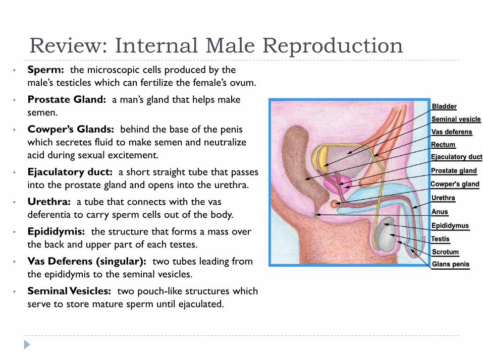
Review: Internal Male Reproduction• Sperm: the microscopic cells produced by the
male’s testicles which can fertilize the female’s ovum.
• Prostate Gland: a man’s gland that helps make
semen.
• Cowper’s Glands: behind the base of the penis
which secretes fluid to make semen and neutralize
acid during sexual excitement.
• Ejaculatory duct: a short straight tube that passes
into the prostate gland and opens into the urethra.
• Urethra: a tube that connects with the vas
deferentia to carry sperm cells out of the body.
• Epididymis: the structure that forms a mass over
the back and upper part of each testes.
• Vas Deferens (singular): two tubes leading from
the epididymis to the seminal vesicles.
• Seminal Vesicles: two pouch-like structures which
serve to store mature sperm until ejaculated.

Other Related Concerns
Circumcision: A process that surgically removes the flap of skin that covers the glans of the penis.
Nocturnal Emissions: normal, involuntary ejaculation of semen and sperm while a male is asleep.
Impotence: the failure to get or maintain an erection
Vasectomy: surgical procedure for sterilization of the male.
Female Timeline
Ages 9-12 Secondary sex characteristics appear
Ages 11-14 Menstrual cycle begins
Late 20-30’s Peak sexual urges
Ages 45-55 Menopause: cycle stops, but sex urge continues

Female Reproductive System
Female reproductive organs are for intercourse, urination,
pregnancy, and childbirth.
External Female Anatomy
• Vulva: woman’s external genital area.
• Pudendum or Pubes: the area in the body where the sex organs are located.
• Mons Pubis: a mound of fatty tissue which covers the pubic bone.
• Labia Majora: (large lips) two folds of skin running from the mons pubis to below the vaginal opening
• Labia Minora: two smaller folds of tissue which lie just within the labia majora.
• Clitoris: a small, pea-shaped bump at the front of the labia that contains erectile tissue (counter part to male penis.)
• Urethra: below the clitoris, the opening to the bladder.

Internal Female Anatomy Hymen: a narrow fold of tissue
encircling the entrance to the vagina.
Vagina: passageway between the uterus and the outside of a woman’s body.
Cervix: Opening from the uterus to the vagina.
Uterus: place where the baby grows in a woman’s abdomen.
Oviducts (Fallopian Tubes): two tubular structures leading from the ovaries to the uterus
Ovaries: organs holding a woman’s eggs.
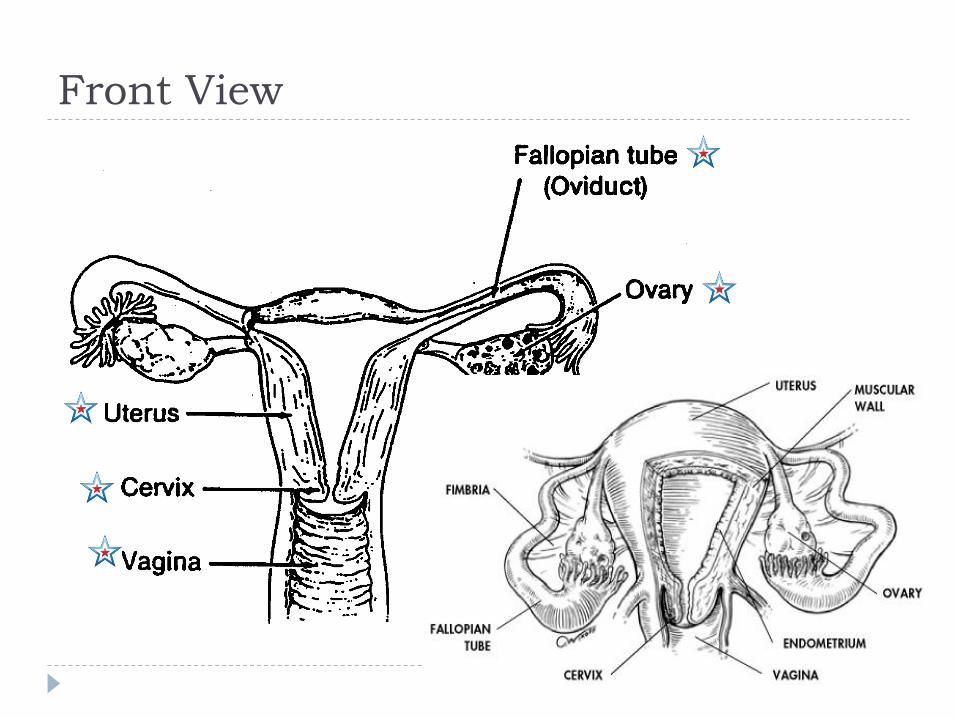
Front View
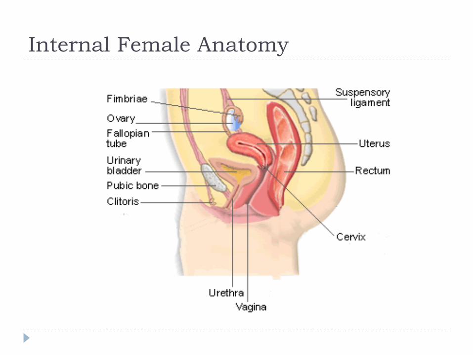
Internal Female Anatomy

Ovaries (b)
• Two solid egg-shaped structures
• They are attached to the uterus by ligaments. They are
the counterpart of the male testicles.
• Ovaries have two main functions:
1. Store and release the ova or female egg cell. Some of
the ova disappear; others are dormant until each is
ripened and released after puberty.
2. Produce female sex hormones ESTROGEN and
PROGESTERONE
Ova
• The female reproductive cell.
• They are the largest cells in the female body. (about the size of a grain
of sand.)
• The female baby is born with all the ova she will ever have (about 200,000 in each ovary).
• About 400-500 ova mature and are released over a lifetime
Estrogen & Progesterone
Estrogen is responsible for the secondary sex
characteristics and the sex drive in females. It spurs the
onset of puberty and is responsible for OVULATION.
Progesterone builds up the lining of the uterus called the
endometrium in preparation for the fertilized ovum
Ovulation
• When the egg is released from the ovary.
• At the age of puberty
• The ovum moves to the surface of the ovary in bursts out
• The ova falls into the fallopian tube and waits for fertilization
• This happens every 28 days
• It happens at about the 14th day of the cycle

Fallopian Tubes (Oviducts) (a)
• Two tubes attached on either side of the uterus.
• They are about four inches long and 3/16 inch in diameter (the
size of a cooked spaghetti noodle).
• The oviducts carry egg cells toward the uterus and sperm cells
toward the egg cell.
• Fertilization takes place in the upper third of the oviduct.
Uterus (c)
• A hollow, muscular organ (shaped somewhat like an upside-down pear,
about the size of a fist).
• The uterus is lined with endometrium (a blood lining.)
• The uterus has one main function—to protect and
nourish a fetus
• The walls of the uterus have the ability to stretch to the
size of a small watermelon.
• After childbirth the uterus shrinks back to the original
shape in 6-8 weeks, but it can take up to nine months for
the uterus to fully recover.
Cervix (d)
• The neck or opening of the uterus.
• A normal healthy cervix is the strongest muscle in the body.
• It dips down about half an inch into the vagina.
• It is normally plugged by mucus. It stays tightly closed during pregnancy, but thins and opens for the delivery of the baby.
• How big does it need to dilate to for birth?
Vagina (k)
• Female organ used for intercourse, it is an empty passageway leading from the vaginal opening to the uterus.
• It is only 3-4 inches long, but will lengthen during arousal.
• The vaginal walls are made of many small folds of membrane that stretch greatly to accommodate a baby during birth.
• The vaginal wall also secrete a fluid that helps to make intercourse easier.
Urethra (j)
The opening to the bladder
Clitoris (g)
• A small, pea shaped bump at the front of the labia.
• It contains a small amount of erectile tissue.
• The clitoris increases sexual pleasure
Endometrium
The lining of the uterus.
During menstruation, it is what sloughs off.
During pregnancy it thickens and provide the place of
implantation for the fertilized ova.

Fertiliztion/Conception
The end purpose for the
ova and the sperm
When the sperm
penetrates the surface of
the ova and enters inside.
The 23 chromosomes from
each sex cell combine and
begin to multiply to begin
to form a new human
being!!!
Menstrual Cycle
• Day 1 – Menstruation begins (bleeding)
• Day 1-5 – Bleeding Continues
• Day 6-9 - Ovum is maturing and endometrial lining is thin
• Day 10 – 14 - Endometrial lining thickens and hormones rise. – Around Day 14 – Ovum bursts out of the ovary
• Day 15 – After 24 hours the egg is done
• Day 15-28 – Egg travels down to thickened lining and either is implanted or it dissolves– Day 26 – In the absence of fertilization, hormone levels drop
and the endometrial lining breaks down
– Day 28 – Menstruation prepares to begin again.

During Menstruation: Days 1-5
Menstruation occurs and the
lining of the uterus, with a
small amount of blood, leaves
the body. At this time, another
egg is maturing in the ovary.
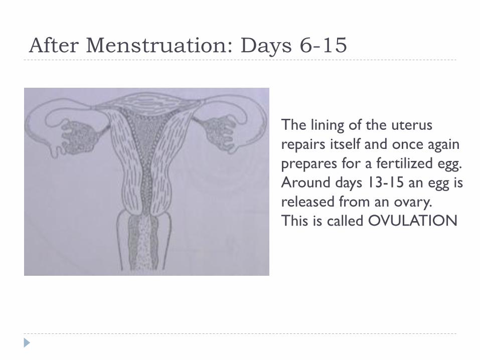
After Menstruation: Days 6-15
The lining of the uterus
repairs itself and once again
prepares for a fertilized egg.
Around days 13-15 an egg is
released from an ovary.
This is called OVULATION
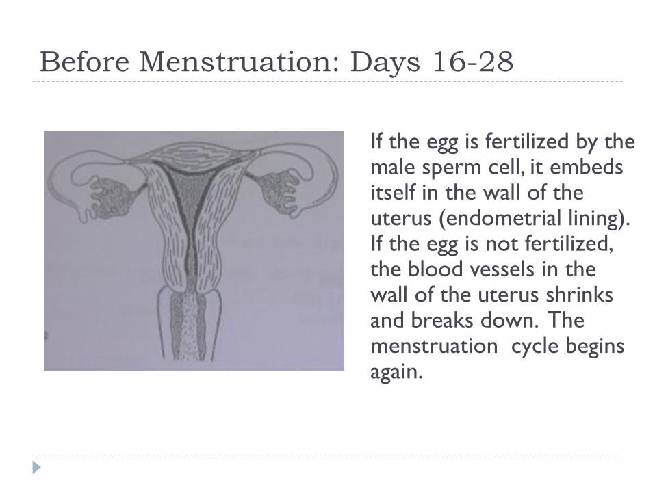
Before Menstruation: Days 16-28
If the egg is fertilized by the male sperm cell, it embeds itself in the wall of the uterus (endometrial lining). If the egg is not fertilized, the blood vessels in the wall of the uterus shrinks and breaks down. The menstruation cycle begins again.
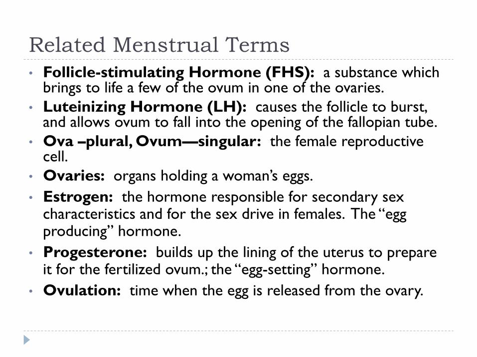
Related Menstrual Terms
• Follicle-stimulating Hormone (FHS): a substance which brings to life a few of the ovum in one of the ovaries.
• Luteinizing Hormone (LH): causes the follicle to burst, and allows ovum to fall into the opening of the fallopian tube.
• Ova –plural, Ovum—singular: the female reproductive cell.
• Ovaries: organs holding a woman’s eggs.
• Estrogen: the hormone responsible for secondary sex characteristics and for the sex drive in females. The ―egg producing‖ hormone.
• Progesterone: builds up the lining of the uterus to prepare it for the fertilized ovum.; the ―egg-setting‖ hormone.
• Ovulation: time when the egg is released from the ovary.

Other Related Concerns
D&C: dilation and curettage, a common minor operation on women.
Endometriosis: fragments of the endometrium in abnormal places.
Orgasm: characterized by the massive release of muscle tension which has built up during excitement.
Dysmenorrhea: painful menstruation
Hysterectomy: surgical removal of uterus.
Tubal Ligation: an operation for sterilization of women.
PMS: premenstrual syndrome.
Menstrual Cycle: the process of passing the blood and tissue lining of the uterus from the body.
Toxic Shock Syndrome: caused by bacteria that live in the vagina, which then multiply and causes infection.
Menopause: the remaining ova no longer ripen or develop.
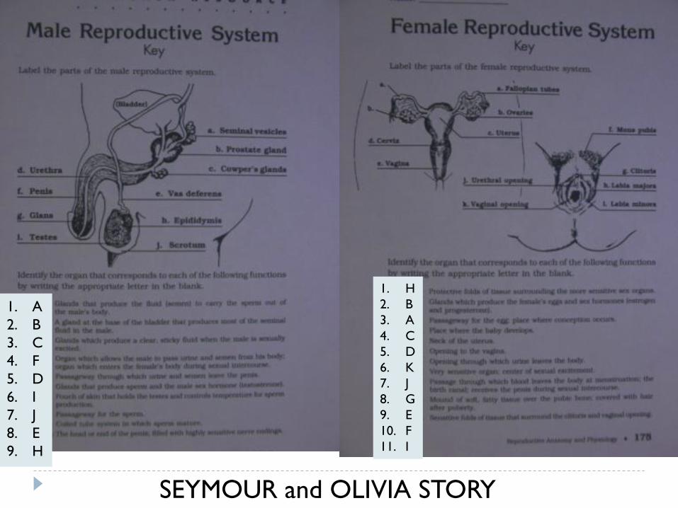
SEYMOUR and OLIVIA STORY
1. A
2. B
3. C
4. F
5. D
6. I
7. J
8. E
9. H
1. H
2. B
3. A
4. C
5. D
6. K
7. J
8. G
9. E
10. F
11. I