Embed Size (px)
Citation preview
Human primary cell culture sourcebook
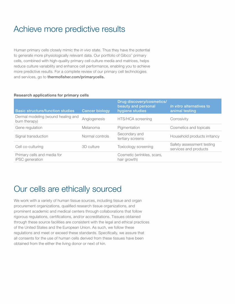
Human primary cells closely mimic the in vivo state. Thus they have the potential to generate more physiologically relevant data. Our portfolio of Gibco™ primary cells, combined with high-quality primary cell culture media and matrices, helps reduce culture variability and enhance cell performance, enabling you to achieve more predictive results. For a complete review of our primary cell technologies and services, go to thermofisher.com/primarycells.
Achieve more predictive results
Our cells are ethically sourced
Research applications for primary cells
Basic structure/function studies Cancer biology
Drug discovery/cosmetics/beauty and personal hygiene studies
In vitro alternatives to animal testing
Dermal modeling (wound healing and burn therapy) Angiogenesis HTS/HCA screening Corrosivity
Gene regulation Melanoma Pigmentation Cosmetics and topicals
Signal transduction Normal controls Secondary and tertiary screens Household products irritancy
Cell co-culturing 3D culture Toxicology screening Safety assessment testing services and products
Primary cells and media for iPSC generation
Cosmetic (wrinkles, scars, hair growth)
We work with a variety of human tissue sources, including tissue and organ procurement organizations, qualified research tissue organizations, and prominent academic and medical centers through collaborations that follow rigorous regulations, certifications, and/or accreditations. Tissues obtained through these source facilities are consistent with the legal and ethical practices of the United States and the European Union. As such, we follow these regulations and meet or exceed these standards. Specifically, we assure that all consents for the use of human cells derived from these tissues have been obtained from the either the living donor or next of kin.
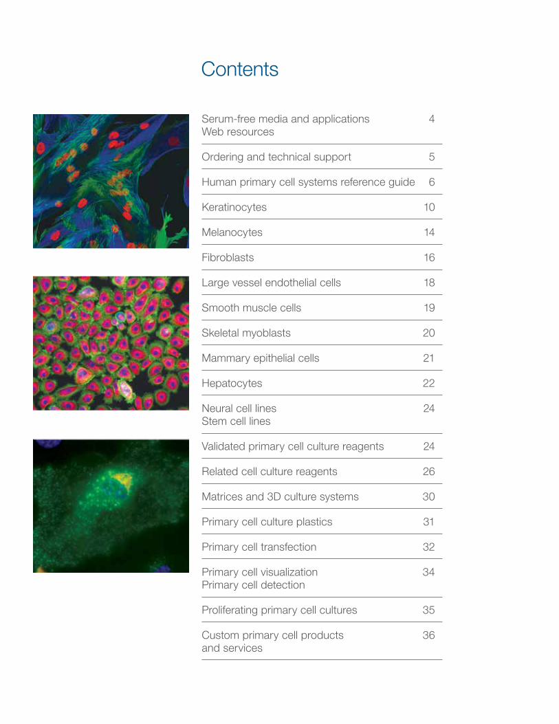
Serum-free media and applications 4 Web resources
Ordering and technical support 5
Human primary cell systems reference guide 6
Keratinocytes 10
Melanocytes 14
Fibroblasts 16
Large vessel endothelial cells 18
Smooth muscle cells 19
Skeletal myoblasts 20
Mammary epithelial cells 21
Hepatocytes 22
Neural cell lines 24 Stem cell lines
Validated primary cell culture reagents 24
Related cell culture reagents 26
Matrices and 3D culture systems 30
Primary cell culture plastics 31
Primary cell transfection 32
Primary cell visualization 34 Primary cell detection
Proliferating primary cell cultures 35
Custom primary cell products 36 and services
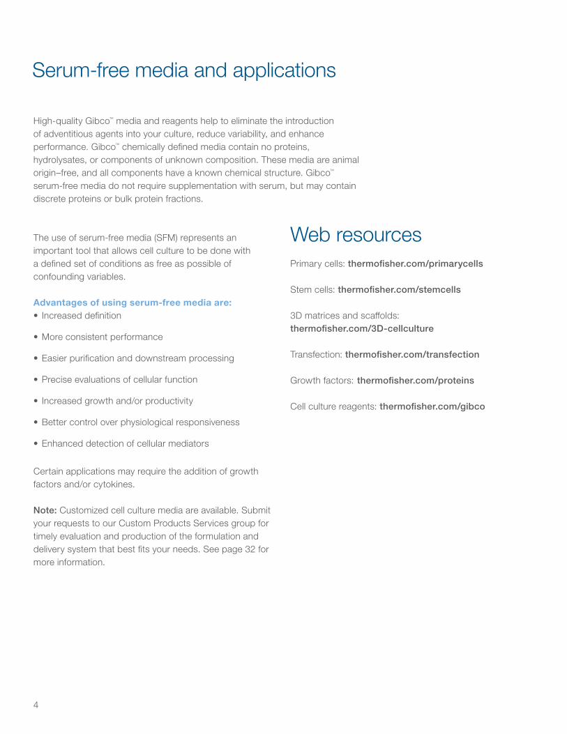
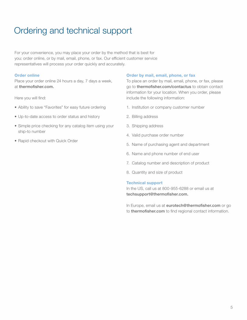
Contents
4
High-quality Gibco™ media and reagents help to eliminate the introduction of adventitious agents into your culture, reduce variability, and enhance performance. Gibco™ chemically defined media contain no proteins, hydrolysates, or components of unknown composition. These media are animal origin–free, and all components have a known chemical structure. Gibco™ serum-free media do not require supplementation with serum, but may contain discrete proteins or bulk protein fractions.
Serum-free media and applications
The use of serum-free media (SFM) represents an important tool that allows cell culture to be done with a defined set of conditions as free as possible of confounding variables.
Advantages of using serum-free media are:• Increased definition
• More consistent performance
• Easier purification and downstream processing
• Precise evaluations of cellular function
• Increased growth and/or productivity
• Better control over physiological responsiveness
• Enhanced detection of cellular mediators
Certain applications may require the addition of growth factors and/or cytokines.
Note: Customized cell culture media are available. Submit your requests to our Custom Products Services group for timely evaluation and production of the formulation and delivery system that best fits your needs. See page 32 for more information.
Web resourcesPrimary cells: thermofisher.com/primarycells
Stem cells: thermofisher.com/stemcells
3D matrices and scaffolds: thermofisher.com/3D-cellculture
Transfection: thermofisher.com/transfection
Growth factors: thermofisher.com/proteins
Cell culture reagents: thermofisher.com/gibco
5
For your convenience, you may place your order by the method that is best for you: order online, or by mail, email, phone, or fax. Our efficient customer service representatives will process your order quickly and accurately.
Ordering and technical support
Order onlinePlace your order online 24 hours a day, 7 days a week, at thermofisher.com.
Here you will find:
• Ability to save “Favorites” for easy future ordering
• Up-to-date access to order status and history
• Simple price checking for any catalog item using your ship-to number
• Rapid checkout with Quick Order
Order by mail, email, phone, or faxTo place an order by mail, email, phone, or fax, please go to thermofisher.com/contactus to obtain contact information for your location. When you order, please include the following information:
1. Institution or company customer number
2. Billing address
3. Shipping address
4. Valid purchase order number
5. Name of purchasing agent and department
6. Name and phone number of end user
7. Catalog number and description of product
8. Quantity and size of product
Technical supportIn the US, call us at 800-955-6288 or email us at [email protected].
In Europe, email us at [email protected] or go to thermofisher.com to find regional contact information.
6
Human primary cell systems quick reference guide
Anatomical location of derivation Cells Acronym
Cryopreserved product Cat. No. Growth medium Cat. No. Growth supplement Acronym Cat. No.
Skin
Human Epidermal Keratinocytes, neonatal (animal origin–free) HEKn-AOF C-020-5CEpiLife* Medium MEPI500CA Supplement S7†§ S7†§ S-017-5
Human Epidermal Keratinocytes, adult (animal origin–free) HEKa-AOF C-021-5CHuman Epidermal Keratinocytes, neonatal HEKn C-001-5C
EpiLife* Medium MEPI500CAEpiLife Defined Growth Supplement† EDGS† S-012-5
Human Epidermal Keratinocytes, adult HEKa C-005-5CHuman Keratinocyte Growth Supplement HKGS S-001-5Human Keratinocyte Growth Supplement Kit HKGS Kit S-001-K
Human Epidermal Keratinocytes, pooled HEKp A13401 Medium 154* M-154-500Human Keratinocyte Growth Supplement HKGS S-001-5Human Keratinocyte Growth Supplement Kit HKGS Kit S-001-K
Human Melanocytes, neonatal, lightly pigmented HEMn-LP C-002-5CMedium 254‡ M-254-500 Human Melanocyte Growth Supplement HMGS S-002-5Human Melanocytes, neonatal, moderately pigmented HEMn-MP C-102-5C
Human Melanocytes, neonatal, darkly pigmented HEMn-DP C-202-5CHuman Melanocytes, adult, lightly pigmented HEMa-LP C-024-5C Medium 254‡ M-254-500 Human Melanocyte Growth Supplement-2 HMGS-2 S-016-5Human Dermal Fibroblasts, neonatal HDFn C-004-5C
Medium 106 M-106-500Low Serum Growth Supplement LSGS S-003-10
Human Dermal Fibroblasts, adult HDFa C-013-5C Low Serum Growth Supplement Kit LSGS Kit S-003-KHuman Microvascular Endothelial Cells, neonatal dermis HMVECnd C-010-5C
MCBD 131 Medium 10372019 Microvascular Growth Supplement MVGS 10372019Human Microvascular Endothelial Cells, adult dermis HMVECad C-011-5C
HeartHuman Aortic Smooth Muscle Cells HASMC C-007-5C Medium 200** M-200-500 Large Vessel Endothelial Supplement LVES A1460801Human Coronary Artery Smooth Muscle Cells HCASMC C-017-5C Medium 231 M-231-500 Smooth Muscle Growth Supplement SMGS S-007-25Human Skeletal Myoblasts, large size A11440 Medium 231 M-231-500 Smooth Muscle Growth Supplement SMGS S-007-25
Umbilical cordHuman Umbilical Vein Endothelial Cells HUVEC C-003-5C
Medium 200** M-200-500 Large Vessel Endothelial Supplement LVES A1460801Human Umbilical Vein Endothelial Cells (pooled, 500,000 cells) HUVEC, pooled C-015-5CHuman Umbilical Vein Endothelial Cells (pooled, 1 million cells) HUVEC, pooled C-015-10C
Breast Human Mammary Epithelial Cells HMEC A10565
Medium 171 M-171-500 Mammary Epithelial Growth Supplement MEGS S-015-5HuMEC Ready Medium 12752-010 Note: Supplements included with mediaHuMEC Basal Serum-Free Medium 12753-018 HuMEC Supplement Kit NA 12755013
BrainHuman Astrocytes††
NAN7805-100 Astrocyte Medium A12613-01 NA
Human Astrocyte Kit†† N7805-200 Note: Included in kit
(continued on next page)
Human primary cell systems quick reference guide

7
Human primary cell systems quick reference guide
Anatomical location of derivation Cells Acronym
Cryopreserved product Cat. No. Growth medium Cat. No. Growth supplement Acronym Cat. No.
Skin
Human Epidermal Keratinocytes, neonatal (animal origin–free) HEKn-AOF C-020-5CEpiLife* Medium MEPI500CA Supplement S7†§ S7†§ S-017-5
Human Epidermal Keratinocytes, adult (animal origin–free) HEKa-AOF C-021-5CHuman Epidermal Keratinocytes, neonatal HEKn C-001-5C
EpiLife* Medium MEPI500CAEpiLife Defined Growth Supplement† EDGS† S-012-5
Human Epidermal Keratinocytes, adult HEKa C-005-5CHuman Keratinocyte Growth Supplement HKGS S-001-5Human Keratinocyte Growth Supplement Kit HKGS Kit S-001-K
Human Epidermal Keratinocytes, pooled HEKp A13401 Medium 154* M-154-500Human Keratinocyte Growth Supplement HKGS S-001-5Human Keratinocyte Growth Supplement Kit HKGS Kit S-001-K
Human Melanocytes, neonatal, lightly pigmented HEMn-LP C-002-5CMedium 254‡ M-254-500 Human Melanocyte Growth Supplement HMGS S-002-5Human Melanocytes, neonatal, moderately pigmented HEMn-MP C-102-5C
Human Melanocytes, neonatal, darkly pigmented HEMn-DP C-202-5CHuman Melanocytes, adult, lightly pigmented HEMa-LP C-024-5C Medium 254‡ M-254-500 Human Melanocyte Growth Supplement-2 HMGS-2 S-016-5Human Dermal Fibroblasts, neonatal HDFn C-004-5C
Medium 106 M-106-500Low Serum Growth Supplement LSGS S-003-10
Human Dermal Fibroblasts, adult HDFa C-013-5C Low Serum Growth Supplement Kit LSGS Kit S-003-KHuman Microvascular Endothelial Cells, neonatal dermis HMVECnd C-010-5C
MCBD 131 Medium 10372019 Microvascular Growth Supplement MVGS 10372019Human Microvascular Endothelial Cells, adult dermis HMVECad C-011-5C
HeartHuman Aortic Smooth Muscle Cells HASMC C-007-5C Medium 200** M-200-500 Large Vessel Endothelial Supplement LVES A1460801Human Coronary Artery Smooth Muscle Cells HCASMC C-017-5C Medium 231 M-231-500 Smooth Muscle Growth Supplement SMGS S-007-25Human Skeletal Myoblasts, large size A11440 Medium 231 M-231-500 Smooth Muscle Growth Supplement SMGS S-007-25
Umbilical cordHuman Umbilical Vein Endothelial Cells HUVEC C-003-5C
Medium 200** M-200-500 Large Vessel Endothelial Supplement LVES A1460801Human Umbilical Vein Endothelial Cells (pooled, 500,000 cells) HUVEC, pooled C-015-5CHuman Umbilical Vein Endothelial Cells (pooled, 1 million cells) HUVEC, pooled C-015-10C
Breast Human Mammary Epithelial Cells HMEC A10565
Medium 171 M-171-500 Mammary Epithelial Growth Supplement MEGS S-015-5HuMEC Ready Medium 12752-010 Note: Supplements included with mediaHuMEC Basal Serum-Free Medium 12753-018 HuMEC Supplement Kit NA 12755013
BrainHuman Astrocytes††
NAN7805-100 Astrocyte Medium A12613-01 NA
Human Astrocyte Kit†† N7805-200 Note: Included in kit
(continued on next page)
8
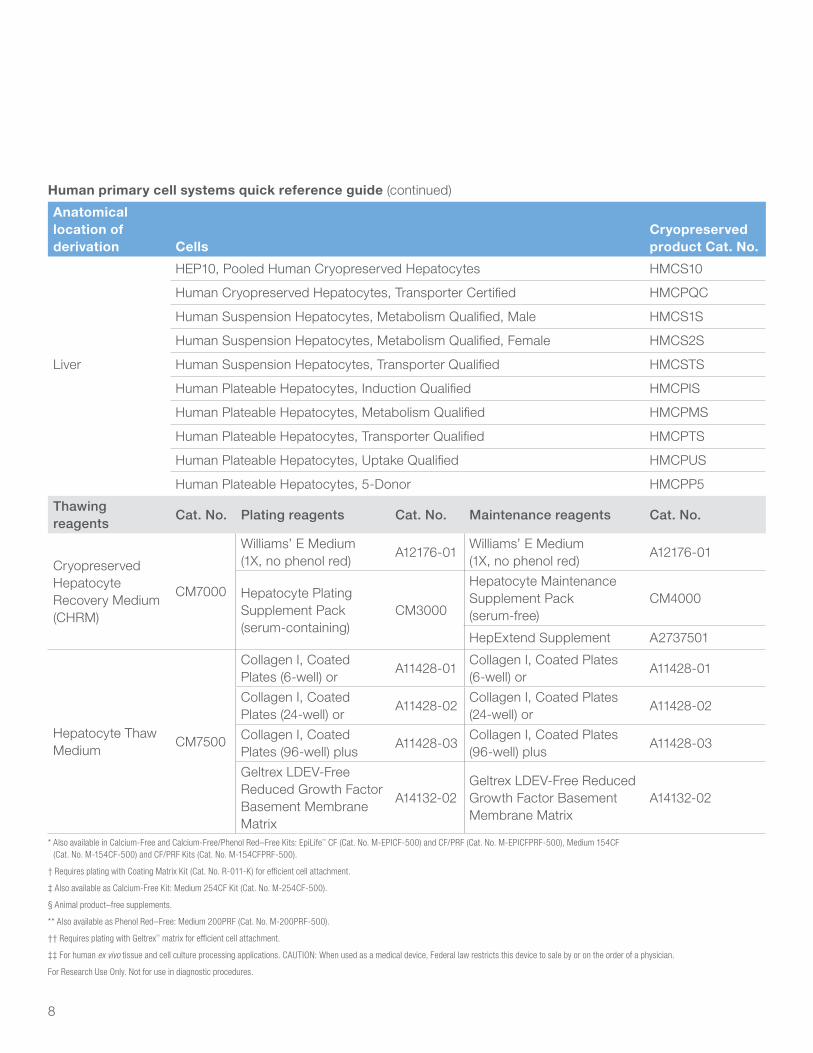
Human primary cell systems quick reference guide (continued)
Anatomical location of derivation Cells
Cryopreserved product Cat. No.
Liver
HEP10, Pooled Human Cryopreserved Hepatocytes HMCS10
Human Cryopreserved Hepatocytes, Transporter Certified HMCPQC
Human Suspension Hepatocytes, Metabolism Qualified, Male HMCS1S
Human Suspension Hepatocytes, Metabolism Qualified, Female HMCS2S
Human Suspension Hepatocytes, Transporter Qualified HMCSTS
Human Plateable Hepatocytes, Induction Qualified HMCPIS
Human Plateable Hepatocytes, Metabolism Qualified HMCPMS
Human Plateable Hepatocytes, Transporter Qualified HMCPTS
Human Plateable Hepatocytes, Uptake Qualified HMCPUS
Human Plateable Hepatocytes, 5-Donor HMCPP5
Thawing reagents
Cat. No. Plating reagents Cat. No. Maintenance reagents Cat. No.
Cryopreserved Hepatocyte Recovery Medium (CHRM)
CM7000
Williams’ E Medium (1X, no phenol red)
A12176-01Williams’ E Medium (1X, no phenol red)
A12176-01
Hepatocyte Plating Supplement Pack (serum-containing)
CM3000
Hepatocyte Maintenance Supplement Pack (serum-free)
CM4000
HepExtend Supplement A2737501
Hepatocyte Thaw Medium
CM7500
Collagen I, Coated Plates (6-well) or
A11428-01Collagen I, Coated Plates (6-well) or
A11428-01
Collagen I, Coated Plates (24-well) or
A11428-02Collagen I, Coated Plates (24-well) or
A11428-02
Collagen I, Coated Plates (96-well) plus
A11428-03Collagen I, Coated Plates (96-well) plus
A11428-03
Geltrex LDEV-Free Reduced Growth Factor Basement Membrane Matrix
A14132-02Geltrex LDEV-Free Reduced Growth Factor Basement Membrane Matrix
A14132-02
* Also available in Calcium-Free and Calcium-Free/Phenol Red–Free Kits: EpiLife™ CF (Cat. No. M-EPICF-500) and CF/PRF (Cat. No. M-EPICFPRF-500), Medium 154CF (Cat. No. M-154CF-500) and CF/PRF Kits (Cat. No. M-154CFPRF-500).
† Requires plating with Coating Matrix Kit (Cat. No. R-011-K) for efficient cell attachment.
‡ Also available as Calcium-Free Kit: Medium 254CF Kit (Cat. No. M-254CF-500).
§ Animal product–free supplements.
** Also available as Phenol Red–Free: Medium 200PRF (Cat. No. M-200PRF-500).
†† Requires plating with Geltrex™ matrix for efficient cell attachment.
‡‡ For human ex vivo tissue and cell culture processing applications. CAUTION: When used as a medical device, Federal law restricts this device to sale by or on the order of a physician.
For Research Use Only. Not for use in diagnostic procedures.
9
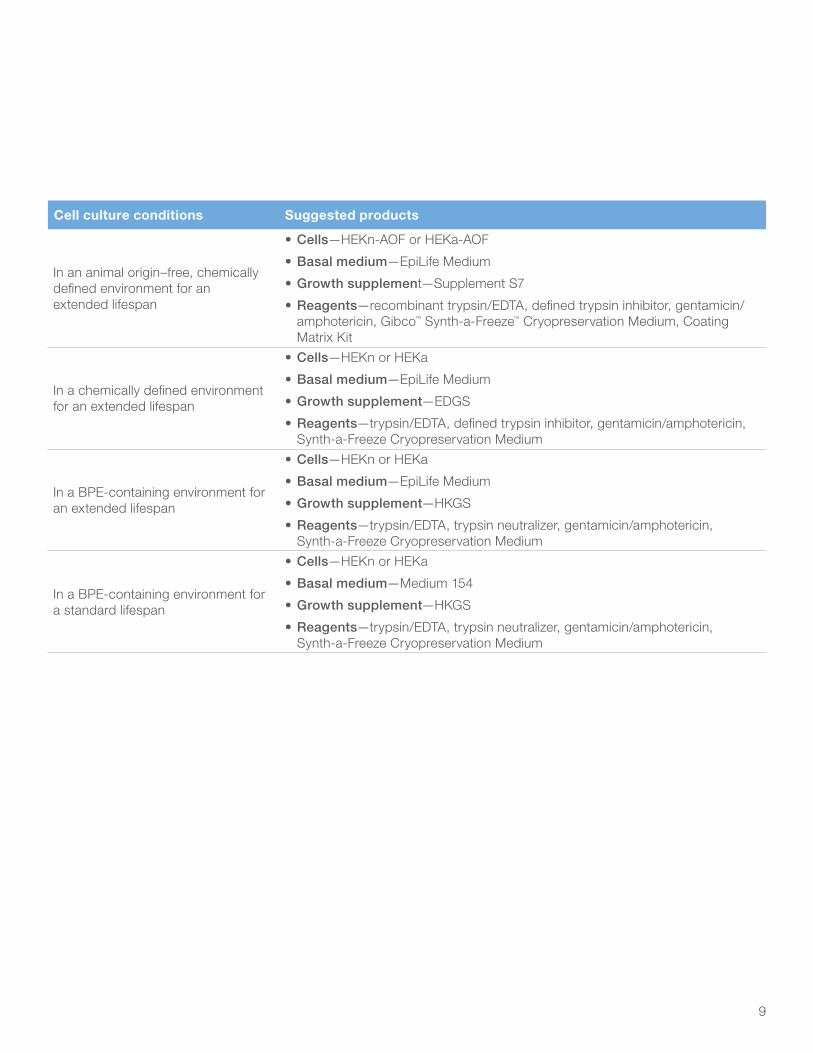
Cell culture conditions Suggested products
In an animal origin–free, chemically defined environment for an extended lifespan
• Cells—HEKn-AOF or HEKa-AOF
• Basal medium—EpiLife Medium
• Growth supplement—Supplement S7
• Reagents—recombinant trypsin/EDTA, defined trypsin inhibitor, gentamicin/amphotericin, Gibco™ Synth-a-Freeze™ Cryopreservation Medium, Coating Matrix Kit
In a chemically defined environment for an extended lifespan
• Cells—HEKn or HEKa
• Basal medium—EpiLife Medium
• Growth supplement—EDGS
• Reagents—trypsin/EDTA, defined trypsin inhibitor, gentamicin/amphotericin, Synth-a-Freeze Cryopreservation Medium
In a BPE-containing environment for an extended lifespan
• Cells—HEKn or HEKa
• Basal medium—EpiLife Medium
• Growth supplement—HKGS
• Reagents—trypsin/EDTA, trypsin neutralizer, gentamicin/amphotericin, Synth-a-Freeze Cryopreservation Medium
In a BPE-containing environment for a standard lifespan
• Cells—HEKn or HEKa
• Basal medium—Medium 154
• Growth supplement—HKGS
• Reagents—trypsin/EDTA, trypsin neutralizer, gentamicin/amphotericin, Synth-a-Freeze Cryopreservation Medium
10
We offer a wide array of Gibco™ products for keratinocyte culture, including products that are free of any animal-derived components such as bovine pituitary extract (BPE), serum, or any other components that are typically purified from animal sources. We refer to these products as being animal origin–free and use the abbreviation AOF to identify them. Gibco™ animal origin–free products do not contain material directly derived from animal tissues, cells, or body fluids of higher eukaryotic organisms, such as mammals (including humans), fish, birds, insects, etc. The term “animal origin” does not refer to other eukaryotic organisms such as the higher plants, fungi, protozoa, and algae, nor does it include prokaryotic organisms such as bacteria or blue-green algae. We offer complete cell culture systems designed and optimized to work together for the study of keratinocytes.
Characterization of human epidermal keratinocytesEach lot of human epidermal keratinocyte (HEK) cells is performance-tested in our laboratory for viability and growth potential. The cells are also tested for potential biological contaminants (HIV-1, hepatitis B and hepatitis C viruses, mycoplasmas, bacteria, yeasts, and other fungi). To be approved for distribution, cells must be at least 70% viable upon thawing, each vial must contain at least 500,000 viable cells, and no potential biological contaminants can be detected. In addition, neonatal cells must be able to grow through at least 30 population doublings when cultured in Gibco™ EpiLife™ Medium supplemented with HKGS (for HEKn) or Supplement S7 (for HEKn-AOF). Adult cells must be able to grow through at least 25 population doublings when cultured in EpiLife Medium supplemented with HKGS (for HEKa) or Supplement S7 (for HEKa-AOF). Certificates of Analysis are available on our website, or by request.
Keratinocytes
11
Cells Human epidermal keratinocytes (HEK), neonatal cells HEKn, cryopreservedNormal human epidermal keratinocytes isolated from neonatal foreskin, cryopreserved at the end of the primary culture.
Quantity Cat. No.
1 vial (>500,000 viable cells) C-001-5C
HEKn-AOF, neonatal cells, cryopreservedNormal human epidermal keratinocytes from neonatal foreskin are isolated, grown, and cryopreserved in an animal origin–free environment at the end of the primary culture. For optimal performance when culturing keratinocytes in an animal origin–free environment, we recommend coating the culture surfaces with our Coating Matrix Kit (Cat. No. R-011-K).
Quantity Cat. No.
1 vial (>500,000 viable cells) C-020-5C
HEKp, neonatal cells, cryopreservedNormal human epidermal keratinocytes isolated and pooled from 4–6 neonatal foreskins and cryopreserved at the end of the primary culture stage in AOF medium containing 10% DMSO.
Quantity Cat. No.
1 vial (>1,000,000 viable cells) A13401
Human epidermal keratinocytes (HEK), adult cells HEKa, cryopreservedNormal human epidermal keratinocytes, isolated from adult skin, cryopreserved at the end of the primary culture.
Quantity Cat. No.
1 vial (>500,000 viable cells) C-005-5C
HEKa-AOF, adult cells, cryopreservedNormal human epidermal keratinocytes from adult skin are isolated, grown, and cryopreserved in an animal origin–free environment. They are cryopreserved at the end of the primary culture. For optimal performance when culturing keratinocytes in an animal origin–free environment, we recommend coating the culture surfaces with our Coating Matrix Kit (Cat. No. R-011-K).
Quantity Cat. No.
1 vial (>500,000 viable cells) C-021-5C
12
plus an appropriate growth supplement prior to use. Calcium chloride is provided as a separate component with each bottle of medium.
Quantity Cat. No.
500 mL M-EPICFPRF-500
† Calcium concentration from other sources is 0.65 μM in unsupplemented EpiLife CF and CF/PRF media.
Medium 154A sterile, liquid medium for the serum-free culture of human epidermal keratinocytes. This basal medium requires the addition of HKGS (Cat. No. S-001-5) or HKGS Kit (Cat. No. S-001-K) prior to use. Contains 200 µM calcium chloride.
Quantity Cat. No.
500 mL M-154-500
Medium 154CF (calcium-free)A sterile, liquid medium for the serum-free culture of human epidermal keratinocytes. Medium 154CF is Medium 154 prepared without calcium chloride.‡ This basal medium requires the addition of calcium plus HKGS (Cat. No. S-001-5) or HKGS Kit (Cat. No. S-001-K) prior to use. Calcium chloride is provided as a separate component with each bottle of medium.
Quantity Cat. No.
500 mL M-154CF-500
Medium 154CF/PRF (calcium-free, phenol red–free)A sterile, liquid medium for the serum-free culture of human epidermal keratinocytes. Medium 154CF/PRF is Medium 154 prepared without calcium chloride‡ or phenol red. This basal medium requires the addition of calcium plus HKGS (Cat. No. S-001-5) or HKGS Kit (Cat. No. S-001-K) prior to use. Calcium chloride is provided as a separate component with each bottle of medium.
Quantity Cat. No.
500 mL M-154CF-500
‡ Calcium concentration from other sources is 0.5 µM in unsupplemented Medium 154CF and Medium 154CF/PRF.
Basal media for keratinocytesEpiLife Medium is designed to extend the lifespan of keratinocytes. If the desired end point is differentiation, Medium 154 may provide better results. Media do not contain antibiotics or antimycotics.
EpiLife MediumGet the most from your cells using EpiLife serum-free, chemically defined, animal origin–free cell culture medium. EpiLife Medium can help extend the in vitro lifespan of primary cells in culture up to twice as long compared to other serum-free formulations. EpiLife Medium contains 60 µM calcium chloride and is convenient and easy to use with single-shot supplementation. It is ideal for supporting the isolation, growth, and survival of both normal human keratinocytes and other types of epithelial cells when combined with appropriate supplements.
Quantity Cat. No.
500 mL M-EPI-500-CA
EpiLife CF (calcium-free) MediumA sterile liquid medium, EpiLife CF Medium is prepared without calcium chloride† for the long-term, serum-free culture of human epidermal keratinocytes. This basal medium requires the addition of calcium plus an appropriate growth supplement prior to use. Calcium chloride is provided as a separate component with each bottle of medium.
Quantity Cat. No.
500 mL M-EPICF-500
EpiLife CF/PRF (calcium-free, phenol red–free) Medium A sterile, liquid medium for the long-term, serum-free culture of human epidermal keratinocytes, EpiLife CF/PRF Medium is prepared without calcium chloride† or phenol red. This basal medium requires the addition of calcium
13
Growth supplements for keratinocytesOnly supplements in kit form contain antibiotics and antimycotics.
Human Keratinocyte Growth Supplement (HKGS)A sterile, concentrated (100X) solution intended for use with EpiLife Medium or Medium 154 to culture human epidermal keratinocytes. Contains bovine pituitary extract (BPE),† human epidermal growth factor, hydrocortisone, recombinant human insulin-like growth factor-1 (IGF-1), and transferrin.
Quantity Cat. No.
5 mL S-001-5
† BPE from New Zealand sources only.
Human Keratinocyte Growth Supplement (HKGS) KitA sterile set of solutions intended for use with EpiLife Medium or Medium 154 to culture human epidermal keratinocytes. The HKGS Kit provides, in separate vials, all the components of complete HKGS: bovine pituitary extract (BPE),* human epidermal growth factor, hydrocortisone, recombinant human insulin-like growth factor-1 (IGF-1), and transferrin. A vial of gentamicin/amphotericin B solution (GA) is also included. Use of GA is optional.
Quantity Cat. No.
1 kit S-001-K
* BPE from New Zealand sources only.
Supplement S7A chemically defined, sterile, animal origin–free, concentrated (100X), ionically balanced solution intended for use with EpiLife Medium to culture human epidermal keratinocytes (not intended for use with Medium 154). Each 5 mL bottle of Supplement S7 is the correct amount of supplement for a 500 mL bottle of Gibco™ EpiLife™ Basal Medium. For optimal performance, we recommend using S7 in conjunction with our Coating Matrix Kit (Cat. No. R-011-K).
Quantity Cat. No.
5 mL S-017-5
EpiLife Defined Growth Supplement (EDGS)A defined, sterile, concentrated (100X) solution intended for use with EpiLife Medium to culture human epidermal keratinocytes (not intended for use with Medium 154). This solution contains BSA, bovine transferrin, rhIGF-1, rhEGF, hydrocortisone, and PGE-2 (synthetic). For optimal performance, we recommend using EDGS in conjunction with our Coating Matrix Kit (Cat. No. R-011-K).
Quantity Cat. No.
5 mL S-012-5
Keratinocyte AOF Growth KitThe Keratinocyte AOF Growth Kit is for the animal origin–free culture of human keratinocytes. The kit contains EpiLife Basal Medium, Supplement S7, and the Coating Matrix Kit.
Quantity Cat. No.
1 kit A1051501
14
Melanocytes Characterization of human epidermal melanocytes
We offer three varieties of human epidermal melanocytes, defined by the degree of pigmentation of the cells in the preparation: light (LP), medium (MP), and dark (DP). Each lot of human epidermal melanocyte (HEM) cells is performance-tested in our laboratory for viability and growth potential. The cells are also tested for potential biological contaminants (HIV-1, hepatitis B and hepatitis C viruses, mycoplasmas, bacteria, yeasts, and other fungi). To be approved for distribution, cells must be at least 70% viable upon thawing, each vial must contain at least 500,000 viable cells, and no potential biological contaminants can be detected. HEMn-MP and HEMn-DP cells must be able to grow through at least 12 population doublings; HEMn-LP and HEMa-LP cells must be able to grow through at least 16 population doublings. HEMa-LP cells stain positively with Mel-5 antibody in the fourth culture after thawing. Certificates of Analysis are available on our website, or by request.
CellsHuman epidermal melanocytes (HEM), neonatal cells HEMn-LP, cryopreservedNormal human epidermal melanocytes isolated from lightly pigmented neonatal foreskin, cryopreserved at the end of the secondary culture.
Quantity Cat. No.
1 vial (>500,000 viable cells) C-002-5C
HEMn-MP, cryopreservedNormal human epidermal melanocytes isolated from moderately pigmented neonatal foreskin, cryopreserved at the end of the secondary culture.
Quantity Cat. No.
1 vial (>500,000 viable cells) C-102-5C
HEMn-DP, cryopreservedNormal human epidermal melanocytes isolated from darkly pigmented neonatal foreskin, cryopreserved at the end of the secondary culture.
Quantity Cat. No.
1 vial (>500,000 viable cells) C-202-5C
Human epidermal melanocytes (HEM), adult cells HEMa-LP, cryopreservedNormal human epidermal melanocytes isolated from lightly pigmented adult skin, cryopreserved at the end of the secondary culture. For optimal performance when culturing adult melanocytes, we recommend using Human Melanocyte Growth Supplement-2 (Cat. No. S-016-5).
Quantity Cat. No.
1 vial (>500,000 viable cells) C-024-5C
15
Basal media for melanocytesMedia do not contain antibiotics or antimycotics.
Medium 254A sterile, liquid medium optimized for the culture of human epidermal melanocytes. This basal medium requires the addition of HMGS (Cat. No. S-002-5) or HMGS-2 (Cat. No. S-016-5) prior to use.
Quantity Cat. No.
500 mL M-254-500
Medium 254CF (calcium-free)A sterile, liquid medium for the culture of human epidermal melanocytes. Medium 254CF is Medium 254 prepared without calcium chloride.* Calcium chloride is provided as a separate component with each bottle of medium. This basal medium requires the addition of calcium plus HMGS (Cat. No. S-002-5) or HMGS-2 (Cat. No. S-016-5) prior to use.
Quantity Cat. No.
500 mL M-254CF-500
*Calcium concentration from other sources is 0.5 μM in unsupplemented Medium 254CF.
Growth supplements for melanocytesSupplements do not contain antibiotics or antimycotics.
Human Melanocyte Growth Supplement (HMGS)HMGS is a sterile, concentrated (100X) solution intended for use with Medium 254 or Medium 254CF to culture human epidermal melanocytes. The solution contains fetal bovine serum, basic fibroblast growth factor, bovine pituitary extract (BPE),† heparin, hydrocortisone, recombinant human insulin-like growth factor-1 (IGF-1), transferrin, and phorbol 12-myristate 13-acetate. It is recommended for either neonatal or adult melanocytes.
Quantity Cat. No.
5 mL S-002-5
Human Melanocyte Growth Supplement-2 (HMGS-2)HMGS-2 is a sterile, concentrated (100X) solution intended for use with Medium 254 or Medium 254CF to culture human epidermal melanocytes. The solution contains fetal bovine serum, basic fibroblast growth factor, bovine pituitary extract (BPE),† heparin, hydrocortisone, recombinant human insulin-like growth factor-1 (IGF-1), transferrin, and endothelin-1. It is recommended for HEMa-LP cells.
Quantity Cat. No.
5 mL S-016-5
† BPE from New Zealand and/or Australian sources only.
16
Fibroblasts Characterization of human dermal fibroblasts
Each lot of human dermal fibroblast (HDF) cells is performance-tested in our laboratory for viability and growth potential. The cells are also tested for potential biological contaminants (HIV-1, hepatitis B and hepatitis C viruses, mycoplasmas, bacteria, yeasts, and other fungi). To be approved for distribution, cells must be at least 70% viable upon thawing, each vial must contain at least 500,000 viable cells, HDFa cells must be able to grow through at least 12 population doublings, and HDFn cells must be able to grow through at least 16 population doublings; no potential biological contaminants can be detected. Certificates of Analysis are available on our website, or by request.
CellsHuman dermal fibroblasts (HDF), neonatal cells HDFn, cryopreservedNormal human dermal fibroblasts isolated from neonatal foreskin, cryopreserved at the end of the primary culture.
Quantity Cat. No.
1 vial (>500,000 viable cells) C-004-5C
Human dermal fibroblasts (HDF), adult cells HDFa, cryopreserved Normal human dermal fibroblasts isolated from adult skin, cryopreserved at the end of the primary culture.
Quantity Cat. No.
1 vial (>500,000 viable cells) C-013-5C
17
Basal medium for fibroblastsMedium does not contain antibiotics or antimycotics.
Medium 106A sterile liquid medium for the culture of human dermal fibroblasts. This basal medium requires the addition of LSGS (Cat. No. S-003-10) or LSGS Kit (Cat. No. S-003-K) prior to use.
Quantity Cat. No.
500 mL M-106-500
Growth supplements for fibroblastsOnly supplements in kit form contain antibiotics or antimycotics.
Low Serum Growth Supplement (LSGS)A sterile, concentrated (50X) solution intended for use with Medium 106 (for fibroblasts) or Medium 200 (for endothelial cells). Optimized for dermal fibroblast culture when paired with Medium 106, LSGS contains fetal bovine serum, basic fibroblast growth factor, heparin, hydrocortisone, and epidermal growth factor.
Quantity Cat. No.
10 mL S-003-10
Low Serum Growth Supplement (LSGS) KitThe LSGS Kit provides, in separate vials, all the components of complete LSGS: fetal bovine serum, hydrocortisone, human epidermal growth factor, and basic fibroblast growth factor/heparin (stabilized with BSA). A vial of gentamicin/amphotericin B solution (GA) is also included. Use of GA is optional.
Quantity Cat. No.
1 kit S-003-K
18
Large vessel endothelial cellsCharacterization of human large vessel endothelial cells
Each lot of cells is performance-tested in our laboratory for viability, growth potential, and differentiation markers. The cells are also tested for potential biological contaminants (HIV-1, hepatitis B and hepatitis C viruses, mycoplasmas, bacteria, yeasts, and other fungi). To be approved for distribution, the cells must be at least 70% viable upon thawing, each vial must contain at least the indicated number of viable cells, the cells must be able to grow through at least 16 population doublings, and no potential biological contaminants can be detected. In addition, during the first culture after thawing, the cells must take up acetylated LDL and express von Willebrand factor (vWF) and CD31 (endothelial cell markers), but not α-actin (a smooth muscle cell marker). Certificates of Analysis are available on our website, or by request.
CellsHUVEC, cryopreservedNormal human umbilical vein endothelial cells, cryopreserved at the end of the primary culture.
Quantity Cat. No.
1 vial (>500,000 viable cells) C-003-5C
HUVECp, cryopreservedNormal human umbilical vein endothelial cells pooled from multiple donors, cryopreserved at the end of the primary culture.
Quantity Cat. No.
1 vial (>500,000 viable cells) C-015-5C
1 vial (>1,000,000 viable cells) C-015-10C
Basal media for large vessel endothelial cellsMedia do not contain antibiotics or antimycotics.
Medium 200A sterile liquid medium for the culture of human large vessel endothelial cells. This basal medium requires the addition of LSGS (Cat. No. S-003-10) or LSGS Kit (Cat. No. S-003-K) prior to use.
Quantity Cat. No.
500 mL M-200-500
Medium 200PRF (phenol red–free)A sterile, liquid medium for the culture of human large vessel endothelial cells. Medium 200PRF is Medium 200 prepared without phenol red. This basal medium requires the addition of LVES (Cat. No. A1460801) prior to use.
Quantity Cat. No.
500 mL M-200PRF-500
Growth supplements for large vessel endothelial cellsSupplements do not contain antibiotics or antimycotics.
Large Vessel Endothelial Supplement (LVES)A sterile, concentrated (50X) solution intended for use with Medium 200 or Medium 200PRF for optimal culture of human large vessel endothelial cells. Contains fetal bovine serum, basic fibroblast growth factor, heparin, hydrocortisone, epidermal growth factor, and ascorbic acid.
Quantity Cat. No.
11 mL A1460801
19
Smooth muscle cellsCharacterization of human smooth muscle cells
Each lot of cells is performance-tested in our laboratory for viability, growth potential, and differentiation markers. The cells are also tested for potential biological contaminants (HIV-1, hepatitis B and hepatitis C viruses, mycoplasmas, bacteria, yeasts, and other fungi). To be approved for distribution, cells must be at least 70% viable upon thawing, each vial must contain at least 500,000 viable cells, the cells must be able to grow through at least 16 population doublings, and no potential biological contaminants can be detected. In addition, when cultured in differentiation medium, cells must express α-actin (a smooth muscle cell marker), but not von Willebrand factor (vWF, an endothelial cell marker). Certificates of Analysis are available on our website, or by request.
CellsHuman smooth muscle cells HASMC, cryopreservedNormal human aortic smooth muscle cells, cryopreserved at the end of the tertiary culture.
Quantity Cat. No.
1 vial (>500,000 viable cells) C-007-5C
HCASMC, cryopreservedNormal human coronary artery smooth muscle cells, cryopreserved at the end of the tertiary culture.
Quantity Cat. No.
1 vial (>500,000 viable cells) C-017-5C
HPASMC, cryopreservedNormal human pulmonary artery smooth muscle cells, cryopreserved at the end of the tertiary culture.
Quantity Cat. No.
1 vial (>500,000 viable cells) C-009-5C
Basal medium for smooth muscle cellsMedium does not contain antibiotics or antimycotics.
Medium 231A sterile, liquid medium for the culture of human smooth muscle cells. For growth of human smooth muscle cells, the medium requires the addition of SMGS (Cat. No. S-007-25) prior to use. For differentiation of human smooth muscle cells, the medium requires the addition of SMDS (Cat. No. S-008-5) prior to use.
Quantity Cat. No.
500 mL M-231-500
Growth and differentiation supplements for smooth muscle cellsSupplements do not contain antibiotics or antimycotics.
Smooth Muscle Cell Growth Supplement (SMGS)A sterile, concentrated (20X) solution intended for use with Medium 231 to culture human smooth muscle cells. The solution contains fetal bovine serum, basic fibroblast growth factor, epidermal growth factor, heparin, recombinant human insulin-like growth factor-1 (IGF-1), and BSA.
Quantity Cat. No.
25 mL S-007-25
Smooth Muscle Cell Differentiation Supplement (SMDS)A sterile, concentrated (100X) solution intended for use with Medium 231 to enhance differentiation of human smooth muscle cells. The solution contains fetal bovine serum and heparin.
Quantity Cat. No.
5 mL S-008-5
20
Skeletal myoblastsCharacterization of human skeletal myoblast cells
Each lot is designed to undergo highly efficient differentiation directly following plating of cryopreserved cells. Each vial contains cells that have been cryopreserved in a medium containing 10% DMSO and will differentiate and form myotubes within 48 hours after plating. These cells do not require expansion prior to differentiation. Cell viability is >70% post-thaw with a ≥50% myogenic index after 48 hours. Each vial of human skeletal myoblast cells (HSkMs) contains a sufficient number of cells to fully seed ¼ of a single multi-well dish (ranging in format from 6-well to 384-well). Cells are thawed and plated in DMEM supplemented with 2% horse serum. Certificates of Analysis are available on our website, or by request.
Human skeletal myoblast cells HSkM, cryopreservedNormal human skeletal myoblast cells, cryopreserved at the end of the tertiary culture.
Quantity Cat. No.
1 vial (1 x 10⁶ cells) A12555
1 vial (5 x 10⁶ cells) A11440
Basal media for corneal epithelial cellsMedia do not contain antibiotics or antimycotics
Keratinocyte-SFMA sterile, serum-free liquid medium that supports the robust growth of human corneal epithelial cells, keratinocytes, and other types of epithelial cells. It contains L-glutamine and is supplied as a kit that includes aliquots of bovine pituitary extract (BPE) and recombinant epidermal growth factor (rEGF).
Quantity Cat. No.
500 mL 17005-042
Defined Keratinocyte-SFMDefined Keratinocyte-SFM is a sterile, defined liquid medium that supports the robust growth of human corneal epithelial cells, keratinocytes and other types of epithelial cells. Defined Keratinocyte-SFM is free of serum and bovine pituitary extract and is supplied as a kit that includes single aliquots of growth supplement–containing factors, including insulin, epidermal growth factor (EGF), and fibroblast growth factor (FGF) from bovine pituitary extract. For optimal performance, we recommend using Defined Keratinocyte-SFM in conjunction with our Coating Matrix Kit (Cat. No. R-011-K).
Quantity Cat. No.
500 mL 10744-019
21
Each lot of human mammary epithelial cells (HMEC) is performance-tested in our laboratory for viability, growth potential, and differentiation markers. The cells are also tested for potential biological contaminants (HIV-1, hepatitis B and hepatitis C viruses, mycoplasmas, bacteria, yeasts, and other fungi). To be approved for distribution, cells must be at least 70% viable upon thawing, each vial must contain at least 500,000 viable cells, the cells must be able to grow through at least 16 population doublings, and no potential biological contaminants can be detected. In addition, when cultured in complete HuMEC Medium, cells must test positive for cytokeratins 5⁄6, 8, 18, and E-cadherin. Certificates of Analysis are available on our website, or by request.
CellsHuman mammary epithelial cells (HMEC), cryopreservedNormal human mammary epithelial cells, cryopreserved at the end of the 6th culture.
Quantity Cat. No.
1 vial (>500,000 viable cells) A10565
Medium for mammary epithelial cellsMedium does not contain antibiotics or antimycotics.
HuMEC Ready MediumA sterile liquid medium for the culture of human mammary epithelial cells. Engineered specifically for culture of human mammary epithelial cells, HuMEC Medium is a serum-free formulation with frozen supplements containing growth factor and other components. This ready medium does not require additional supplementation.
Quantity Cat. No.
500 mL 12752-010
Growth supplement for mammary epithelial cellsSupplements do not contain antibiotics or antimycotics.
HuMEC Supplement KitHuMEC Medium supplemented with this kit promotes superior growth of normal human mammary epithelial cells. The HuMEC Supplement Kit includes 5 mL of a supplement mix containing epidermal growth factor, hydrocortisone, isoproterenol, transferrin, and insulin, and 25 mg of bovine pituitary extract (BPE).*
Quantity Cat. No.
1 kit 12755013
*BPE from New Zealand and/or Australian sources only.
Mammary epithelial cells Characterization of human mammary epithelial cells
22
HepatocytesCharacterization of human hepatocytes
Gibco™ hepatocytes have in vivo–like enzyme expression levels, and, if qualified as plateable cells, form confluent monolayers that contribute to polarization and functioning cell–cell contacts. Extensive lot selection options from a variety of donors are available. Many lots have >100 vials, are fully characterized for phase I and phase II drug metabolizing enzyme activities, and have viabilities routinely above 80%.
CellsCryopreserved hepatocytes, transporter qualifiedEach of our transporter-qualified lots (suspension and plateable) have been functionally tested for the activities of the NTCP, OATP1B3, and OATP transporter pathways. They have also been tested for phase I and phase II metabolic activities. Short-term transporter–qualified lots are tested at a 24-hour time point. Long-term uptake–qualified lots are tested at a 120-hour (5-day) time point. The cells have stringent release specifications and are guaranteed* to show ≥80% viability and ≥80% confluency (if cells are plated under appropriate conditions).
Product Quantity Cat. No.
Human Cryopreserved Plateable Hepatocytes, Transporter Qualified
4–8 million viable cells/vial HMCPTS
Human Cryopreserved Plateable Hepatocytes, Uptake Qualified
4–8 million viable cells/vial HMCPUS
Human Cryopreserved Suspension Hepatocytes, Transporter Qualified
4–8 million viable cells/vial HMCSTS
Human Cryopreserved Hepatocytes, Transporter Certified
4-8 million viable cells/vial HMCPQC
Induction qualifiedOur induction-qualified hepatocytes have passed our test for specific activity and mRNA levels in response to prototypical inducers.
Minimum specific activities:
• 10-fold induction of CYP1A2
• 5-fold induction of CYP2B6
• 3-fold induction of CYP3A4
The cells have stringent release specifications and are guaranteed* to show ≥80% viability and ≥80% confluency (if appropriately plated).
Product Quantity Cat. No.
Human Cryopreserved Plateable Hepatocytes, Induction Qualified
4–8 million viable cells/vial HMCPIS
Metabolism qualifiedOur metabolism-qualified hepatocytes have been tested for enzymatic functions using intrinsic clearance (CLint) or assays using known substrates for P450 activity. The disappearance of parent compound is monitored by LC/MS/MS and CLint values determined by linear regression. Gibco™ HEP10™ Pooled Hepatocytes give a representative population of 10 or more different donors (male and female).
Cells Characterization
Single donor, plated CYP3A4, CYP2C9, CYP2D6
Single donor, suspensionCYP1A2, CYP2B6, CYP2C8, CYP2C9, CYP2C19, CYP2D6, CYP3A, ECOD, 7-HCG, and 7-HCS
HEP10 pooled, suspension
CYP1A2, CYP2B6, CYP2C8, CYP2C9, CYP2C19, CYP2D6, CYP2E1, CYP3A4, CYP3A4/5, FMO activity, and NTCP, OATP1B3, and OATP transporter pathways
The cells have stringent release specifications and are guaranteed* to show ≥80% viability and ≥75% attachment efficiency (when plated).
Product Quantity Cat. No.
Human Cryopreserved Plateable Hepatocytes, Metabolism Qualified
4–8 million viable cells/vial HMCPMS
Human Cryopreserved Suspension Hepatocytes, Metabolism Qualified, Male Donor
4–8 million viable cells/vial HMCS1S
Human Cryopreserved Suspension Hepatocytes, Metabolism Qualified, Female Donor
4–8 million viable cells/vial HMCS2S
HEP10 Pooled Cryopreserved Human Hepatocytes
4–8 million viable cells/vial HMCS10
* For data on viability, go to thermofisher.com/us/en/home/industrial/pharma-biopharma/drug-discovery-development/adme-tox/gibco-hepatocytes/human-hepatocytes/characterization-of-human-hepatocytes.html.
23
Basal media for hepatocytesHepatocyte Thaw MediumGibco™ Hepatocyte Thaw Medium is a ready-to-use medium specifically designed for use with thawed cryopreserved hepatocytes to enhance their viability and overall quality.
Product Quantity Cat. No.
Hepatocyte Thaw Medium 45 mL CM7500
Cryopreserved Hepatocyte Recovery Medium (CHRM)A proprietary formulation designed to enhance the recovery of viable hepatocytes while removing cryoprotectant after cell cryopreservation, yielding consistently higher viability.
Product Quantity Cat. No.
Cryopreserved Hepatocyte Recovery Medium (CHRM) 50 mL CM7000
Cryopreserved Hepatocyte Plating Medium (CHPM)Product Quantity Cat. No.
Cryopreserved Hepatocyte Plating Medium (CHPM) 50 mL CM9000
Williams’ E MediumProduct Quantity Cat. No.
Williams’ E Medium (1X, no phenol red) 500 mL A12176-01
Growth supplements for hepatocytesHepExtend SupplementGibco™ HepExtend™ Supplement (50X) has been optimized for use with cryopreserved primary hepatocytes to improve cell viability, function, and number of days in culture. This enables researchers to perform metabolic and toxicity experiments not achievable using standard culture conditions. The supplement does not contain any small molecules or fetal bovine serum, components that are known to interfere with primary hepatocyte function.
Product Quantity Cat. No.
HepExtend Supplement 10 mL A2737501
Hepatocyte Plating Supplement PackHepatocyte Plating Supplement Packs contain prequalified fetal bovine serum, dexamethasone, and a cocktail solution of penicillin-streptomycin, human recombinant insulin, Gibco™ GlutaMAX™ Supplement, and HEPES to supplement up to 500 mL of Williams’ E Medium without phenol red, or a suitable alternative basal medium, for the purpose of plating fresh or cryopreserved hepatocytes.
Product Quantity Cat. No.
Hepatocyte Plating Supplement Pack (serum-containing)
1 kit for 500 mL medium CM3000
Hepatocyte Maintenance Supplement PackHepatocyte Maintenance Supplement Packs contain dexamethasone and a cocktail solution of penicillin-streptomycin, ITS+ (insulin, transferrin, selenium complex, BSA, and linoleic acid), GlutaMAX Supplement, and HEPES to supplement up to 500 mL of Williams’ E Medium without phenol red, or a suitable alternative basal medium, for the purpose of incubating hepatocytes in suspension or plated cultures.
Product Quantity Cat. No.
Hepatocyte Maintenance Supplement Pack (serum-free)
1 kit for 500 mL medium CM4000
24
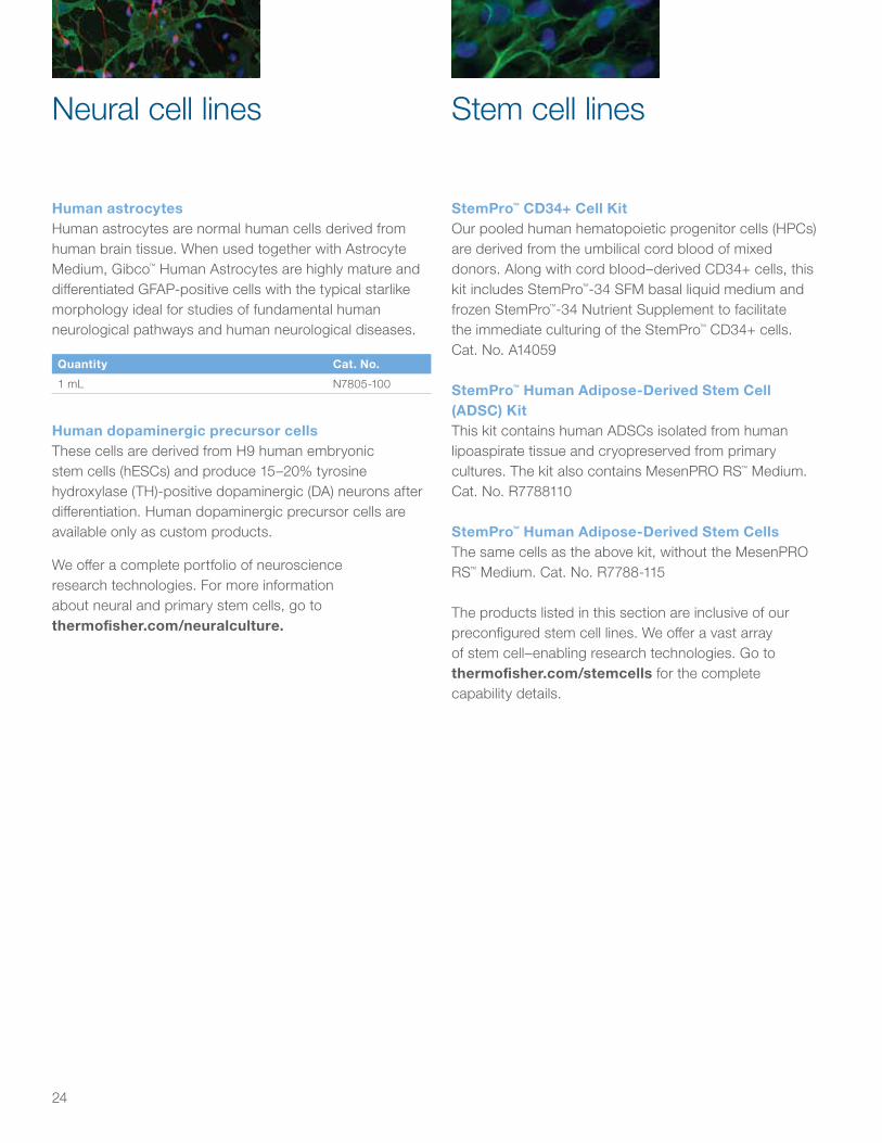
StemPro™ CD34+ Cell KitOur pooled human hematopoietic progenitor cells (HPCs) are derived from the umbilical cord blood of mixed donors. Along with cord blood–derived CD34+ cells, this kit includes StemPro™-34 SFM basal liquid medium and frozen StemPro™-34 Nutrient Supplement to facilitate the immediate culturing of the StemPro™ CD34+ cells. Cat. No. A14059
StemPro™ Human Adipose-Derived Stem Cell (ADSC) KitThis kit contains human ADSCs isolated from human lipoaspirate tissue and cryopreserved from primary cultures. The kit also contains MesenPRO RS™ Medium.Cat. No. R7788110
StemPro™ Human Adipose-Derived Stem CellsThe same cells as the above kit, without the MesenPRO RS™ Medium. Cat. No. R7788-115
The products listed in this section are inclusive of our preconfigured stem cell lines. We offer a vast array of stem cell–enabling research technologies. Go to thermofisher.com/stemcells for the complete capability details.
Stem cell linesNeural cell lines
Human astrocytesHuman astrocytes are normal human cells derived from human brain tissue. When used together with Astrocyte Medium, Gibco™ Human Astrocytes are highly mature and differentiated GFAP-positive cells with the typical starlike morphology ideal for studies of fundamental human neurological pathways and human neurological diseases.
Quantity Cat. No.
1 mL N7805-100
Human dopaminergic precursor cellsThese cells are derived from H9 human embryonic stem cells (hESCs) and produce 15–20% tyrosine hydroxylase (TH)-positive dopaminergic (DA) neurons after differentiation. Human dopaminergic precursor cells are available only as custom products.
We offer a complete portfolio of neuroscience research technologies. For more information about neural and primary stem cells, go to thermofisher.com/neuralculture.
25
For optimal performance when culturing the primary cells found in this sourcebook, we recommend using the reagents found in this section, which have been validated for these culture systems. Go to thermofisher.com/culturereagents for the complete capability details
Trypsin/EDTA Solution (TE)A sterile, phosphate buffered saline solution (1X) containing trypsin and EDTA for the subculture of attachment- dependent cells.
Quantity Cat. No.
100 mL R-001-100
Gentamicin/Amphotericin B Solution (GA), 10-PackA sterile, concentrated (500X) solution of gentamicin and amphotericin B (10 x 1 mL vials/pkg). One GA 10-Pack is enough to supplement ten 500 mL bottles of basal medium.
Quantity Cat. No.
10 x 1 mL R-015-10
Trypsin Neutralizer Solution (TN)A sterile, phosphate-buffered saline solution (1X) containing calf serum as a trypsin inhibitor.
Quantity Cat. No.
100 mL R-002-100
Defined Trypsin Inhibitor (DTI)A sterile, phosphate-buffered saline solution (1X) containing purified soybean trypsin inhibitor.
Quantity Cat. No.
100 mL R-007-100
Coating Matrix KitThe Coating Matrix Kit is a convenient combination of Coating Matrix and Dilution Medium for treatment of tissue culture labware for keratinocyte culture. The kit contains enough material to coat approximately thirty T-25 flasks.
Quantity Cat. No.
1 kit R-011-K
Synth-a-Freeze Cryopreservation MediumA defined, protein-free, sterile cryopreservation medium containing 10% DMSO. This solution is suitable for the cryopreservation of all cell types, with the exception of melanocytes.
Quantity Cat. No.
50 mL A12542-01
TrypLE Express, Stable Trypsin ReplacementEnzyme (1X), liquidTrypLE™ Express, Stable Trypsin Replacement Enzyme is formulated in DPBS with 1 mM EDTA. It is currently validated for use with human corneal epithelial cells. It contains no phenol red.
Quantity Cat. No.
100 mL 12604-013
500 mL 12604-021
20 x 100 mL (case) 12604-039
Validated primary cell culture reagents
26
The following products have not been validated with the primary cells listed in this sourcebook. We are continuing to build our application database. For a complete list of cell culture reagents, please go to thermofisher.com/gibco.
Collagenase Type I, lyophilizedPrepared from Clostridium histolyticum for dissociation of tissues. Suggested uses are for epithelial, lung, fat, and adrenal tissue cell preparations.
Quantity Cat. No.
500 mg 17018-029
1 g 17100-017
Collagenase Type IIPrepared from Clostridium histolyticum for dissociation of tissues. Suggested use is for preparation of cells from liver, bone, thyroid, heart, and salivary gland tissue.
Quantity Cat. No.
1 g 17101-015
Collagenase Type IVPrepared from Clostridium histolyticum for dissociation of cells from pancreas tissue. The suggested use is for isolation of pancreatic islets.
Quantity Cat. No.
1 g 17104-019
Related cell culture reagents
Dispase IIPrepared from Bacillus polymyxa. It can be used for gentle dissociation of animal tissues to release individual cells.
Quantity Cat. No.
5 g 17105-041
Fungizone™ Antimycotic, liquidContains 250 µg of amphotericin B and 205 µg of sodium deoxycholate per mL as a solubilizer in distilled water.
• Spectrum: fungi and yeasts.
• Recommended concentration: 0.25 to 2.5 µg/mL.
• Attention: This material is hazardous. All persons using this product should review the Material Safety Data Sheet before handling.
Quantity Cat. No.
20 mL 15290-018
27
Gentamicin Reagent Solution, liquidContains 50 mg/mL gentamicin sulfate in distilled water.
• Spectrum: gram-positive and gram-negative bacteria.
• Recommended concentration: 0.5 to 50 μg/mL.
• Attention: This material is hazardous. All persons using this product should review the Material Safety Data Sheet before handling.
Concentration Quantity Cat. No.
10 mg/mL 10 mL 15710-064
10 x 10 mL (case) 15710-072
50 mg/mL 10 mL 15750-060
10 x 10 mL (case) 15750-078
Recovery Cell Culture Freezing MediumA complete cryopreservation medium for mammalian cell cultures. Recover more cells with Gibco™ Recovery™ Cell Culture Freezing Medium, which offers these advantages:
• Increases cell viability an average of 25%
• Saves time, allowing recovery of more cells sooner
• A convenient, fully supplemented formulation
• Safety—no messy mixing of DMSO required
Contains Dulbecco’s Modified Eagle Medium (DMEM), fetal bovine serum, bovine serum, and 10% DMSO.
Quantity Cat. No.
50 mL 12648-010
TrypLE Select, Stable Trypsin Replacement Enzyme (1X), liquidAn animal-origin free cell dissociation enzyme. This recombinant enzyme is an alternative to animal trypsin for the dissociation of adherent mammalian cells from plasticware. The enzyme is formulated in DPBS with 1 mM EDTA. It contains no phenol red. It is stable at 15°C to 30°C for 6 months.
Quantity Cat. No.
100 mL 12563-011
500 mL 12563-029

28
Trypsin-EDTA(0.5% trypsin, EDTA•4Na) (10X), liquid Trypsin-EDTA is porcine parvovirus– and mycoplasma-tested. It contains 5.0 g/L of trypsin (1:250), 2.0 g/L of EDTA•4Na, and 8.5 g/L of NaCl, but no phenol red.
• Dilution: Aseptically prepare 1X solution in balanced salt solution without Ca++ or Mg++.
• Final concentration: 0.5 g of trypsin and 0.2 g EDTA•4Na per liter (1:250).
Quantity Cat. No.
100 mL 15400-054
Trypsin-EDTA (0.05% trypsin, EDTA•4Na) (1X), liquidTrypsin-EDTA is porcine parvovirus– and mycoplasma-tested. It contains phenol red, 0.5 g/L of trypsin (1:250), and 0.2 g/L of EDTA•4Na in Hanks’ Balanced Salt Solution without CaCl2, MgCl2•6H2O, or MgSO4•7H2O.
Quantity Cat. No.
100 mL 25300-054
20 x 100 mL (case) 25300-120
500 mL 25300-062
Trypsin-EDTA (0.25% trypsin, EDTA•4Na) (1X), liquidTrypsin-EDTA is porcine parvovirus– and mycoplasma-tested. It contains phenol red, 2.5 g/L of trypsin (1:250), and 0.38 g/L of EDTA•4Na in Hanks’ Balanced Salt Solution without CaCl2, MgCl2•6H2O, or MgSO4•7H2O.
Quantity Cat. No.
100 mL 25200-056
20 x 100 mL (case) 25200-114
500 mL 25200-072
TrypLE Express, Stable Trypsin Replacement Enzyme (1X), liquidA pure enzyme for gentler cell dissociation. This recombinant enzyme is an alternative to animal trypsin for the dissociation of adherent mammalian cells from plasticware. TrypLE Express is based on the same enzyme found in TrypLE Select, but is manufactured with economies of scale. The enzyme is formulated in DPBS with 1 mM EDTA. It contains phenol red. It is stable at 15°C to 30°C for 6 months.
Quantity Cat. No.
100 mL 12605-010
500 mL 12605-028
20 x 100 mL (case) 12605-036
Trypsin, 0.25% (1X), liquidTrypsin, 0.25% (1X), liquid is porcine parvovirus– and mycoplasma-tested. It contains phenol red, 2.5 g/L of trypsin (1:250) in Hanks’ Balanced Salt Solution without CaCl2, MgCl2•6H2O, and MgSO4•7H2O.
• Source: Porcine
Quantity Cat. No.
100 mL 15050-065 US
25050-014 EU
500 mL 15050-057
Trypsin, 2.5% (10X), liquidTrypsin, 2.5% (10X), liquid is porcine parvovirus– and mycoplasma-tested. It contains 25 g/L of trypsin (1:250) and 8.5 g/L of NaCl, but no phenol red.
• Source: Porcine
• Dilution: Aseptically prepare 1X solution in balanced salt solution without Ca++ or Mg++. Final concentration: 2.5 g/L of trypsin (1:250).
Quantity Cat. No.
100 mL 15090-046
29
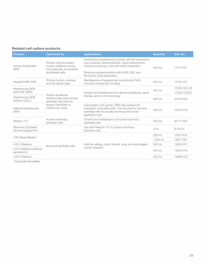
Related cell culture products
Product Optimized for Applications Quantity Cat. No.
Human Endothelial-SFM*
Primary and secondary human umbilical venous, microvascular, and arterial endothelial cells
Growth and maintenance to study cell-cell interactions, injury analysis, atherosclerosis, signal transduction, cytokine production, and cell matrix interaction
Requires supplementation with bFGF, EGF, and fibronectin (sold separately).
500 mL 11111-044
HepatoZYME-SFM Primary human, monkey, and rat hepatocytes
Maintenance of hepatocytes (cytochrome P450 induction maintained >9 days) 500 mL 17705-021
Keratinocyte-SFM (with EGF, BPE)*
Human epidermal keratinocytes and cervical epithelial cells (will not support fibroblast or melanocyte cells)
Growth and maintenance for dermal substitutes, gene therapy, and in vitro toxicology
500 mL17005-042 US
17005-075 EU
Keratinocyte-SFM (without CaC2)*
500 mL 37010-022
Defined Keratinocyte-SFM*
Low-protein (<25 µg/mL), BPE-free medium for cultivation of keratinocytes. Can be used for cervical epithelial cells for studies involving the human papilloma virus
500 mL 10744-019
Medium 171 Human mammaryepithelial cells
Growth and maintenance of human mammary epithelial cells 500 mL M-171-500
Mammary Epithelial Growth Supplement
Use with Medium 171 to culture mammary epithelial cells 5 mL S-015-5
LHC Basal Medium
Bronchial epithelial cells Asthma, allergy, cystic fibrosis, lung, and esophageal cancer research
500 mL 12677-019
1,000 mL 12677-027
LHC-8 Medium 500 mL 12678-017
LHC-8 Medium (without gentamicin) 500 mL 12679-015
LHC-9 Medium 500 mL 12680-013
* Drug master file available.
30
Gibco products including extracellular matrices, laminin, scaffolds, and proteins allow in vivo–like morphology and physiologically relevant environments for more realistic cell biology and better intercellular interactions.
Geltrex LDEV-Free Reduced Growth Factor Basement Membrane MatrixGeltrex™ LDEV-free matrix is a soluble form of reduced growth factor (RGF) basement membrane extract (BME) purified from a continuous sheet of specialized extracellular matrix that forms an interface between Engelbreth-Holm-Swarm (EHS) tumor cells. It is free of viruses, including lactose dehydrogenase elevating virus (LDEV), sometimes found in this type of preparation. The major components of Geltrex matrix include laminin, collagen IV, entactin, and heparan sulfate proteoglycan, which provide the foundation for three-dimensional (3D) culture studies.
Geltrex matrix can be used for promotion and maintenance of a differentiated phenotype in a variety of cell cultures including primary epithelial cells, endothelial cells, hepatocytes, and smooth muscle cells. It has been used in angiogenesis assays, neurite outgrowth assays, and tumor cell invasion assays.
Following appropriate dilutions, Geltrex matrix is a ready-to-use soluble form of basement membrane matrix that requires no additional treatment before use. Depending on the application, different thicknesses and concentrations of Geltrex matrix are required.
Quantity Cat. No.
1 mL A1413201
5 mL A1413202
AlgiMatrix 3D Culture SystemWe developed the AlgiMatrix™ 3D Culture System to be the first user-friendly, animal-free bioscaffold available for the development of higher-fidelity cell culture models that are more predictive of disease states and drug responses. The AlgiMatrix 3D Culture System is available in 6-well, 24-well, and 96-well formats that integrate easily into any workflow. Cells can be inoculated directly into sterile microtiter
plates preloaded with lyophilized alginate sponge; no pretreatment or other preparation is required.
The AlgiMatrix 3D Culture System is suitable for a broad range of procedures, including multicellular tumor spheroid assays (MCTS) [1], hepatocyte [2–4], and cardiomyocyte organogenesis studies [5], as well as co-culture studies [6], high-throughput drug screening assays [1], and embryonic stem cell 3D differentiation [7]. This broad applicability fulfills a critical need for cell-based screening, drug discovery, and in vitro human cell therapy research.
References1. Kunz-Schughart LA, Freyer JP, Hofstaedter F et al. (2004) The use of 3-D cultures
for high-throughput screening: the multicellular spheroid model. J Biomol Screen 9:273–285.
2. Dvir-Ginzberg M, Gamlieli-Bonshtein I, Agbaria R et al. (2003) Liver tissue engineering within alginate scaffolds: effects of cell-seeding density on hepatocyte viability, morphology, and function. Tissue Eng 9:757–766.
3. Elkayam T, Amitay-Shaprut S, Dvir-Ginzberg M et al. (2006) Enhancing the drug metabolism activities of C3A--a human hepatocyte cell line—by tissue engineering within alginate scaffolds. Tissue Eng 12:1357–1368.
4. Kedem A, Perets A, Gamlieli-Bonshtein I et al. (2005) Vascular endothelial growth factor-releasing scaffolds enhance vascularization and engraftment of hepatocytes transplanted on liver lobes. Tissue Eng 11:715–722.
5. Dar A, Shachar M, Leor J et al. (2002) Optimization of cardiac cell seeding and distribution in 3D porous alginate scaffolds. Biotechnol Bioeng 80:305–312.
6. Kim J (2005) Three-dimensional tissue culture models in cancer biology. Semin Cancer Biol 15:365–377.
7. Gerecht-Nir S, Cohen S, Ziskind A et al. (2004) Three-dimensional porous alginate scaffolds provide a conducive environment for generation of well-vascularized embryoid bodies from human embryonic stem cells. Biotechnol Bioeng 88:313–320.
Quantity Cat. No.
1 x 6-well plate A10982-01
4 x 6-well plates A10982-02
1 x 24-well plate 12684-023
4 x 24-well plates 12684-049
1 x 96-well plate 12684-015
5 x 96-well plates 12684-031
Firming buffer (50 mL) A1091501
Dissolving buffer (50 mL) A1134001
Collagen I, Rat Tail and Collagen I, BovineCollagen is the cell culture tissue workhorse of basic coating and gel preparations for growing attachment cells, supporting migration, and tissue morphogenesis. We offer Collagen I Rat Tail (in liquid form and on precoated
Matrices and 3D culture systems
31
tissue culture plates) and Collagen I Bovine (in liquid form) for 3D applications and as coating solution. The precoated plates can be stored at room temperature for a maximum shelf life of 2 years, and are ready to use with no additional preparation.
Product Quantity Cat. No.
Collagen I, Rat Tail 20 mL A1048301
Collagen I, Bovine 10 mL A10644-01
Collagen I, coated 6-well plate 5 plates A11428-01
Collagen I, coated 24-well plate 5 plates A11428-02
Collagen I, coated 96-well plate 5 plates A11428-03
Angiogenesis Starter KitThe Angiogenesis Starter Kit contains the reagents and protocols necessary to perform various angiogenesis assays. Designed to address the needs of both the occasional user and the most experienced researcher, this combination of high-quality products is easy to use and cost-effective. A detailed protocol for the tube formation assay is provided with the kit. The kit includes Large Vessel Endothelial Supplement (LVES), 50X, 11 mL; Medium 200 basal medium, 500 mL; Human Umbilical Vein Endothelial Cells (HUVEC), 1 vial, 5 x 10⁵ cells; and Geltrex LDEV-Free Reduced Growth Factor Basement Membrane Matrix, 5 mL.
Quantity Cat. No.
1 kit A1460901
Natural Mouse LamininLaminin is the major glycoprotein component of basement membranes, with a cross-like shape containing three short arms and one long arm. This structure allows for the modulation of such functions as cell attachment, cell spreading, cell growth and motility, and binding to itself and other matrix components to promote epithelial differentiation, modification of leukocyte function, and stimulation of neurite outgrowth. This natural preparation of mouse laminin is isolated from Engelbreth-Holm-Swarm (EHS) sarcoma.
Quantity Cat. No.
1 mg 23017-015
Choose the culture vessels, surfaces, and tools you need for preparing, growing, and analyzing more physiologically relevant primary tissues, with reproducible results from start to finish. Thermo Scientific™ Nunc™ cell culture research products have been used by researchers for more than 60 years in labs all over the world. Our longstanding commitment to exacting product quality standards and testing helps ensure that you can advance your primary cell research quickly, efficiently, and with confidence.
Product Cat. No.
Nunclon Delta EasYDish 35 mm 150460
Nunclon Delta EasYDish 60 mm 150462
Nunclon Delta EasYDish 100 x 15 mm 150464
Nunclon Delta EasYDish 100 x 20 mm 150466
Nunclon Delta EasYDish 150 x 20 mm 150468
Nunclon Delta 6-well multidish 140675
Nunclon Delta 12-well multidish 150628
Nunclon Delta 24-well multidish 142475
Nunclon Delta 48-well multidish 150687
Nunclon Delta 96-well microplate 167008
Nunclon Delta Edge 2.0 96-well microplate 167425
Product Cat. No.
Nunc Collagen I–coated 6-well multidish 152034
Nunc Collagen I–coated 96-well microplate 152038
Nunc PDL-coated 6-well multidish 152035
Nunc PDL-coated 96-well microplate 152039
Nunc cell scraper, 23 cm, small 179693
Nunc cell scraper, 32 cm, large 179707
Gibco Collagen I–coated 24-well plate A1142802
Gibco Collagen I–coated 96 well plate A1142803
Primary cell culture plastics
32
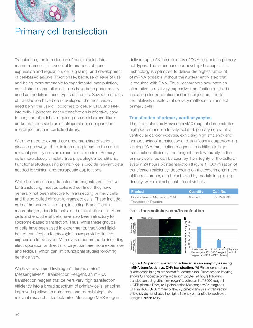
Figure 1. Superior transfection achieved in cardiomyocytes using mRNA transfection vs. DNA transfection. (A) Phase-contrast and fluorescence images are shown for comparison. Fluorescence imaging shows GFP-positive primary cardiomyocytes 24 hours following transfection using either Invitrogen™ Lipofectamine™ 3000 reagent + GFP plasmid DNA, or Lipofectamine MessengerMAX reagent + GFP mRNA. (B) Summary of flow cytometry analysis of transfection efficiency demonstrates the high efficiency of transfection achieved using mRNA delivery.
Phase contrast GFP
Lip
ofe
ctam
ine
30
00 r
eag
ent
+ G
FP
pla
smid
DN
A
Lip
ofe
ctam
ine
M
esse
nger
MA
X
reag
ent
+ G
FP
mR
NA
A
010
203040
5060
Lipofectamine MessengerMAX reagent + mRNA
Lipofectamine 3000 reagent+ GFP plasmid
Negative controlTr
ansf
ecti
on
e�ci
ency
(%) 70
Transfection, the introduction of nucleic acids into mammalian cells, is essential to analyses of gene expression and regulation, cell signaling, and development of cell-based assays. Traditionally, because of ease of use and being more amenable to experimental manipulation, established mammalian cell lines have been preferentially used as models in these types of studies. Several methods of transfection have been developed, the most widely used being the use of liposomes to deliver DNA and RNA into cells. Liposome-based transfection is effective, easy to use, and affordable, requiring no capital expenditure, unlike methods such as electroporation, sonoporation, microinjection, and particle delivery.
With the need to expand our understanding of various disease pathways, there is increasing focus on the use of relevant primary cells as experimental models. Primary cells more closely simulate true physiological conditions. Functional studies using primary cells provide relevant data needed for clinical and therapeutic applications.
While liposome-based transfection reagents are effective for transfecting most established cell lines, they have generally not been effective for transfecting primary cells and the so-called difficult-to-transfect cells. These include cells of hematopoietic origin, including B and T cells, macrophages, dendritic cells, and natural killer cells. Stem cells and endothelial cells have also been refractory to liposome-based transfection. Thus, while these groups of cells have been used in experiments, traditional lipid-based transfection technologies have provided limited expression for analysis. Moreover, other methods, including electroporation or direct microinjection, are more expensive and tedious, which can limit functional studies following gene delivery.
We have developed Invitrogen™ Lipofectamine™ MessengerMAX™ Transfection Reagent, an mRNA transfection reagent that delivers very high transfection efficiency into a broad spectrum of primary cells, enabling improved application outcomes and more biologically relevant research. Lipofectamine MessengerMAX reagent
delivers up to 5X the efficiency of DNA reagents in primary cell types. That’s because our novel lipid nanoparticle technology is optimized to deliver the highest amount of mRNA possible without the nuclear entry step that is required with DNA. Thus, researchers now have an alternative to relatively expensive transfection methods including electroporation and microinjection, and to the relatively unsafe viral delivery methods to transfect primary cells.
Transfection of primary cardiomyocytes The Lipofectamine MessengerMAX reagent demonstrates high performance in freshly isolated, primary neonatal rat ventricular cardiomyocytes, exhibiting high efficiency and homogeneity of transfection and significantly outperforming leading DNA transfection reagents. In addition to high transfection efficiency, the reagent has low toxicity to the primary cells, as can be seen by the integrity of the culture system 24 hours posttransfection (Figure 1). Optimization of transfection efficiency, depending on the experimental need of the researcher, can be achieved by modulating plating density, with minimal effect on cell viability.
Product Quantity Cat. No.
Lipofectamine MessengerMAX Transfection Reagent
0.75 mL LMRNA008
Go to thermofisher.com/transfection
Primary cell transfection
B
33
Neon Transfection SystemFor simple transfection of primary and stem cells, we also offer the Invitrogen™ Neon™ Transfection System, a next-generation electroporation technology for highly efficient delivery (~80%) across payloads (mRNA, DNA, siRNA, Cas9 protein) into virtually any animal cell type.
• The Neon Transfection System has been demonstrated to transfect many difficult-to-transfect cells, including stem cells and primary cells
• Using a fail-safe optimization experiment, conditions are easily adjusted to maximize delivery efficiency and cell viability
Product Cat. No.
Neon Transfection System MPK5000
Go to thermofisher.com/neon
BacMam technologyBacMam technology is based on an insect virus (baculovirus) to help efficiently deliver and express genes in mammalian cells. The baculovirus has been modified to include an expression cassette for transgene expression in mammalian cells.
• Efficient transduction of mammalian cell lines, including primary cells (fibroblasts, hepatocytes, cardiovascular cells, and epithelial cells) and stem cells (neuronal and mesenchymal)
• Safe transduction (nonreplicating in mammalian cells) and lack of observable cytopathic effect
• Frozen storage of pre-transduced cells generates assay-ready cells
• Assay development speed is increased (no need to spend time generating a stable cell line)
Go to thermofisher.com/bacmam
Table 1. Comparison between mRNA and DNA delivery by the Neon Transfection System.
Electroporation parameters (10 µL tips)
Cell lineNumber of cells
Transfection efficiency with mRNA
Transfection efficiency with DNA
mRNA 24-well optimization protocol (program #) Cas9/gRNA
Primary T cells 2 x 10⁵ 96% 84% 1,600 V/10 ms/3 pulses (#24) 1,000 ng/240 ng
Jurkat 2 x 10⁵ 95% 86% 1,400 V/20 ms/2 pulses (#16) 1,500 ng/350 ng
NK-92 2 x 10⁵ 98% 52% 1,300 V/10 ms/3 pulses (#21) 1,000 ng/250 ng
KG-1 2 x 10⁵ 95% 82% 1,600 V/20 ms/1 pulse (#4) 1,000 ng/250 ng
THP-1 1.5 x 10⁵ 88% 42% 1,400 V/20 ms/2 pulses (#16) 1,000 ng/250 ng
SC-1 2 x 10⁵ 78% 54% 1,700 V/20 ms/1 pulse (#5) 1,000 ng/250 ng
SC 2 x 10⁵ 89% 70% 1,600 V/20 ms/1 pulse (#4) 2,000 ng/500 ng
J774A.1 2 x 10⁵ 85% ND 1,700 V/20 ms/1 pulse (#5) 1,000 ng/250 ng
Human primary T cells were isolated from LeukoPak blood product from healthy donors using Ficoll-Paque PLUS medium and the Invitrogen Dynabeads Untouched Human T Cells Kit. The cells were then cultured in OpTmizer CTS T-Cell Expansion medium with 2% human serum, and activated with Dynabeads Human T-Expander CD3/CD28. The transfection experiments with the Neon system were performed 3 days after activation. Each cell line was maintained with Gibco medium, serum, and growth factors according to ATCC guidelines. Cell lines were prepared at a density of 1.5–2.0 x 10⁵ cells per 10 µL tip, for electroporation in Buffer R (component of Neon Transfection System Kits), with 1–1.5 µg of mRNA encoding GFP. The 24-well optimization protocols were performed using the 10 µL Neon tip, and the cells were dispensed into 0.5 mL prewarmed medium in a 24-well plate. Cells were analyzed with the Attune NxT Flow Cytometer 24 hours posttransfection. ND = not determined.
34
EVOS Cell Imaging SystemsDesigned in collaboration with microscopy users, Invitrogen™ EVOS™ Cell Imaging Systems capture high-quality phase contrast or fluorescent cell images right at your benchtop, with interfaces that are so simple, even novice users can collect data in just a few seconds. EVOS Cell Imaging Systems can be used in primary cell experimental protocols during passaging to easily observe and monitor cell dissociation.
Countess II FL Automated Cell CounterWith the option for a reusable slide and fluorescence capabilities—brightfield and two user-changeable fluorescence channels—the Invitrogen™ Countess™ II FL Automated Cell Counter can count cells, monitor fluorescent protein expression, and measure cell viability. The Countess II FL Automated Cell Counter is excellent for counting primary cells during the plating and passaging steps of our culture protocols (Cat. No. AMQAF1000).
Product Cat. No.
EVOS XL Core for cell culture AMEX1000
EVOS FL for easy fluorescence visualization AMF4300
EVOS FL Auto 2 for integrated analysis software AMAFD2000
Primary cell visualization Primary cell detection
We offer a host of products that are designed to enable further engineering of primary cells, as well as analysis of activity and function within primary cell models. These include detection technologies such as Invitrogen™ CellLight™ reagents and Premo™ Cameleon reagents.
CellLight reagentsCellLight reagents are ready-to-use fluorescent protein constructs targeted to specific subcellular structures. These reagents provide a simple and effective method for introducing targeted intracellular labels within living cells. Simply add the reagent to your cells, incubate overnight, and you’re ready to image your cells. CellLight reagents come in a variety of colors and targets, including actin, endosomes, lysosomes, and tubulin, for convenient multiplexing and colocalization studies. Cellular labeling with CellLight reagents employs BacMam technology, which uses a modified insect cell baculovirus coupled with a mammalian promoter as a vehicle to efficiently deliver and express genes in mammalian cells. Unlike expression vectors, BacMam reagents enable titratable and reproducible expression and offer high co-transduction efficiency, enabling multiple BacMam reagents to be used in the same cell.
Go to thermofisher.com/celllight
Premo Cameleon Calcium SensorPremo Cameleon Calcium Sensor is a no-wash, nonorganic dye indicator for intracellular calcium signal measurements. It is provided as a ready-to-use baculovirus stock containing the cameleon DNA, which is efficiently delivered to target cells prior to cell plating. The stock is used in conjunction with a Premo™ enhancer for increased sensor expression in multiple cell types, including primary cells. The Premo Cameleon Calcium Sensor readily and accurately detects intracellular calcium flux from different receptors.
Go to thermofisher.com/premocameleon
35
Tissue Product Quantity Cat. No.
Keratinocytes, neonatalHEKn, prepared in EpiLife Medium and EDGS 6 x T-25 flasks C-001-25P-B
HEKn, prepared in Medium 154 and HKGS 6 x T-25 flasks C-001-25P-G
Keratinocytes, adultHEKa, prepared in EpiLife Medium and HKGS 6 x T-25 flasks C-005-25P-A
HEKa, prepared in Medium 154 and HKGS 6 x T-25 flasks C-005-25P-G
Melanocytes, neonatal, lightly pigmented
HEMn-LP, prepared in Medium 254 and HMGS 3 x T-25 flasks C-002-25P-A
HEMn-LP, prepared in Medium 254 and HMGS-2 3 x T-25 flasks C-002-25P-B
Melanocytes, neonatal, moderately pigmented
HEMn-MP, prepared in Medium 254 and HMGS 3 x T-25 flasks C-102-25P-A
HEMn-MP, prepared in Medium 254 and HMGS-2 3 x T-25 flasks C-102-25P-B
Melanocytes, adult, lightly pigmented HEMa-LP, prepared in Medium 254 and HMGS-2 3 x T-25 flasks C-024-25P-B
Fibroblasts, neonatal HDFn, prepared in Medium 106 and LSGS 6 x T-25 flasks C-004-25P-A
Fibroblasts, adult HDFa, prepared in Medium 106 and LSGS 3 x T-25 flasks C-013-25P-A
Aortic endothelial cells HUVEC, prepared in Medium 200 and LSGS 6 x T-25 flasks C-003-25P-A
Umbilical vein endothelial cells
HUVEC, prepared in Medium 200PRF and LSGS 6 x T-25 flasks C-003-25P-B
HUVEC-pooled, prepared in Medium 200PRF and LSGS 6 x T-25 flasks C-015-25P-B
Aortic smooth muscle cells HASMC, prepared in Medium 231 and SMGS 6 x T-25 flasks C-007-25P-A
We offer many of the human primary cell types included in this sourcebook as proliferating cultures, sourced from the industry’s largest and most reliable tissue sourcing network. These cultures are currently ONLY AVAILABLE IN THE UNITED STATES. Setup is required. Information on availability (including isolation schedules) of these cells can be obtained through our technical service team.
Proliferating primary cell cultures
Contact your account manager or technical sales specialist, or email our custom services team at [email protected] for more details.
For Research Use Only. Not for use in diagnostic procedures. © 2016, 2018 Thermo Fisher Scientific Inc. All rights reserved. All trademarks are the property of Thermo Fisher Scientific and its subsidiaries unless otherwise specified. Fungizone is a trademark of E.R. Squibb & Sons, LLC. CHRM is a trademark of Advanced Pharmaceutical Sciences, Inc. COL16143 0418
These products may be covered by one or more Limited Use Label Licenses (see our product catalog at thermofisher.com). By use of these products you accept the terms and conditions of all applicable Limited Use Label Licenses.
We welcome requests for custom preparations of cell culture products and contract research. Please contact us and we will work with you to develop a solution that meets your research and budgetary needs.
The custom order process is designed on an individual basis, enabling us to tailor the process to suit each request. Once we determine the specifications for the project, we will provide you with a quote for all work and a timeframe for its completion. As always, our technical support and customer service staff are available to assist you every step of the way—from developing the initial specifications to final packaging and delivery.
We have the technical and manufacturing capabilities to produce a wide variety of customized cell culture media and reagents. From slight formulation modifications to complicated engineered-to-order products, custom products are available in both standard and highly specialized packaging configurations. Wherever possible, we can formulate custom media with non–animal origin components and offer developmental support to help you reengineer formulations to allow you to meet regulatory and performance goals. The Gibco brand is the only brand that offers four distinct formats for media: ready-to-use (1X) liquid media, dry powder media (DPM), liquid media concentrates (LMCs), and Advanced Granulation Technology™ (AGT™) media.
Custom cell culture products and services• Custom cell isolations and configurations
• Custom medium and supplement formulations
• Cell pellets suitable for RNA isolation and other purposes
• Additional cell characterization and virus testing
• SynerGy™ Selector—online bag design tool
Custom primary cell products and services