Embed Size (px)
Citation preview

Human Mas-Related G Protein-Coupled Receptors-X1Induce Chemokine Receptor 2 Expression in Rat DorsalRoot Ganglia Neurons and Release of Chemokine Ligand2 from the Human LAD-2 Mast Cell LineHans Jurgen Solinski1, Franziska Petermann2, Kathrin Rothe1, Ingrid Boekhoff1, Thomas Gudermann1,
Andreas Breit1*
1 Walther-Straub-Institut fur Pharmakologie und Toxikologie, Ludwig-Maximilians-Universitat Munchen, Munich, Germany, 2 Department of Neurology, Klinikum rechts
der Isar, Technical University Munich, Munich, Germany
Abstract
Primate-specific Mas-related G protein-coupled receptors-X1 (MRGPR-X1) are highly enriched in dorsal root ganglia (DRG)neurons and induce acute pain. Herein, we analyzed effects of MRGPR-X1 on serum response factors (SRF) or nuclear factorsof activated T cells (NFAT), which control expression of various markers of chronic pain. Using HEK293, DRG neuron-derivedF11 cells and cultured rat DRG neurons recombinantly expressing human MRGPR-X1, we found activation of a SRF reportergene construct and induction of the early growth response protein-1 via extracellular signal-regulated kinases-1/2 known toplay a significant role in the development of inflammatory pain. Furthermore, we observed MRGPR-X1-induced up-regulation of the chemokine receptor 2 (CCR2) via NFAT, which is considered as a key event in the onset of neuropathic painand, so far, has not yet been described for any endogenous neuropeptide. Up-regulation of CCR2 is often associated withincreased release of its endogenous agonist chemokine ligand 2 (CCL2). We also found MRGPR-X1-promoted release ofCCL2 in a human connective tissue mast cell line endogenously expressing MRGPR-X1. Thus, we provide first evidence tosuggest that MRGPR-X1 induce expression of chronic pain markers in DRG neurons and propose a so far unidentifiedsignaling circuit that enhances chemokine signaling by acting on two distinct yet functionally co-operating cell types. Giventhe important role of chemokine signaling in pain chronification, we propose that interruption of this signaling circuit mightbe a promising new strategy to alleviate chemokine-promoted pain.
Citation: Solinski HJ, Petermann F, Rothe K, Boekhoff I, Gudermann T, et al. (2013) Human Mas-Related G Protein-Coupled Receptors-X1 Induce ChemokineReceptor 2 Expression in Rat Dorsal Root Ganglia Neurons and Release of Chemokine Ligand 2 from the Human LAD-2 Mast Cell Line. PLoS ONE 8(3): e58756.doi:10.1371/journal.pone.0058756
Editor: Roland Seifert, Medical School of Hannover, United States of America
Received January 18, 2013; Accepted February 6, 2013; Published March 7, 2013
Copyright: � 2013 Solinski et al. This is an open-access article distributed under the terms of the Creative Commons Attribution License, which permitsunrestricted use, distribution, and reproduction in any medium, provided the original author and source are credited.
Funding: This work was supported by a grant from the ‘‘Deutsche Forschungsgemeinschaft’’ [grant BR 3346/3–1]. The funders had no role in study design, datacollection and analysis, decision to publish, or preparation of the manuscript.
Competing Interests: The authors have declared that no competing interests exist.
* E-mail: [email protected]
Introduction
Primate-specific Mas-related G protein-coupled receptors-X1
(MRGPR-X1) have originally been described to be selectively
expressed in small-diameter dorsal root ganglia (DRG) neurons
[1,2]. However, recently significant MRGPR-X1 mRNA levels
were also detected in connective tissue mast cells (CTMC) and the
leukaemia-derived human mast cell line (LAD)-2 [3,4]. The
endogenous agonist of MRGPR-X1, bovine adrenal medulla
(BAM) peptide 8–22, is cleaved from pro-enkephalin, and several
studies reported activation of the Gq pathway by MRGPR-X1 in
over-expression systems [1,5,6,7]. Studies from our laboratory
revealed that MRGPR-X1 engage phospholipase-Cb to release
calcium form the endoplasmatic reticulum and activate the
proalgetic transient receptor potential cation channel V1. In sharp
contrast to most if not all Gq-coupled receptors MRGPR-X1 do
not undergo agonist-promoted endocytosis [6,8]. In line with
direct TRPV1 activation by MRGPR-X1 observed at the cellular
level, application of BAM8-22 to healthy human volunteers
provoked pain-like sensations pointing to acute nociceptive
functions of MRGPR-X1 [9]. In contrast, over-expression of
MRGPR-X1 in rat dorsal root ganglia (DRG) neurons resulted in
BAM8-22-mediated inhibition of voltage-gated calcium currents
via Gi/o proteins believed to blunt pain perception [10]. Thus,
MRGPR-X1 play a significant role in acute human pain
perception, but the underlying signaling pathways are still poorly
defined. Furthermore, the impact of MRGPR-X1 on gene
expression still remains largely elusive. This is of particular
interest, because alterations in gene expression are often associated
with chronic pain syndromes. In general, G protein-activating
neuropeptides have been reported to affect gene expression via
cAMP response elements (CRE) or serum response elements
(SRE). CRE is activated by means of its interaction with the CRE
binding protein (CREB) [11], whereas SRE activity is enhanced
after binding to serum response factors (SRF) and to ternary
complex factors (TCF) such as the E twenty-six-like transcription
factor-1 (ELK-1) [12]. Interactions between CRE and CREB are
enhanced after phosphorylation of the latter protein by numerous
PLOS ONE | www.plosone.org 1 March 2013 | Volume 8 | Issue 3 | e58756
andreas.

down-stream kinases of GPCR signaling such as protein kinase A
or extracellular signal-regulated kinases-1/2 (ERK-1/2) [13].
Likewise, the affinity of the ELK-1/SRF/SRE complex is
increased after phosphorylation of ELK-1 by ERK-1/2 [14].
Recent data also suggested a role for calcium/calcineurin-induced
activation of nuclear factors of activated T cells (NFAT) in G
protein-coupled receptor (GPCR)-promoted gene expression
[15,16]. Of note, CREB-, TCF/SRF- or NFAT-dependent gene
expression is thought to induce maladaptive processes leading to
neuronal dysfunction or pain chronification [16,17,18,19,20,21].
Given the strong link between alterations in gene expression and
pain chronification we herein analyzed effects of BAM8-22 on
gene expression-regulating signaling pathways in previously
reported human HEK293 or F11 (rat DRG neurons 6 murine
NG18TG-2) cells stably expressing MRGPR-X1 and in cultured
rat DRG neurons transiently expressing MRGPR-X1 [6,8]. We
observed that MRGPR-X1 induce gene expression in all three cell
models tested and of particular interest that BAM8-22-induced
expression of chemokine receptors 2 (CCR2), which has been
linked to neuropathic pain syndromes [22,23]. In LAD-2 mast
cells endogenously expressing MRGPR-X1, we detected signifi-
cant release of the endogenous CCR2 agonist chemokine ligand 2
(CCL2) after BAM8-22 stimulation. Thus, we propose a MRGPR-
X1-induced chemokine signaling circuit that involves induction of
CCR2 expression in DRG neurons and CCL2 release in mast
cells.
Materials and Methods
Materials and PlasmidAll cell culture reagents were purchased from Invitrogen
(Darmstadt, Germany). PromoFectinH was from PromoCell
(Heidelberg, Germany). Anti-pERK-1/2 (E-4), anti-EGR-1 (C-
19) and anti-ERK-2 antiserum (C-14) were obtained from Santa
Cruz Biotechnology (Heidelberg, Germany). Monoclonal anti-
mouse/rat-CCR2 antibody, conjugated to allophycocyanin (APC)
and the CCL2 ELISA kit were obtained from R&D systems
(Wiesbaden, Germany) and peroxidase-conjugated anti-mouse or
anti-rabbit antibody, both raised in goat, were obtained from
Sigma-Aldrich (Deisenhofen, Germany). BSA, forskolin (FSK),
poly-L-ornithine and poly-L-lysine were from Sigma-Aldrich and
fura2-acetoxymethyl ester was obtained from Fluka (Deisenhofen,
Germany). Coelenterazine H was purchased from Biaffin (Kassel,
Germany) and firefly luciferase substrate was from Promega
(Mannheim, Germany). [3H]-adenine was obtained from Perki-
nElmer (Rodgau, Germany). BAM8-22 and bradykinin (BK) were
purchased from Biotrend (Cologne, Germany) and rat CCL2 from
Peprotech (Hamburg, Germany). Cyclosporine A (CsA) was
purchased from Sigma-Aldrich and PD-184352 was from Enzo
life science (Lorrach, Germany). The generation of expression
vectors encoding the human MRGPR-X1 (accession number:
AF474990), the rat MRGPR-C (accession number: AF518245) or
the murine MRGPR-C (accession number: AY152435) fused at
the N-terminus to the Xpress epitope (pcDNA4-MRGPR-X1,
pcDNA4-rMRGPR-C and pcDNA4-mMRGPR-C) was previous-
ly described [6]. The plasmid pG5a [24] encoding aequorin was
kindly provided by Dr. Vladimir Chubanov (Walther-Straub-
Institute, Munich, Germany). The reporter gene vector p5xSRE,
carrying five repeats of the TCF/SRF-activated SRE-site (59-
GGATGTCCATATTAGGAC-ATC-39) was kindly provided by
Dr. Susanne Muehlich (Walther-Straub-Institute, Munich, Ger-
many). The reporter gene vector p6xCRE carrying six repeats of
the CREB-activated CRE site and the reporter gene vector
Table 1. Primer sequences used for RTQ-PCR experiments.
gene number name forward primer reverse primer
NM_031144.2 b-actin Ggctcctagcaccatgaaga atctgctggaaggtggacag
NM_022197.2 c-Fos Gggacagcctttcctactacc gatctgcgcaaaagtcctgt
NM_001013146.1 FosB Acgccgagtcctactccag tctcctcctcttcgggagac
NM_001109302.1 SRF Gcacagacctcacgcaga atgtggccacccacagtt
NM_021836.2 JunB Gggactgggagctcatacc aaagggtggtgcatgtgg
NM_012551.2 EGR-1 Cgaacaaccctacgagcac gcgccttctcgttattcaga
NM_053633.1 EGR-2 Ctacccggtggaagacctc aatgttgatcatgccatctcc
NM_017086.1 EGR-3 Caatctgtaccccgaggaga ccgatgtccatcacattctct
NM_019137.1 EGR-4 Gccctcttcaacctcatgtct ggggaaagggacatccag
NM_024388.1 Nur77 Tgttgatgttcctgcctttg ggaggccatgtcgatcag
NM_001106009.1 Ldb2 Cagtgctgggaacacaacc ttggctctcctaccaccatc
NM_017334.1 Crem2 Ctgctttgccacaaggtgt cgacattctttagcagcttcc
NM_001110860.1 Crem3 Actagcacggggcaatacat accatcagatcctgggttagaa
NM_153626.1 Npas4 Agggtttgctgatgagttgc tcccctccacttccatctt
NM_031327.2 Cyr61 Ggatctgtgaagtgcgtcct ctgcatttcttgcccttttt
NM_022266.2 Ctgf gctgacctagaggaaaacattaaga ccggtaggtcttcacactgg
NM_021866.1 CCR2 aagaagtatccaagagcttgatgag tcaccatcatcatagtcatacgg
NM_001007235.1 IP3R-1 Catccagtatggcaacgtga tcatggcgttcttctccagt
NM_031530.1 CCL2 Agcatccacgtgctgtctc gatcatcttgccagtgaatgag
NM_017232.3 COX-2 tccaacctctcctactacaccag tccagaacttcttttgaatcagg
NM_012513.3 BDNF Agcgcgaatgtgttagtggt gcaattgtttgcctctttttct
doi:10.1371/journal.pone.0058756.t001
MRGPR-X1 Enhance CCR2/CCL2 Signaling
PLOS ONE | www.plosone.org 2 March 2013 | Volume 8 | Issue 3 | e58756
pGL4.30 carrying four repeats of the NFAT-activated transcrip-
tion site were obtained from Promega.
Cell culture and transfectionsHEK293 cells were cultured in DMEM (10% FBS, 2 mM L-
glutamine, penicillin/streptomycin) and F11 (rat DRG neurons 6murine NG18TG-2) cells in HAM’s F-12 (15% FBS, 2 mM L-
glutamine, hypoxanthine-aminopterin-thymidine, G-418 and pen-
icillin/streptomycin). F11 cells were generated by Francel et al.,
[25] and kindly provided by Dr. Mederos y Schnitzler (Philipps
University, Marburg, Germany). The CTMC line LAD-2 [26]
was kindly provided from Dr. Arnold S. Kirshenbaum (National
Institute of Allergy and Infectious Diseases, Bethesda, USA) and
cultured in StemPro 34 (2 mM L-glutamine, nutrient supplement,
100 ng/ml recombinant human stem cell factor, penicillin/
streptomycin). HEK293-MRGPR-X1, HEK293-rMRGPR-C,
HEK293-mMRGPR-C and F11-MRGPR-X1 cells were de-
scribed previously [6]. HEK293 cells stably expressing either
receptor were subjected to a second selection (250 mg/ml G418
and 50 mg/ml zeocin) after transfection with the calcium sensing
aequorin-eGFP construct pG5a to give HEK293-MRGPR-X1-
Ae, HEK293-rMRGPR-C-Ae and HEK293-mMRGPR-C-Ae
cells, respectively. The expression of the fusion proteins was
controlled by measuring agonist-induced calcium releases and by
fluorescence microscopy. Primary DRG neurons from day 18
embryonic Sprague/Dawley rats were purchased from Innoprot
(Derio – Bizkaia, Spain) and cultured in NeuroBasal medium (B-
27 supplement, 10% FBS, 2 mM L-glutamine, penicillin/strepto-
mycin/neomycin) on poly-L-ornithine-coated well plates. For
transient protein expression in HEK293 cells or DRG neurons
PromoFectinH reagent was used according to the manufacturer’s
protocol. F11 cells were transfected using the NeonH transfection
system from Invitrogen accordingly to settings provided by the
manufacturer.
Fura2-based single cell calcium imaging24 h prior to the experiment F11-MRGPR-X1 cells were
seeded at a density of 4–66104 cells on glass cover slips in 6-well
plates coated with 0.1% poly-L-lysine. Cells were loaded with
5 mM fura2-AM in HEPES-buffered saline (HBS, 10 mM
HEPES, 5 mM KCl, 1 mM MgCl2, 140 mM NaCl, 0.1% glucose
and 2 mM CaCl2 adjusted to pH 7.4 with 1 M NaOH)
supplemented with 0.02% pluronic F-127. Cover slips were
washed twice with HBS without BSA and placed in a recording
chamber. Cells were analyzed using a Polychrome 5000 mono-
chromator (Till-Photonics, Grafelfing, Germany) and an Andor
charge-coupled device camera coupled to an inverted microscope
(IX71, Olympus, Hamburg, Germany) by alternately exciting with
340 nm or 380 nm, respectively, every 0.5 s. Normalized fura2-
ratios (340/380) were then plotted against the time in seconds.
Fura2-based calcium measurements with a plate readerCalcium transients in MRGPR-X1 or rodent MRGPR-C
expressing HEK293, F11-MRGPR-X1 or LAD-2 cells were
measured with a FLUOstarH Omega plate reader (BMG Labtech,
Ortenberg, Germany) as described previously [6] using the
fluorescent calcium indicator fura2. To determine concentration-
response curves, integrated calcium signals were calculated,
normalized to responses to maximal ligand concentrations
(100%) and then subjected to curve fitting.
Aequorin-based calcium measurementsTotal luminescence in HEK293-MRGPR-Ae cells or DRG
neurons transfected with the aequorin encoding plasmid G5a was
measured using a FLUOstarH Omega plate reader at 37uC after
labelling of the cells with coelenterazine H (5 mM) for 30 min at
room temperature. HBS as a control or HBS including BAM8-22
was automatically injected 5 s after starting the measurement. In
intervals of 1 s total emission was measured and is given as
arbitrary units against the time in seconds.
Western-BlottingCells were serum-starved for 24 h, stimulated for the indicated
period of time with BAM8-22 or BK and then lysed in laemmli
buffer. After SDS-PAGE and protein transfer to nitrocellulose,
ERK-1/2 phosphorylation or EGR-1 expression was analyzed by
using either a phospho-specific ERK-1/2 (p-ERK-1/2: 1:3,000) or
an EGR-1 specific (EGR1: 1:10,000) antiserum and the corre-
sponding peroxidase-coupled secondary antibody (1:10,000).
Membranes were stripped and then re-probed with an anti-
ERK-2 antibody (t-ERK-2: 1:10,000) and the corresponding
peroxidase-coupled secondary antibody (1:10,000). Immuno-reac-
tivity was quantified by densitometry, ratios between p-ERK-1/2
or EGR-1 and t-ERK-2 signals were calculated, and ligand-
induced ERK-1/2 phosphorylation/EGR-1 expression was nor-
malized to not stimulated cells.
Reporter gene assaysAfter transfection of a firefly luciferase reporter gene construct
DRG neurons were serum-starved for 1 h and F11 or HEK293
cells for 24 h. Afterwards cells were stimulated with BAM8-22, BK
or FSK for the indicated time in serum-free medium. Small
molecule inhibitors (concentration as indicated) and the respective
carrier control were pre-incubated for 30 minutes before addition
of agonist and present during ligand stimulation. After stimulation
cells were lysed (25 mM Tris/HCl pH 7.4, 4 mM EGTA, 8 mM
MgCl2, 1 mM DTT and 1% Triton-X-100) and luciferase activity
measured in white bottom 96-well plates after automatically
injecting luciferase substrate and simultaneous detection of total
light emission for 7.5 s post injection in a FLUOstarH Omega
plate reader. Maximal light emission during this period was
recorded and is indicated relative to not stimulated cells.
RT-PCRDRG neurons were serum-starved for 1 h and F11 or HEK293
cells for 24 h. Afterwards cells were stimulated with BAM8-22 or
BK for the indicated period of time in serum-free medium. Total
RNA from these cells or from not stimulated LAD-2 cells was
isolated using the Tri reagentH (Sigma-Aldrich) according to the
manufacturer’s instructions. First strand synthesis was carried out
with oligo(dT)18 primer using 2 mg of total RNA and the
RevertAidTM
H Minus First Strand cDNA Synthesis Kit (Fermen-
tas, Sankt-Leon Roth, Germany). Quantitative RT-PCR (RTQ-
PCR) was done using the LightCyclerH 480 SybrGreen I Master
Mix (Roche, Mannheim, Germany), 0.08 ml of the first strand
synthesis reaction and 1 mM of the primer pairs shown in table 1.
Samples were analyzed in a LightCyclerH 480 (Roche) using the
following conditions: initial denaturation for 15 min at 94uC, 55
cycles of 94uC for 10 sec, 55uC for 10 sec and 72uC for 10 sec.
Crossing points (Cp) were determined by the software supplied
with the LightCyclerH 480 and ligand-induced modulation of
target gene expression was calculated relative to b-actin using the
DDCp method. Non-quantitative RT-PCR was conducted in
cDNA from LAD-2 cells using specific primers for MRGPR-X1
MRGPR-X1 Enhance CCR2/CCL2 Signaling
PLOS ONE | www.plosone.org 3 March 2013 | Volume 8 | Issue 3 | e58756

(forward: 59-tgagtctctgatctgccctct-39, reverse: 59-tcaccagctgtat-
gatctctgatt-39) or b-actin (forward: 59-ccaaccgcgagaagatga-39,
reverse: 59-ccagaggcgtacagggatag-39). PCR settings were the same
as in RTQ-PCR and the number of amplification cycles is
indicated in the figure legend.
Flow cytometryCells were serum-starved for 4 h and stimulated for 20 h in
serum-free medium with BAM8-22, detached with PBS/2 mM
EDTA, washed once with PBS/10% FBS and stained with anti-
rat-CCR2-APC antibody or the corresponding isotype for
30 minutes at room temperature. After two washing steps cells
were resuspended in PBS/10% FBS and analyzed by flow
cytometry (CyAn ADP, Beckman Coulter).
cAMP accumulation assayTo determine CCL2-promoted inhibition of forskolin-induced
cAMP accumulation, F11 cells were seeded in 12-plates dishes
coated with 0.1% polyl-L-lysine 24 h prior to the experiment.
Afterwards cells were serum-starved for 4 h, stimulated for 20 h
with BAM8-22 or BK in serum-free HAM’s F-12 medium
containing 2 mCi/ml of [3H] adenine. Cells were stimulated for
30 min at 37uC in HAM’s F-12 containing 100 mM HEPES,
pH 7.4, 2.5 mM IBMX, 5 mM forskolin alone or along with
100 ng/ml CCL2. The reaction was terminated by removing the
medium and adding ice-cold 5% trichloroacetic acid to the cells.
[3H] cAMP was then purified by sequential chromatography over
dowex resin and aluminium oxide, and the cAMP accumulation
expressed as the total amount of [3H] cAMP in DPM/well.
Data analysisData were analyzed using Prism4.0 (GraphPad Software Inc.,
San Diego, CA). Statistical significance of differences was assessed
by the two-tailed Student’s t-test (two groups) or by one-way
analysis of variance and Tukey’s honest significance post-hoc test
(more than two groups). Asterisks (***p,0.001, **p,0.01,
*p,0.05) or dagger signs ({{{p,0.001, {{p,0.01, {p,0.05) were
used to indicate significant differences between different experi-
mental groups.
Results
BAM8-22-induced calcium release and ERK-1/2 activationin HEK293-MRGPR-X1 and F11-MRGPR-X1 cells
Binding of the specific agonist BAM8-22 to MRGPR-X1 has
been described to activate Gq proteins and thus, to increase
intracellular calcium concentrations [1,6,7]. Therefore, we first
analyzed BAM8-22-induced calcium signals in previously reported
HEK293-MRGPR-X1 or F11-MRGPR-X1 cells that stably
express low levels of MRGPR-X1 [6,8]. Stimulation of
MRGPR-X1 with BAM8-22 elicited calcium transients with a
potency of ,200 nM (Fig. 1A+B) consistent with previous reports
[1,5]. ERK-1/2 play a central role in GPCR-promoted gene
induction, because ERK-1/2 enhance CREB- and TCF/SRF-
dependent gene expression. Thus, we also analyzed BAM8-22-
promoted ERK-1/2 phosphorylation in HEK293-MRGPR-X1
and F11-MRGPR-X1 cells. We observed marked, but transient
phosphorylation of ERK-1/2 in both cell pools, with a peak at
5 min and a decline thereafter (Fig. 1C+D). Given that calcium
has been reported to enhance NFAT-dependent gene expression
via calcineurin, and ERK-1/2 via TCF/SRF or CREB, we
postulated that BAM8-22 might induce CREB-, TCF/SRF- or
NFAT-dependent gene expression in MRGPR-X1 expressing
cells.
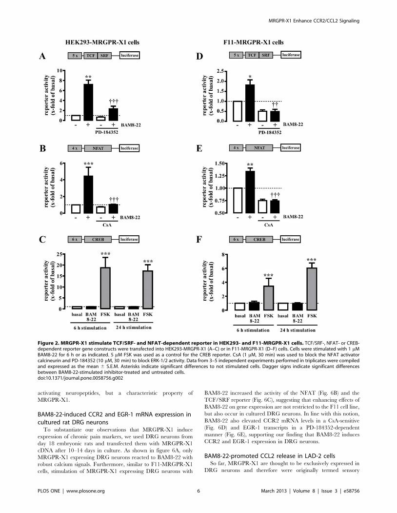
BAM8-22-induced activation of TCF/SRF- and NFAT-dependent reporters in HEK293-MRGPR-X1 and F11-MRGPR-X1 cells
To test the ability of BAM8-22 to induce gene expression via
MRGPR-X1, we used HEK293-MRGPR-X1 and F11-MRGPR-
X1 cells to perform reporter gene assays employing constructs that
contained binding sites for TCF/SRF, NFAT or CREB.
Stimulation of both cell types with BAM8-22 led to activation of
the TCF/SRF reporter (Fig. 2A+D) preventable by a specific
ERK-1/2 inhibitor (PD-184352), and of the NFAT reporter
sensitive to CsA, an inhibitor of the NFAT activator calcineurin
(Fig. 2B+E). In contrast, BAM8-22-induced activation of the
CREB reporter was not observed in either MRGPR-X1
expressing cell type (Fig. 2C+F).
BAM8-22-induced expression of c-Fos and EGR-1 via ERK-1/2 in F11-MRGPR-X1 cells
To gain deeper insight into BAM8-22-induced expression of
TCF/SRF-dependent genes, we analyzed mRNA levels of 15
genes reported to be up-regulated by TCF/SRF in neurons [18].
As shown in figure 3A, mRNA levels of c-Fos and EGR-1 were
significantly elevated in F11-MRGPR-X1 cells after 1 h of BAM8-
22 stimulation and total EGR-1 protein levels significantly
increased after challenging the cells with BAM8-22 for 1 to 2 h
(Fig. 3B). Because c-Fos and EGR-1 mRNA induction was blocked
by PD-184352 (Fig. 3C and D), we concluded that ERK-1/2
activity is required for BAM8-22-induced expression of TCF/
SRF-dependent genes.
BAM8-22-induced expression of IP3R-1 and CCR2 viacalcineurin in F11-MRGPR-X1 cells
Recently, it has been reported that CCR2, IP3R-1, CCL2,
cyclooxygenase-2 (COX-2) or the brain-derived neurotrophic
factor are up-regulated by NFAT in neurons [16,17,27]. Thus, we
analyzed effects of BAM8-22 on the mRNA levels of these genes in
F11-MRGPR-X1 cells. As shown in figure 4, IP3R-1 and CCR2
were up-regulated after 6 h of BAM8-22 incubation in a CsA-
dependent manner, indicating that BAM8-22 induces expression
of both genes via calcium/calcineurin-mediated NFAT activation
in F11-MRGPR-X1 cells. BAM8-22-induced expression of CCR2
is of particular interest, because enhanced CCR2 expression in
DRG neurons is strongly associated with various neuropathic pain
syndromes [28]. To further corroborate our observation of BAM8-
22-induced CCR2 mRNA expression, we performed flow
cytometry experiments with F11-MRGPR-X1 cells and an APC-
conjugated CCR2 specific antibody (Fig. 4D). Stimulation of cells
with BAM8-22 increased the amount of CCR2 positive cells by
10.861.4%, demonstrating that BAM8-22-promoted induction of
CCR2 mRNA is translated into increased expression of the CCR2
protein. To test whether BAM8-22-induced CCR2 expression was
sufficient to affect the sensitivity of F11-MRGPR-X1 cells to
CCL2, we took advantage of the Gi/o coupling of CCR2 and
performed cAMP accumulation assays with BAM8-22-treated
cells. As shown in figure 4E, untreated F11-MRGPR-X1 cells did
not respond to CCL2, whereas BAM8-22-pre-treated F11-
MRGPR-X1 cells significantly responded to the peptide, such
that the CCR2 specific agonist reduced FSK-induced cAMP
accumulation by 23.864.3%, comparable to the effects of various
opioids acting on endogenous opioid receptors constitutively
expressed in F11 cells [29]. Therefore, we concluded that
BAM8-22 enhances the expression of functional CCR2 in F11-
MRGPR-X1 cells, thus enabling CCL2 to engage cognate
signaling pathways in DRG neuron-derived cells.
MRGPR-X1 Enhance CCR2/CCL2 Signaling
PLOS ONE | www.plosone.org 4 March 2013 | Volume 8 | Issue 3 | e58756

Bradykinin induces EGR-1 expression via ERK-1/2 but notCCR2 expression via NFAT
Next we asked whether other Gq protein activating neuropep-
tides would also induce EGR-1 and CCR2 expression in a DRG
neuron-derived cell model. F11 cells have been reported to
endogenously express Gq activating bradykinin-2-receptors (B2R)
[25]. Thus, we first compared calcium transients triggered by
BAM8-22 or BK. As shown in figure 5A, calcium signals in F11-
MRGPR-X1 cells induced by saturated concentrations (EC95) of
BK or BAM8-22 were almost identical illustrating that activation
of recombinant MRGPR-X1 did not elicit unphysiological high
calcium transients. Furthermore, BK induced EGR-1 expression
(Fig. 5B+C) in accord with previously observed effects of BK on
EGR-1 expression in human fibroblasts or NG108-15 neuroblas-
toma cells [30,31]. BK also activated the NFAT reporter (Fig. 5D)
and also induced IP3R-1 mRNA expression via NFAT (Fig. 5E).
However, despite these similarities, BK treatment did neither
induce CCR2 mRNA expression (Fig. 5E), nor did it impart
CCL2 sensitivity to F11-MRGPR-X1 cells (Fig. 5F), suggesting
that CCR2 induction is not a common feature among Gq
Figure 1. MRGPR-X1 stimulate ERK-1/2 in HEK293- and F11-MRGPR-X1 cells. Concentration response curves of BAM8-22-promoted calciumsignals in fura2-loaded HEK293-MRGPR-X1 (A) or in F11-MRGPR-X1 (B) cells (squares) or mock cells (circles) are shown. BAM8-22-induced (1 mM)phosphorylation of ERK-1/2 in HEK293-MRGPR-X1 (C) or in F11-MRGPR-X1 (D) cells was analyzed by western-blotting using a phospho-specificantibody (p-ERK-1/2). Afterwards blots were stripped and re-probed with an antibody against ERK-2 (t-ERK-2). One representative blot is shown.Ligand-induced ERK-1/2 phosphorylation was quantified by densitometry and is given normalized to not stimulated cells. Data from 4 experimentswere compiled and expressed as the mean 6 S.E.M. Asterisks indicate significant differences to not stimulated cells.doi:10.1371/journal.pone.0058756.g001
MRGPR-X1 Enhance CCR2/CCL2 Signaling
PLOS ONE | www.plosone.org 5 March 2013 | Volume 8 | Issue 3 | e58756
activating neuropeptides, but a characteristic property of
MRGPR-X1.
BAM8-22-induced CCR2 and EGR-1 mRNA expression incultured rat DRG neurons
To substantiate our observations that MRGPR-X1 induce
expression of chronic pain markers, we used DRG neurons from
day 18 embryonic rats and transfected them with MRGPR-X1
cDNA after 10–14 days in culture. As shown in figure 6A, only
MRGPR-X1 expressing DRG neurons reacted to BAM8-22 with
robust calcium signals. Furthermore, similar to F11-MRGPR-X1
cells, stimulation of MRGPR-X1 expressing DRG neurons with
BAM8-22 increased the activity of the NFAT (Fig. 6B) and the
TCF/SRF reporter (Fig. 6C), suggesting that enhancing effects of
BAM8-22 on gene expression are not restricted to the F11 cell line,
but also occur in cultured DRG neurons. In line with this notion,
BAM8-22 also elevated CCR2 mRNA levels in a CsA-sensitive
(Fig. 6D) and EGR-1 transcripts in a PD-184352-dependent
manner (Fig. 6E), supporting our finding that BAM8-22 induces
CCR2 and EGR-1 expression in DRG neurons.
BAM8-22-promoted CCL2 release in LAD-2 cellsSo far, MRGPR-X1 are thought to be exclusively expressed in
DRG neurons and therefore were originally termed sensory
Figure 2. MRGPR-X1 stimulate TCF/SRF- and NFAT-dependent reporter in HEK293- and F11-MRGPR-X1 cells. TCF/SRF-, NFAT- or CREB-dependent reporter gene constructs were transfected into HEK293-MRGPR-X1 (A–C) or in F11-MRGPR-X1 (D–F) cells. Cells were stimulated with 1 mMBAM8-22 for 6 h or as indicated. 5 mM FSK was used as a control for the CREB reporter. CsA (1 mM, 30 min) was used to block the NFAT activatorcalcineurin and PD-184352 (10 mM, 30 min) to block ERK-1/2 activity. Data from 3–5 independent experiments performed in triplicates were compiledand expressed as the mean 6 S.E.M. Asterisks indicate significant differences to not stimulated cells. Dagger signs indicate significant differencesbetween BAM8-22-stimulated inhibitor-treated and untreated cells.doi:10.1371/journal.pone.0058756.g002
MRGPR-X1 Enhance CCR2/CCL2 Signaling
PLOS ONE | www.plosone.org 6 March 2013 | Volume 8 | Issue 3 | e58756

neuron-specific GPCR [1]. Recently, significant mRNA levels of
MRGPR-X1 were detected in native CTMC and in the human
LAD-2 mast cell line [4]. In theses lines, we detected significant
MRGPR-X1 transcripts in LAD-2-derived cDNA samples
(Fig. 7A). However, activation of the MRGPR-X1 protein by its
specific agonist BAM8-22 has not been demonstrated yet. Thus,
we took advantage of LAD-2 cells and performed fura2-based
calcium measurements in order to monitor activation of the
MRGPR-X1 protein by its specific agonist BAM8-22. As shown in
figure 7B, BAM8-22 induced robust calcium transients in LAD-2
cells, indicating endogenous expression of functional MRGPR-X1.
Next, we asked whether BAM8-22-induced signaling would affect
Figure 3. MRGPR-X1 induce EGR-1 via ERK-1/2 in F11-MRGPR-X1 cells. RTQ-PCR experiments were performed with cDNAs derived fromserum-starved F11-MRGPR-X1 cells stimulated or not with BAM8-22 (1 mM) for 1 h (A, C and D) using specific primers for 15 distinct genes asindicated in table 1. The name of the analyzed gene is listed beneath the corresponding bar. Genes with a p value ,0.05 are show in black bars. In (Cand D) cells were treated or not with PD-184352 (30 min, 10 mM). Relative BAM8-22-induced gene expression was normalized to b-actin andcalculated using the DDCp method. In (B) serum-starved F11-MRGPR-X1 cells were stimulated or not with BAM8-22 (1 mM) for the indicated period oftime and expression of EGR-1 was determined by western-blotting. Afterwards blots were stripped and re-probed with an antibody against ERK-2 (t-ERK-2). One representative blot is shown. Ligand-induced EGR-1 expression was quantified by densitometry and is given normalized to notstimulated cells. Data from 4 independent experiments were compiled and expressed as the mean 6 S.E.M. Asterisk indicate a significant differenceto not stimulated cells. Dagger signs indicate a significant difference between BAM8-22-stimulated inhibitor-treated and untreated cells.doi:10.1371/journal.pone.0058756.g003
MRGPR-X1 Enhance CCR2/CCL2 Signaling
PLOS ONE | www.plosone.org 7 March 2013 | Volume 8 | Issue 3 | e58756

Figure 4. MRGPR-X1 induce CCR2 via NFAT in F11-MRGPR-X1 cells. RTQ-PCR experiments were performed with cDNAs derived from serum-starved F11-MRGPR-X1 cells stimulated or not with BAM8-22 (1 mM) for 6 h (A-C) using specific primers for 5 distinct genes as indicated in table 1. Thename of the analyzed gene is listed beneath the corresponding bar. Genes with a p value ,0.05 are show in black bars. In (B and C) cells were treatedor not with CsA (1 mM, 30 min). Relative BAM8-22-induced gene expression was normalized to b-actin and calculated using the DDCp method. Datafrom 4 independent experiments were compiled and expressed as the mean 6 S.E.M. BAM8-22-induced (1 mM, 20 h) CCR2 protein expression in F11-MRGPR-X1 cells was assessed by flow cytometry (D) or by CCL2-promoted (100 ng/ml) inhibition of FSK-induced (5 mM) cAMP accumulation (E). In (D)
MRGPR-X1 Enhance CCR2/CCL2 Signaling
PLOS ONE | www.plosone.org 8 March 2013 | Volume 8 | Issue 3 | e58756

CCL2 release in LAD-2 cells. Notably, when performing ELISA
assays with an antibody specific to human CCL2, we observed that
the chemokine was released into the supernatant of LAD-2 cells in
response to BAM8-22 (Fig. 7C), indicating that MRGPR-X1 exert
their action in cells of the peripheral nervous and the immune
system.
Discussion
Primate-specific MRGPR-X1 are selectively expressed in DRG
neurons and therefore represent promising targets for innovative
pain therapies [1,2]. Interestingly, disease-causing genes are
strongly enriched among primate-specific genes [32] and the
ligand-binding domain of MRGPR-X1 has been described as a
product of strong positive selection [33,34], further underscoring
the potential of MRGPR-X1 as therapeutic targets. Despite the
high therapeutic potential of MRGPR-X1, hardly any information
about the role of this receptor subtype in pain chronification is
available. Herein, we analyzed BAM8-22-induced gene expression
in HEK293 and F11 cells stably expressing MRGPR-X1. We
observed activation of a NFAT- and TCF/SRF-, but not of a
CREB-dependent reporter. In-depth analysis of F11-MRGPR-X1
cells confirmed these findings, because BAM8-22-induced ERK-
1/2 or calcineurin activation led to significant up-regulation of c-
Fos, EGR-1, IP3R-1 and CCR2 mRNA. Thus, our data provide
first evidence of BAM8-22-induced gene expression in DRG
neuron-derived cells. F11 cells emerged as a valuable biological
tool for the analysis of DRG neuron-related signaling pathways,
since many studies reported striking similarities to cultured DRG
neurons [25,27,35,36,37,38,39,40,41,42,43]. Along these lines,
data from our laboratory revealed morphine-induced expression of
the early growth response protein-4 via m opioid receptors in F11
cells and cultured rat DRG neurons [29]. In the same vein, we
confirmed BAM8-22-promoted activation of the NFAT or TCF/
SRF reporter and enhanced CCR2 or EGR-1 mRNA expression
in cultured rat DRG neurons transiently expressing MRGPR-X1.
BAM8-22-induced EGR-1 expression via ERK-1/2Activated TCF/SRF complexes have been shown to induce
immediate early genes such as c-Fos, c-jun or members of the
EGR family in neurons of the peripheral or central nervous system
and thereby modulate synaptic activity, memory and learning
[18]. Herein, we observed BAM8-22-promoted up-regulation of c-
Fos and EGR-1 in F11-MRGPR-X1 cells. Induction of c-Fos in
the spinal dorsal horn is an established marker of neuronal activity
or noxious stimulation [44]. However, less is known about the role
of c-Fos induction in DRG neurons. A recent study correlated
surgery- or opioid-induced hyperalgesia with ERK-1/2 phosphor-
ylation and c-Fos expression in DRG neurons [45], suggesting that
BAM8-22-promoted c-Fos induction is in good agreement with
enhanced pain sensations previously reported after BAM8-22
application in humans [9]. So far, induction of the zinc-finger
transcription factor EGR-1 (also zif/268) in DRG neurons has
been described for neurotrophins such as the nerve growth factor
or tumor necrosis factor-a [46,47]. Tumor necrosis factor-a-
promoted EGR-1 expression plays a significant role in carrageen-
an-induced inflammatory hyperalgesia [46] and selective knock-
down of the EGR-1 protein significantly reduced inflammatory
pain induced by complete Freund’s adjuvants [48]. Furthermore,
elevated EGR-1 protein levels correlate with inflammatory pain in
sheep [49], strongly indicating a major contribution of EGR-1 to
inflammatory pain in various species.
BAM8-22-induced CCR-2 expression via calcineurinNFAT-induced gene expression significantly contributes to the
differentiation and proliferation of lymphocytes. Recent work
suggested that NFAT activation also increases the expression of
pro-nociceptive genes such as COX-2 or CCR2 in neurons
[17,27], indicating a significant role of NFAT-dependent gene
induction for pain chronification [28]. Induction of IP3R-1 via
NFAT, as shown herein for Gq-coupled MRGPR-X1, has so far
been linked to the activation of receptor tyrosine kinases by
neurotrophins in DRG, spinal cord and hippocampal pyramidal
neurons [50] or to potassium-induced depolarisation in rat
cerebellar neurons [51]. Thus, IP3R-1 is one of the best described
targets of NFAT-dependent gene expression in non-immune cells,
but cellular or physiological consequences of enhanced IP3R-1
expression levels in neurons have not been sorted out. Chemically
induced calcium influx in F11 cells or DRG neurons by ionomycin
or high potassium concentrations have been reported to induce
CCR2 mRNA expression via NFAT [27]. However, G protein-
promoted CCR2 induction via NFAT has not yet been described.
Here, we report that stimulation of F11-MRGPR-X1 cells with
BAM8-22 not only increased CCR2 mRNA and protein levels,
but also imparted sensitivity to the endogenous CCR2 agonist,
CCL2, to these cells. MRGPR-X1-induced CCR2 expression is of
particular interest, considering that CCL2-induced activation of
CCR2 in DRG neurons plays a major role in distinct neuropathic
pain syndromes, as demonstrated by significantly diminished
neuropathic pain responses found in mice deficient of the CCR2
gene [52] and by abolished neuropathy-induced hyperalgesia and
allodynia after application of CCR2 specific antagonists to wild-
type mice [22,53]. Therefore, inhibition of CCL2-promoted
CCR2 signaling is a promising approach towards a future therapy
of neuropathic pain. However, because of versatile physiological
roles of chemokines significant side effects are expected when
blocking CCR2-induced signaling in vivo. Of note, CCR2-deficient
mice exhibited normal pain behaviour under physiological
conditions and many studies showed that, similar to F11-
MRGPR-X1 cells, CCR2 expression levels in DRG neurons are
rather low under non-neuropathic conditions [27,28]. Hence,
CCR2 up-regulation in DRG neurons emerged as a key event that
initiates neuropathic pain syndromes or enhances their progress
[22,23,54,55,56,57]. Within this model, controlling CCR2 ex-
pression selectively in DRG neurons would be a promising
approach to block chemokine-promoted signaling and thus, to
alleviate neuropathic pain syndromes without affecting CCL2/
CCR2 signaling in other cell types. Herein, we define MRGPR-
X1 as endogenous inducers of CCR2 in DRG neurons. Thus,
although our conclusions still have to await ultimate proof by in
vivo studies in primates, we propose a role of MRGPR-X1 in
chemokine-promoted pain chronification and consider MRGPR-
X1 as promising targets to selectively manipulate CCR2
expression in DRG neurons.
one representative experiment is shown. Accumulation of the data from 5 independent experiments revealed an increase in the number of CCR2positive cells by 10.861.4% after BAM8-22 stimulation. In (E, left panel) one representative experiment is shown and in (E, right panel) data from 5independent experiments performed in triplicates were compiled and expressed as the mean 6 S.E.M. Asterisks indicate a significant difference tonot stimulated cells. Dagger signs indicate a significant difference between BAM8-22-stimulated inhibitor-treated and untreated cells.doi:10.1371/journal.pone.0058756.g004
MRGPR-X1 Enhance CCR2/CCL2 Signaling
PLOS ONE | www.plosone.org 9 March 2013 | Volume 8 | Issue 3 | e58756
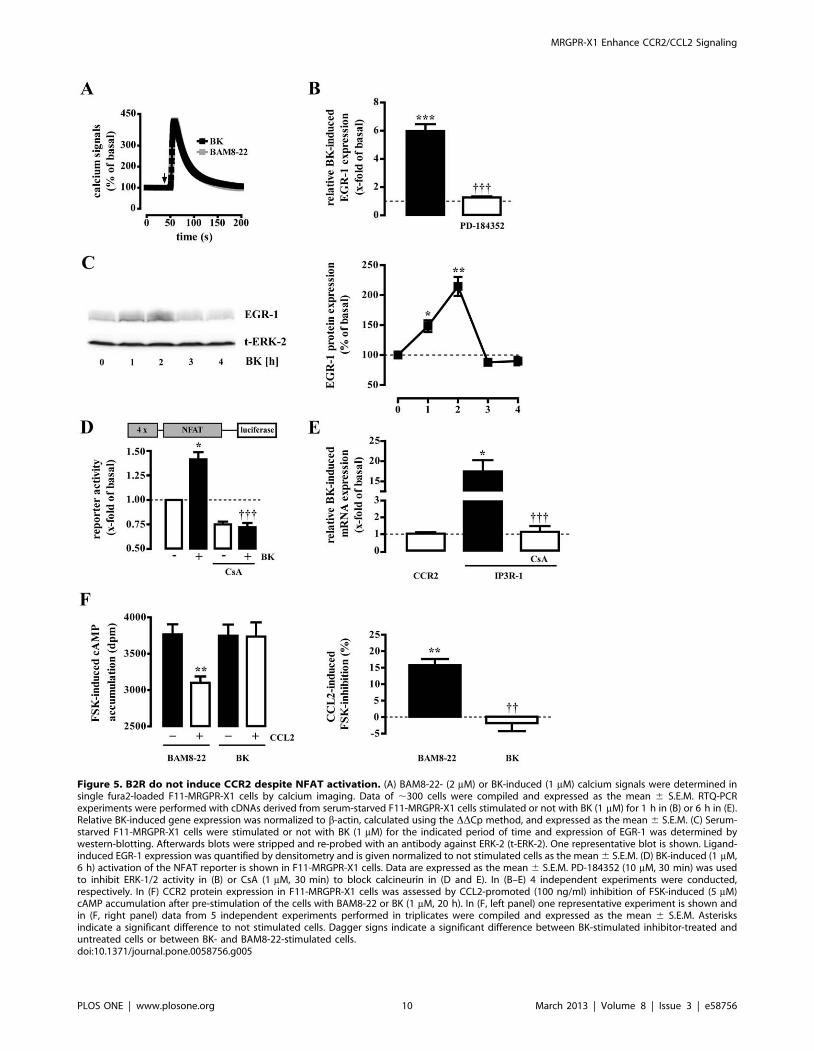
Figure 5. B2R do not induce CCR2 despite NFAT activation. (A) BAM8-22- (2 mM) or BK-induced (1 mM) calcium signals were determined insingle fura2-loaded F11-MRGPR-X1 cells by calcium imaging. Data of ,300 cells were compiled and expressed as the mean 6 S.E.M. RTQ-PCRexperiments were performed with cDNAs derived from serum-starved F11-MRGPR-X1 cells stimulated or not with BK (1 mM) for 1 h in (B) or 6 h in (E).Relative BK-induced gene expression was normalized to b-actin, calculated using the DDCp method, and expressed as the mean 6 S.E.M. (C) Serum-starved F11-MRGPR-X1 cells were stimulated or not with BK (1 mM) for the indicated period of time and expression of EGR-1 was determined bywestern-blotting. Afterwards blots were stripped and re-probed with an antibody against ERK-2 (t-ERK-2). One representative blot is shown. Ligand-induced EGR-1 expression was quantified by densitometry and is given normalized to not stimulated cells as the mean 6 S.E.M. (D) BK-induced (1 mM,6 h) activation of the NFAT reporter is shown in F11-MRGPR-X1 cells. Data are expressed as the mean 6 S.E.M. PD-184352 (10 mM, 30 min) was usedto inhibit ERK-1/2 activity in (B) or CsA (1 mM, 30 min) to block calcineurin in (D and E). In (B–E) 4 independent experiments were conducted,respectively. In (F) CCR2 protein expression in F11-MRGPR-X1 cells was assessed by CCL2-promoted (100 ng/ml) inhibition of FSK-induced (5 mM)cAMP accumulation after pre-stimulation of the cells with BAM8-22 or BK (1 mM, 20 h). In (F, left panel) one representative experiment is shown andin (F, right panel) data from 5 independent experiments performed in triplicates were compiled and expressed as the mean 6 S.E.M. Asterisksindicate a significant difference to not stimulated cells. Dagger signs indicate a significant difference between BK-stimulated inhibitor-treated anduntreated cells or between BK- and BAM8-22-stimulated cells.doi:10.1371/journal.pone.0058756.g005
MRGPR-X1 Enhance CCR2/CCL2 Signaling
PLOS ONE | www.plosone.org 10 March 2013 | Volume 8 | Issue 3 | e58756

CCR-2 expression via calcineurin is not common amongGq activating neuropeptides
Interestingly, BK-promoted NFAT activation did not induce
CCR2 expression in F11 cells, although IP3R-1 expression was
found to be significantly induced as was COX-2 expression in
DRG neurons [16]. Vice versa, BAM8-22-induced NFAT activation
led to increased CCR2, but not to COX-2 expression (Fig. 4A).
Thus, we postulate that NFAT up-regulates pro-nociceptive genes
in DRG neurons in concert with other transcription factors and
therefore is required, but not sufficient to mediate CCR2
expression via MRGPR-X1 or COX-2 induction via B2R.
Further, in contrast to B2R, MRGPR-X1 are resistant against
agonist-promoted internalisation and thus, do not interact with
arrestins, which have been reported to affect GPCR-promoted
gene induction [6,58,59]. Therefore, distinct effects of B2R and
MRGPR-X1 on gene expression could either be due to the
sustained effects of non-desensitizing MRGPR-X1-promoted
signaling or caused by the functional interactions between B2R
and arrestins. At last, it has to be mentioned that B2R were
endogenously and MRGPR-X1 recombinantly expressed in F11
Figure 6. MRGPR-X1 induce EGR-1 via ERK-1/2 and CCR2 via NFAT in primary DRG neurons. BAM8-22-induced (2 mM) calcium signals inrat DRG neurons co-expressing MRGPR-X1 and aequorin or solely aequorin are presented in (A). BAM8-22-induced (2 mM, 8 h) activation of the NFAT(B) or TCF/SRF (C) reporter is shown in rat DRG neurons transiently co-expressing MRGPR-X1. RTQ-PCR experiments were performed with cDNAsderived from serum-starved MRGPR-X1 expressing rat DRG neurons stimulated or not with BAM8-22 (2 mM) for 6 h (D) or 40 min (E). CsA (1 mM,30 min) was used to block calcineurin in (B and D) or PD-184352 (10 mM, 30 min) to inhibit ERK-1/2 activity in (C and E). Relative BAM8-22-inducedgene expression was normalized to b-actin and calculated using the DDCp method. Data from 4 independent experiments were compiled andexpressed as the mean 6 S.E.M. Asterisks indicate a significant difference to not stimulated cells. Dagger signs indicate a significant differencebetween BAM8-22-stimulated inhibitor-treated and untreated cells.doi:10.1371/journal.pone.0058756.g006
MRGPR-X1 Enhance CCR2/CCL2 Signaling
PLOS ONE | www.plosone.org 11 March 2013 | Volume 8 | Issue 3 | e58756

cells. Recombinant protein expression often leads to significant
higher protein levels compared to endogenous expression.
Unfortunately, absolute receptor numbers of MRGPR-X1 cannot
be determined, because no radiolabeled BAM8-22 is commercially
available. Thus, we cannot completely rule out that distinct
protein expression levels are responsible for the differences
observed. However, it is noteworthy that calcium signaling
induced by both receptors and saturated agonist concentrations
were almost identical, suggesting comparable numbers of both
receptors. Hence, additional work is required to find out how
distinct neuropeptides differentially induce NFAT-dependent
genes.
BAM8-22-induced CCL2 release in mast cellsUp-regulation of CCR2 in the process of pain chronification is
often associated with increased release of its endogenous agonist
CCL2 in DRG neurons or immune cells. As shown in figure 4A,
BAM8-22 did not induce CCL2 expression in F11-MRGPR-X1
cells, excluding autocrine F11 cell activation via CCL2. Cp have
been reported to be associated with DRG neurons and to produce
CCL2, leading to paracrine stimulation of CCR2 expressing DRG
neurons [60]. Herein, we provide first functional data, indicating
that MRGPR-X1 are expressed in the CTMC-derived LAD-2 cell
line and thus, exert their action in cells of the peripheral nervous
and the immune system. However, so far no data about the
physiological role of MRGPR-X1-induced signaling in mast cells
are available. Interestingly, we found BAM8-22-promoted CCL2
release in LAD-2 cells and thus, describe the first putatively
biological relevant effect of MRGPR-X1 in mast cells. Further-
more our data suggest that MRGPR-X1 may induce a chemokine
signaling circuit involving two different mechanisms in distinct, yet
functionally co-operating cell types (see figure 8). As mentioned
earlier, MRGPR-X1 belong to the small group of GPCR that do
not undergo agonist-promoted desensitisation or internalisation
[6]. Thus, the predicted effects of BAM8-22 on the chemokine
signaling circuit would not be diminished by cellular mechanisms
that usually down-regulate GPCR signaling. Hence, one may
postulate that prolonged MRGPR-X1-promoted signaling may
have sustained effects on CCR2/CCL2 signaling and thus, on
chemokine-induced pain chronification in humans.
Pitfalls and PerspectivesRodents play a central role as in vivo models for pain research.
Primate-specific expression is therefore the major limitation in the
work with human MRGPR-X1, as no ortholog rodent in vivo
models exist. Of note, there is one receptor subtype in rodents,
named mas-related G protein-coupled receptor-C (MRGPR-C)
that is also expressed in DRG neurons, activated by BAM8-22 and
shares ,50% sequence homology with MRGPR-X1. In addition
to BAM8-22, murine MRGPR-C are activated by c2-melanocyte
stimulating hormone (c2-MSH), dynorphin-14 and neuropeptides
NPFF or NPAF [61,62]. Overall it is difficult to determine
whether rodent MRGPR-C are suitable MRGPR-X1 counter-
parts. Thus, it has been under debate whether the function of
human MRGPR-X1 can be accurately extrapolated from studies
with rodent MRGPR-C [7,10]. To obtain more information about
the functional characteristics of different MRGPR, we determined
the ligand profile of human MRGPR-X1, rat and mouse
MRGPR-C in HEK293 cells stably expressing either receptor
[6], by monitoring changes in intracellular calcium levels induced
by various concentrations of all peptides reported to activate the
murine MRGPR-C (Supp. Fig. S1). Thereby we observed that
BAM8-22, c2-MSH and dynorphin-14 are full agonists and
NPFF/NFAF partial agonists of the murine MRGPR-C. Surpris-
ingly, rat MRGPR-C were activated only by c2-MSH and BAM8-
22, with c2-MSH being more potent than BAM8-22, but not by
dynorphin-14 or NPFF/NFAF, suggesting differences in the ligand
profile between mice and rats. Of all tested peptides, BAM8-22
was the only one that induced significant calcium signals in
MRGPR-X1 expressing cells. Thus, these experiments clearly
indicate striking differences in the ligand profile between rodent
and human MRGPR. Interestingly, differences in the pharmaco-
dynamics of GPCR from distinct species are not restricted to
MRGPR but have also been observed for other GPCR such as
histamine receptors [63], free fatty acid receptors 2 and 3 [64], G-
protein-coupled receptor 35 [65], trace amine-associated receptors
1 [66], kappa opioid receptors [67] and beta-adrenergic receptors
[68] and thus, might be an underestimated problem for the
development of GPCR-targeting drugs. However, in conjunction
with fundamental differences in ligand-promoted desensitization
[6] and the lack of MRGPR-C expression in murine mast cells [3],
Figure 7. MRGPR-X1-induced CCL2 release in LAD-2 mast cells.(A) MRGPR-X1 (40 cycles) or b-actin (30 cycles) mRNA expression wasdetermined in LAD-2 cells by RT-PCR. As a negative control, RT-PCR wasconducted without addition of cDNA (H2O). (B) Calcium signals in fura2-loaded LAD-2 cells are shown after injection of BAM8-22 (5 mM) orionomycin (5 mM) or HBS as positive or negative control, respectively.(C) CCL2 release in LAD-2 cells after stimulation with BAM8-22 (5 mM,18 h) was determined by ELISA. Data from 4 independent experimentsperformed in triplicates were compiled and expressed as the mean 6S.E.M. Asterisks indicate a significant difference to not stimulated cells.doi:10.1371/journal.pone.0058756.g007
MRGPR-X1 Enhance CCR2/CCL2 Signaling
PLOS ONE | www.plosone.org 12 March 2013 | Volume 8 | Issue 3 | e58756

we reasoned that rodents would not qualify as suitable model
systems for our study. Thus, our results obtained at the cellular
level still have to await final physiological proof.
Our data define MRGPR-X1 as modulators of gene expression
and thereby as inducers of chronic pain markers such CCR2.
Furthermore, we identified MRGPR-X1 as positive regulators of
CCL2 release in mast cells. Thus, MRGPR-X1 may not only be
therapeutic targets for the treatment of acute, but also for chronic
pain. Given their exclusive expression in DRG neurons and mast
cells, MRGPR-X1-based pain therapy would selectively affect cell
types that are pertinent to pain chronification, thereby minimizing
the risk of side effects. To validate this hypothesis in the future, it is
imperative to establish primate-based, DRG neuron-derived
model systems. Furthermore, testing recently described
MRGPR-X1 specific antagonists in humans suffering from
neuropathic pain may represent an invaluable approach to
delineate the pathophysiological role of MRGPR-X1 signaling in
human pain chronification [69].
Supporting Information
Figure S1 Distinct ligand profile of human MRGPR-X1and rodent MRGPR-C. Calcium concentration-response
curves in fura2-loaded stably MRGPR-X1 (A), murine
MRGPR-C (B) or rat MRGPR-C (C) expressing cells and in
coelenterazine H-loaded HEK293-Ae cells stably expressing
MRGPR-X1 (D), murine MRGPR-C (E) or rat MRGPR-C (F)
were monitored, after injection of various concentrations of
BAM8-22 (blue), c2-MSH (orange), dynorphin-14 (green), dynor-
phin-A (black), neuropeptide FF (brown) or neuropeptide AF (red).
Background signals induced by injection of HBS were subtracted
and responses normalized by defining the calcium transients
elicited by the full agonist as 100%. Results are expressed as the
mean 6 S.E.M. of at least 4 independent experiments performed
in duplicates. Curve fittings were carried out using the sigmoid
dose-response (variable slope) algorithm of Prism4.0.
(TIF)
Author Contributions
Conceived and designed the experiments: HJS FP IB TG AB. Performed
the experiments: HJS FP KR. Analyzed the data: HJS FP AB. Wrote the
paper: HJS IB TG AB.
References
1. Lembo PM, Grazzini E, Groblewski T, O’Donnell D, Roy MO, et al. (2002)
Proenkephalin A gene products activate a new family of sensory neuron –
specific GPCRs. Nat Neurosci 5: 201–209.
2. Dong X, Han S, Zylka MJ, Simon MI, Anderson DJ (2001) A diverse family of
GPCRs expressed in specific subsets of nociceptive sensory neurons. Cell 106:
619–632.
3. Tatemoto K, Nozaki Y, Tsuda R, Konno S, Tomura K, et al. (2006)
Immunoglobulin E-independent activation of mast cell is mediated by Mrg
receptors. Biochem Biophys Res Commun 349: 1322–1328.
4. Subramanian H, Kashem SW, Collington SJ, Qu H, Lambris JD, et al. (2011)
PMX-53 as a dual CD88 antagonist and an agonist for Mas-related gene 2
(MrgX2) in human mast cells. Mol Pharmacol 79: 1005–1013.
5. Breit A, Gagnidze K, Devi LA, Lagace M, Bouvier M (2006) Simultaneous
activation of the delta opioid receptor (deltaOR)/sensory neuron-specific
receptor-4 (SNSR-4) hetero-oligomer by the mixed bivalent agonist bovine
adrenal medulla peptide 22 activates SNSR-4 but inhibits deltaOR signaling.
Mol Pharmacol 70: 686–696.
6. Solinski HJ, Boekhoff I, Bouvier M, Gudermann T, Breit A (2010) Sensory
neuron-specific MAS-related gene-X1 receptors resist agonist-promoted endo-
cytosis. Mol Pharmacol 78: 249–259.
Figure 8. MRGPR-X1-induced signaling in DRG neurons and CTMCs. A cartoon illustrating the signaling circuit by which MRGPR-X1 affectchemokine signaling in DRG neurons and connective tissue mast cells is given.doi:10.1371/journal.pone.0058756.g008
MRGPR-X1 Enhance CCR2/CCL2 Signaling
PLOS ONE | www.plosone.org 13 March 2013 | Volume 8 | Issue 3 | e58756

7. Burstein ES, Ott TR, Feddock M, Ma JN, Fuhs S, et al. (2006) Characterization
of the Mas-related gene family: structural and functional conservation of human
and rhesus MrgX receptors. Br J Pharmacol 147: 73–82.
8. Solinski HJ, Zierler S, Gudermann T, Breit A (2012) Human sensory neuron-
specific Mas-related G protein-coupled receptors-X1 sensitize and directly
activate transient receptor potential cation channel V1 via distinct signaling
pathways. J Biol Chem.
9. Sikand P, Dong X, Lamotte RH (2011) BAM8-22 Peptide Produces Itch and
Nociceptive Sensations in Humans Independent of Histamine Release.
J Neurosci 31: 7563–7567.
10. Chen H, Ikeda SR (2004) Modulation of ion channels and synaptic transmission
by a human sensory neuron-specific G-protein-coupled receptor, SNSR4/
mrgX1, heterologously expressed in cultured rat neurons. J Neurosci 24: 5044–
5053.
11. Andrisani OM (1999) CREB-mediated transcriptional control. Crit Rev
Eukaryot Gene Expr 9: 19–32.
12. Buchwalter G, Gross C, Wasylyk B (2004) Ets ternary complex transcription
factors. Gene 324: 1–14.
13. Shaywitz AJ, Greenberg ME (1999) CREB: a stimulus-induced transcription
factor activated by a diverse array of extracellular signals. Annu Rev Biochem
68: 821–861.
14. Davis RJ (1995) Transcriptional regulation by MAP kinases. Mol Reprod Dev
42: 459–467.
15. Seybold VS, Coicou LG, Groth RD, Mermelstein PG (2006) Substance P
initiates NFAT-dependent gene expression in spinal neurons. J Neurochem 97:
397–407.
16. Jackson JG, Usachev YM, Thayer SA (2007) Bradykinin-induced nuclear factor
of activated T-cells-dependent transcription in rat dorsal root ganglion neurons.
Mol Pharmacol 72: 303–310.
17. Groth RD, Coicou LG, Mermelstein PG, Seybold VS (2007) Neurotrophin
activation of NFAT-dependent transcription contributes to the regulation of pro-
nociceptive genes. J Neurochem 102: 1162–1174.
18. Knoll B, Nordheim A (2009) Functional versatility of transcription factors in the
nervous system: the SRF paradigm. Trends Neurosci 32: 432–442.
19. Maldonado R, Blendy JA, Tzavara E, Gass P, Roques BP, et al. (1996)
Reduction of morphine abstinence in mice with a mutation in the gene encoding
CREB. Science 273: 657–659.
20. Kerr N, Pintzas A, Holmes F, Hobson SA, Pope R, et al. (2010) The expression
of ELK transcription factors in adult DRG: Novel isoforms, antisense transcripts
and upregulation by nerve damage. Mol Cell Neurosci 44: 165–177.
21. Taylor DA, Fleming WW (2001) Unifying perspectives of the mechanisms
underlying the development of tolerance and physical dependence to opioids.
J Pharmacol Exp Ther 297: 11–18.
22. Bhangoo S, Ren D, Miller RJ, Henry KJ, Lineswala J, et al. (2007) Delayed
functional expression of neuronal chemokine receptors following focal nerve
demyelination in the rat: a mechanism for the development of chronic
sensitization of peripheral nociceptors. Mol Pain 3: 38.
23. Bhangoo SK, Ripsch MS, Buchanan DJ, Miller RJ, White FA (2009) Increased
chemokine signaling in a model of HIV1-associated peripheral neuropathy. Mol
Pain 5: 48.
24. Baubet V, Le Mouellic H, Campbell AK, Lucas-Meunier E, Fossier P, et al.
(2000) Chimeric green fluorescent protein-aequorin as bioluminescent Ca2+reporters at the single-cell level. Proc Natl Acad Sci U S A 97: 7260–7265.
25. Francel PC, Harris K, Smith M, Fishman MC, Dawson G, et al. (1987)
Neurochemical characteristics of a novel dorsal root ganglion 6neuroblastoma
hybrid cell line, F-11. J Neurochem 48: 1624–1631.
26. Kirshenbaum AS, Akin C, Wu Y, Rottem M, Goff JP, et al. (2003)
Characterization of novel stem cell factor responsive human mast cell lines
LAD 1 and 2 established from a patient with mast cell sarcoma/leukemia;
activation following aggregation of FcepsilonRI or FcgammaRI. Leuk Res 27:
677–682.
27. Jung H, Miller RJ (2008) Activation of the nuclear factor of activated T-cells
(NFAT) mediates upregulation of CCR2 chemokine receptors in dorsal root
ganglion (DRG) neurons: a possible mechanism for activity-dependent
transcription in DRG neurons in association with neuropathic pain. Mol Cell
Neurosci 37: 170–177.
28. White FA, Bhangoo SK, Miller RJ (2005) Chemokines: integrators of pain and
inflammation. Nat Rev Drug Discov 4: 834–844.
29. Rothe K, Solinski HJ, Boekhoff I, Gudermann T, Breit A (2012) Morphine
activates the ELK-1/SRF pathway via ERK-1/2 in F11 cells derived from
dorsal root ganglia neurons. J Pharmacol Exp Ther 342: 41–52.
30. Katayama N, Iwata E, Sakurai H, Tsuchiya T, Tsuda M (1993) Additive
induction of Egr-1 (zif/268) mRNA expression in neuroblastoma 6 glioma
hybrid NG108-15 cells via cholinergic muscarinic, alpha 2-adrenergic, and
bradykinin receptors. J Neurochem 60: 902–907.
31. Jamieson GA Jr., Mayforth RD, Villereal ML, Sukhatme VP (1989) Multiple
intracellular pathways induce expression of a zinc-finger encoding gene (EGR1):
relationship to activation of the Na/H exchanger. J Cell Physiol 139: 262–268.
32. Hao L, Ge X, Wan H, Hu S, Lercher MJ, et al. (2012) Human functional
genetic studies are biased against the medically most relevant primate-specific
genes. BMC Evol Biol 10: 316.
33. Fatakia SN, Costanzi S, Chow CC (2011) Molecular evolution of the
transmembrane domains of G protein-coupled receptors. PLoS One 6: e27813.
34. Vallender EJ, Lahn BT (2004) Positive selection on the human genome. Hum
Mol Genet 13 Spec No 2: R245–254.
35. Fan SF, Shen KF, Scheideler MA, Crain SM (1992) F11 neuroblastoma6DRGneuron hybrid cells express inhibitory mu- and delta-opioid receptors which
increase voltage-dependent K+ currents upon activation. Brain Res 590: 329–333.
36. Jow F, He L, Kramer A, Hinson J, Bowlby MR, et al. (2006) Validation of
DRG-like F11 cells for evaluation of KCNQ/M-channel modulators. Assay
Drug Dev Technol 4: 49–56.
37. Puttfarcken PS, Manelli AM, Arneric SP, Donnelly-Roberts DL (1997) Evidencefor nicotinic receptors potentially modulating nociceptive transmission at the
level of the primary sensory neuron: studies with F11 cells. J Neurochem 69:930–938.
38. McIlvain HB, Baudy A, Sullivan K, Liu D, Pong K, et al. (2006) Pituitary
adenylate cyclase-activating peptide (PACAP) induces differentiation in the
neuronal F11 cell line through a PKA-dependent pathway. Brain Res 1077: 16–23.
39. Boland LM, Allen AC, Dingledine R (1991) Inhibition by bradykinin of voltage-
activated barium current in a rat dorsal root ganglion cell line: role of proteinkinase C. J Neurosci 11: 1140–1149.
40. Li Y, Ji A, Weihe E, Schafer MK (2004) Cell-specific expression and
lipopolysaccharide-induced regulation of tumor necrosis factor alpha (TNFal-pha) and TNF receptors in rat dorsal root ganglion. J Neurosci 24: 9623–9631.
41. Gaudioso C, Hao J, Martin-Eauclaire MF, Gabriac M, Delmas P (2012)Menthol pain relief through cumulative inactivation of voltage-gated sodium
channels. Pain 153: 473–484.
42. Eijkelkamp N, Heijnen CJ, Willemen HL, Deumens R, Joosten EA, et al. (2010)GRK2: a novel cell-specific regulator of severity and duration of inflammatory
pain. J Neurosci 30: 2138–2149.
43. Fioravanti B, De Felice M, Stucky CL, Medler KA, Luo MC, et al. (2008)Constitutive activity at the cannabinoid CB1 receptor is required for behavioral
response to noxious chemical stimulation of TRPV1: antinociceptive actions of
CB1 inverse agonists. J Neurosci 28: 11593–11602.
44. Bullitt E (1989) Induction of c-fos-like protein within the lumbar spinal cord andthalamus of the rat following peripheral stimulation. Brain Res 493: 391–397.
45. Romero A, Gonzalez-Cuello A, Laorden ML, Campillo A, Vasconcelos N, et al.
(2012) Effects of surgery and/or remifentanil administration on the expression ofpERK1/2, c-Fos and dynorphin in the dorsal root ganglia in mice. Naunyn
Schmiedebergs Arch Pharmacol 385: 397–409.
46. Utreras E, Futatsugi A, Rudrabhatla P, Keller J, Iadarola MJ, et al. (2009)
Tumor necrosis factor-alpha regulates cyclin-dependent kinase 5 activity duringpain signaling through transcriptional activation of p35. J Biol Chem 284: 2275–
2284.
47. Kendall G, Ensor E, Brar-Rai A, Winter J, Latchman DS (1994) Nerve growthfactor induces expression of immediate-early genes NGFI-A (Egr-1) and NGFI-B
(nur 77) in adult rat dorsal root ganglion neurons. Brain Res Mol Brain Res 25:
73–79.
48. Ko SW, Vadakkan KI, Ao H, Gallitano-Mendel A, Wei F, et al. (2005) Selectivecontribution of Egr1 (zif/268) to persistent inflammatory pain. J Pain 6: 12–20.
49. Dolan S, Hastie P, Crossan C, Nolan AM (2011) Co-induction of cyclooxygen-
ase-2 [correction of cyclooxyenase-2] and early growth response gene (Egr-1) inspinal cord in a clinical model of persistent inflammation and hyperalgesia. Mol
Pain 7: 91.
50. Groth RD, Mermelstein PG (2003) Brain-derived neurotrophic factor activation
of NFAT (nuclear factor of activated T-cells)-dependent transcription: a role forthe transcription factor NFATc4 in neurotrophin-mediated gene expression.
J Neurosci 23: 8125–8134.
51. Genazzani AA, Carafoli E, Guerini D (1999) Calcineurin controls inositol 1,4,5-trisphosphate type 1 receptor expression in neurons. Proc Natl Acad Sci U S A
96: 5797–5801.
52. Abbadie C, Lindia JA, Cumiskey AM, Peterson LB, Mudgett JS, et al. (2003)
Impaired neuropathic pain responses in mice lacking the chemokine receptorCCR2. Proc Natl Acad Sci U S A 100: 7947–7952.
53. Serrano A, Pare M, McIntosh F, Elmes SJ, Martino G, et al. (2012) Blocking
spinal CCR2 with AZ889 reversed hyperalgesia in a model of neuropathic pain.Mol Pain 6: 90.
54. Sun JH, Yang B, Donnelly DF, Ma C, LaMotte RH (2006) MCP-1 enhances
excitability of nociceptive neurons in chronically compressed dorsal root ganglia.
J Neurophysiol 96: 2189–2199.
55. White FA, Sun J, Waters SM, Ma C, Ren D, et al. (2005) Excitatory monocytechemoattractant protein-1 signaling is up-regulated in sensory neurons after
chronic compression of the dorsal root ganglion. Proc Natl Acad Sci U S A 102:14092–14097.
56. Wang CH, Zou LJ, Zhang YL, Jiao YF, Sun JH (2010) The excitatory effects of
the chemokine CCL2 on DRG somata are greater after an injury of the ganglion
than after an injury of the spinal or peripheral nerve. Neurosci Lett 475: 48–52.
57. Jung H, Bhangoo S, Banisadr G, Freitag C, Ren D, et al. (2009) Visualization ofchemokine receptor activation in transgenic mice reveals peripheral activation of
CCR2 receptors in states of neuropathic pain. J Neurosci 29: 8051–8062.
58. Zimmerman B, Simaan M, Akoume MY, Houri N, Chevallier S, et al. (2011)Role of ssarrestins in bradykinin B2 receptor-mediated signalling. Cell Signal 23:
648–659.
59. Pierce KL, Lefkowitz RJ (2001) Classical and new roles of beta-arrestins in the
regulation of G-protein-coupled receptors. Nat Rev Neurosci 2: 727–733.
MRGPR-X1 Enhance CCR2/CCL2 Signaling
PLOS ONE | www.plosone.org 14 March 2013 | Volume 8 | Issue 3 | e58756

60. Skaper SD, Giusti P, Facci L (2012) Microglia and mast cells: two tracks on the
road to neuroinflammation. Faseb J.61. Grazzini E, Puma C, Roy MO, Yu XH, O’Donnell D, et al. (2004) Sensory
neuron-specific receptor activation elicits central and peripheral nociceptive
effects in rats. Proc Natl Acad Sci U S A 101: 7175–7180.62. Brinkmann M, Lutkemeyer D, Gudermann F, Lehmann J (2002) New
technologies for automated cell counting based on optical image analysis; TheCellscreen’. Cytotechnology 38: 119–127.
63. Strasser A, Wittmann HJ, Buschauer A, Schneider EH, Seifert R (2013) Species-
dependent activities of G-protein-coupled receptor ligands: lessons fromhistamine receptor orthologs. Trends Pharmacol Sci 34: 13–32.
64. Hudson BD, Tikhonova IG, Pandey SK, Ulven T, Milligan G (2012)Extracellular ionic locks determine variation in constitutive activity and ligand
potency between species orthologs of the free fatty acid receptors FFA2 andFFA3. J Biol Chem 287: 41195–41209.
65. Jenkins L, Brea J, Smith NJ, Hudson BD, Reilly G, et al. (2012) Identification of
novel species-selective agonists of the G-protein-coupled receptor GPR35 that
promote recruitment of beta-arrestin-2 and activate Galpha13. Biochem J 432:
451–459.
66. Wainscott DB, Little SP, Yin T, Tu Y, Rocco VP, et al. (2007) Pharmacologic
characterization of the cloned human trace amine-associated receptor1
(TAAR1) and evidence for species differences with the rat TAAR1.
J Pharmacol Exp Ther 320: 475–485.
67. Li J, Li JG, Chen C, Zhang F, Liu-Chen LY (2002) Molecular basis of
differences in (2) (trans)-3,4-dichloro-N-methyl-N-[2-(1-pyrrolidiny)-cyclohex-
yl]benzeneacetamide - induced desensitization and phosphorylation between
human and rat kappa-opioid receptors expressed in Chinese hamster ovary cells.
Mol Pharmacol 61: 73–84.
68. Moore JJ Jr., Whitsett JA (1982) The beta-adrenergic receptor in mammalian
placenta: species differences and ontogeny. Placenta 3: 257–268.
69. Kunapuli P, Lee S, Zheng W, Alberts M, Kornienko O, et al. (2006)
Identification of small molecule antagonists of the human mas-related gene-X1
receptor. Anal Biochem 351: 50–61.
MRGPR-X1 Enhance CCR2/CCL2 Signaling
PLOS ONE | www.plosone.org 15 March 2013 | Volume 8 | Issue 3 | e58756