Embed Size (px)
Citation preview

Proc. Nat. Acad. Sci. USAVol. 72, No. 5, pp. 1797-1801, May 1975
Human Gene Expression in Rodent Cells after Uptake of IsolatedMetaphase Chromosomes
(gene mapping/X-linked genes/hypoxanthine and adenine phosphoribosyltransferases/human lymphoblasts/mouse A9 cells)
JOHN W. BURCH AND 0. WESLEY McBRIDE
Laboratory of Biochemistry, National Cancer Institute, National Institutes of Health, Bethesda, Maryland 20014
Communicated by C. B. Anfinsen, February 24, 1975
ABSTRACT Permanent transfer of genetic informa-tion from chromosomes isolated from human diploidcells to recipient cells has been demonstrated. Humanmetaphase chromosomes were incubated with mouse A9fibroblasts deficient in hypoxanthine phosphoribosyl-transferase (IMP: pyrophosphate phosphoribosyltransfer-ase, EC 2.4.2.8) and adenine phosphoribosyltransferase(AMP: pyrophosphate phosphoribosyltransferase, EC 2.4.-2.7). Colonies of cells containing hypoxanthine phospho-ribosyltransferase appeared during growth in a selectivemedium. The hypoxanthine phosphoribosyltransferasegene product in four independent colonies was identifiedas human donor species by both gel electrophoresis andisoelectric focusing; hence these colonies did not resultfrom reversion of A9 parental cells. Other X-linked humangenes, glucose-6-phosphate dehydrogenase (D-glucose-6-phosphate:NAD+ I-oxidoreductase, EC 1.1.1.49) and phos-phoglycerate kinase (ATP:3-phospho-D-glycerate I-phos-photransferase, EC 2.7.2.3), were not expressed in thesesame colonies. Dissociation of expression of these X-linkedgenes probably results from chromosomal fragmentationduring uptake, but other mechanisms have not been ex-cluded.
The stable transfer of genetic information from isolatedmammalian metaphase chromosomes into fibroblasts in cellculture has been demonstrated in several laboratories (2-5).This technique could provide a useful method for the mappingof genes in eukaryotic cells which would be complementary,and in some respects superior, to the currently used inter-species somatic cell hybridization. It could also provide abasis for mapping integration sites of oncogenic viruses inmammalian cells (4).The utility of this technique for genetic mapping will depend
to some extent on the amount of information actually trans-ferred. Previous studies, employing isolated Chinese hamsterchromosomes and recipient mouse fibroblasts, suggested thatonly a small amount of genetic information is incorporatedand expressed by the recipient cell (2). This suggestion could
Abbreviations: HPRT, hypoxanthine phosphoribosyltransferase(IMP:pyrophosphate phosphoribosyltransferase, EC 2.4.2.8);APRT, adenine phosphoribosyltransferase (AMP:pyrophosphatephosphoribosyltransferase, EC 2.4.2.7); hprt or aprt, gene direct-ing synthesis of HPRT or APRT, respectively; HAAT, hypoxan-thine (50 AM), adenine (50 uM), amethopterin (0.4 uM), thymi-dine (16 AM), glycine (3 AM); MEM, Eagle's minimal essentialmnedium (1); G6PD, glucose-6-phosphate dehydrogenase (n-glucose-6-phosphate:NADP + l-oxidoreductase, EC 1.1.1.49);PGK, phosphoglycerate kinase (ATP:3-phospho-D-glycerate 1-
phosphotransferase, EC 2.7.2.3).
1797
not be fully evaluated, however, because of inadequatemethods for the resolution and identification of the variousX-linked gene products under consideration. The amount ofinformation transferred by this technique could be estimatedmore readily using a system involving the transfer of isolatedhuman metaphase chromosomes into mouse recipient cells.Several human X-linked gene products can be readily dis-tinguished electrophoretically from their murine counterparts(6). Moreover, these X-linked genes have been assigned torelatively specific regions along the human X chromosome (7).Therefore, the present study was undertaken to extend thetechnique by the use of human donor chromosomes from celllines of relatively normal karyotype, and to further charac-terize the unit of genetic information actually transferred inthis process.
MATERIALS AND METHODS
Cell Cultures. Cells used were: (1) two human lympho-blastoid cell lines, RAJI (CCL 86) and CCRF-SB (CCL 120),obtained from the American Type Culture Collection; (2)HeLa cells; (3) mouse fibroblasts (L929); and (4) mouse Lcells, A9, (8, 9) deficient in hypoxanthine phosphoribosyl-transferase (HPRT) and adenine phosphoribosyltransferase(APRT). Fibroblasts were maintained in monolayer culturesat 370 in a gas-flow (7% C02-air) humidified incubator, inEagle's minimal essential medium (MEM) containing twicethe usual concentrations of amino acids and vitamins. Lym-phoblasts were grown in suspension culture in Eagle's mediumwithout calcium (spinner medium). All media were supple-mented with 10% fetal calf serum, 4 mM\I glutamine, penicillin(50 ,g/ml), and streptomycin (50 ,g/ml).
Isolation and Purification of Mlletaphase Chromosomes.Human chromosomes were isolated under sterile conditionsusing a modification of the procedure of Mendelsohn et al.(10). Washed cells (2 X 10⁶/ml) were incubated for 20 min atroom temperature in 0.075 M KCl. After centrifugation, thecells (107/ml) were lysed in pH 3 buffer with a WaringBlendor®. The chromosomes were separated from cellulardebris and nuclei by differential, isopycnic, and unit gravitysedimentation (3).
Information Transfer to A9 Cells by Chromosome Uptake.Purified human metaphase chromosomes were incubated withmouse A9 fibroblasts (6 X 10⁶/ml) for 2 hr at 370 in Eagle'sMEM spinner medium containing 12 ,ug/ml of poly(L-ornithine) (molecular weight 70,000), as described previously
Dow
nloa
ded
by g
uest
on
Dec
embe
r 1,
202
1
1798 Genetics: Burch and McBride
CCRF
3-i
8-2
8-1
0!
8-3
FIG. 1. Starch gel electrophoresis of HPRT activity. Mmouse L929 extract. Colonies are labeled numerically as in text.
CCRF and HeLa are extracts from the respective human cell
lines. 0 = origin; arrow points to anode.
(2). Aliquots (5 X 101 cells) were plated in 100 mm plasticdishes (Falcon) containing 10 ml of MEM. The medium was
replaced with hypoxanthine/adenine/amethopterin/thymi..dine/glycine (HAAT) selective medium after 3 days, and
plates were refed twice weekly for 6 weeks. (It is possible to
simultaneously select for the non-linked hprt and aprt loci in
this medium.) Colonies appearing on separate plates were
cloned and subcultured in HAAT medium. Large numbers of
A9 fibroblasts (>108 cells per exp.), which had not been
exposed to chromosomes, were similarly cultured in HAAT for
6 weeks to determine the frequency of spontaneous reversion
to the wild-type phenotype for hprt and aprt.
Enzyme Extracts and Assays. Cells were washed and soni-
cated (00) in 0.01 MI Tris-HCl, pH 7.4 (5 X 101 cells per ml).
High-speed supernatants (100,000 X g for 1 hr) were assayed
for HPRT and APRT activity by the method of Harris and
Cook (11). Extracts for glucose-6-phosphate dehydrogenase
(G6PD) analysis were similarly prepared, except that the
"~extraction buffer" of Bakay and Nyhan (12) was employed,and the assay procedure was that of Motulsky et al. (13).
Phosphoglycerate kinase (PGK) extracts were prepared from
sonicates of washed cell pellets suspended in equal volumes of
buffer containing 0.1 MI, Tris - HCl, pH 7.0; 1.1 mM 2-mer-
captoethanol; 1 mM Na2EDTA; and 1 mM ATP (14).
Gel Electrophoresis. Mobility of APRT was determined by
disc electrophoresis in 5% polyacrylamide gel slabs using a
slight modification of the procedure of Bakay and Nyhan
(15). Slabs were photopolymerized in the presence of riboflavin
and a 4% stacking gel was added. Extracts were electro-
phoresed at 5-150 for 3.5 hr at 300 V until the bromophenolblue tracking dye reached the bottom of the separating gel.The gel was reacted with substrate and the ['4C]AMP product
was precipitated with LaCl3 and detected by autoradiography
(2). Electrophoresis of HPRT was performed at 40 in 12%
starch gels at 120 V for 14-18 hr and evaluated by the method
of Nichols and Ruddle (16). Electrophoresis of G6PD was
carried out on Cellogel® (Chemetron) according to MeeraKhan (6), with the addition of NADP (3 mg/liter) to the
electrophoresis buffer. Electrophoresis of PGLciwas performedat 40 for 18-24 hr at 150 V in 12% starch gels containing 3 mM
ATP (17). The sliced gel was reacted for 1 hr at 370 with an
0.5% agar overlay containing a reaction mixture slightly
modified from Omenn and Cohen (14). The position of PGK
TABLE 1. Colonies arising in gene transfer and inreversion experiments
No. of Positivecells No. of plates/ DNA§
Exp. plated* chromosomes total mol. wt.no. (X 10-7) added, donort platest (X 10-6)
A. Gene transfer experiments1 1.2 1.8 X CCRF 2/242 2.4 2 X RAJI 7/48 -3 1.8 5 X HeLa 4/36 5.54 1.8 1 X CCRF 4/36 -5 1.8 2 X RAJI 2/366 0.6 1.2 X RAJI 0/12 2.07 1.2 0.4 X RAJI 4/12 2.68 4.6 2 X CCRF 8/96 11.69 3.7 1 X CCRF 9/68 11.910 1.8 1 X CCRF 0/36 7.011 1.8 1.3 X HeLa 5/36 14.012 7.2 1.3 X HeLa 11/44 19.5
B. Reversion experimentsA 26 0 4/57B 31 0 3/100C 51 0 3/90D 38 0 2/106E 21 0 4/48F 37 0 0/100 -
* Total number of A9 cells incubated and subsequently plated.t Number of cell equivalents of chromosomes from the respec-
tive donor incubated per A9 cell. Cell equivalent = number ofchromosomes isolated from a single mitotic cell.
I Number of plates with one or more colonies per total numberof plates inoculated.
§ Molecular weight of single-stranded DNA in the chromosomepreparations (3).
of fluorescence (conversion of NADH to NAD) or it wasobserved in visible light by flooding the gel-agar overlay witha phenazine methosulfate/MTT tetrazolium mixture (6).Photographs of G6PD and PGK gels were made with aPolaroid® camera using visible light.
Isoelectric Focusing. Five percent polyacrylamide gel slabs,115 X 250 X 1 mm, containing 2% Ampholines® (LKB) wereprepared (18). Enzyme extracts were either applied in cylin-drical wells molded into the gel or directly onto the gel surfaceusing square (6 X 6 X 2 mm) glass tubing chambers. Electro-phoresis was performed at 5° along the long axis of the gel for10-16 hr up to a voltage of 1200 V (maximum power of 5 W).Suitable controls demonstrated focusing of the proteins withinthis time period. After focusing, disks (6 mm diameter) werecut from one edge of the gel and extracted overnight beforepH measurement at room temperature. Positions of HPRTwere determined on the remainder of the gel by reaction with["4C]hypoxanthine substrate and autoradiography (2).
Karyotypes. Cells were arrested with colcemid (0.2 /Ag/ml)for 5 hr, swollen in 0.075 AI KCl at room temperature for 30min, fixed in 3:1 (v/v) methanol: acetic acid, and spread oncold, moist slides by flaming. Slides were stained with 1%crystal violet and photomicrographed. Total number ofchromosomes, number of biarmed chromosomes (metacentricplus acrocentric), and number in other classes (teleocentric
Proc. Nat. Acad. Sci. USA 72 (1975)
activity appeared in long-wave ultraviolet light as an absence
Dow
nloa
ded
by g
uest
on
Dec
embe
r 1,
202
1
Gene Transfer by Isolated Human Chromosomes 1799
t
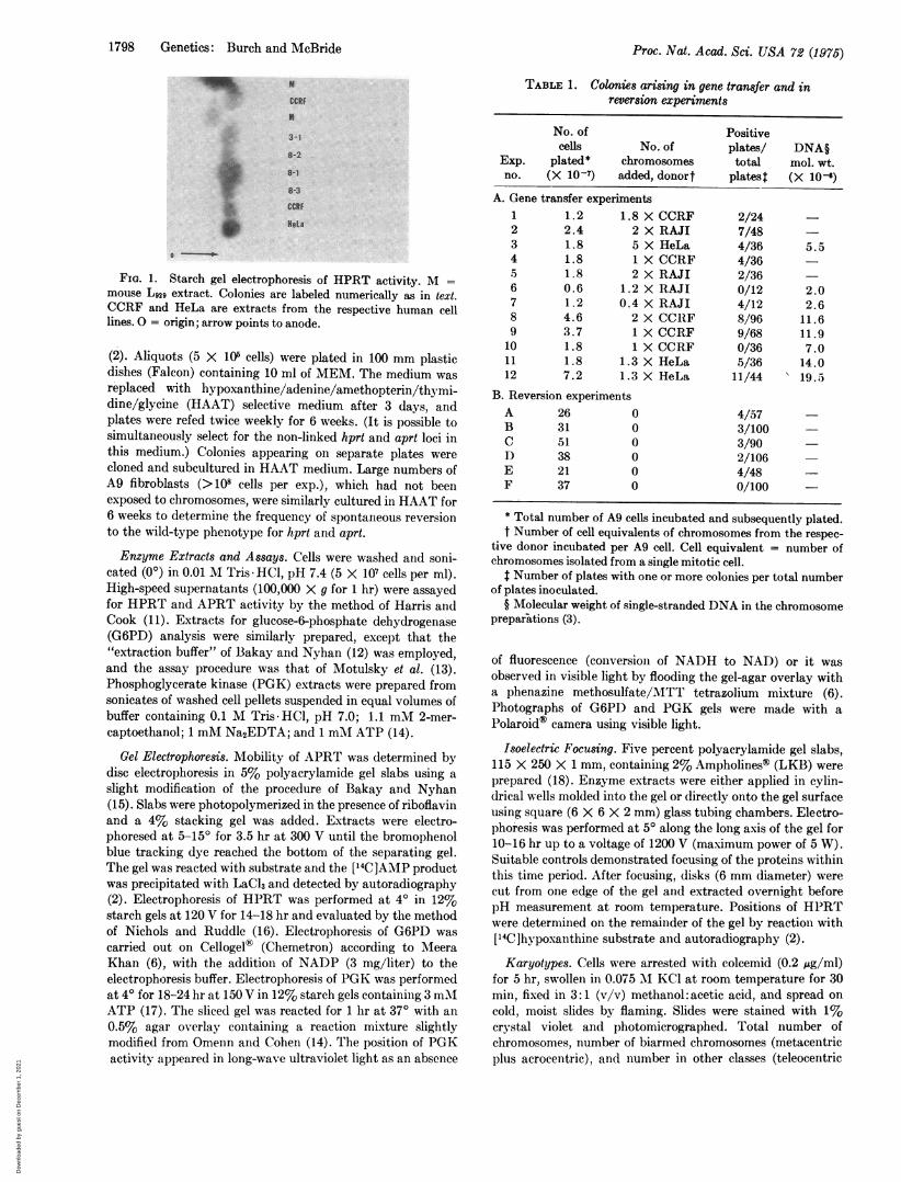
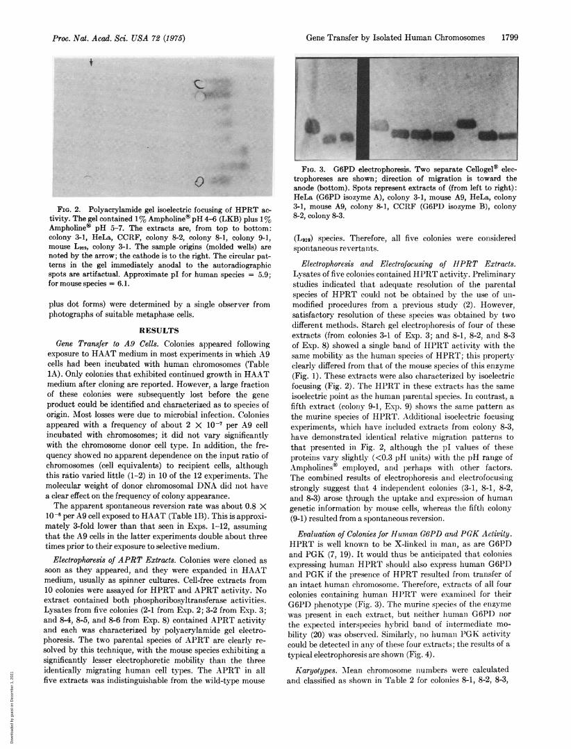
FIG. 2. Polyacrylamide gel isoelectric focusing of HPRT ac-tivity. The gel contained 1% Ampholine® pH 4-6 (LKB) plus 1%Ampholine® pH 5-7. The extracts are, from top to bottom:colony 3-1, HeLa, CCRF, colony 8-2, colony 8-1, colony 9-1,mouse L929, colony 3-1. The sample origins (molded wells) arenoted by the arrow; the cathode is to the right. The circular pat-terns in the gel immediately anodal to the autoradiographicspots are artifactual. Approximate pI for human species = 5.9;for mouse species = 6.1.
plus dot forms) were determined by a single observer fromphotographs of suitable metaphase cells.
RESULTSGene Transfer to A9 Cells. Colonies appeared following
exposure to HAAT medium in most experiments in which A9cells had been incubated with human chromosomes (Table1A). Only colonies that exhibited continued growth in HAATmedium after cloning are reported. However, a large fractionof these colonies were subsequently lost before the geneproduct could be identified and characterized as to species oforigin. Most losses were due to microbial infection. Coloniesappeared with a frequency of about 2 X 10⁻⁷ per A9 cellincubated with chromosomes; it did not vary significantlywith the chromosome donor cell type. In addition, the fre-quency showed no apparent dependence on the input ratio ofchromosomes (cell equivalents) to recipient cells, althoughthis ratio varied little (1-2) in 10 of the 12 experiments. Themolecular weight of donor chromosomal DNA did not havea clear effect on the frequency of colony appearance.The apparent spontaneous reversion rate was about 0.8 X
10-8 per A9 cell exposed to HAAT (Table 1B). This is approxi-mately 3-fold lower than that seen in Exps. 1-12, assumingthat the A9 cells in the latter experiments double about threetimes prior to their exposure to selective medium.
Electrophoresis of APRT Extracts. Colonies were cloned assoon as they appeared, and they were expanded in HAATmedium, usually as spinner cultures. Cell-free extracts from10 colonies were assayed for HPRT and APRT activity. Noextract contained both phosphoribosyltransferase activities.Lysates from five colonies (2-1 from Exp. 2; 3-2 from Exp. 3;and 84, 8-5, and 8-6 from Exp. 8) contained APRT activityand each was characterized by polyacrylamide gel electro-phoresis. The two parental species of APRT are clearly re-solved by this technique, with the mouse species exhibiting asignificantly lesser electrophoretic mobility than the threeidentically migrating human cell types. The APRT in allfive extracts was indistinguishable from the wild-type mouse
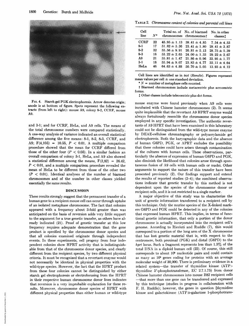
FIG. 3. G6PD electrophoresis. Two separate Cellogel® elec-trophoreses are shown; direction of migration is toward theanode (bottom). Spots represent extracts of (from left to right):HeLa (G6PD isozyme A), colony 3-1, mouse A9, HeLa, colony3-1, mouse A9, colony 8-1, CCRF (G6PD isozyme B), colony8-2, colony 8-3.
(L929) species. Therefore, all five colonies were consideredspontaneous revertants.
Electrophoresis and Electrofocusing of HPRT Extracts.Lysates of five colonies contained HPRT activity. Preliminarystudies indicated that adequate resolution of the parentalspecies of HPRT could not be obtained by the use of un-modified procedures from a previous study (2). However,satisfactory resolution of these species was obtained by twodifferent methods. Starch gel electrophoresis of four of theseextracts (from colonies 3-1 of Exp. 3; and 8-1, 8-2, and 8-3of Exp. 8) showed a single band of HPRT activity with thesame mobility as the human species of HPRT; this propertyclearly differed from that of the mouse species of this enzyme(Fig. 1). These extracts were also characterized by isoelectricfocusing (Fig. 2). The HPRT in these extracts has the sameisoelectric point as the human parental species. In contrast, afifth extract (colony 9-1, Exp. 9) shows the same pattern asthe murine species of HPRT. Additional isoelectric focusingexperiments, which have included extracts from colony 8-3,have demonstrated identical relative migration patterns tothat presented in Fig. 2, although the pI values of theseproteins vary slightly (<0.3 pH units) with the pH range ofAmpholines® employed, and perhaps with other factors.The combined results of electrophoresis and electrofocusingstrongly suggest that 4 independent colonies (3-1, 8-1, 8-2,and 8-3) arose through the uptake and expression of humangenetic information by mouse cells, whereas the fifth colony(9-1) resulted from a spontaneous reversion.
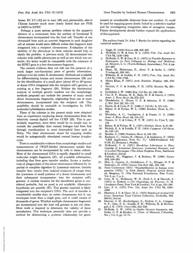
Evaluation of Colonies for Hiuman G6PD and PGK Activity.HPRT is well known to be X-linked in man, as are G6PDand PGK (7, 19). It would thus be anticipated that coloniesexpressing human HPRT should also express human G6PDand PGK if the presence of HPRT resulted from transfer ofan intact human chromosome. Therefore, extracts of all fourcolonies containing human HJPRT were examined for theirG6PD phenotype (Fig. 3). The murine sl)ecies of the enzymewas present in each extract, but neither human G6PD northe expected interspecies hybrid band of intermediate mo-bility (20) was observed. Similarly no human PGK activitycould be detected in any of these four extracts; the results of a
typical electrophoresis are shown (Fig. 4).
Karyotypes. MNean chromosome numbers were calculatedand classified as shown in Table 2 for colonies 8-1, 8-2, 8-3,
Proc. Nat. Acad. Sci. USA 72 (1975)
Dow
nloa
ded
by g
uest
on
Dec
embe
r 1,
202
1
1800 Genetics: Burch and McBride
TABLE 2. Chromosome content of colonies and parental cell lines
Cell Total no. of No. of biarmed No. in otherline N* chromosomes chromosomest classeslCCRF 22 45.95 i 1.13 38.41 ± 4.85 7.54 ± 4.538-1 17 51.82 4± 5.36 23.41 ± 1.80 28.41 ± 3.878-2 32 55.56 ± 9.91 26.81 ± 5.13 28.75 ± 5.398-3 18 53.22 ± 2.65 24.00 ±- 1.33 29.22± 2.67A9 21 55.81 ± 1.47 21.86 ±i 0.96 33.95 ±t 1.773-1 18 55.94 ± 9.07 23.83± 4.77 32.11 ± 4.64HeLa 46 64.63±i4.88 50.70 ±- 5.05 13.93 ± 4.12
Cell lines are identified as in text (Results). Figures representmean values per cell ± one standard deviation.
* N = number of metaphase cells counted.t Biarmed chromosomes include metacentric plus acrocentric
forms.t Other classes include teleocentric plus dot forms.
FIG. 4. Starch gel PGK electrophoresis. Arrow denotes origin;anode is at bottom of figure. Spots represent the following ex-tracts (from left to right): mouse A9, colony 8-2, CCRF, mouseA9.
and 3-1; and for CCRF, HeLa, and A9 cells. The means ofthe total chromosome numbers were compared statistically.A one-way analysis of variance indicated an overall statisticaldifference among the five means: 8-1, 8-2, 8-3, CCRF, andA9; F(4,105) = 10.55, P < 0.01. A multiple comparisonprocedure showed that the mean for CCRF differed fromthose of the other four (P < 0.05). In a similar fashion anoverall comparison of colony 3-1, HeLa, and A9 also showeda statistical difference among the means, F(2,82) = 26.42,P < 0.01, and a multiple comparison procedure revealed themean of HeLa to be different from those of the other two(P < 0.01). Identical analyses of the number of biarmedchromosomes and of the number in other classes yieldedessentially the same results.
DISCUSSION
These results strongly suggest that the permanent transfer of ahuman gene to a recipient mouse cell can occur through uptakeof an isolated metaphase chromosome. The fact that coloniesappeared with a frequency about 3-fold greater than wasanticipated on the basis of reversion adds very little supportto the argument for a true genetic transfer, as others have al-ready indicated (21). Proof of genetic transfer at this lowfrequency requires adequate demonstration that the geneproduct is specified by the chromosome donor species andthat all colonies examined originate through independentevents. In these experiments, cell progeny from four inde-pendent colonies show HPRT activity that is indistinguish-able from that of the chromosome donor species, and clearlydifferent from the recipient species, by two different physicalcriteria. It must be recognized that a revertant enzyme wouldnot necessarily be identical in physical properties with thewild-type species. However, the fact that the HPRT productfrom these four colonies cannot be distinguished by eitherstarch gel electrophoresis or electrofocusing from the HPRTin their respective human chromosome donor lines indicatesthat reversion is a very improbable explanation for these re-sults. Moreover, chromosome donor species of HPRT withdifferent physical properties than either human or wild-type
mouse enzyme were found previously when A9 cells wereincubated with Chinese hamster chromosomes (2). It seemsquite implausible that the revertant A9 HPRT enzyme wouldalways fortuitously resemble the chromosome donor speciesemployed in any specific investigation. The authentic rever-tants of A9 HPRT that have been examined in this laboratorycould not be distinguished from the wild-type mouse enzymeby DEAE-cellulose chromatography or polyacrylamide gelelectrophoresis. Both the karyographic data and the absenceof human G6PD, PGK, or APRT excludes the possibilitythat these colonies could have arisen through contaminationof the cultures with human cells. These observations, par-ticularly the absence of expression of human G6PD and PGK,also diminish the likelihood that colonies arose through spon-taneous fusion of A9 cells with human cells or nuclei. Otherarguments to support the nature of this transfer have beenpresented previously (2). Our findings support and extendthe results of reported studies (2-4); the combined observa-tions indicate that genetic transfer by this method is notdependent upon the species of the chromosome donor orrecipient cells, and it is not restricted to a single marker.A major objective of this study was to characterize the
unit of genetic information transferred to a recipient cell bythis technique. Only the murine species of the X-linked mark-ers G6PD and PGK could be detected in any of the coloniesthat expressed human HPRT. This implies, in terms of func-tional genetic information, that only a portion of the donorchromosome is actually incorporated into the recipient cell'sgenome. According to Ricciuti and Ruddle (7), this wouldcorrespond to a portion of the long arm of the X chromosomethat has lost genetic material that is, with respect to thecentromere, both proximal (PGK) and distal (G6PD) to thehprt locus. Such a fragment represents less than 1.5% of thetotal DNA in a diploid human cell (22). Of course, this stillcorresponds to about 108 nucleotide pairs and could containas many as 105 genes coding for proteins with an averagemolecular weight of 30,000. There is preliminary evidence in arelated system-the transfer of thymidine kinase (ATP:-thymidine 5'-phosphotransferase, EC 2.7.1.75) from donorChinese hamster chromosomes into mouse B82 recipient cells-that more than one gene can be transferred and expressedby this technique (studies in progress in collaboration withF. H. Ruddle); however, the genes in question [thymidinekinase and galactokinase (ATP:D-galactose 1-phosphotrans-
Proc. Nat. Acad. Sci. USA 72 (1975)
Dow
nloa
ded
by g
uest
on
Dec
embe
r 1,
202
1
Gene Transfer by Isolated Human Chromosomes 1801
ferase, EC 2.7.1.6)] are in man (26) and, presumably, also inChinese hamster much more closely linked than are PGKor G6PD to HPRT.
Perhaps a more serious problem relates to the apparentabsence of a centromere from the portion of functional Xchromosome incorporated into the host cell. Transfer of onechromatid from such an acentric fragment to each daughtercell at mitosis would seem difficult unless this fragment wereintegrated into a recipient chromosome. Evaluation of thestability of the phenotype in these colonies should help toclarify the problem. A previous study (2) demonstrated twocolonies with stable phenotypes as well as one which was un-stable; the latter would be compatible with the existence ofthe HPRT gene in a free chromosome fragment.The current evidence does not rule out the presence of a
much larger non-functional piece of genetic information,perhaps even the entire X chromosome. Methods are availablefor differentiating human and mouse chromosomes (23) andfor the identification of a small piece (about 103 to 104 genes)of donor DNA integrated into the recipient cell's genome, orexisting as a free fragment (24). Neither the biochemicalanalysis of multiple genetic markers nor the morphologicmethods proposed can exclude the presence of many smalldonor chromosome fragments, originating from one or morechromosomes, incorporated into the recipient cell. Thispossibility should be amenable to investigation by DNAmolecular hybridization studies.Three of the colonies in the present study were obtained
from an experiment employing donor chromosomes from therelatively normal diploid cell line CCRF (25). This is par-ticularly important, since there would be no simple way toexclude the possibility that normal linkages had been lostthrough translocations in most heteroploid lines such asHeLa. The ideal chromosome donor for mapping studieswould be mitogenically stimulated normal human lympho-cytes.There is considerable evidence from morphologic studies and
measurements of [3H ]dT-labeled chromosome uptake thatchromosomes can be incorporated by cells in tissue culture.Most of the chromosomal DNA is rapidly degraded to smallmolecular weight fragments (27). All available information,including that from gene transfer studies, favors a mecha-nism of phagocytosis of the donor chromosome followed by itspartial or complete digestion by lysosomal enzymes. Genetictransfer then results from isolated instances of escape fromthe lysosomes of small portions of a donor chromosome andtheir subsequent incorporation into the recipient cell'sgenome. A nuclear location for the transferred genes is con-sidered probable, but no proof is yet available. Alternativehypotheses are possible (27). This genetic material is likelyintegrated into the recipient's DNA. The unit of transfer isconsiderably smaller than an entire chromosome but it canencompass more than a single gene, and it could consist ofthousands of genes. Whether multiple chromosome fragmentsare incorporated into the host cell genome is not yet clear.More work is required to determine the validity of thesespeculations. This technique presently does not provide amethod for determining a syntenic relationship for genes
located at considerable distances from one another. It couldbe used for mapping genes closely linked to a selective markerand for investigating integration sites of oncogenic viruses.Future developments should further expand the applicationsof this approach.
The authors thank Dr. John T. Bartko for advice regarding the3tatistical analyses.
1. Eagle, H. (19.59) Science 130, 432-437.2. McBride, 0. W. & Ozer, H. L. (1973) Proc. Nat. Acad. Sci.
USA 70, 1258-1262.3. McBride, 0. W. & Ozer, H. L. (1973) in Possible Episomes in
Eukaryotes: Le Petit Colloquia on Biology and Medicine,ed. Silvestri, L. G. (North-Holland, Amsterdam), Vol. 4, pp.255-267.
4. Shani, M., Huberman, E., Aloni, Y. & Sachs, L. (1974)Virology 61, 303-305.
5. Willecke, K. & Ruddle, F. H. (1975) Proc. Nat. Acad. Sci.USA 72, 1792-1796.
6. Meera Khan, P. (1971) Arch. Biochem. Biophys. 145, 470-483.
7. Ricciuti, F. C. & Ruddle, F. H. (1973) Genetics 74, 661-678.
8. Littlefield, J. W. (1966) Exp. Cell Res. 41, 190-196.9. Cox, R. P., Krauss, M. R., Balis, M. E. & Dancis, J. (1974)
Amer. J. Hum. Genet. 26, 272-273.10. Mendelsohn, J., Moore, I). E. & Salzman, N. P. (1968)
J. Mfol. Biol. 32, 101-112.11. Harris, H. & Cook, P. R. (1969) J. Cell Sci. 5, 121-133.12. Bakay, B. & Nyhan, W. L. (1969) Biochem. Genet. 3, 571-
582.13. Motulsky, A. G., Vandepitte, J. & Fraser, G. R. (1966)
Amer. J. Hum. Genet. 18, 514-537.14. Omenn, G. S. & Cohen, P. T. W. (1971) In Vitro 7, 132-
139.15. Bakay, B. & Nyhan, W. L. (1971) Biochem. Genet. 5, 81-90.16. Nichols, E. A. & Ruddle, F. H. (1974) Cytogenet. Cell Genet.
13, 132-135.17. Beutler, E. (1969) Biochem. Genet. 3, 189-195.18. Karlsson, C., 1)avies, H., Ohman, J. & Andersson, U. (1973)
"LKB Application Note No. 75," LKB-Produkter AB,8-161 25, Bromma 1, Sweden.
19. McKusick, V. A. (1971) Miendelian Inheritance in Man:Catalogs of Autosomal Dominant, Autosomal Recessive, andX-Linked Phenotypes (The Johns Hopkins Press, Baltimore& London), 3rd ed.
20. Nabholk, M., Miggiano, V. & Bodmer, W. (1969) Nature223, 358-363.
21. Shin, S., Caneva, 1t., Schildkraut, C. L., Klinger, H. P. &Siniscalco, M. (1973) Nature New Biol. 241, 194-196.
22. Paris Conference (1971) "Standardization in human cyto-genetics (1972)," in Birth Defects: Original Article Series,ed. Bergsma, D. (The National Foundation, New York).Vol. 8, no. 7, p. 4.
23. Lubs, H. A., McKenzie, W. H., Patil, S. R. & Merrick, S.(1972) in Methods iln Cell Physiology, ed. Prescott, D. M.(Academic Press, New York & London), Vol. 6, pp. 345- 380.
24. Latt, S. A. (1973) Proc. Nat. Acad. Sci. USA 70, 3395-3399.
25. Shannon, J. E. & Macy, M. L. (1972) Registry of Animal CellLines (American Type Culture Collection, Rockville, Md.20852), 2nd ed.
26. Elsevier, S. M., Kucherlapati, iR., Nichols, E. A., Creagan,R. P., Giles, R. E., Ruddle, F. H., Willecke, K. & McDou-gall, J. K. (1974) Nature 251, 633-636.
27. McBride, 0. W. (1974) in Stadler Genetics Symposia, eds.R6dei, G. P. & Kimber, G. (Univ. of Missouri, Columbia,Mo.), Vol. 6, pp. 53-75.
Proc. Nat. Acad. Sci. USA 72 (1975)
Dow
nloa
ded
by g
uest
on
Dec
embe
r 1,
202
1