Embed Size (px)
Citation preview
Human Erythrocyte Hexokinase Deficiency
CHARACTERIZATIONOF A MUTANTENZYMEWITH ABNORMAL
REGULATORYPROPERTIES
GERTRIJKSEN and GERARDE. J. STAAL, Unit of Medical Enzymology, HaematologicalDepartment, State University Hospital, Utrecht, The Netherlands
A B S T RA C T In the erythrocytes of a patient withhereditary nonspherocytic hemolytic anemia, a homo-zygous expression of hexokinase deficiency was de-tected. The mutant enzyme was characterized bynormal kinetic parameters with respect to its substrates,glucose and MgATP2-, normal pH optimum, normalheat stability at 40°C, but abnormal behavior withrespect to its regulation by glucose-1,6-diphosphateand inorganic phosphate, and an altered electropho-retic pattern. Interpretation of the results revealed thepresence of two different hexokinases type I in normalhuman erythrocytes: one enzyme with a high affinityfor glucose-1,6-diphosphate, the inhibition of whichis regulated by inorganic phosphate; and anotherenzyme with a lower affinity for the inhibitor, not reg-ulated by inorganic phosphate. The former enzymewas not detectable in the erythrocytes of the patient,whereas the presence of the latter enzyme could bedemonstrated.
INTRODUCTION
Hexokinase deficiency was first described in 1965by L6hr et al. (1) in the blood cells of three patientswith a panmyelopathy (type Fanconi) with multiplemalformations. The residual hexokinase activity in theerythrocyte was 25-60% of normal and the enzymeshowed decreased affinities for its substrates glucoseand MgATP2-.
However, the metabolic defects in these patientsare acquired rather than truly genetic. Hexokinasedeficiency of human erythrocytes was first related tohereditary nonspherocytic hemolytic anemia in 1967by Valentine et al. (2). The hexokinase activity of thepatienit's erythrocytes was only slightly decreased,but was markedly lowered when the degree of reticulo-cytosis was taken into account. The kinetic (2) and
Received for publication 9 September 1977 and in revisedformn 3 April 1978.
electrophoretic (3) behavior of the enzyme showedno abnormalities. Since the report of Valentine et al.(2), some other variants of hexokinase deficiency re-lated to hereditary nonspherocytic hemolytic anemiahave been reported (4-7). Most variants showed in-creased Michaelis-Menten constants for the substratesglucose and MgATP2- (5-7), and in one case a decreasedheat stability was found (5). The hexokinase deficiencydescribed by Necheles et al. (4) was kinetically andelectrophoretically characterized by the absence of the"low-Kmn" hexokinase type III, which is normally pre-sent in low concentrations, whereas hexokinase type I,which is the predominant form of hexokinase in humanerythrocytes (8), was present in a normal amount. Hexo-kinase deficiency appears to be inherited as an auto-somal recessive disorder (1, 2).
Recently (9, 10), we described the regulation ofpurified erythrocyte hexokinase type I by hexose-phosphate and diphosphate and inorganic phosphate(Pi).' Hexokinase is strongly inhibited by glucose-6-phosphate (glc-6-P) and glucose-1,6-diphosphate (glc-1,6-P2), whereas Pi is able at least partly to overcomethis inhibition.
To explain the results, two alternative models weresuggested: (a) Hexokinase exists in equilibrium be-tween a free and a phosphate-associated form, thelatter having a reduced affinity for glc-1,6-P2 with re-spect to the first. (b) There are two different hexoki-nases: one enzyme with a high affinity for glc-1,6-P2,the inhibition of which is competitively influencedby Pi; the other enzyme with a lower affinity for theinhibitor and insensitive to regulation by Pi.
In this paper we present a patient with nonsphero-cytic hemolytic anemia associated with a homozygoushexokinase deficiency. The defective enzyme couldbe characterized by a different electrophoretic pattern
'Abbreviations used in this paper: glc-1,6-P2, glucose-1,6-diphosphate; glc-6-P, glucose-6-phosphate; Pi, inorganicphosphate.
J. Clini. Intvest. ©) The American Society for Clinical Investigation, Inc., 0021-9738/78/0801-294 $1.00294
and altered regulatory properties. The results are usedto discriminate between the models proposed for theregulation of the normal erythrocyte enzyme.
Case Report
The patient is a 30-yr-old woman suffering fromchronic hemolysis since birth. During the first yearsof life many blood transfusions were necessary. At theage of 2 yr, splenectomy was performed, after whichthe need for blood transfusions decreased.
The parents of the patient are second cousins. Theyboth had a normal blood picture except for a slightlyincreased packed cell volume and hemoglobin con-tent in the mother's blood. These increases were con-sistent on two different occasions; the cause of thesedeviations remains unclear. The patient is their onlychild.
In 1967 a hexokinase deficiency of the erythrocyteswas found as the possible cause of the hemolytic anemia(11). The hexokinase activity of the erythrocytes wasonly slightly lowered with respect to normal, but wasmarkedly decreased when the degree of reticulocy-tosis was taken into account. The most recent hemato-logical data, obtained at the time of the present exten-sive enzyme studies in the patient, are shown in TableI. There was a slight anemia and pronounced reticulo-cytosis, the latter accounting for the markedly increasedmean corpuscular volume and mean corpuscular hemo-globin. The blood smear showed 60 erythroblasts per100 white cells, marked anisocytosis, macrocytosis,polychromasia, and basophilic stippling of the erythro-cytes. Many Howell-Jolly bodies and a few target cellsand spherocytes were present.
METHODS
Substrates, coenzymes, and auxiliary enzymes for determina-tion of glycolytic enzymes and intermediates, except forthe determination of 2,3-diphosphoglycerate, were obtainedfrom Boehringer (Boehringer & Soehne, Mannheim, Ger-many). Sulfoethyl-cellulose SE-23 was supplied by Serva
(Heidelberg, Germany) and Sephadex G-25 (coarse) andDEAE-Sephadex A-50 by Pharmacia Fine Chemicals (Pis-cataway, N. J.). Acrylamide and bisacrylamide were obtainedfrom Ega-chemie (Albuch, Germany) and phenazine metho-sulfate and MTT-tetrazolium (bimethyl-thiozolyl diphenyl-tetrazolium bromide) from Sigma Chemical Co. (St. Louis,Mo.). All other chemicals were of analytical grade pu-rity. Venous blood was collected with heparin (30 U/ml)from the patient and her parents, from a normal healthy donor,and from two control patients with reticulocytosis. The firstcontrol (reticulocytosis control I) had a glucosephosphateisomerase deficiency of the erythrocytes with a very highreticulocyte count (72%) and was splenectomized (12). Thesecond control (reticulocytosis control II) was suffering froma hemolytic anemia probably of viral origin. No metabolicabnormalities were found. This patient was not splenecto-mized and had a reticulocyte count of 20%.
Immediately after collection of the blood, a part of it wasdeproteinized for the determination of glycolytic interme-diates. Deproteinization and determination of all glycolyticintermediates except 2,3-diphosphoglycerate were performedaccording to the methods of Minakami et al. (13). The 2,3-diphosphoglycerate content of the acid extract was deter-mined with a Sigma-testkit (Sigma Technical Bulletin 35-UV,December 1974). All determinations were performed within5 h after extract preparation.
The leucocytes and platelets of the remaining blood cellswere removed according to the method of Nakao et al. (14)by filtrating through a mixture of sulfoethyl-cellulose andSephadex G-25. However, instead of phosphate buffer weused 0.154 M NaCl which does not affect the results of theseparation (15).
The erythrocyte glycolytic enzymes were determined bythe methods of Beutler (16). Erythrocyte hexokinase waspartly purified by batchwise treatment with DEAE-SephadexA-50 and by ammonium sulfate precipitation as previouslydescribed (17). The ammonium sulfate precipitate was dis-solved in 0.2 M Tris-HCl (pH 8.0) and 3 mM,B-mercapto-ethanol. Before enzyme characterization the sample wasdesalted by gel filtration on Sephadex G-25 equilibratedwith the same buffer. The procedure resulted in a 30- to 50-fold purification with an overall yield of approximately 50%.Hexokinase activity of the partially purified preparations wasdetermined at 37°C in a glc-6-P dehydrogenase-coupledassay as previously described (17).
MgCl2 was added in concentrations necessary to maintainan excess of 5 mMMg2+ over ATP. Routinely, the concen-tration of glucose was 10 mM. The pH of the assay bufferwas 7.15 at 37°C. The reaction was started by adding 0.03 Uhexokinase. 1 U of enzyme activity is defined as the amount of
TABLE ISome Routine Hematological Data of the Patient and Her Parents
NormalPatient
(9) Father Mother
Erythrocytes, x 1012/liter 2.39 4.91 5.23 3.7-5.0 4.1-5.6Hemoglobin, gldl 11.3 15.3 16.6 11.9-15.5 13.8-17.6Packed cell volume, mlll00 ml 37 48 52 36-46 41-51Mean corpuscular volume,fl 156 98 100 84-104Mean corpuscular hemoglobin, pg 47.8 31.9 32.5 28.2-35.7Mean corpuscular hemoglobin concentration,
g/100 ml erythrocyte 30.6 32.5 32.9 32.3-37Reticulocytes, % 39 1.6 1.2 0.4-1.8
A New Variant of Hexokinase Deficiency 295
enzyme that catalyzes the formation of 1 ,umol glc-6-P permin at 37°C.
The heat stability of the partly purified enzyme prepara-tions was determined by measuring the decrease of activityvs. the time of incubation at 40°C.
Disk-electrophoresis on poly-acrylamide gels was per-formed by method II of Davis (18) at pH 8.8. No sample gel wasused and some modifications were made. The buffer contained3 mM 13-mercaptoethanol to protect the enzyme fromoxidation. Glucose was incorporated in the gels at the sameconcentration as that used in the staining mixture to stabilizethe enzyme. The staining mixture contained in a final volumeof 80 ml: 0.1 MTris/HCl (pH 7.2), 7.5 mMMgCl2, 0.2 mMNADP+, 35 IU glc-6-P dehydrogenase, 12 IU phosphogluconatedehydrogenase, 2 mMATP, 1.25 mMEDTA, 2 mMKCN, 6 mgphenazine methosulfate, 16 mg MTT, and glucose at theconcentrations as indicated in the results.
The staining was stopped with 2.5% acetic acid and the gelswere scanned at 625 nmon a Zeiss spectrophotometer modelPMQ3 provided with a ZK5 Disc Attachment (Carl Zeiss, Inc.,New York).
RESULTS
Erythrocyte metabolism. Hexokinase activity ofthe erythrocytes of the patient was only slightlydecreased (Table II). The enzyme activity was loweredto about 15-20% of normal, however, when the degreeof reticulocytosis was taken into account. Due to thevery high percentage of reticulocytes, the enzymespyruvate kinase, 6-phosphofructokinase, glc-6-P de-hydrogenase and aldolase showed a high activity andwere comparable to the values found for the "reticulo-cytosis controls." The activities of the other enzymeswere in the normal range.
Hexokinase activity in the erythrocytes of bothparents was decreased to 50-80% of normal, indicatingthat both parents are heterozygous for the defect.
The contents of glycolytic intermediates of thepatient's erythrocytes are shown in Table III. The dataof reticulocytosis control I are of no use here, because ofthe metabolic nature of the defect in this patient. The2,3-diphosphoglycerate level in the patient's erythro-cytes was strongly decreased, whereas the content offructose-1,6-diphosphate was increased. The decreased2,3-diphosphoglycerate content had caused a shift in theoxygen dissociation curve to the left. The P50(mm Hg of oxygen at 50% saturation) was shiftedto 20.4 mm Hg (normal range 26-28 mm Hg),which implied that the release of oxygen to the tissueswas impaired.
Enzyme characterization. At two opportunitieserythrocyte hexokinase was purified from the erythro-cytes of the patient and the reticulocytosis controls.The results of the enzyme characterization on bothoccasions were comparable. The parents' erythrocytehexokinase was investigated only once.
Kinetics. The partially purified hexokinase prepara-tion of the patient's erythrocytes showed normal Mi-chaelis-Menten kinetics. In agreement with otherreports on human erythrocyte hexokinase (17, 19),the results were consistent with a rapid equilibriumrandom reaction mechanism. Km for the substrate glu-cose was 0.050 mM, SD = 0.027, n = 3 (normal: 0.064mM, SD = 0.016, n = 30), and for the other substrate,MgATP2-, 1.19 mM, SD = 0.20, n = 5. The latter valuewas slightly increased with respect to normal (0.72 mM,SD = 0.19, n = 30); however, the significance of thisincrease is doubtful.
The kinetic parameters obtained for the enzymesfrom the parents and the reticulocytosis controls fellin the normal range (Table IV).
TABLE IIErythrocyte Enzyme Activities
Reticulocytosis controls Normal controls,mean-SD
Patient I II Father Mother (n = 50)
Hexokinase 0.70 6.7 2.7 0.77 0.81 1.31±+0.23Phosphoglucose isomerase 24.2 8.0 30.4 21.8 23.2 26.2+4.76-Phosphofructokinase 15.0 16.2 16.2 7.8 9.5 10.7±+1.5Aldolase 3.0 5.0 4.8 1.8 1.8 2.54+0.57Triosephosphate isomerase 2,659 2,596 1,850 2,217 1,955 1,950±700Glyceraldehyde dehydrogenase 79.4 38.1 91 25 39.3 79+56Phosphoglycerate kinase 150 244 223 133 140 179+59Biphosphoglyceromutase 34.6 40.8 30.7 25.8 25.6 25.0+5.2Enolase 13.8 14.4 10.1 6.5 7.0 10.0+4.3Pyruvate kinase 13.2 22.6 9.5 5.4 5.1 6.0±+1.2Glucose-6-P dehydrogenase 19.6 26.5 17.8 9.4 10.0 10.6+2.0Gluconate dehydrogenase 12.4 ND* 13.4 6.1 6.1 8.5±+1.7Gluthathione reductase 7.0 ND* 8.3 6.7 6.6 5.7±1.6Reticulocytes, % 39 72 20 1.6 1.2 0.4-1.8
Enzyme activities are expressed as units per gram hemoglobin.* Not determined.
296 G. Rijksen and G. E. J. Staal
TABLE IIIErt throcyte Glycolytic Intermnediates
Normal controls,Reticulocytosis mean±SD
Patient control II (n = 20)
Glc-6-P 34.5 62 35.7+6.6Fructose-6-
phosphate 17.3 28.3 13.6±2.5Frucitose- 1,6-
diphosphate 35.5 27.7 10.2+3.4Dihydroxyacetone-
phosphate 10.0 21.9 12.1+5.1GCINceraldehyde-
phosphate 5.0 6.5 4.8 +1.62,3-Diphospho-
glycerate 3,000 5,100 5,250+4003-Phospho-
glycerate 69.8 ND* 55+352-Phospho-
glycerate 10.0 ND* 9.1+5.32-Phosphoenol-
pyruvate 22.1 22.2 13.4+8.0Pyruvate 78.2 63.9 125+75Lactate 1,500 ND* 990±340ATP 1,700 2,300 1,300+250
Concentrations are expressed as nanomoles per millilitererythrocytes except for pyruvate and lactate (in nanomolesper milliliter of blood).* Not determined.
Influence of glc-1,6-P, and inorganic phosphate.Human erythrocyte hexokinase activity is stronglyregulated by glc-6-P (9, 19-21) and glc-1,6-P2 (10, 19,21). The inhibition by both ligands is regulated byinorganic phosphate in the same manner (9, 10). Hexo-kinase activity in the presence of glc-6-P cannot bemeasured in the glc-6-P dehydrogenase-coupled assay,but is determined in the pyruvate kinase-lactate dehy-
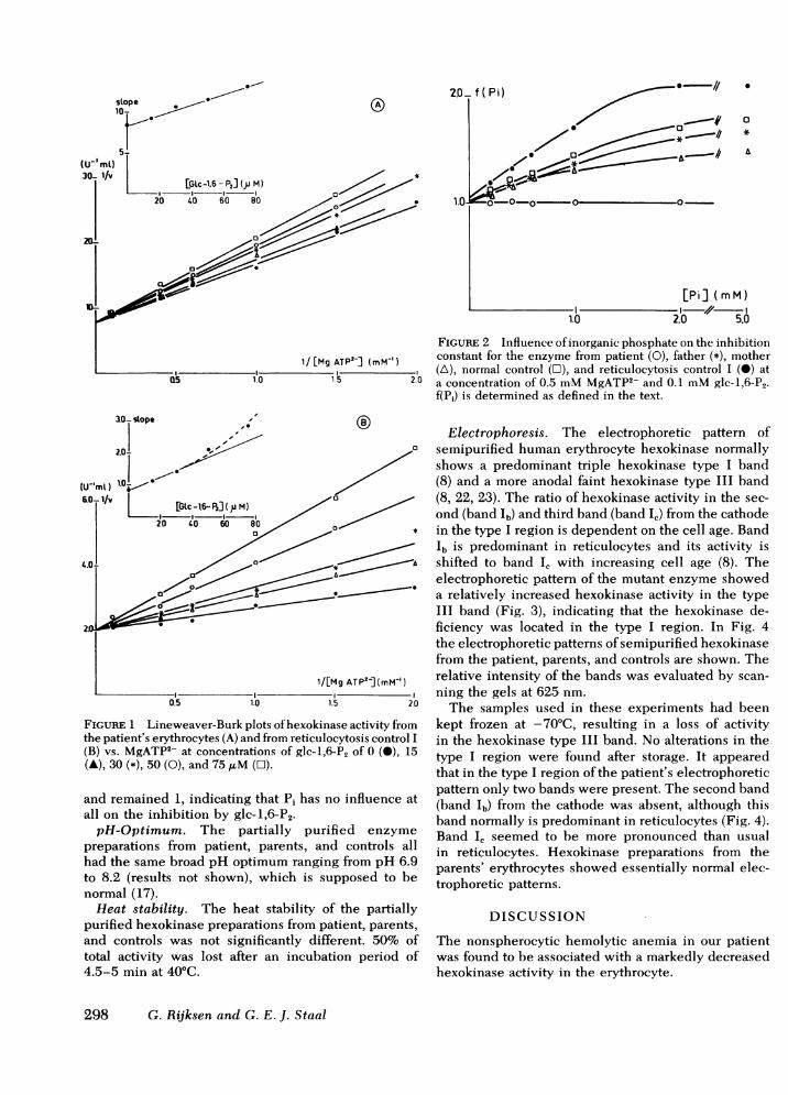
drogenase-coupled assay (9). However, because of theinterference of contaminating enzymes in crude hexo-kinase preparations with the latter assay, we investi-gated only the influence of glc-1,6-P2. The inhibitionof the partially purified mutant enzyme by glc-1,6-P2is shown in Fig. IA. The inhibition was competitivewith respect to MgATP2-. The secondary plot of theslope of the Lineweaver-Burk plot vs. the inhibitorconcentration was linear, and a Ki (glc-1,6-P2) of 115 ,uMcould be calculated. This value was increased withrespect to the reticulocyte control enzyme (Fig. 1B).The secondary plot of Fig. lB is deviating from linearityat higher concentrations of inhibitor. From the linearpart a Ki (glc-1,6-P2) of 40 AM was calculated.
About the same results were obtained for the partiallypurified enzymes from the parents and normal controls.The values obtained (Table IV) are the same as for thepure erythrocyte enzyme at pH = 7.15 (10). The inhibi-tion of pure human erythrocyte hexokinase by glc-1,6-P2was partly counteracted by Pi (10). This influence ofPi was found to be competitive with respect to glc-1,6-P2. To evaluate the influence of inorganic phosphate,a function, f (Pi), was derived describing the influenceof Pi on the inhibition constant of the enzyme for anyinhibitor as a function of phosphate concentration (9).
The influence of Pi on the inhibition by glc-1,6-P2of partially purified hexokinase from patient, parents,and controls was evaluated by calculating f (Pi) (Fig. 2).For the parents and controls f(Pi) was linear up to con-centrations of about 2 mMPi, at higher concentrationsof Pi a maximum was reached. This behavior reflectsthe inability of Pi to overcome completely the inhibi-tion by glc-1,6-P2.
These results were comparable to those obtained forthe pure enzyme (10) although the maxima of the func-tions vary. The function f(Pi) of the patient's enzymebehaved entirely differently: f(Pi) did not increase
TABLE IVProperties of Partially Purified Hexokinase
Reticulocytosis controls
Patient Father Mother I II Normal controls
Km IMgATP2-, mM 1.19 0.75 0.89 0.61 0.60 0.72SD =0.20 (n =5) SD =0.19 (n =30)
Km glucose, mM 0.050 0.035 0.079 0.066 0.053 0.064SD = 0.027 (n = 3) SD = 0.016 (n = 30)
Ki glc-1,6-P2, mM 0.115 0.063-0.029 0.043 0.025-0.040 0.038 0.039(n = 2) (n = 2) SD= 0.006 (it = 4)
pH Optimum Normal Normal Normal
Heat stability Normal Normal Normal
Electrophoresis Altered Normal Normal
A New Variant of Hexokinase Deficiency 297
slope
10-
5-
[Glc-1.6 - P2J (, M)1| 1.
2.0._
1.0-20 40 60 80
f(Pi) -l//
/~~~~~I *
0 ~ ~0-0./ -0 h
[Pi] (mM)
1.0 2.0 5.0
I/[Mg ATP2] (mM-')
FIGuRE 2 Influence of inorganic phosphate on the inhibitionconstant for the enzyme from patient (0), father (*), mother(A), normal control (O), and reticulocytosis control I (0) ata concentration of 0.5 mMMgATP2- and 0.1 mMglc-1,6-P2.f(Pi) is determined as defined in the text.
FIGURE 1 Lineweaver-Burk plots of hexokinase activity fromthe patient's erythrocytes (A) and from reticulocytosis control I(B) vs. MgATP2- at concentrations of glc-1,6-P2 of 0 (0), 15(A), 30 (*), 50 (0), and 75 ,uM (C1).
and remained 1, indicating that Pi has no influence atall on the inhibition by glc-1,6-P2.
pH-Optimum. The partially purified enzyme
preparations from patient, parents, and controls allhad the same broad pH optimum ranging from pH 6.9to 8.2 (results not shown), which is supposed to benormal (17).
Heat stability. The heat stability of the partiallypurified hexokinase preparations from patient, parents,and controls was not significantly different. 50% oftotal activity was lost after an incubation period of4.5-5 min at 40°C.
Electrophoresis. The electrophoretic pattern ofsemipurified human erythrocyte hexokinase normallyshows a predominant triple hexokinase type I band(8) and a more anodal faint hexokinase type III band(8, 22, 23). The ratio of hexokinase activity in the sec-
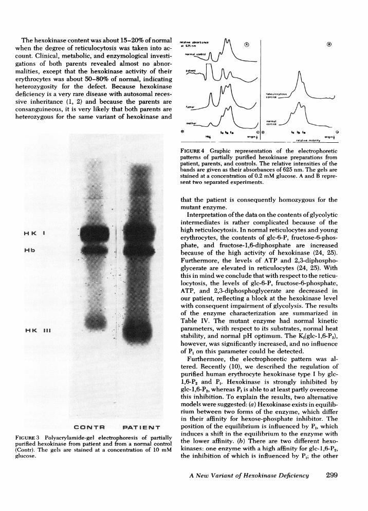
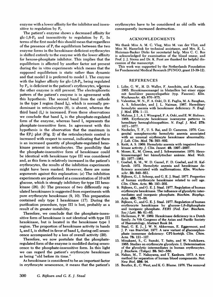
ond (band Ib) and third band (band IC) from the cathodein the type I region is dependent on the cell age. Bandlb is predominant in reticulocytes and its activity isshifted to band IC with increasing cell age (8). Theelectrophoretic pattern of the mutant enzyme showeda relatively increased hexokinase activity in the typeIII band (Fig. 3), indicating that the hexokinase de-ficiency was located in the type I region. In Fig. 4the electrophoretic patterns of semipurified hexokinasefrom the patient, parents, and controls are shown. Therelative intensity of the bands was evaluated by scan-
ning the gels at 625 nm.
The samples used in these experiments had beenkept frozen at -70°C, resulting in a loss of activityin the hexokinase type III band. No alterations in thetype I region were found after storage. It appearedthat in the type I region of the patient's electrophoreticpattern only two bands were present. The second band(band Ib) from the cathode was absent, although thisband normally is predominant in reticulocytes (Fig. 4).Band IC seemed to be more pronounced than usualin reticulocytes. Hexokinase preparations from theparents' erythrocytes showed essentially normal elec-trophoretic patterns.
DISCUSSION
The nonspherocytic hemolytic anemia in our patientwas found to be associated with a markedly decreasedhexokinase activity in the erythrocyte.
298 G. Rijksen and G. E. J. Staal
S
0
A
The hexokinase content was about 15-20% of normalwhen the degree of reticulocytosis was taken into ac-count. Clinical, metabolic, and enzymological investi-gations of both parents revealed almost no abnor-malities, except that the hexokinase activity of theirerythrocytes was about 50-80% of normal, indicatingheterozygosity for the defect. Because hexokinasedeficiency is a very rare disease with autosomal reces-sive inheritance (1, 2) and because the parents areconsanguineous, it is very likely that both parents areheterozygous for the same variant of hexokinase and
IC lb Is GorigiXn +
rotative mobitity
HKI
Hb
CONTR PATIENT
FIGuRE 3 Polyacrylamide-gel electrophoresis of partially
purified hexokinase from patient and from a normal control
(Contr). The gels are stained at a concentration of 10 mM
glucose.
FIGuRE 4 Graphic representation of the electrophoreticpatterns of partially purified hexokinase preparations frompatient, parents, and controls. The relative intensities of thebands are given as their absorbances of 625 nm. The gels are
stained at a concentration of 0.2 mMglucose. A and B repre-
sent two separated experiments.
that the patient is consequently homozygous for themutant enzyme.
Interpretation of the data on the contents of glycolyticintermediates is rather complicated because of thehigh reticulocytosis. In normal reticulocytes and young
erythrocytes, the contents of glc-6-P, fructose-6-phos-phate, and fructose-1,6-diphosphate are increasedbecause of the high activity of hexokinase (24, 25).Furthermore, the levels of ATP and 2,3-diphospho-glycerate are elevated in reticulocytes (24, 25). Withthis in mind we conclude that with respect to the reticu-locytosis, the levels of glc-6-P, fructose-6-phosphate,ATP, and 2,3-diphosphoglycerate are decreased inour patient, reflecting a block at the hexokinase levelwith consequent impairment of glycolysis. The resultsof the enzyme characterization are summarized inTable IV. The mutant enzyme had normal kineticparameters, with respect to its substrates, normal heatstability, and normal pH optimum. The K1(glc-1,6-P2),however, was significantly increased, and no influenceof Pi on this parameter could be detected.
Furthermore, the electrophoretic pattern was al-tered. Recently (10), we described the regulation ofpurified human erythrocyte hexokinase type I by glc-1,6-P2 and Pi. Hexokinase is strongly inhibited byglc-1,6-P2, whereas Pi is able to at least partly overcome
this inhibition. To explain the results, two alternativemodels were suggested: (a) Hexokinase exists in equilib-rium between two forms of the enzyme, which differin their affinity for hexose-phosphate inhibitor. Theposition of the equilibrium is influenced by Pi, whichinduces a shift in the equilibrium to the enzyme withthe lower affinity. (b) There are two different hexo-kinases: one enzyme with a high affinity for glc-1,6-P2,the inhibition of which is influenced by Pi; the other
A New Variant of Hexokinase Deficiency 299
folate,o absorbanceat 625 nm/
no rmaco
pat.ont
ret,culocyto escont rot
nofmnal \contrtot
H64
la lb laorigin .
(0
enzyme with a lower affinity for the inhibitor and insen-sitive to regulation by Pi.
The patient's enzyme shows a decreased affinity forglc-1,6-P2 and insensitivity to regulation by Pi. Interms of the first model this should mean that regardlessof the presence of Pi the equilibrium between the twoenzyme forms in the hexokinase-deficient erythrocytesis shifted entirely to the enzyme with the lower affinityfor hexose-phosphate inhibitor. This implies that theequilibrium is affected by another factor not presentduring the in vitro experiments which means that thesupposed equilibrium is static rather than dynamicand that model 2 is preferred to model 1. The enzymewith the higher affinity for glc-1,6-P2, being regulatedby Pi, is deficient in the patient's erythrocytes, w ereasthe other enzyme is still present. The electrophoreticpattern of the patient's enzyme is in agreement withthis hypothesis. The second band from the cathodein the type I region (band Ib), which is normally pre-dominant in reticulocytes (8), is absent, whereas thethird band (I,) is increased. From these observationswe conclude that band Ib is the phosphate-regulatedform of the enzyme, whereas band IC represents thephosphate-insensitive form. In agreement with thishypothesis is the observation that the maximum inthe f(Pi) plot (Fig. 2) of the reticulocytosis control isincreased with respect to normal, indicating that thereis an increased quantity of phosphate-regulated hexo-kinase present in reticulocytes. The possibility thatthe phosphate-insensitive form of hexokinase couldbe identical with hexokinase type III was consideredand, as this form is relatively increased in the patient'serythrocytes, the results of the inhibition experimentsmight have been masked. However, there are somearguments against this explanation: (a) The inhibitionexperiments are performed at a concentration of 10 mMglucose, which is strongly inhibitory for type III hexo-kinase (26). (b) The presence of two differently reg-ulated hexokinases is suggested from experiments withpure erythrocyte hexokinase (9, 10). This preparationcontained only type I hexokinase (17). During thepurification procedure, type III is lost, probably as aresult of its greater instability.
Therefore, we conclude that the phosphate-insen-sitive form of hexokinase is not identical with type IIIhexokinase, but is located in the hexokinase type Iregion. The proportion of hexokinase activity in bandsIb and Ic is shifted in favor of band Ic during cell senes-cence accompanied by a loss of overall activity (20).
Therefore, we now postulate that the phosphate-regulated form of the enzyme is modified during senes-cence to the phosphate-insensitive form. In this lightwe can regard the patient's erythrocyte hexokinaseas being "old before its time."
As hexokinase is considered to be an important factorin erythrocyte senescence, it means that the patient's
erythrocytes have to be considered as old cells withconsequently increased destruction.
ACKNOWLEDGMENTSWe thank Miss A. M. C. Vlug, Miss M. van der Vlist, andMiss M. Hamelink for technical assistance, and Mrs. E. L.Huisman-Backer Dirks for secretarial help. Miss C. C. Bosis acknowledged for examination of the blood smear, andProf. J. J. Sixma and Dr. K. Punt are thanked for helpful dis-cussion of the manuscript.
This work was supported by the Netherlands Foundationfor Fundamental Medical Research (FUNGO, grant 13-39-12).
REFERENCES
1. Lohr, G. W., H. D. Waller, F. Anschutz, and A. Knopp.1965. Hexokinasemangel in blutzellen bei einer sippemit familiarer panmyelopathie (typ. Fanconi). Klin.Wochenschr. 43: 870-875.
2. Valentine, W. N., F. A. Oski, D. E. Paglia, M. A. Baughan,A. S. Schneider, and J. L. Naiman. 1967. Hereditaryhemolytic anemia with hexokinase deficiency. N. Engl.
J. Med. 276: 1-11.3. Malone, J. I., A. I. Winegrad, F. A. Oski, and E. W. Holmes.
1968. Erythrocyte hexokinase isoenzyme patterns inhereditary hemoglobinopathies. N. Engl. J. Med. 279:1071-1077.
4. Necheles, T. F., U. S. Rai, and D. Cameron. 1970. Con-genital nonspherocytic hemolytic anemia associatedwith an unusual erythrocyte hexokinase abnormality.
J. Lab. Clin. Med. 76: 593-602.5. Keitt, A. S. 1969. Hemolytic anemia with impaired hexo-
kinase activity. J. Clin. Invest. 48: 1997-2007.6. Moser, K., M. Ciresa, and J. Schwarzmeier. 1970. Hexo-
kinasemangel bei hamolytischer anamie. Med. Welt.21: 1977-1981.
7. Goebel, K. M., W. D. Gassel, F. D. Goebel, and H. Kaf-farnik. 1972. Hemolytic anemia and hexokinase de-ficiency associated with malformations. Klin. Wochen-schr. 50: 849-851.
8. Rijksen, G., I. Schoop, and G. E. J. Staal. 1977. Propertiesof human erythrocyte hexokinase related to cell age.Clin. Chim. Acta. 80: 193-202.
9. Rijksen, G., and G. E. J. Staal. 1977. Regulation of humanerythrocyte hexokinase. The influence of glycolytic inter-mediates and inorganic phosphate. Biochim. Biophys.Acta. 485: 75-86.
10. Rijksen, G., and G. E. J. Staal. 1977. Regulation of humanerythrocyte hexokinase by glucose-1,6-diphosphateand inorganic phosphate. FEBS (Fed. Eur. Biochem.Soc.) Lett. 80: 61-65.
11. Helleman, P. W. 1969. Hexokinase deficiency in a Dutchfamily. In Vth Congress of the Asian and Pacific Societyof Haematology, Abstract Book 24.
12. Staal, G. E. J., J. W. N. Akkerman, E. Eggermont, andJ. P. van Biervliet. 1977. A new variant of glucosephos-phate isomerase deficiency: GPI-kortrijk. Clin. Chim.Acta. 78: 121-127.
13. Minakami, S., C. Suzuki, T. Saito, and W. Yoshikawa.1965. Studies on erythrocyte glycolysis. I. Determinationof the glycolytic intermediates in human erythrocytes.
J. Biochem. (Tokyo). 58: 543-550.14. Nakao, M., T. Nakayama, and T. Kankura. 1973. A new
method for separation of human blood components. Nat.New Biol. 256: 94.
15. Beutler, E., C. West, and K. G. Blume. 1976. The removal
300 G. Rijksen and G. E. J. Staal
of leucocytes and platelets from whole blood. J. Lab.Clin. Invest. 88: 328-333.
16. Beutler, E. 1975. Red cell metabolism: a manual of bio-chemical methods. Grune & Stratton Inc., New York.2nd edition. 38-70.
17. Rijksen, G., and G. E. J. Staal. 1976. Purification and someproperties of human erythrocytes hexokinase. Biochim.Biophys. Acta. 445: 330-341.
18. Davis, B. J. 1964. Disc electrophoresis. II. Method andapplication to human serum proteins. Ann. N. Y. Acad.Sci. 121: 404-427.
19. Gerber, G., H. Preissler, R. Heinrich, and S. M. Rapoport.1974. Hexokinase of human erythrocytes. Purification,kinetic model and its application to the conditions inthe cell. Eur. J. Biochem; 45: 39-52.
20. Kosow, D. P., F. A. Oski, J. V. B. Warms, and I. A. Rose.1973. Regulation of mammalian hexokinase: Regulatorydifferences between isoenzyme I and II. Arch. Biochem.Biophys. 157: 114-124.
21. Rose, I. A., J. V. B. Warms, and D. P. Kosow. 1974. Spec-ificity for the glucose-6-P inhibition site of hexokinase.Arch. Biochem. Biophys. 164: 729-735.
22. Kaplan, J. C., and E. Beutler. 1968. Hexokinase isoen-zymes in human erythrocytes. Science (Wash. D. C.).159: 215-216.
23. Rogers, P. A., R. A. Fisher, and H. Harris. 1975. An exami-nation of the age-related patterns of decay of the hexo-kinases of human red cells. Clin. Chim. Acta. 65: 291-298.
24. Buc, H. A., J. P. Leroux, H. Garreau, J. C. Marchand,and P. Cartier. 1974. Metabolic regulation in enzyme-deficient red cells. Enzyme (Basel). 18: 19-36.
25. Oelshlegel, F. J., G. J. Brewer, and C. F. Sing. 1977.Red cell glycolytic intermediates in sickle cell anemia.I. Values and first analyses. J. Mol. Med. 2: 51-60.
26. Katzen, H. M., and R. T. Schimke. 1965. Multiple formsof hexokinase in the rat: tissue distribution, age depend-ency and properties. Proc. Natl. Acad. Soc. U. S. A. 54:1218-1225.
A New Variant of Hexokinase Deficiency 301