Embed Size (px)
Citation preview

Human Body Systems

Multi-cellular Organisms
• Organisms that made up of many cells are called multi-cellular.
• Multi-cellular organism depend of a variety of different kinds of cells to accomplish all the functions of living things.
• Cell specialization: The development of cells indifferent ways to perform different tasks
in an organism.

• The levels of organization in a multi-cellular organism are individual cells tissues, organs, and organ systems.
• Tissue: Similar cells grouped into units that perform a particular function.
• Organ: Different types of tissue working together to perform a specific task.
• Organ system: A group of organs that work together to perform a specific function.
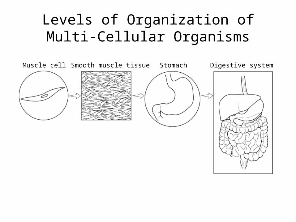
Muscle cell Smooth muscle tissue Stomach Digestive system
Section 7-4
Levels of OrganizationLevels of Organization of Multi-Cellular Organisms
Homeostasis
• Homeostasis is the process by which organisms maintain relatively stable internal conditions despite a changing external environment.
Human Body Systems
• There are 11 major human body systems• These 11 systems work together to maintain
homeostasis in humans.
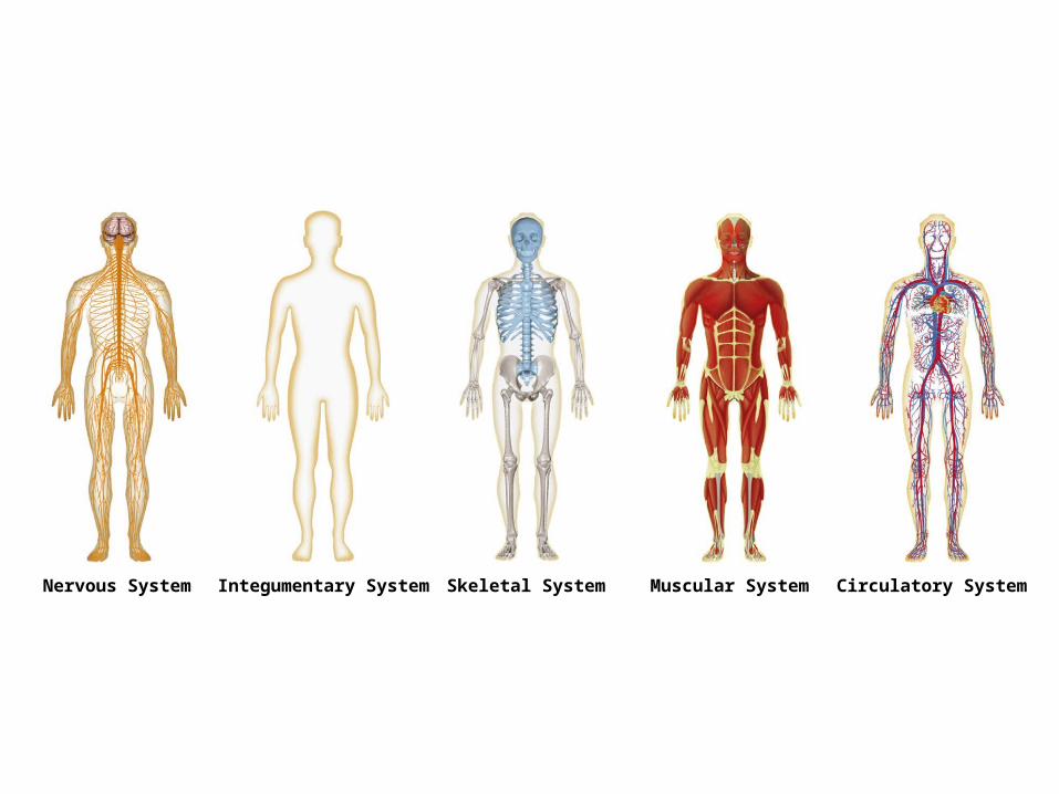
Nervous System Integumentary System Skeletal System Muscular System Circulatory System
Section 35-1
Figure 35-2 Human Organ Systems Part I
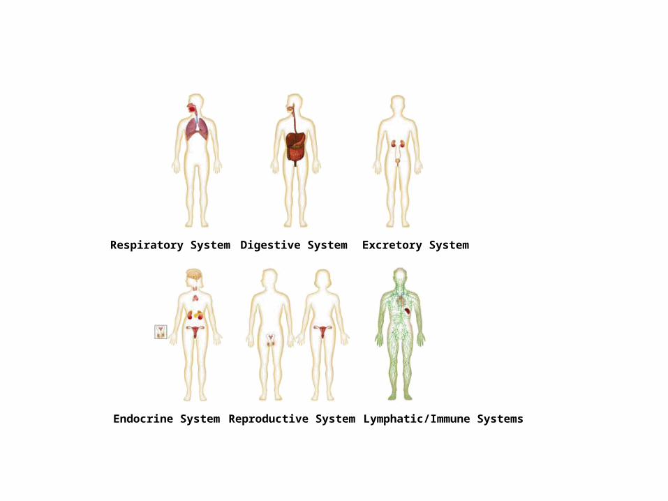
Respiratory System Digestive System Excretory System
Endocrine System Reproductive System Lymphatic/Immune Systems
Section 35-1
Figure 35-2 Human Organ Systems Part 2
Nervous System
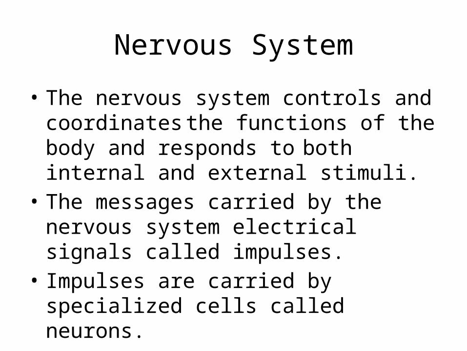
• The nervous system controls and coordinatesthe functions of the body and responds
to both internal and external stimuli. • The messages carried by the nervous system
electrical signals called impulses.• Impulses are carried by specialized cells called
neurons.
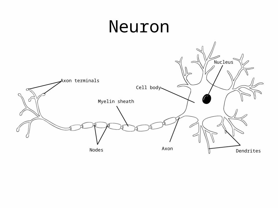
Axon terminals
Myelin sheath
Nodes
Cell body
Axon
Nucleus
Dendrites
Section 35-2
A Neuron
Neuron
Divisions of the Nervous System
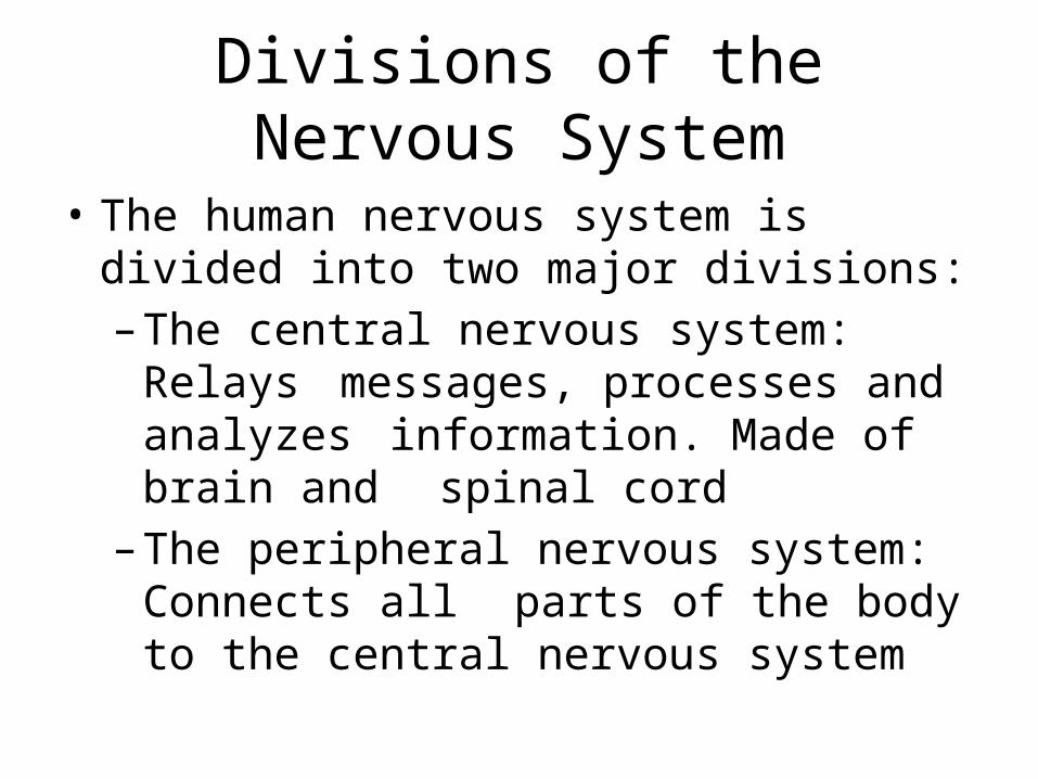
• The human nervous system is divided into two major divisions:– The central nervous system: Relays
messages, processes and analyzes information. Made of brain and spinal cord
– The peripheral nervous system: Connects all parts of the body to the central
nervous system
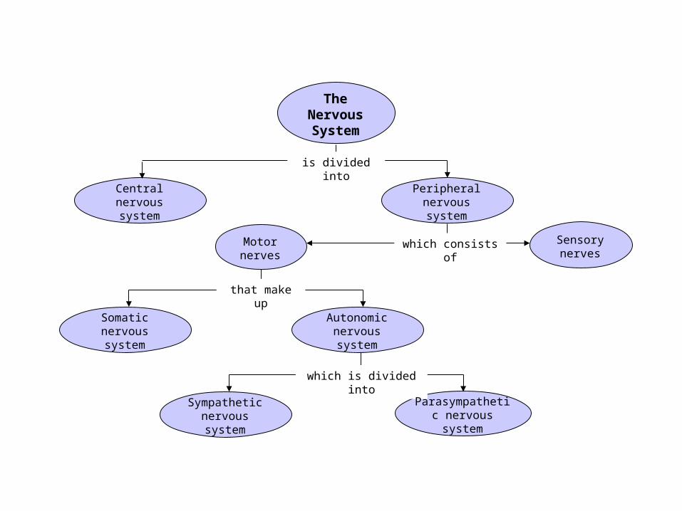
Concept Map
which consists of
is divided into
that make up
which is divided into
Section 35-3
The Nervous System
Sensory nerves
Motor nerves
Autonomic nervous system
Somatic nervous system
Central nervous system
Peripheral nervous system
Sympathetic nervous system
Parasympathetic nervous system
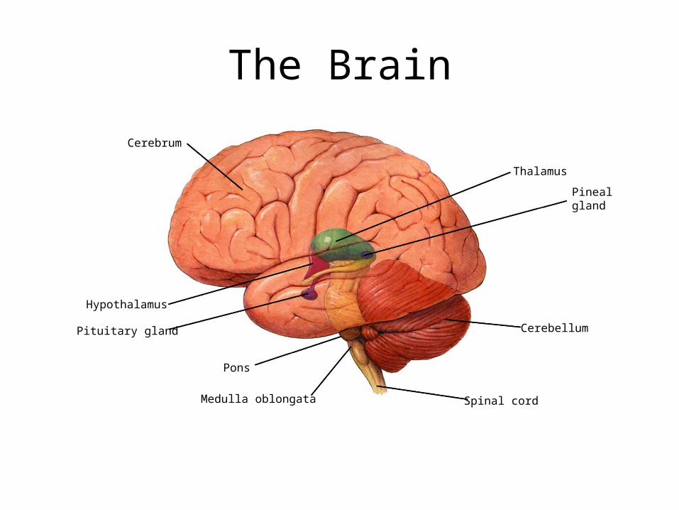
Pons
Pituitary gland
Hypothalamus
Cerebrum
Medulla oblongata Spinal cord
Cerebellum
Pineal gland
Thalamus
Section 35-3
Figure 35-9 The Brain
The Brain

The Circulatory System
• The human circulatory system consists of the heart, a series of blood vessels, and the blood that flows through them.
• The myocardium is the muscle in the heart thatpumps blood through the body.
• The human heart has 4 chambers. The upperchambers are called the atrium and the lower chambers are called ventricles.
• The right side of the heart receives oxygen poorblood from the body and sends it the lungs. The left side of the heart receives oxygen rich blood from the lungs and sends it to the body.
Section 37-1
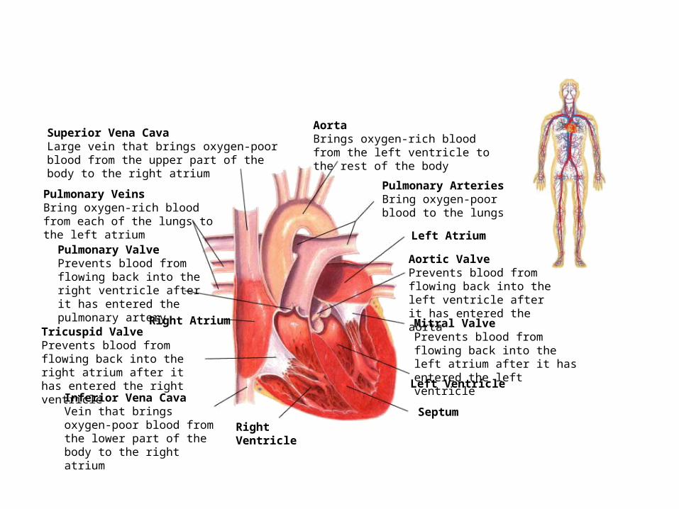
Figure 37-3 The Structures of the Heart
Right Ventricle
Right Atrium
Left Atrium
Inferior Vena CavaVein that brings oxygen-poor blood from the lower part of the body to the right atrium
Tricuspid ValvePrevents blood from flowing back into the right atrium after it has entered the right ventricle
Pulmonary ValvePrevents blood from flowing back into the right ventricle after it has entered the pulmonary artery
Pulmonary VeinsBring oxygen-rich blood from each of the lungs to the left atrium
Superior Vena CavaLarge vein that brings oxygen-poor blood from the upper part of the body to the right atrium
AortaBrings oxygen-rich blood from the left ventricle to the rest of the body
Pulmonary ArteriesBring oxygen-poor blood to the lungs
Aortic ValvePrevents blood from flowing back into the left ventricle after it has entered the aorta
Mitral ValvePrevents blood from flowing back into the left atrium after it has entered the left ventricle
Left Ventricle
Septum

Circulation
• As blood flows through the circulatory system it moves through 3 different types of bloodvessels. Arteries, capillaries and veins.
• Arteries carry oxygen rich blood away from the heart to the body.
• Capillaries carry blood from arteries to individual cells
• Veins carry oxygen poor blood back to the heart.

Section 37-1
Figure 37-5 The Three Types of Blood Vessels
Capillary
Connective tissue
Connective tissue
Smooth muscle
Smooth muscle
Endothelium
Endothelium
Valve
Venule
Endothelium
Arteriole
VeinArtery
Section 37-1
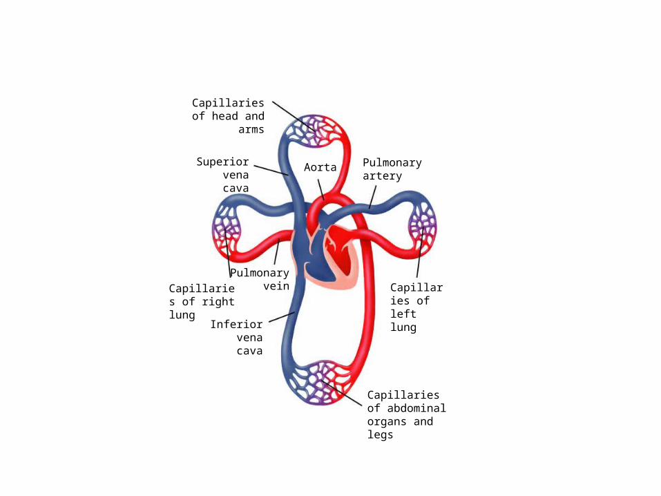
Figure 37-2 The Circulatory System
Capillaries of head and arms
Capillaries of abdominal organs and legs
Inferior vena cava
Pulmonary veinCapillaries of
right lung
Superior vena cava
Aorta Pulmonary artery
Capillaries of left lung
• Click the image to play the video segment.
Video 1
Human Circulation
Blood Pressure
• When the heart pumps blood through the arteries it exerts force on the arterial wall. This is known as blood pressure.
• When the heart contracts it increases bloodpressure on the arterial walls. When it relaxes the blood pressure increases.
• Normal blood pressure 120/80
Cardiovascular Disease
• High blood pressure and atherosclerosis are the two leading causes of heart disease
• Atherosclerosis is the build of fatty tissues on the walls of arteries called plaque.
• Atherosclerosis in the coronary arteries can prevent blood from getting to the muscles of the heart, which can cause the muscle to start die.
• If enough muscle is damaged a heart attack occurs.
Stroke
• Atherosclerosis can cause blood clots to form. • When one of clots break free it can then travel
through the circulatory system.• Stroke occurs when one of these blood clots
blocks a capillary in the brain.

Hypertension
• High blood pressure can lead to a number of medical problems.
• High blood pressure makes the heart work harder to circulate the blood.
• Over long periods of time high blood pressurecan weaken the heart and increase the
risk of heart attack, stroke, or other circulatory diseases.
Disease
• A disease is any change other than injury thatdisrupts normal body function.
• Some disease are produced by agents such asbacteria, viruses, fungi or protists.
• Other diseases are caused by environmetal factors, such as chemical exposure or UV light.
• Some diseases are genetic.• Pathogen: Disease causing agents.

Viruses
Protists
Worms
Fungi
Disease Agent That Causes Disease
Method of Transmission
Common coldInfluenza
Chickenpox
Measles
Tuberculosis
MeningitisCholeraTetanusAfrican sleeping sicknessMalariaAmoebic dysenterySchistosomiasisBeef tapewormAthlete’s foot
Ringworm
RhinovirusTwo types (A, B), plus subtypesVaricella
Paramyxovirus
Mycobacterium tuberculosisNeisseria meningitidisVibrio choleraeClostridium tetaniTrypanosoma
PlasmodiumEntamoeba histolyticaSchistosomaTaenia saginataImperfect fungi
Imperfect fungi
Airborne; direct contact with infected personAirborne; droplet infection; direct contact with infected personAirborne; direct contact with infected personDroplets in air; direct contact with secretions of infected personDroplets in air; contaminated milk and dairy products
Direct contact with a carrierContaminated drinking waterContaminated wound; usually puncture woundSpread by tsetse fly
Spread by Anopheles mosquitoesContaminated drinking waterFreshwater streams and rice paddies Contaminated meatContact with infected personExchange of hats, combs, or athletic head gear with infected person
Section 40-1
Pathogen Types
Pathogens and Disease
Bacteria
Immune System
• The function of the immune system is fight infection through the production of cells that inactivate foreign substances or cells.
• The immune system has two general responsecategories. Non specific defense and
specific defense
Non Specific Defense
• These defenses do not discriminate between one threat and another.
• First line of defense: Keep pathogens out of the body. The skin is primarily responsible of this.
• Second line of defense: If pathogens enter thebody the inflammatory response ifactivated
The Inflammatory response
• The inflammatory response is a non specificresponse to damage or injury to bodilytissue.
• During the inflammatory response white blood cell production rises. These cells fight infection.
• Many of these white blood cells are phagocytes which will engulf and destroybacteria.
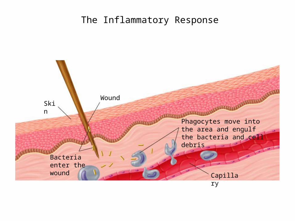
SkinWound
Bacteria enter the wound
Phagocytes move into the area and engulf the bacteria and cell debris
Capillary
Section 40-2 The Inflammatory Response

• Click the image to play the video segment.
Video 1
Inflammatory Response
Specific Defense
• IF a pathogen gets past the non specific defenses of the immune system, it willtrigger a specific response that targets thattype of pathogen.
• A substance that triggers this response is called an antigen. Viruses, bacteria andother pathogens can serve as antigens
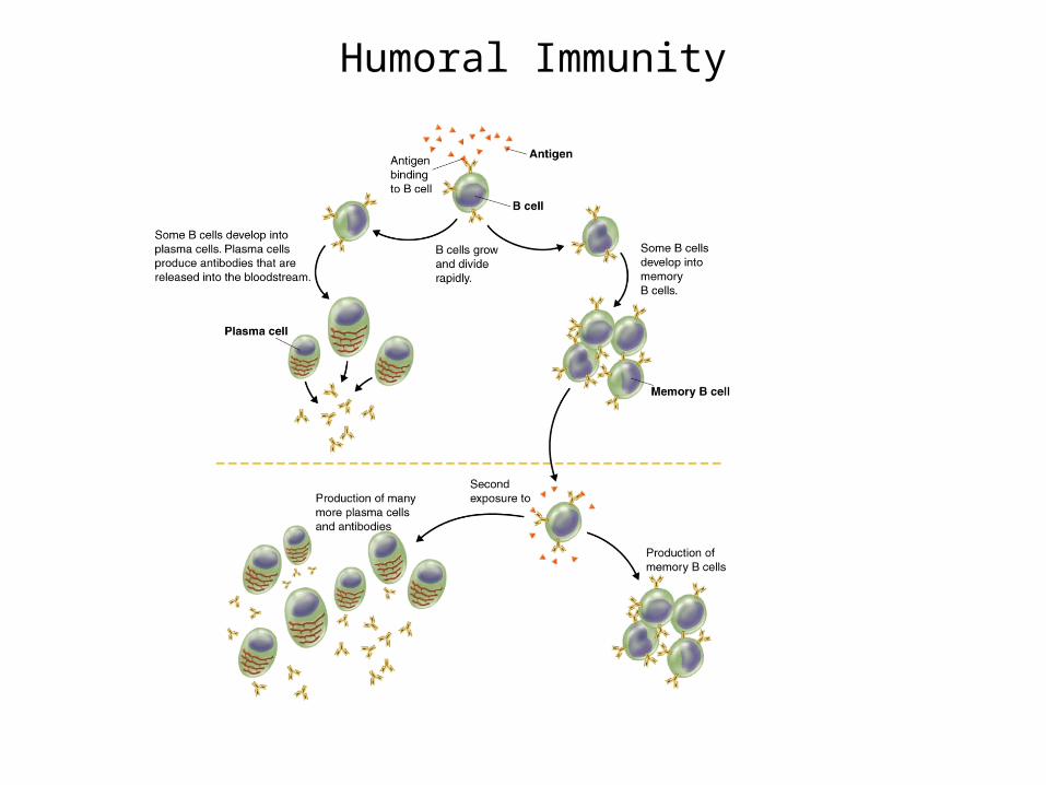
Humoral Activity
• Antigens are recognized by B cells of the immune system.
• These B cells start to produce proteins called antibodies
• Antibodies will attack the antigens.• Some B cells become memory B cells and if
that specific antigen ever returns the body can respond quicker.
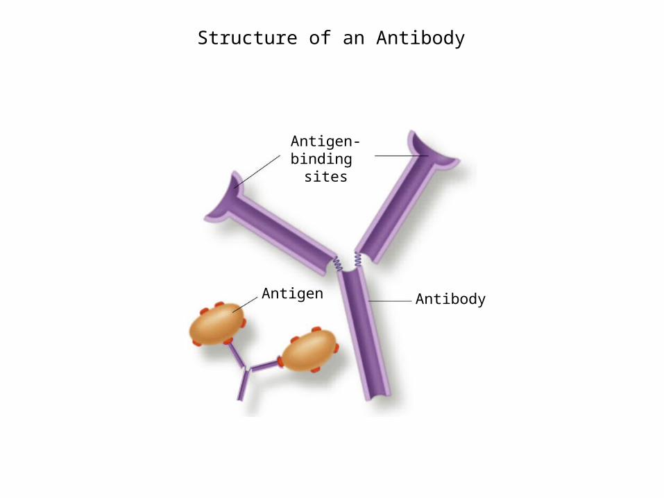
Antigen-binding
sites
Antigen Antibody
Section 40-2
Structure of an Antibody
Section 40-2
Humoral Immunity
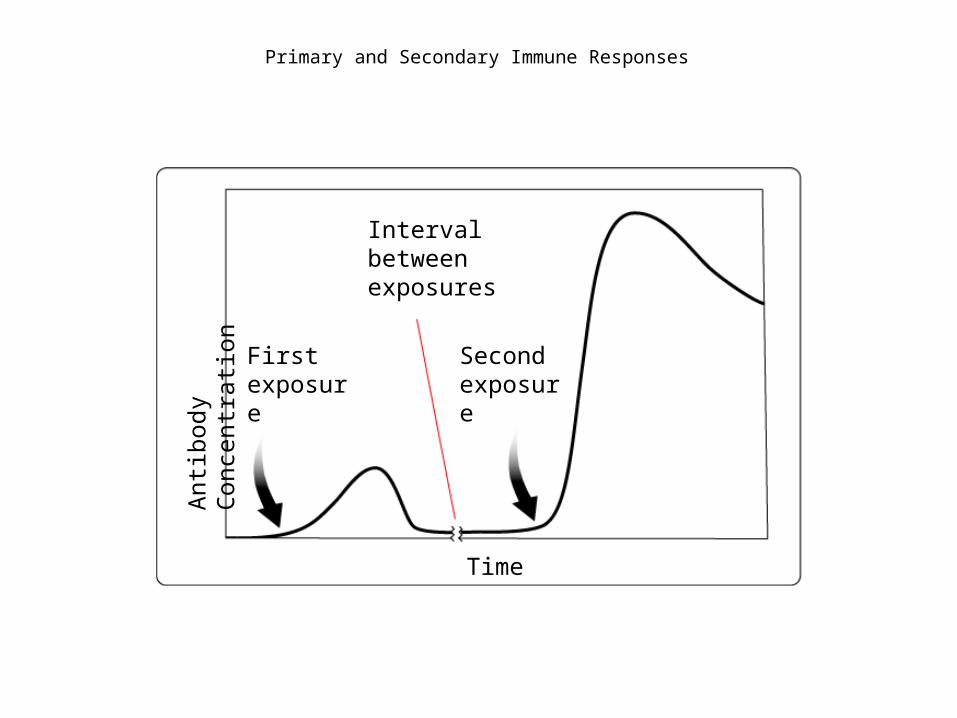
Interval between exposures
First exposure
Second exposure
Time
Antib
ody
Conc
entr
ation
Section 40-2
Primary and Secondary Immune Responses

Click the image to play the video segment.
Video 2
Humoral Immunity
Cell-mediated response
• Once a pathogen has invaded cell the humoralresponse doesn’t work.
• During cell mediated response T cells attack infected cells and destroys them.
• There are 4 kinds of T cells involved in this process. Helper T cells, Killer T cells, suppressor T cells and Memory T cells
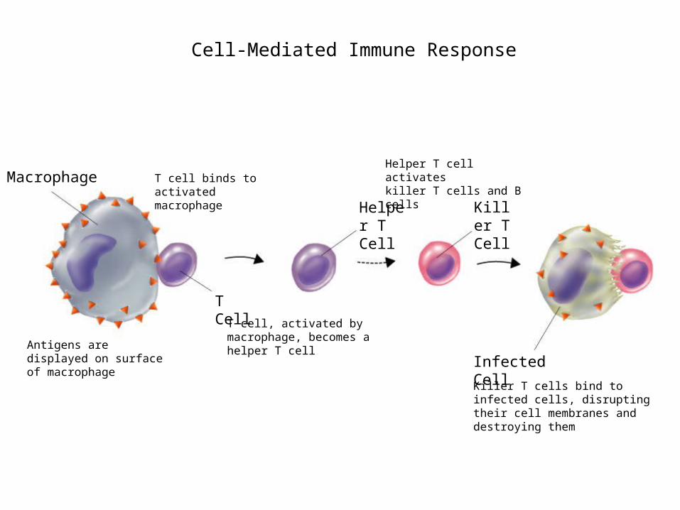
Macrophage
T Cell
Helper T Cell
Killer T Cell
Infected CellAntigens are displayed on surface of macrophage
T cell binds to activated macrophage
T cell, activated by macrophage, becomes a helper T cell
Helper T cell activates killer T cells and B cells
Killer T cells bind to infected cells, disrupting their cell membranes and destroying them
Section 40-2Cell-Mediated Immune Response

Click the image to play the video segment.
Video 3
Cell-Mediated Immunity
Vaccines
• Vaccination: The injection of a weaken pathogen to produce immunity.
• Active immunity: The immune system produces antibodies in response to a antigen.
• Passive immunity: Antibodies are introduced to the body.
Cancer
• When a signal cell loses the ability to control division it can result in the formation oflarge masses of cells called tumors. This disorder is called cancer.
• Cancer is caused by mutations to the genes that regulate the cell cycle. Different agents that can cause these mutations may be found in chemicals, radiation, or viruses.

The Reproductive System
• The reproductive system is used to produce new individuals.
• The male reproductive cells are made in thetestes and the female reproductive cells are made in the ovaries.

The male reproductive system
• The Function of the male reproductive system is to produce and deliver sperm cells.
• Sperm cells are produced in the testes of a male. The testes are contained within thescrotum.
• Sperm is stored inside of the epididymis. Fromthere sperm cells are transported
through the vas deferens to the urethra and then out of the body through the penis.
The Female Reproductive System
• The main function of the female reproductive system is to produce eggs and prepare
the females body to nourish and carry the developing embryo.
• Eggs are produced in the ovaries and are released once a month. They travel through the fallopian tubes to the uterus and then out of the body if fertilization doesn’t occur
Fertilization
• The process of joining a sperm cell and a eggis called fertilization.
• The Fertilized egg is called a zygote.• Fertilization tends to occur in the fallopian tubes.• As the zygote moves through the fallopian tubes
the zygote starts to undergo rapid cell division• After 4 days the embryo is a hollow ball of 64
cells called a morula.
Early Development
• The early stages of development include implantation, gastrulation and neurulation.
• After 6 to 7 days the developing embryoattaches itself to the wall of the uterus in
a process called implantation.• After implantation the embryonic cells begin
to specialize in a process called differentiation.

Implantation of blastocyst
Day 7 Fertilization
Day 4
Day 3 Day 2
Day 1
Day 0
Egg released by ovary
Fertilization and ImplantationSection 39-4
Uterine wall
Blastocyst
Morula4 cells 2 cells Zygote
Ovary
Fallopian tube
• During gastrulation 3 distinct cell layers form:Ectoderm, endoderm and mesoderm.
• After gastrulation, neurulation occurs. Duringneurulation the nervous system begins
to form.
• The placenta is a connection between themother and developing embryo. The placenta is the embryo’s organ of respiration, nourishment and excretion.
• The embryo is attached to the placenta by theumbilical cord
• After 8 weeks of development the development the embryo is called a fetus.
• After 3 months most of the major organs and tissues are fully formed.

Later Development
• During the fourth, fifth and sixth months of development the tissues of the fetus become more complex and start to function.
• During the last 3 months organ systemsmature and prepare the developing fetus to survive on it own.