Embed Size (px)
Citation preview
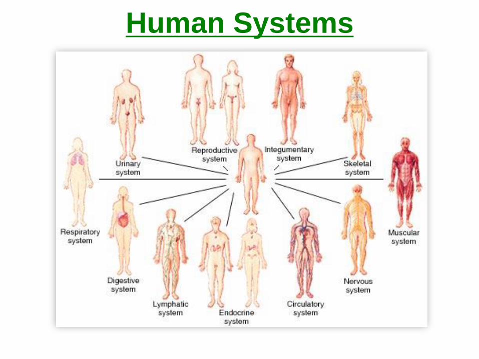
Human Systems
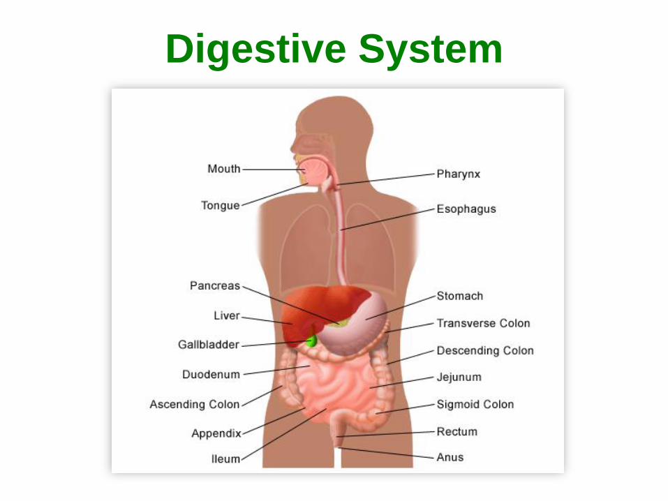
Digestive System
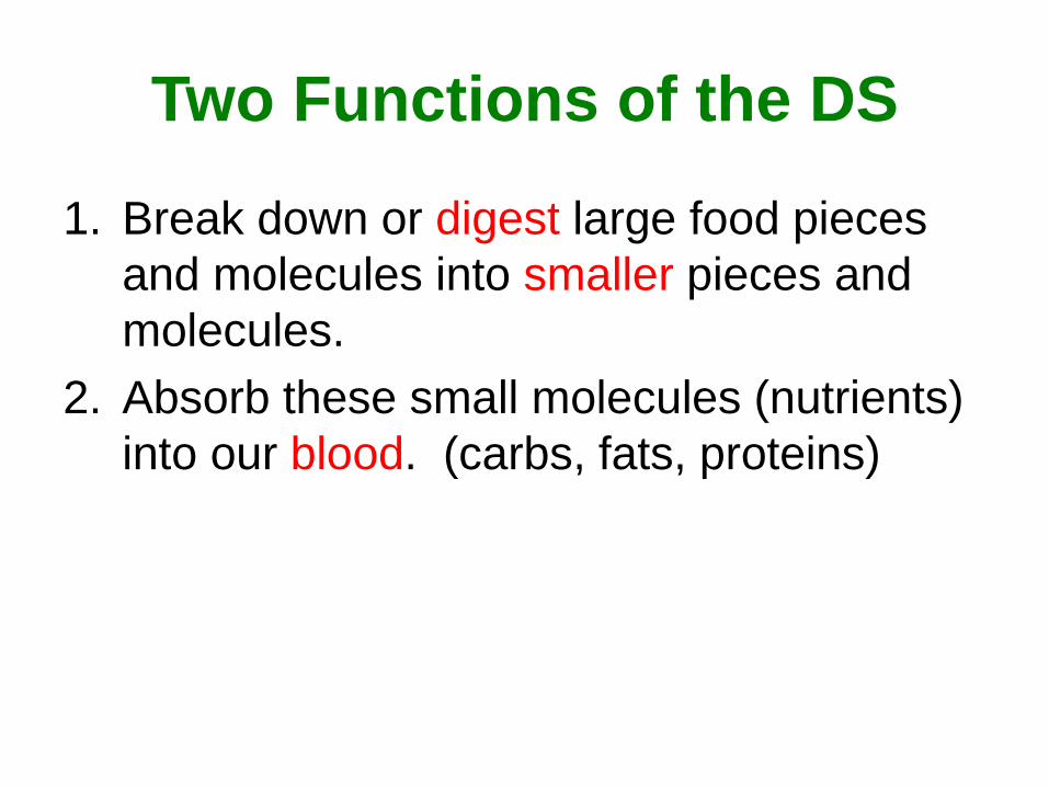
Two Functions of the DS
1. Break down or digest large food pieces
and molecules into smaller pieces and
molecules.
2. Absorb these small molecules (nutrients)
into our blood. (carbs, fats, proteins)
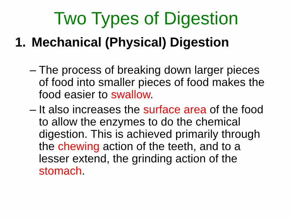
Two Types of Digestion
1. Mechanical (Physical) Digestion
– The process of breaking down larger pieces of food into smaller pieces of food makes the food easier to swallow.
– It also increases the surface area of the food to allow the enzymes to do the chemical digestion. This is achieved primarily through the chewing action of the teeth, and to a lesser extend, the grinding action of the stomach.
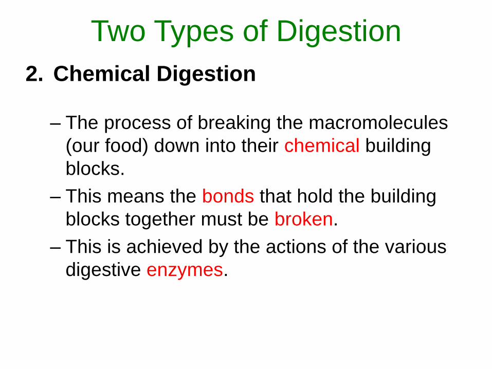
2. Chemical Digestion
– The process of breaking the macromolecules
(our food) down into their chemical building
blocks.
– This means the bonds that hold the building
blocks together must be broken.
– This is achieved by the actions of the various
digestive enzymes.
Two Types of Digestion
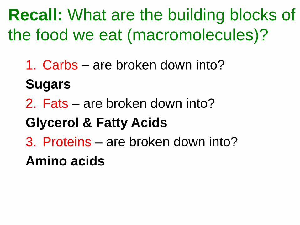
Recall: What are the building blocks of
the food we eat (macromolecules)?
1. Carbs – are broken down into?
Sugars
2. Fats – are broken down into?
Glycerol & Fatty Acids
3. Proteins – are broken down into?
Amino acids
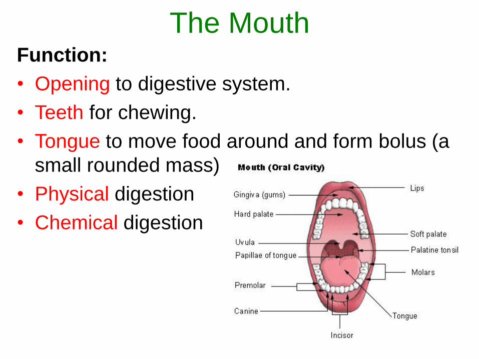
The MouthFunction:
• Opening to digestive system.
• Teeth for chewing.
• Tongue to move food around and form bolus (a
small rounded mass)
• Physical digestion
• Chemical digestion
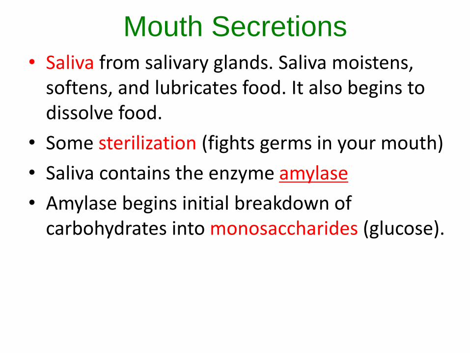
Mouth Secretions• Saliva from salivary glands. Saliva moistens,
softens, and lubricates food. It also begins to dissolve food.
• Some sterilization (fights germs in your mouth)
• Saliva contains the enzyme amylase
• Amylase begins initial breakdown of carbohydrates into monosaccharides (glucose).
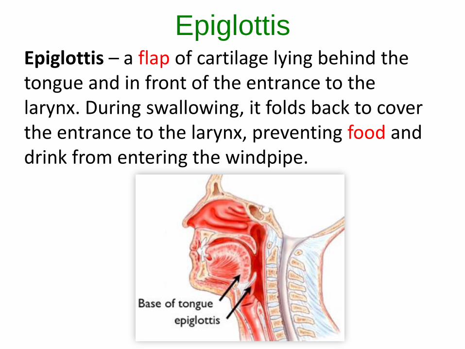
EpiglottisEpiglottis – a flap of cartilage lying behind the tongue and in front of the entrance to the larynx. During swallowing, it folds back to cover the entrance to the larynx, preventing food and drink from entering the windpipe.
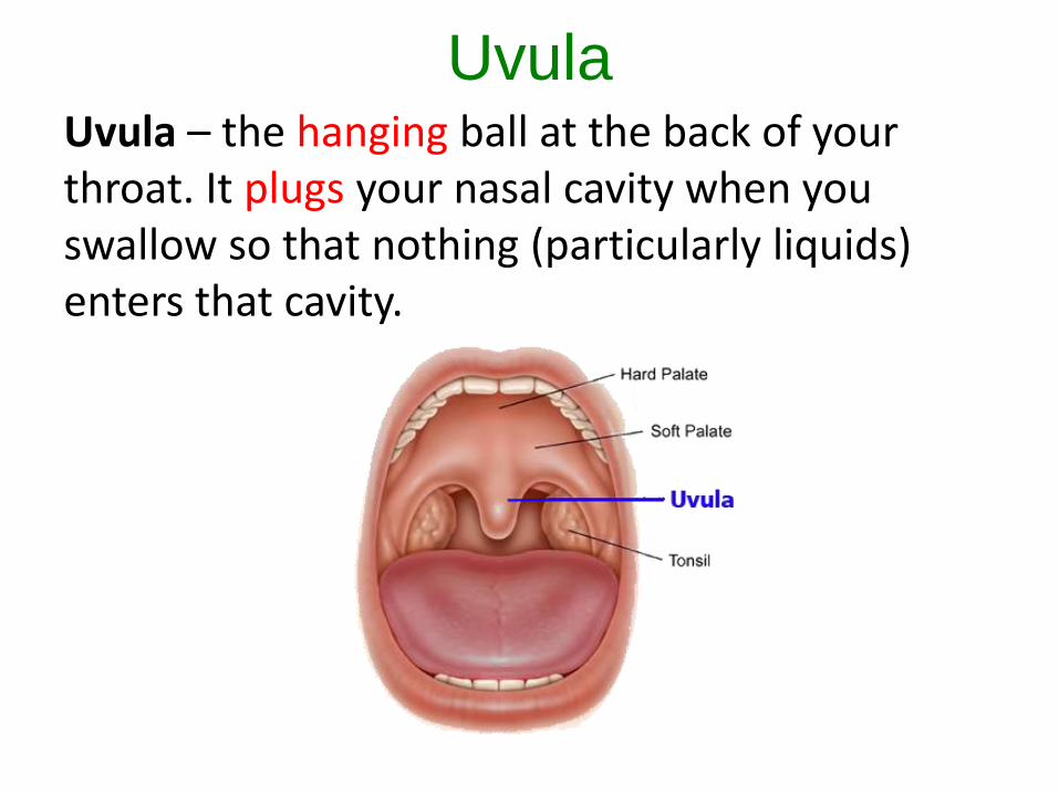
UvulaUvula – the hanging ball at the back of your throat. It plugs your nasal cavity when you swallow so that nothing (particularly liquids) enters that cavity.
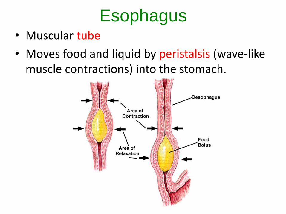
Esophagus• Muscular tube
• Moves food and liquid by peristalsis (wave-like muscle contractions) into the stomach.
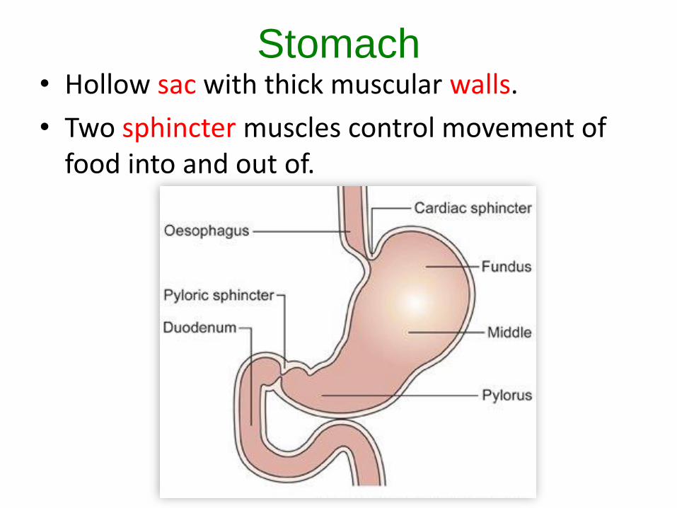
Stomach• Hollow sac with thick muscular walls.
• Two sphincter muscles control movement of food into and out of.
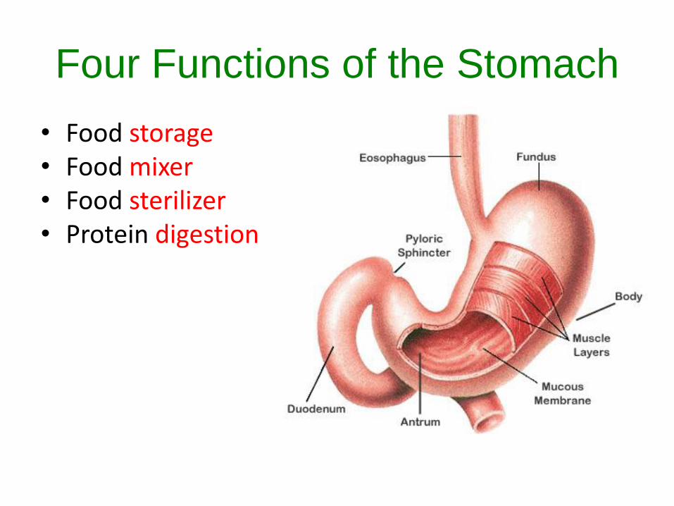
Four Functions of the Stomach
• Food storage• Food mixer• Food sterilizer• Protein digestion
Stomach Secretions
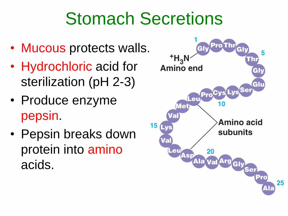
• Mucous protects walls.
• Hydrochloric acid for
sterilization (pH 2-3)
• Produce enzyme
pepsin.
• Pepsin breaks down
protein into amino
acids.
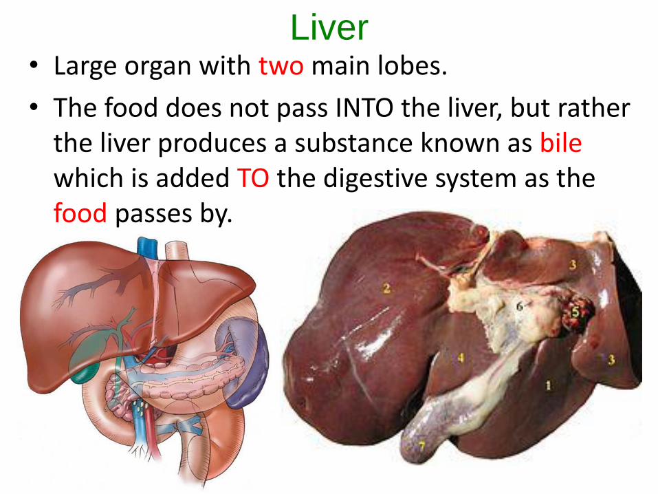
Liver• Large organ with two main lobes.
• The food does not pass INTO the liver, but rather the liver produces a substance known as bilewhich is added TO the digestive system as the food passes by.
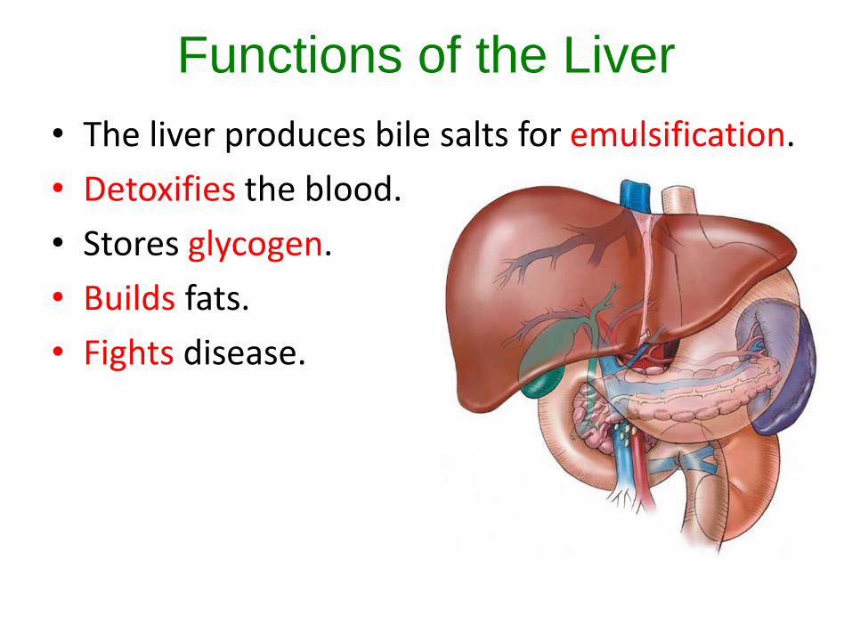
Functions of the Liver
• The liver produces bile salts for emulsification.
• Detoxifies the blood.
• Stores glycogen.
• Builds fats.
• Fights disease.
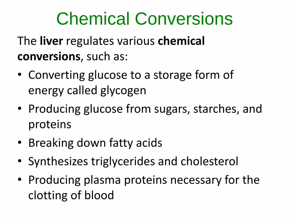
Chemical ConversionsThe liver regulates various chemical conversions, such as:
• Converting glucose to a storage form of energy called glycogen
• Producing glucose from sugars, starches, and proteins
• Breaking down fatty acids
• Synthesizes triglycerides and cholesterol
• Producing plasma proteins necessary for the clotting of blood
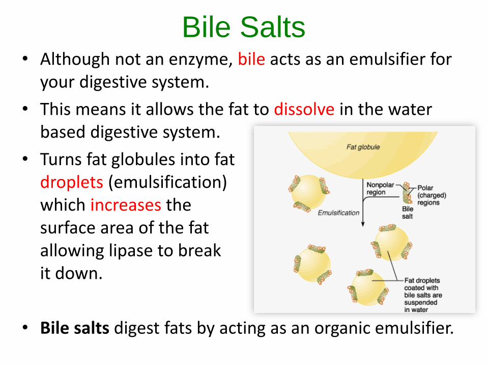
Bile Salts• Although not an enzyme, bile acts as an emulsifier for
your digestive system.
• This means it allows the fat to dissolve in the water based digestive system.
• Turns fat globules into fat droplets (emulsification) which increases the surface area of the fat allowing lipase to break it down.
• Bile salts digest fats by acting as an organic emulsifier.
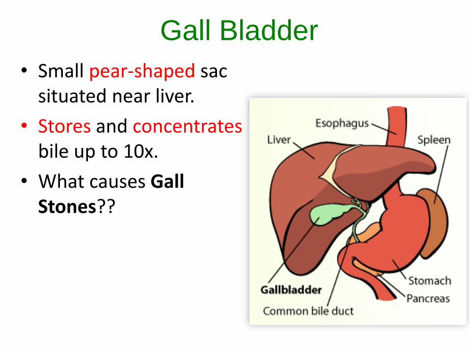
Gall Bladder
• Small pear-shaped sac situated near liver.
• Stores and concentratesbile up to 10x.
• What causes Gall Stones??
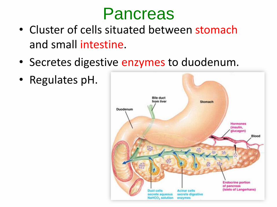
Pancreas• Cluster of cells situated between stomach
and small intestine.
• Secretes digestive enzymes to duodenum.
• Regulates pH.
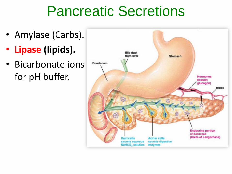
Pancreatic Secretions
• Amylase (Carbs).
• Lipase (lipids).
• Bicarbonate ions for pH buffer.

Small Intestine• Measures roughly 20 feet in length and
about 2.5 cm (1 in) in diameter
• Lipid breakdown begins here (Lipids broken into glycerol and fatty acids).
• Protein and Carb breakdown continues.
Three part tube:
1) duodenum
2) jejunum
3) ileum
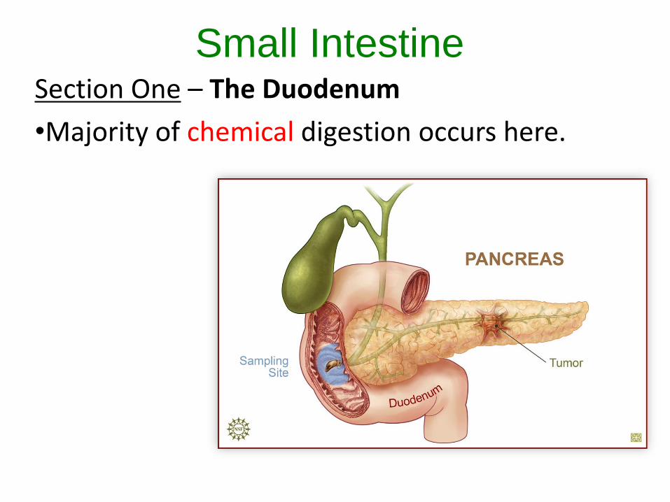
Small IntestineSection One – The Duodenum
•Majority of chemical digestion occurs here.
Small Intestine
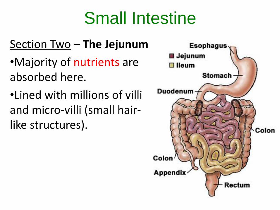
Section Two – The Jejunum
•Majority of nutrients are absorbed here.
•Lined with millions of villi and micro-villi (small hair-like structures).
Small Intestine
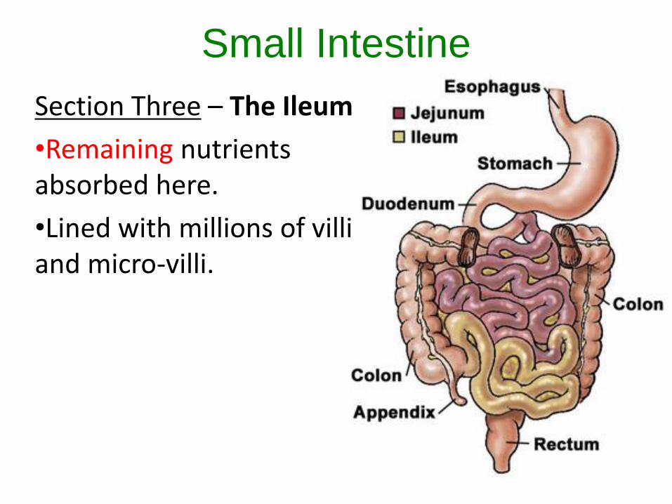
Section Three – The Ileum
•Remaining nutrients absorbed here.
•Lined with millions of villi and micro-villi.
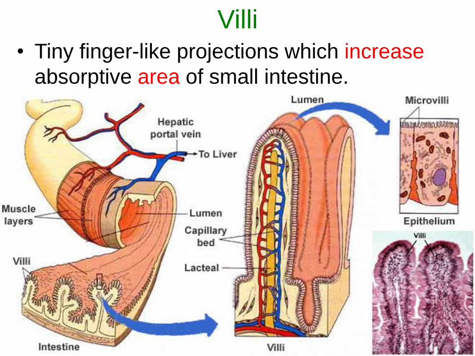
Villi• Tiny finger-like projections which increase
absorptive area of small intestine.
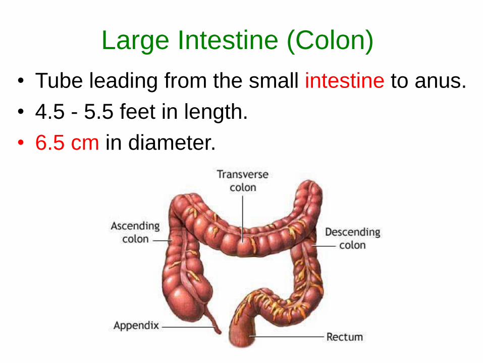
Large Intestine (Colon)
• Tube leading from the small intestine to anus.
• 4.5 - 5.5 feet in length.
• 6.5 cm in diameter.
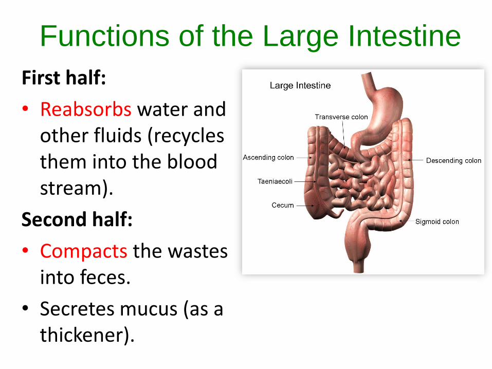
Functions of the Large Intestine
First half:
• Reabsorbs water and other fluids (recycles them into the blood stream).
Second half:
• Compacts the wastes into feces.
• Secretes mucus (as a thickener).
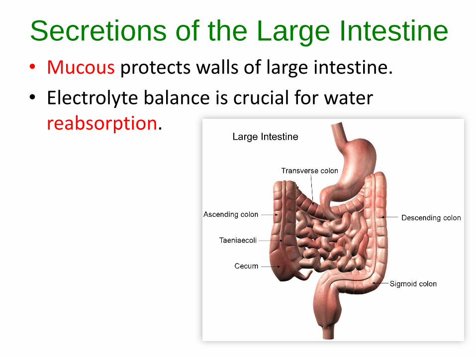
Secretions of the Large Intestine• Mucous protects walls of large intestine.
• Electrolyte balance is crucial for water reabsorption.
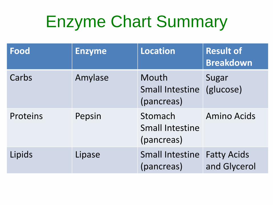
Enzyme Chart Summary
Food Enzyme Location Result of Breakdown
Carbs Amylase MouthSmall Intestine (pancreas)
Sugar(glucose)
Proteins Pepsin StomachSmall Intestine (pancreas)
Amino Acids
Lipids Lipase Small Intestine (pancreas)
Fatty Acidsand Glycerol
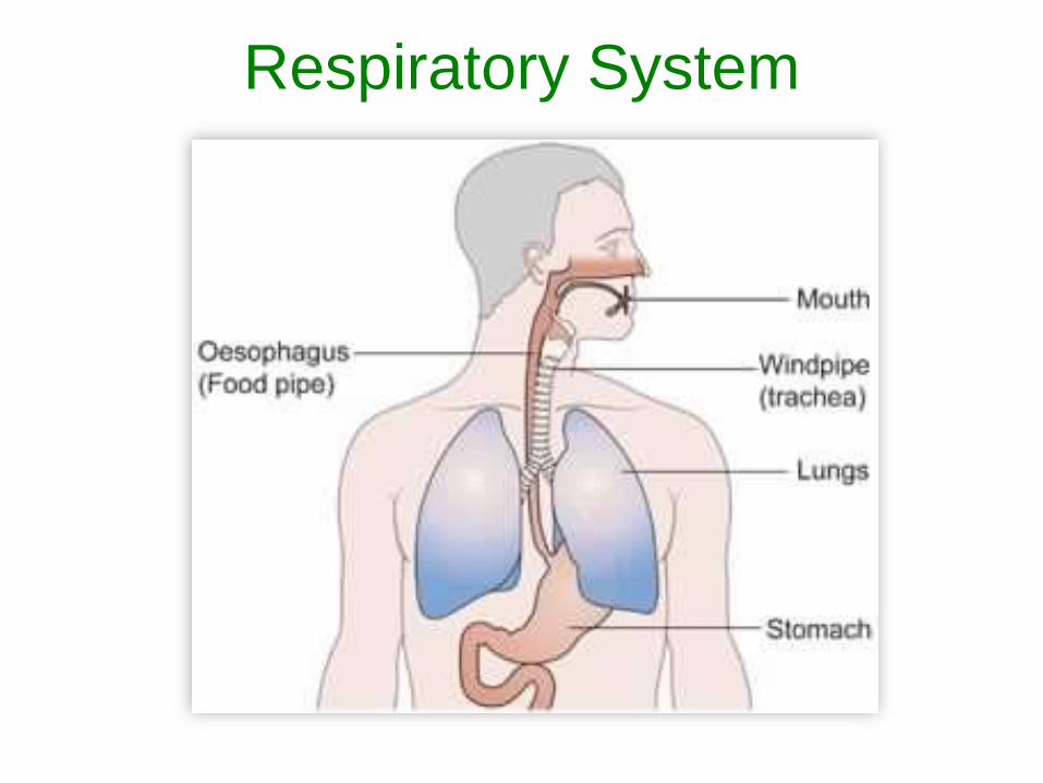
Respiratory System
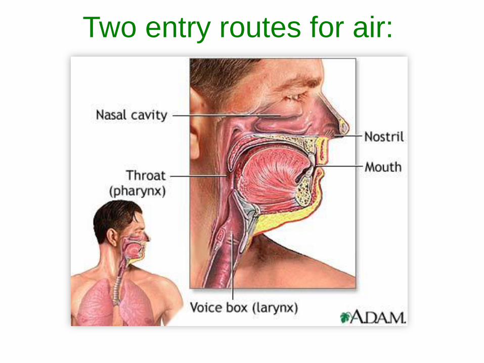
Two entry routes for air:
Goals of the Respiratory System
• Deliver oxygen to the body
• Take away carbon dioxide
Note: The lungs are the main organs of the respiratory system.
Summary of the RS
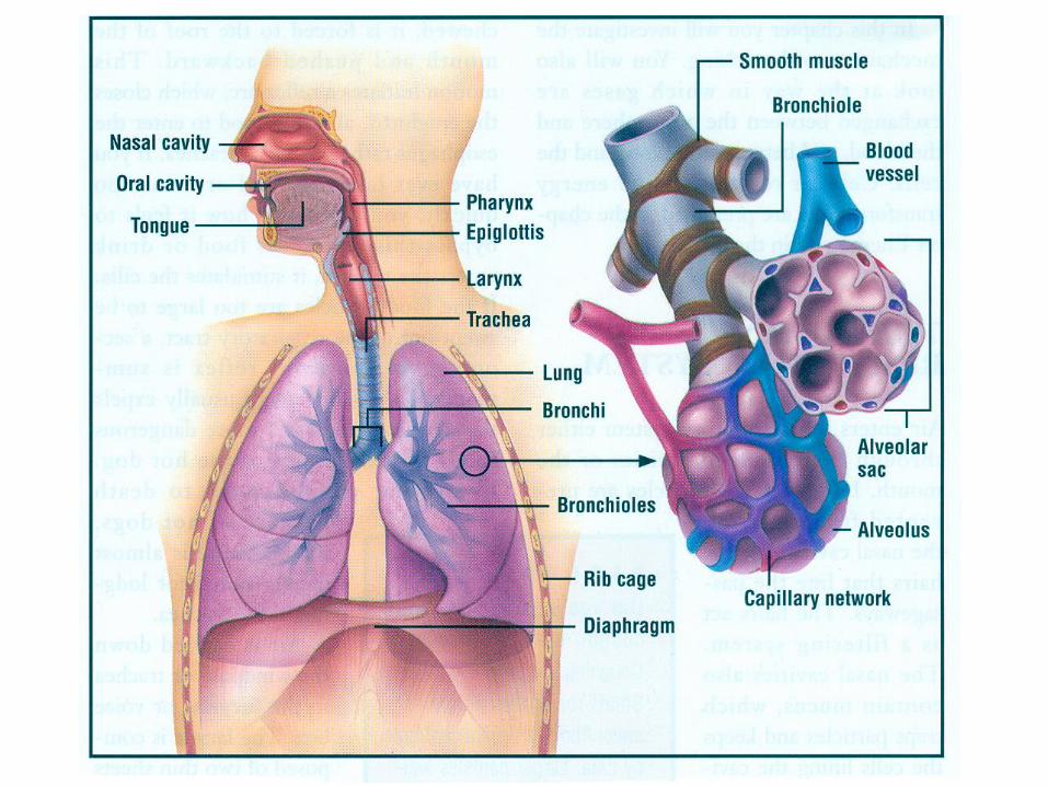
• Air enters through the mouth or nose.
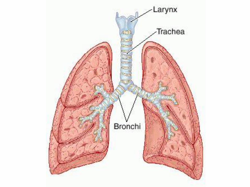
• Air travels down the trachea (windpipe). The trachea filters the air we breathe.
• Air enters the bronchi, which are two air tubes that branch off of the trachea. They carry air directly into the lungs.
• In the lungs, oxygen is taken into the body and carbon dioxide is breathed out. This is facilitated by red blood cells.
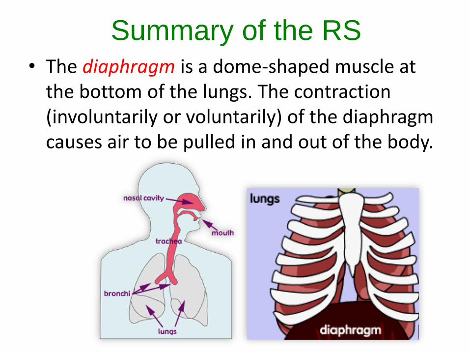
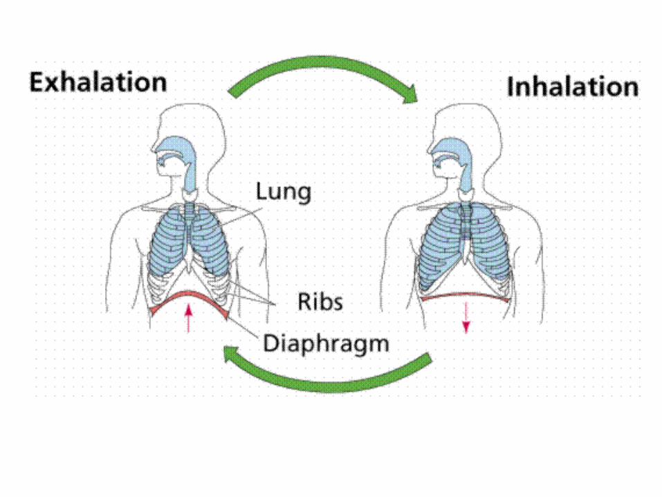
Summary of the RS• The diaphragm is a dome-shaped muscle at
the bottom of the lungs. The contraction (involuntarily or voluntarily) of the diaphragm causes air to be pulled in and out of the body.
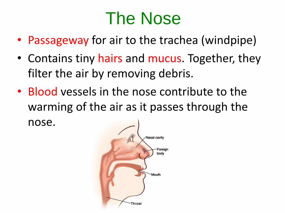
The Nose• Passageway for air to the trachea (windpipe)
• Contains tiny hairs and mucus. Together, they filter the air by removing debris.
• Blood vessels in the nose contribute to the warming of the air as it passes through the nose.
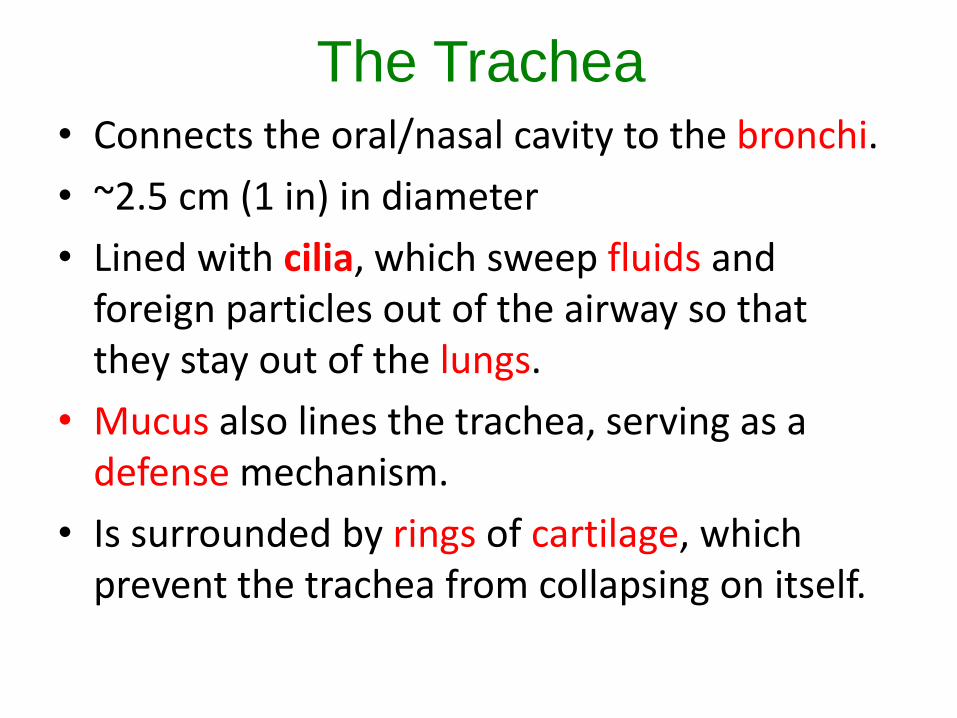
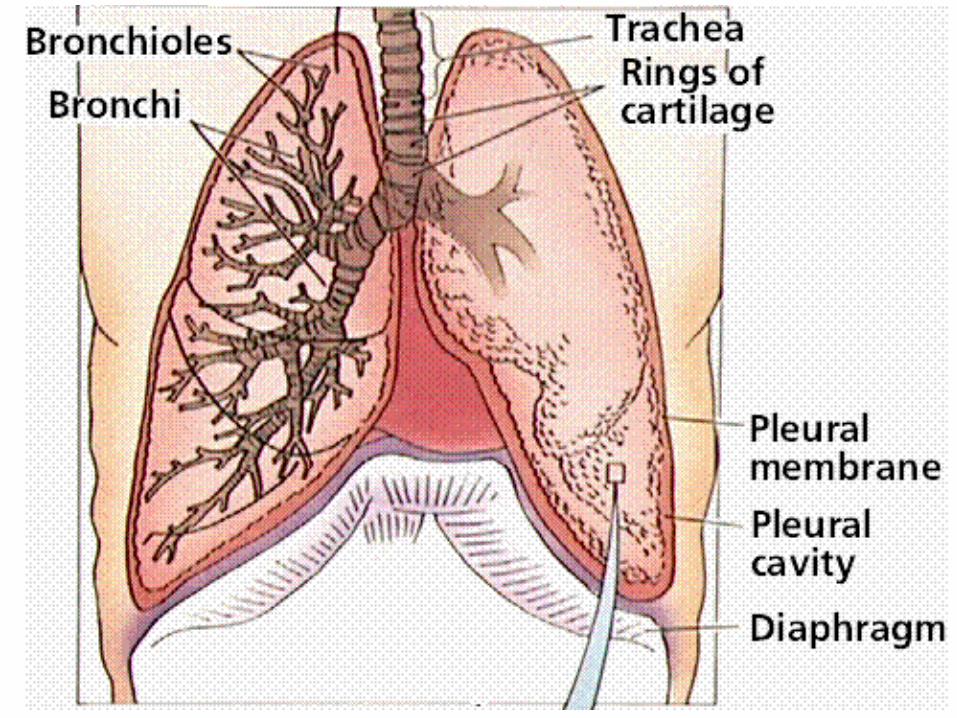
The Trachea• Connects the oral/nasal cavity to the bronchi.
• ~2.5 cm (1 in) in diameter
• Lined with cilia, which sweep fluids and foreign particles out of the airway so that they stay out of the lungs.
• Mucus also lines the trachea, serving as a defense mechanism.
• Is surrounded by rings of cartilage, which prevent the trachea from collapsing on itself.
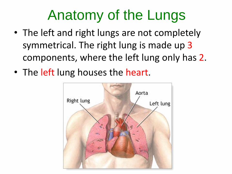
Anatomy of the Lungs• The left and right lungs are not completely
symmetrical. The right lung is made up 3components, where the left lung only has 2.
• The left lung houses the heart.
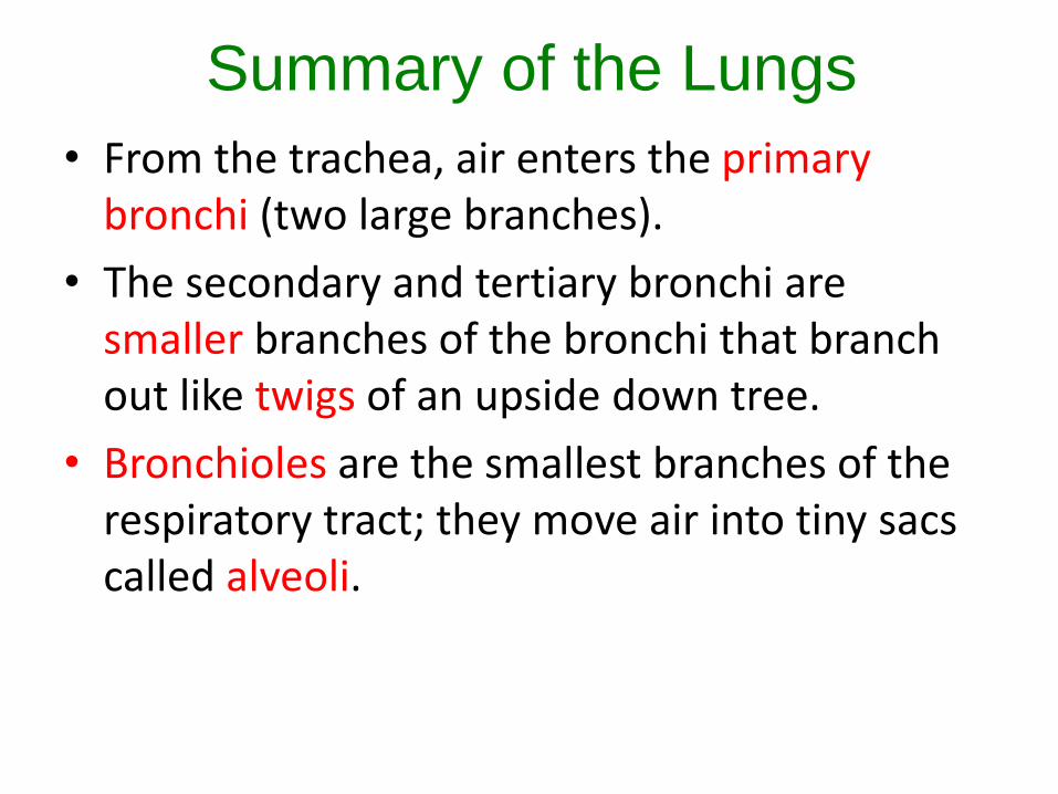
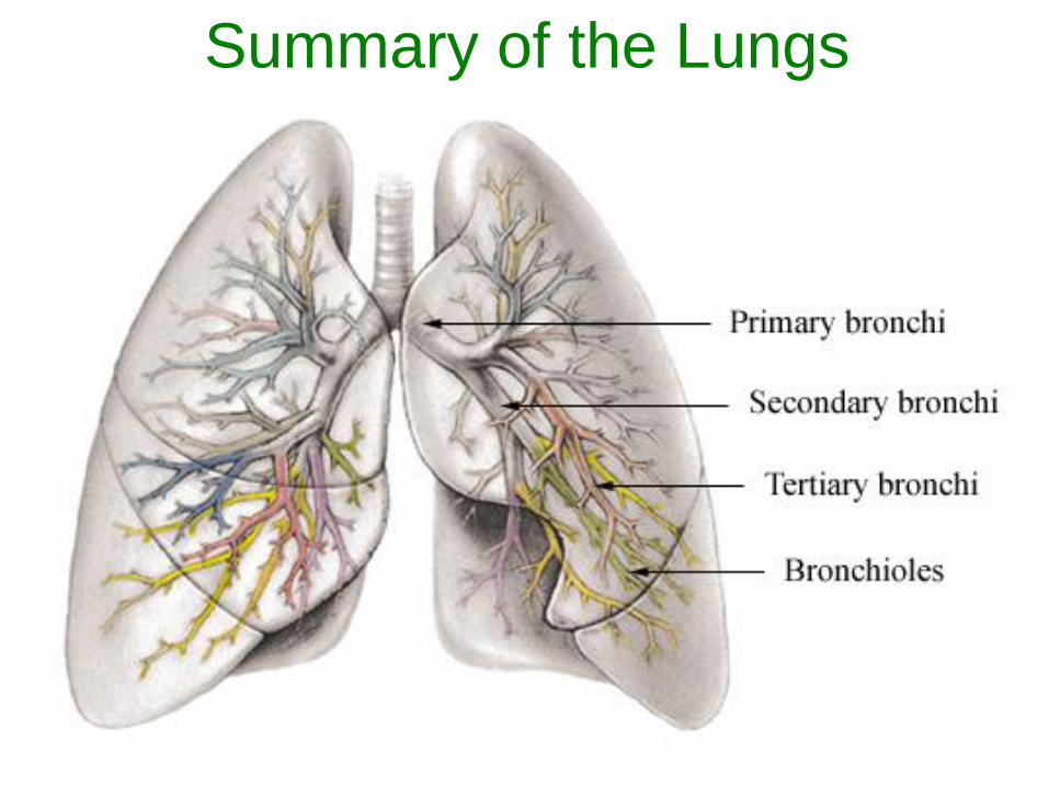
Summary of the Lungs
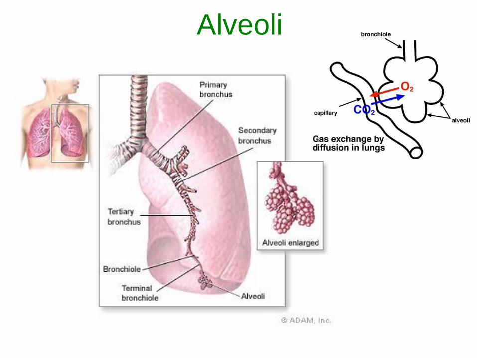
• From the trachea, air enters the primarybronchi (two large branches).
• The secondary and tertiary bronchi are smaller branches of the bronchi that branch out like twigs of an upside down tree.
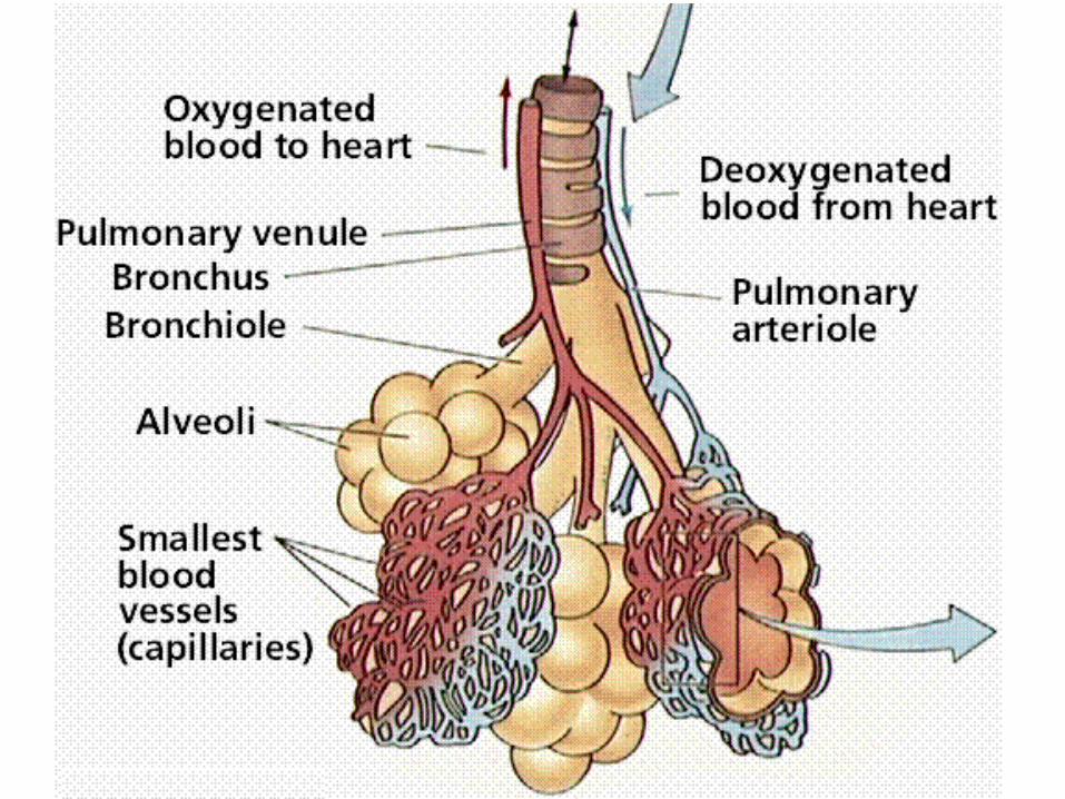
• Bronchioles are the smallest branches of the respiratory tract; they move air into tiny sacs called alveoli.
Summary of the Lungs
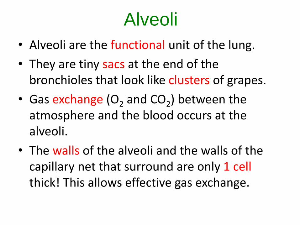
Alveoli
• Alveoli are the functional unit of the lung.
• They are tiny sacs at the end of the bronchioles that look like clusters of grapes.
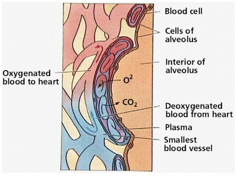
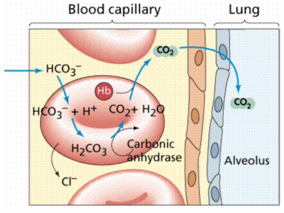
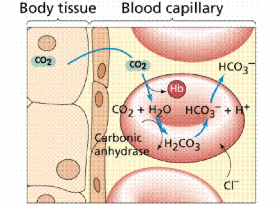
• Gas exchange (O2 and CO2) between the atmosphere and the blood occurs at the alveoli.
• The walls of the alveoli and the walls of the capillary net that surround are only 1 cell thick! This allows effective gas exchange.
Alveoli
Alveoli
Three main factors allow for the exchange of gases between the blood cells in the capillaries and the alveoli:
1. The difference in the partial pressures of oxygen and carbon dioxide result in a diffusion gradient.
2. Thin single celled walls.
3. A moist membrane.
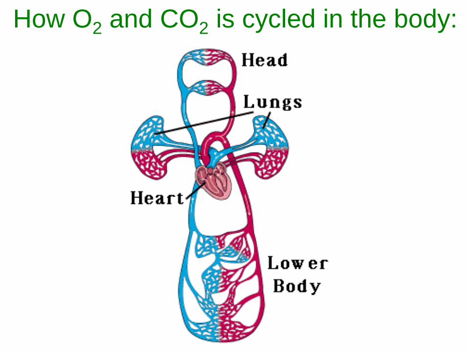
How O2 and CO2 is cycled in the body:
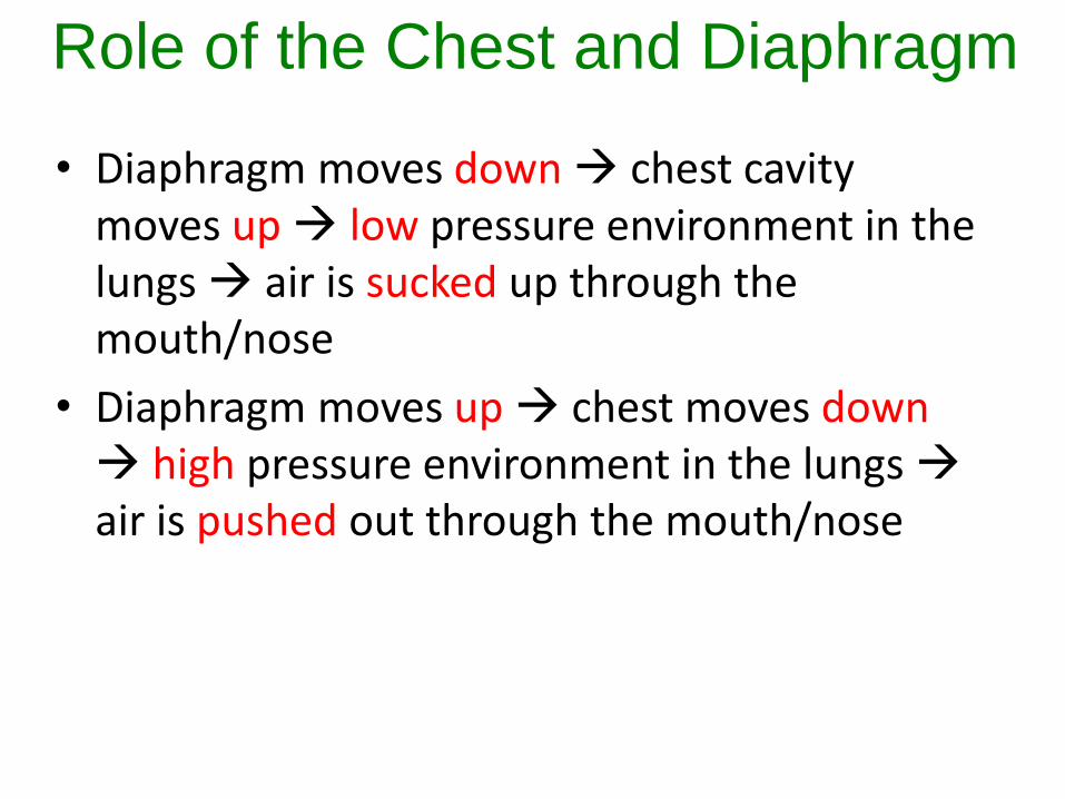
• Diaphragm moves down chest cavity moves up low pressure environment in the lungs air is sucked up through the mouth/nose
• Diaphragm moves up chest moves down high pressure environment in the lungs air is pushed out through the mouth/nose
Role of the Chest and Diaphragm

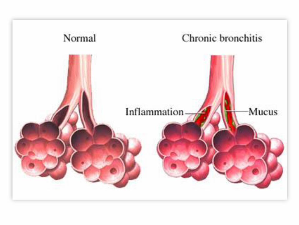
Disorders in the RS
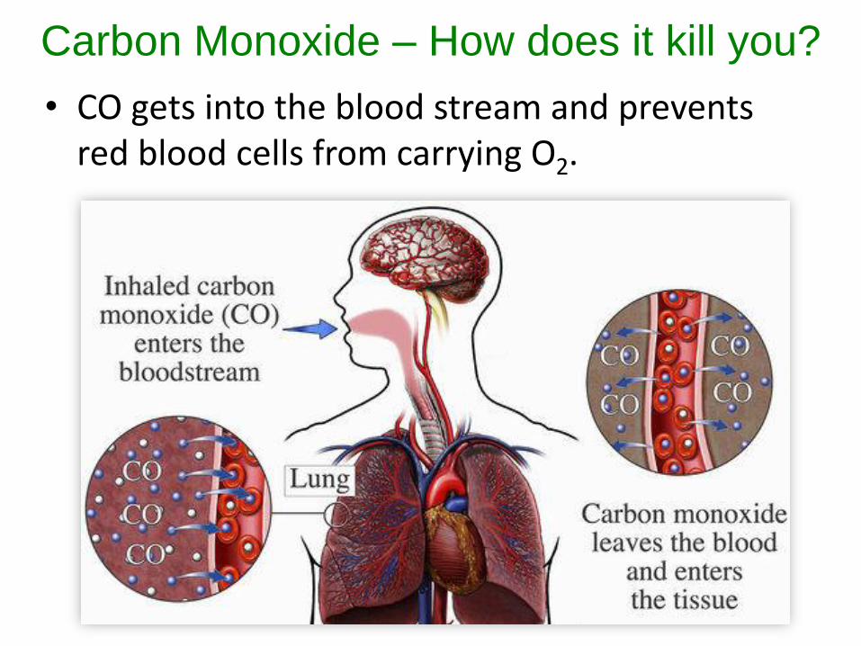
Carbon Monoxide – How does it kill you?
• CO gets into the blood stream and prevents red blood cells from carrying O2.
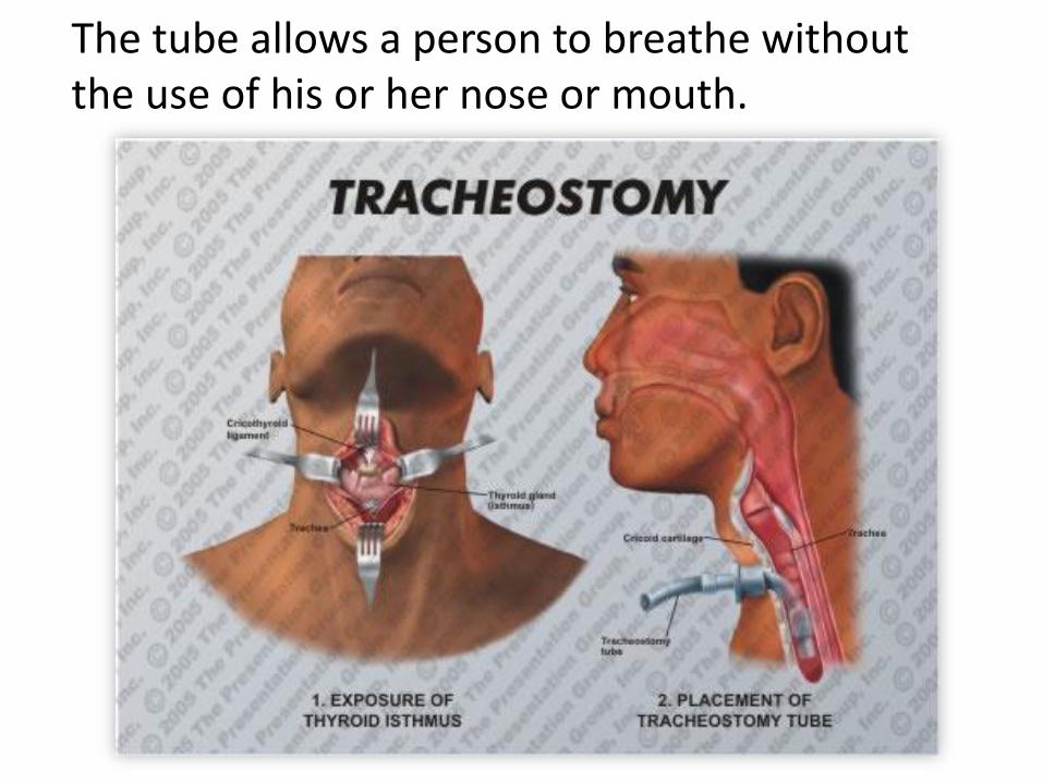
The tube allows a person to breathe without the use of his or her nose or mouth.
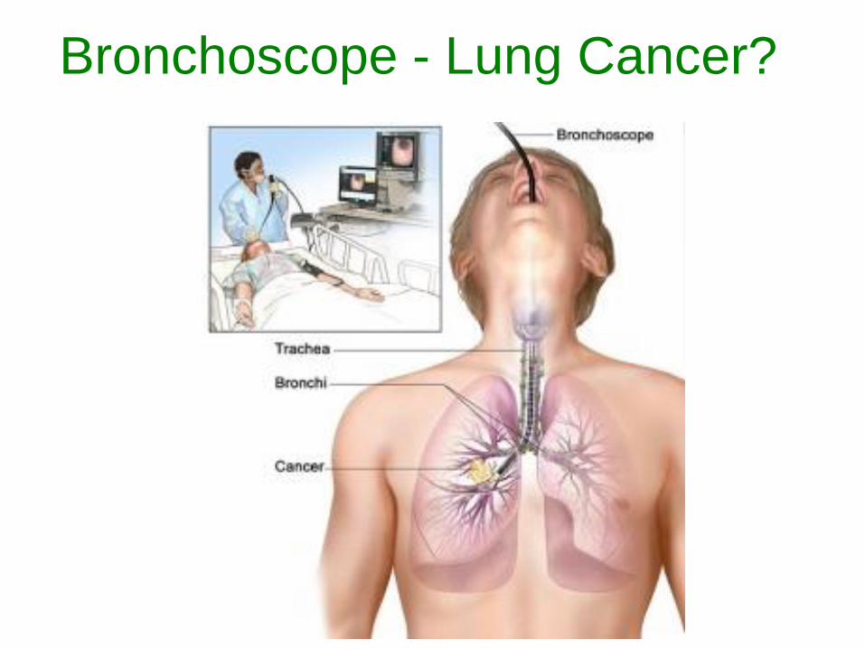
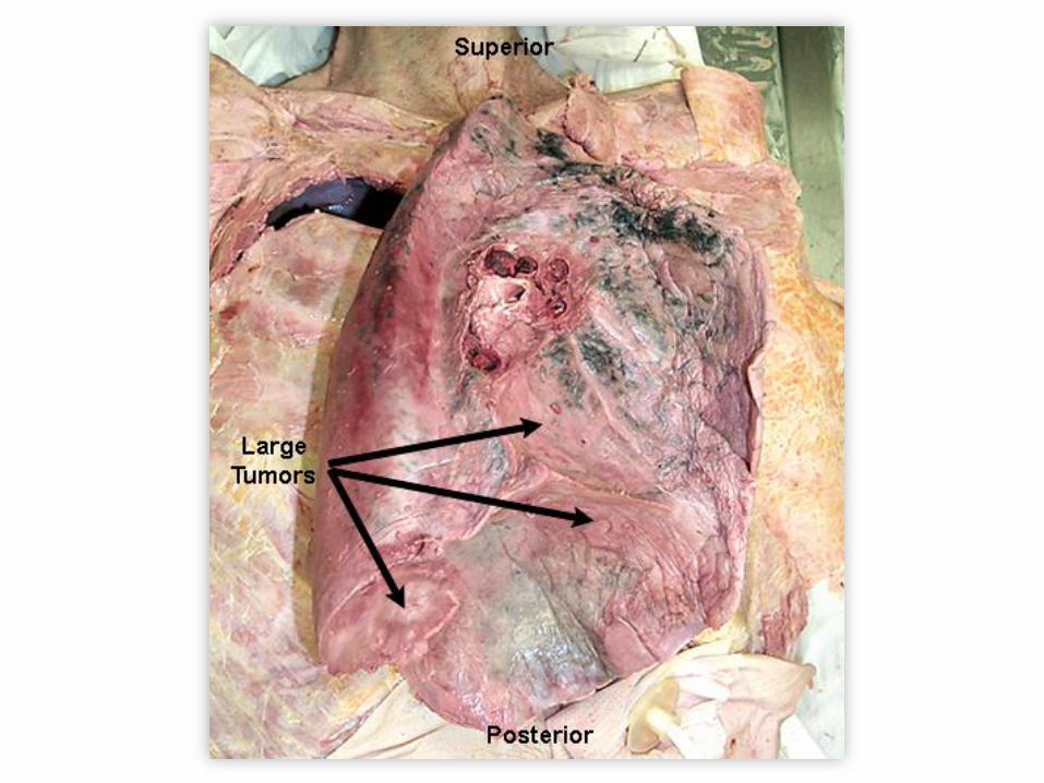
Bronchoscope - Lung Cancer?
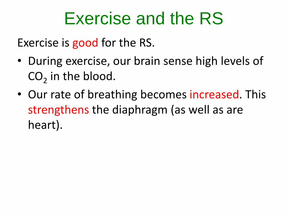
Exercise and the RS
Exercise is good for the RS.
• During exercise, our brain sense high levels of CO2 in the blood.
• Our rate of breathing becomes increased. This strengthens the diaphragm (as well as are heart).
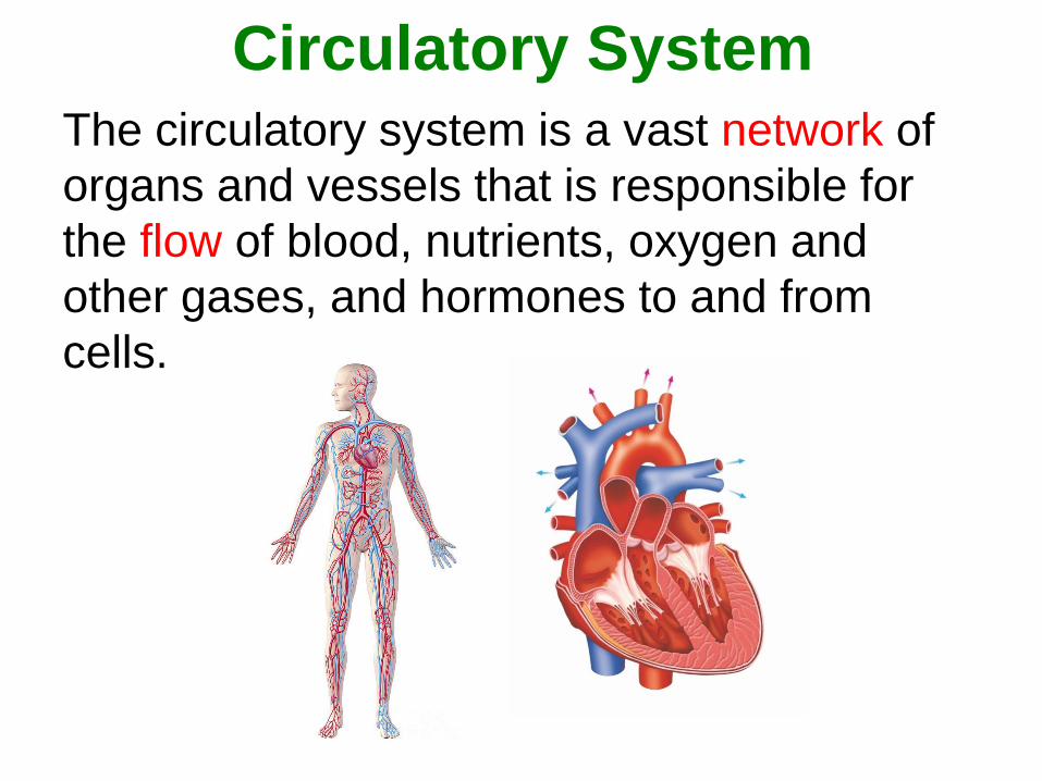
Circulatory System
The circulatory system is a vast network of
organs and vessels that is responsible for
the flow of blood, nutrients, oxygen and
other gases, and hormones to and from
cells.
Circulatory System
• In the average human, about 2,000 gallons (7,572 liters) of blood travel daily through about 60,000 miles (96,560 kilometers) of blood vessels.
• An average adult has 5 to 6 quarts (4.7 to 5.6 liters) of blood, which is made up of plasma, red blood cells, white blood cells and platelets.
• In addition to blood, the circulatory system moves lymph, which is a clear fluid that helps rid the body of unwanted material.
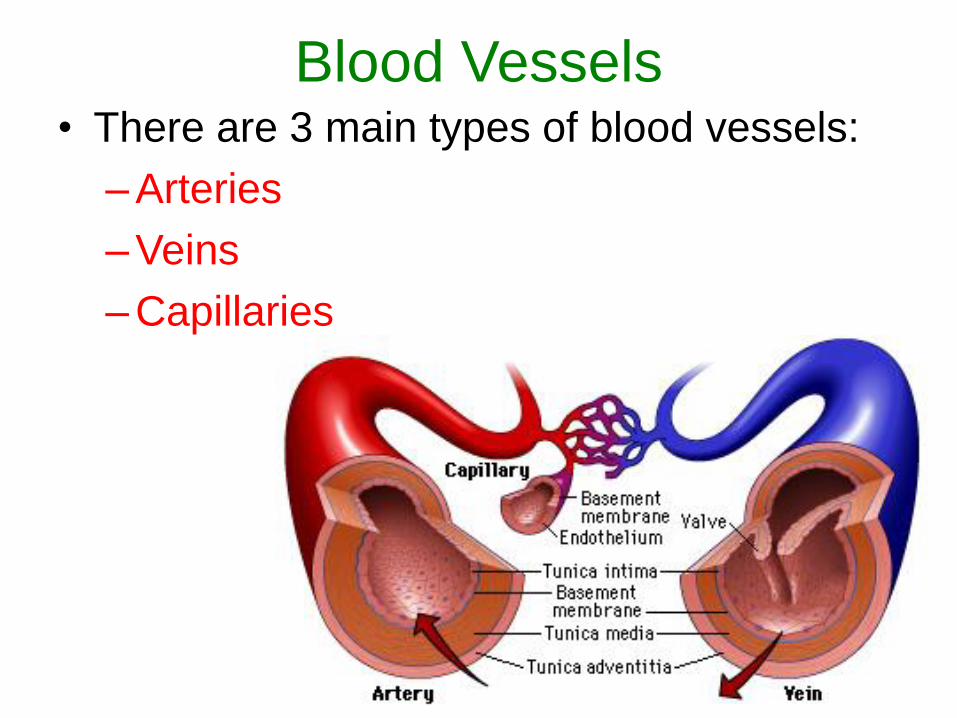
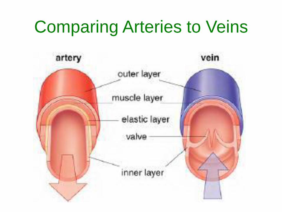
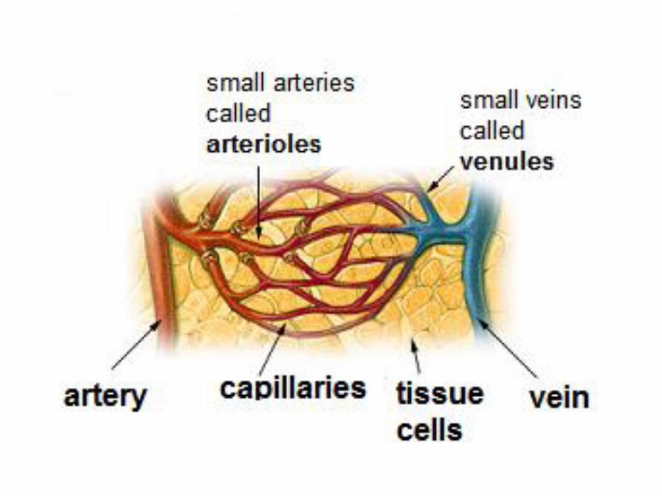
Blood Vessels• There are 3 main types of blood vessels:
– Arteries
– Veins
– Capillaries
Arteries• Carry blood AWAY from the heart.
• Have thick muscular walls.
• Expand and contract as blood pulses
through them.
• Arteries tend to travel along the bones,
deeper in the tissue.
• Do arteries contain blood with high or low
oxygen levels?
– HIGH and LOW oxygen levels
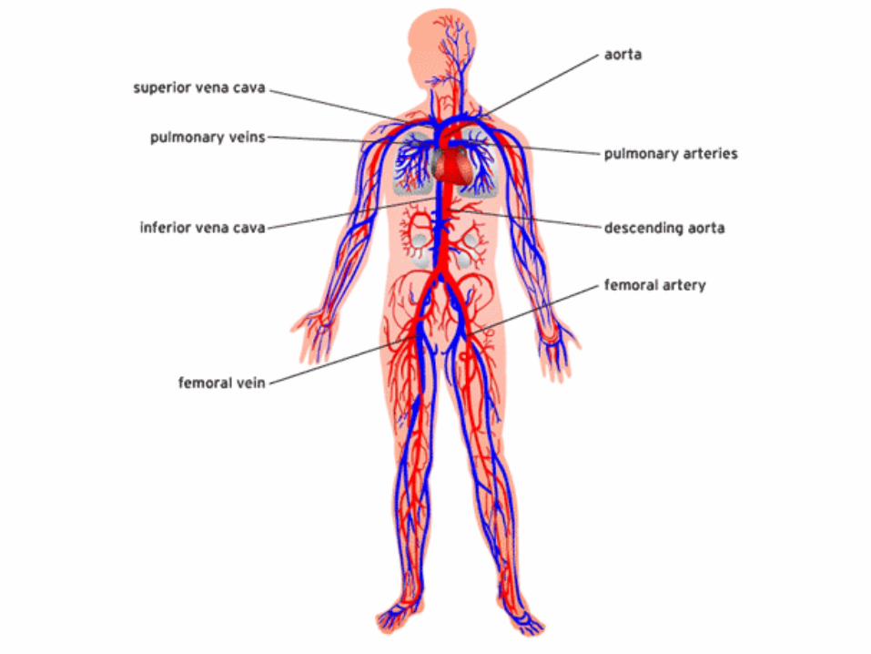
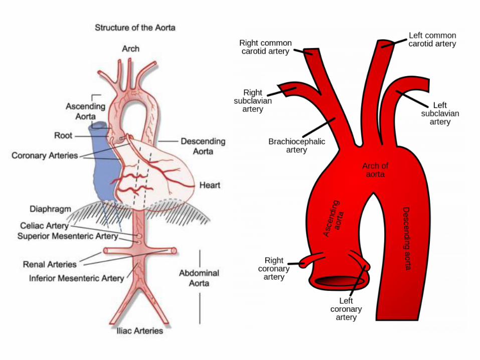
Arteries• The largest artery in the human body is the
aorta.
• Your aorta is approximately the diameter of
your thumb.
• The aorta branches into three main arteries
which carry blood to the upper half of the
body (arms and head), while the rest of the
blood heads down the descending aorta
which wraps around and behind the heart.
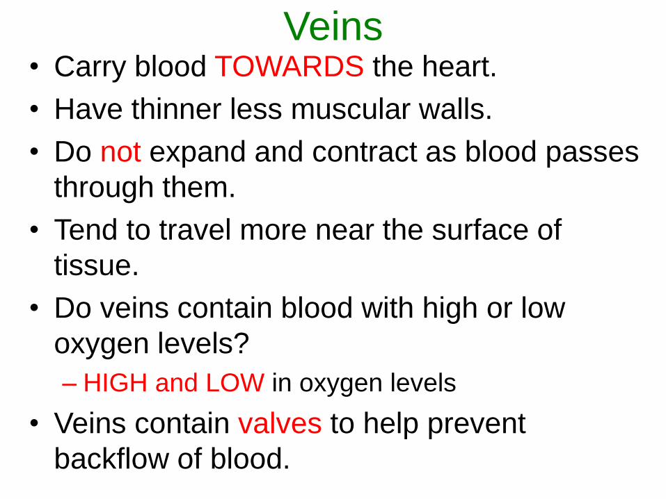
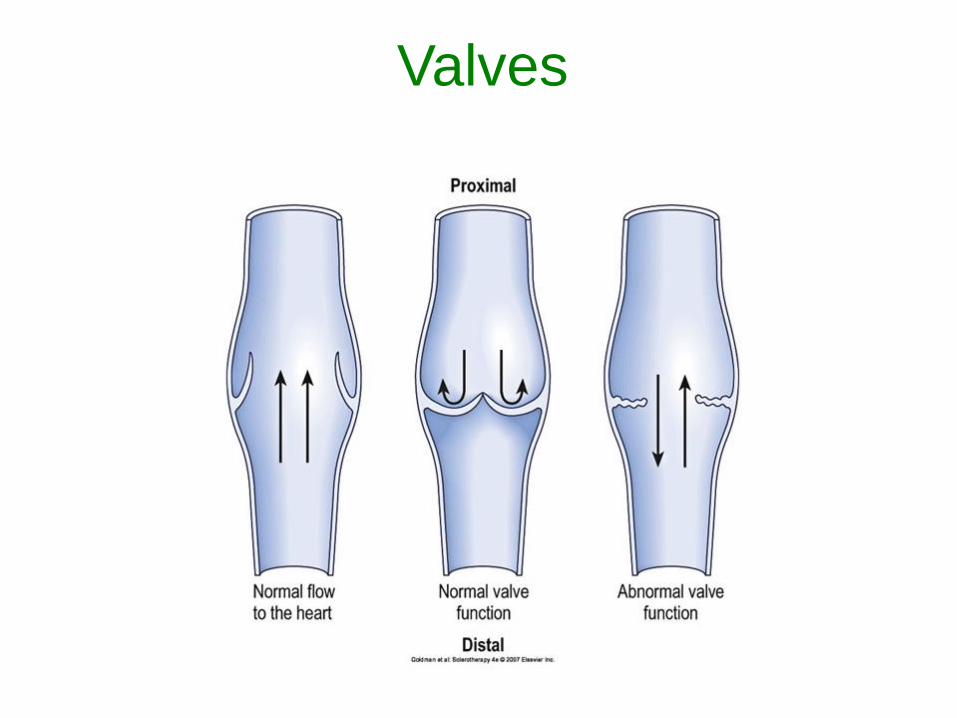
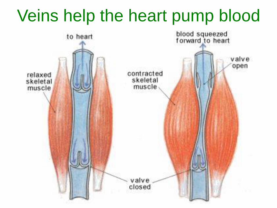
Veins• Carry blood TOWARDS the heart.
• Have thinner less muscular walls.
• Do not expand and contract as blood passes
through them.
• Tend to travel more near the surface of
tissue.
• Do veins contain blood with high or low
oxygen levels?
– HIGH and LOW in oxygen levels
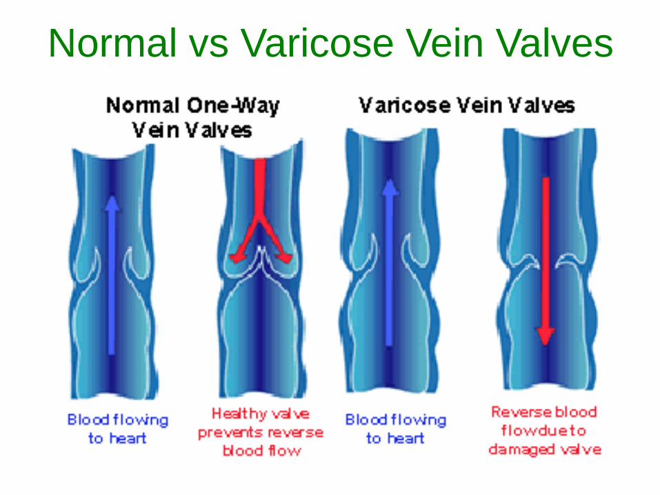
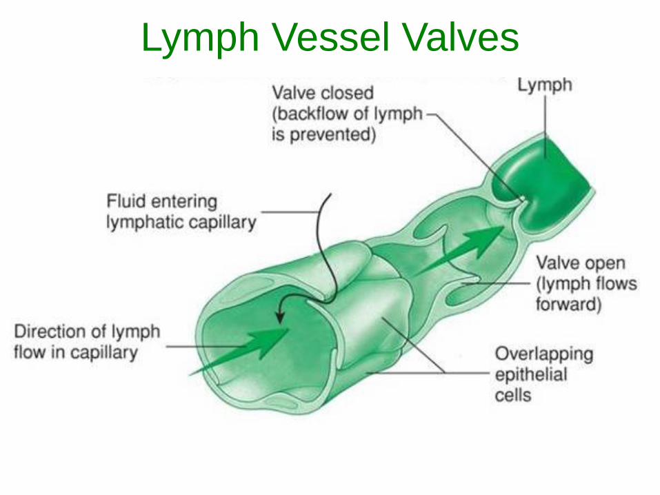
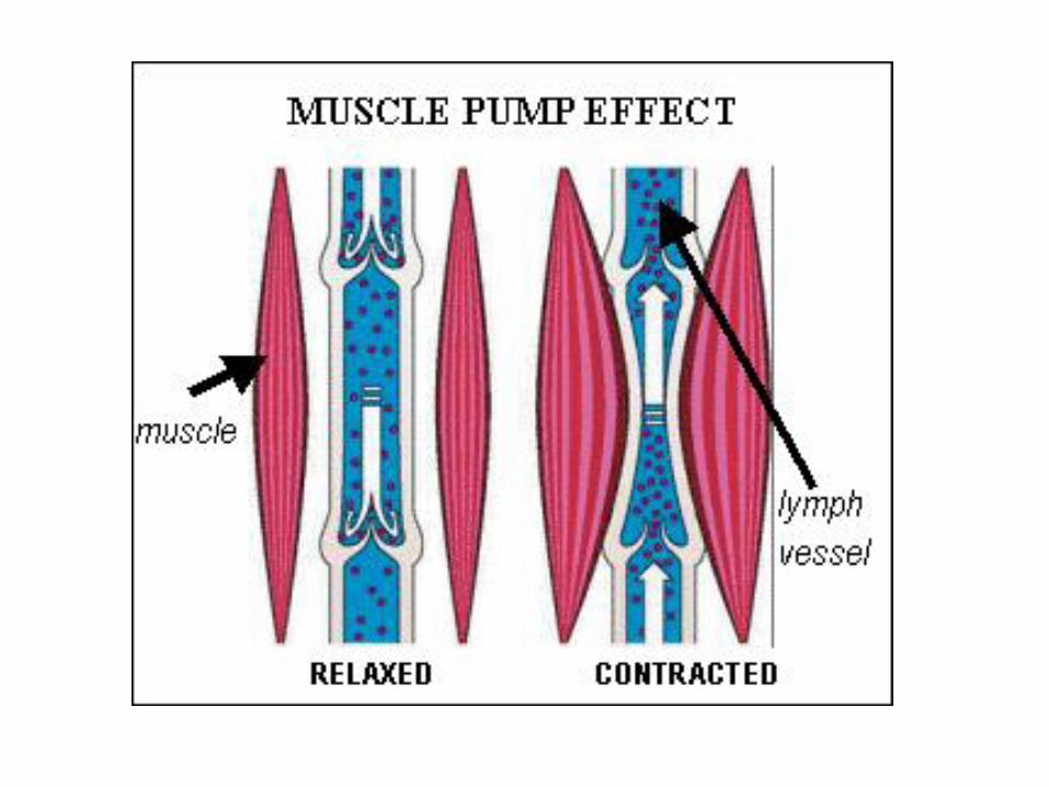
• Veins contain valves to help prevent
backflow of blood.
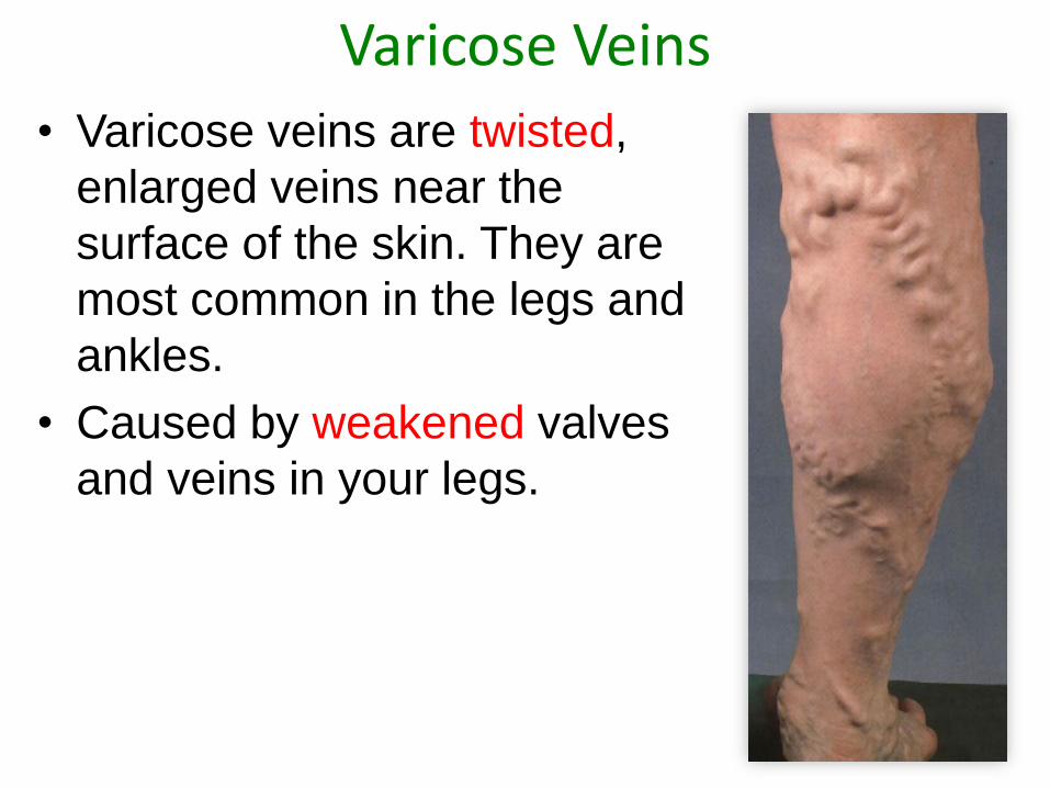
Varicose Veins
• Varicose veins are twisted,
enlarged veins near the
surface of the skin. They are
most common in the legs and
ankles.
• Caused by weakened valves
and veins in your legs.
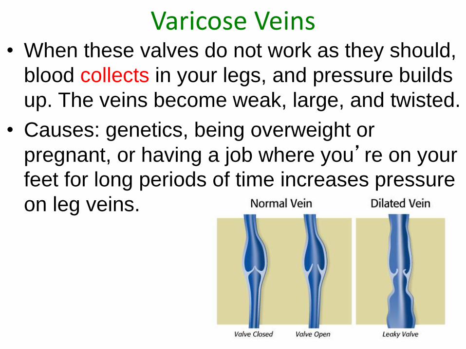
Varicose Veins• When these valves do not work as they should,
blood collects in your legs, and pressure builds
up. The veins become weak, large, and twisted.
• Causes: genetics, being overweight or
pregnant, or having a job where you’re on your
feet for long periods of time increases pressure
on leg veins.
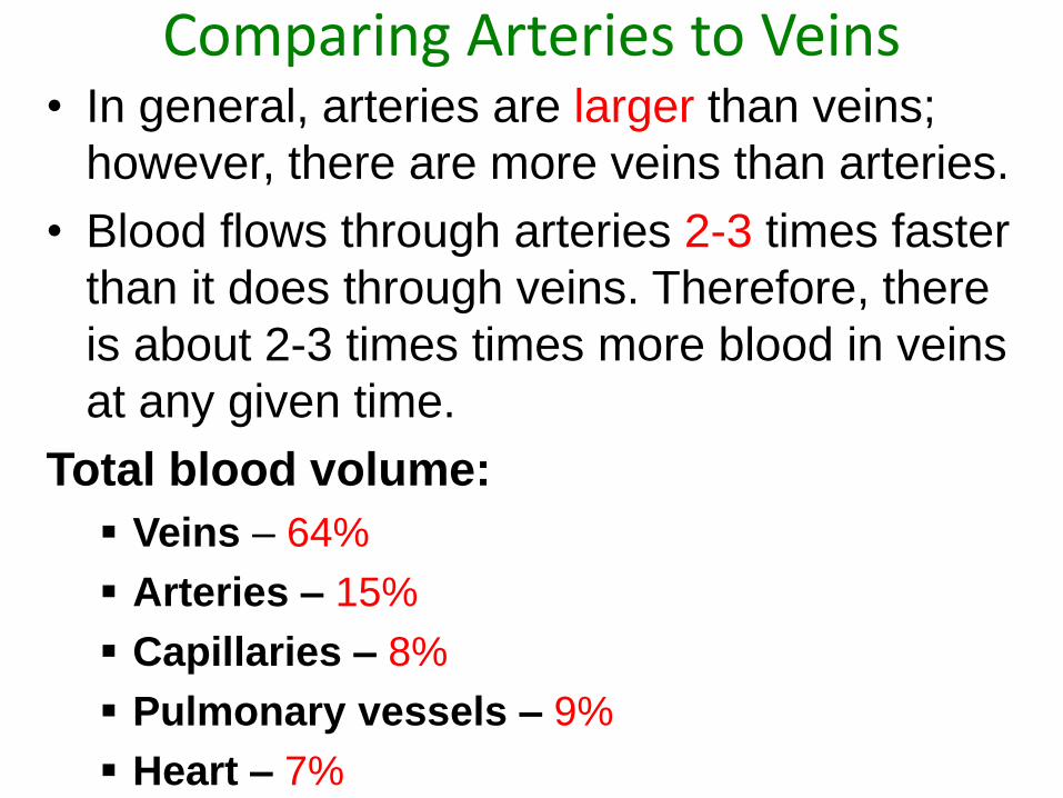
Comparing Arteries to Veins• In general, arteries are larger than veins;
however, there are more veins than arteries.
• Blood flows through arteries 2-3 times faster
than it does through veins. Therefore, there
is about 2-3 times times more blood in veins
at any given time.
Total blood volume:
Veins – 64%
Arteries – 15%
Capillaries – 8%
Pulmonary vessels – 9%
Heart – 7%
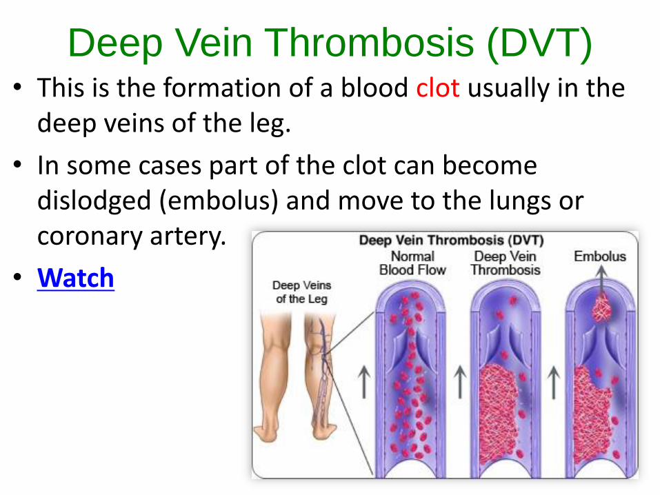
Deep Vein Thrombosis (DVT)• This is the formation of a blood clot usually in the
deep veins of the leg.
• In some cases part of the clot can become dislodged (embolus) and move to the lungs or coronary artery.
• Watch
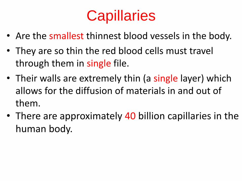
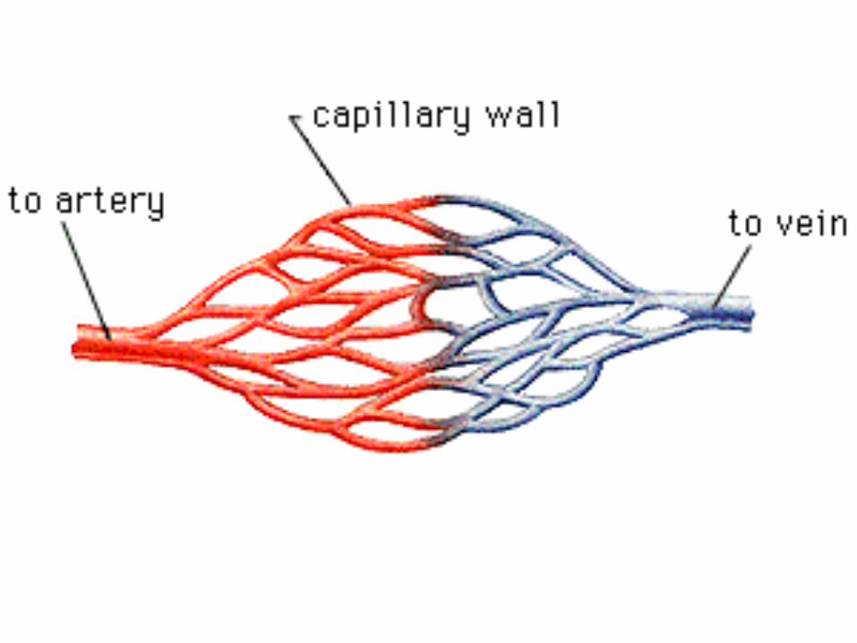
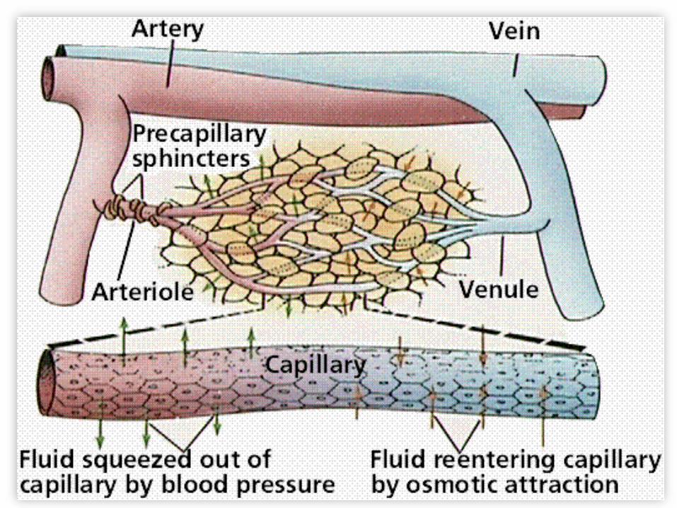
Capillaries
• Are the smallest thinnest blood vessels in the body.
• They are so thin the red blood cells must travel through them in single file.
• Their walls are extremely thin (a single layer) which allows for the diffusion of materials in and out of them.
• There are approximately 40 billion capillaries in the human body.
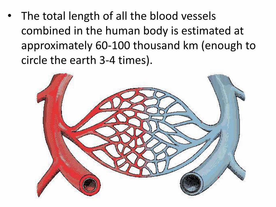
• The total length of all the blood vessels combined in the human body is estimated at approximately 60-100 thousand km (enough to circle the earth 3-4 times).
The Heart• The muscle responsible for pumping blood
throughout the circulatory system.
• Your heart is approximately the size of your fist.
• The muscle cells that comprise the heart are referred to as cardiac muscles.
• What is unique about cardiac muscle as compared to regular skeletal muscle?
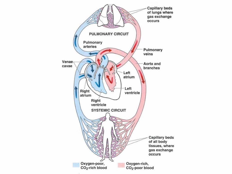
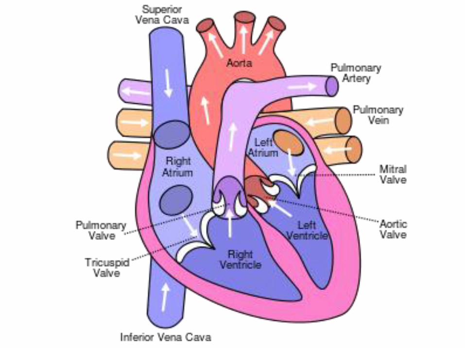
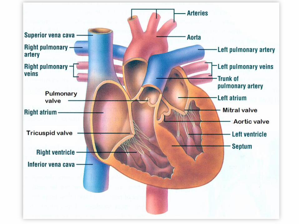
The Heart• A human heart has four chambers.
• This prevents the oxygen rich blood from
mixing with the oxygen poor blood.
• There are also several key valves inside
the heart that help to move the blood
along by preventing backflow.
• The two upper cambers are called atria
and the lower chambers are called
ventricles.
Chambers of the Heart• Top chambers: The Atrium
– Have thin walls and receive blood returning
through the veins to the heart.
• Bottom chambers: The Ventricles
– Have thick muscular walls and force blood out
of the heart into the arteries.
• Left atrium - The left atrium receives oxygen rich blood from the lungs and pumps it to the left ventricle.
• Left ventricle - The left ventricle receives blood from the left atrium and pumps it to the body through the aorta.
• Right atrium - The right atrium receives oxygen poor blood from the superior and inferior vena cava and pumps it to the right ventricle.
• Right ventricle - The right ventricle receives blood from the right atrium and pumps it to the lungs through the left pulmonary artery.
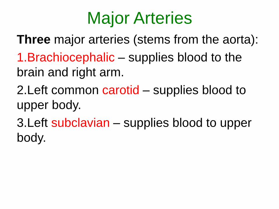
Major ArteriesThree major arteries (stems from the aorta):
1.Brachiocephalic – supplies blood to the
brain and right arm.
2.Left common carotid – supplies blood to
upper body.
3.Left subclavian – supplies blood to upper
body.
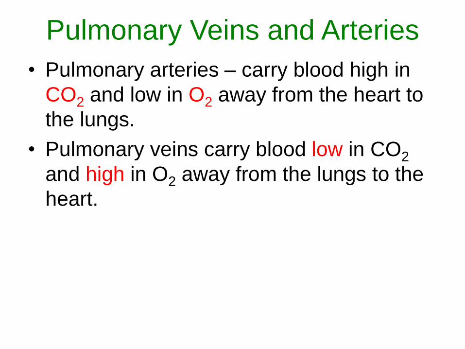
Pulmonary Veins and Arteries
• Pulmonary arteries – carry blood high in
CO2 and low in O2 away from the heart to
the lungs.
• Pulmonary veins carry blood low in CO2
and high in O2 away from the lungs to the
heart.
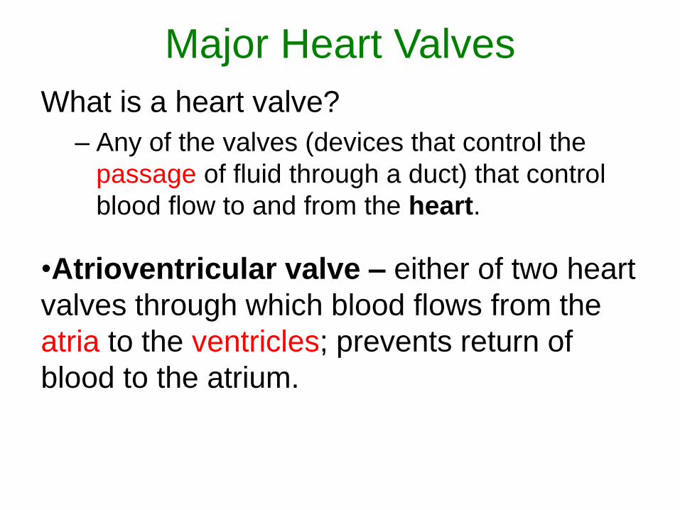
Major Heart Valves
What is a heart valve?
– Any of the valves (devices that control the
passage of fluid through a duct) that control
blood flow to and from the heart.
•Atrioventricular valve – either of two heart
valves through which blood flows from the
atria to the ventricles; prevents return of
blood to the atrium.
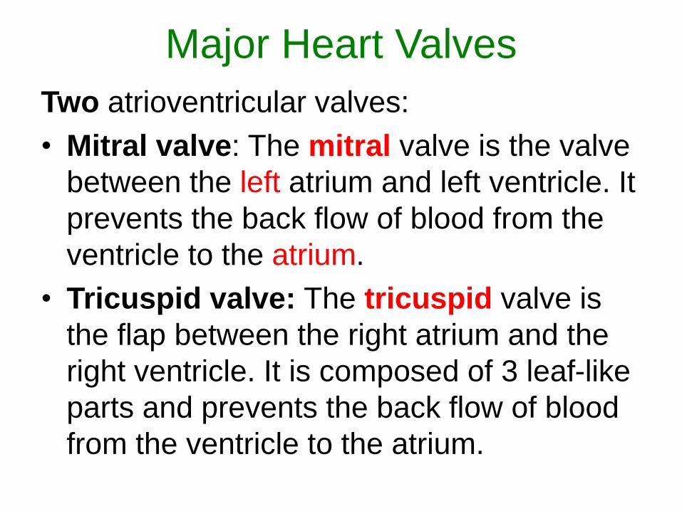
Major Heart Valves
Two atrioventricular valves:
• Mitral valve: The mitral valve is the valve
between the left atrium and left ventricle. It
prevents the back flow of blood from the
ventricle to the atrium.
• Tricuspid valve: The tricuspid valve is
the flap between the right atrium and the
right ventricle. It is composed of 3 leaf-like
parts and prevents the back flow of blood
from the ventricle to the atrium.
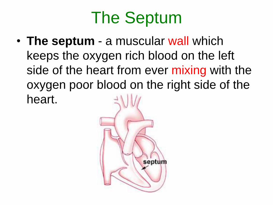
The Septum
• The septum - a muscular wall which
keeps the oxygen rich blood on the left
side of the heart from ever mixing with the
oxygen poor blood on the right side of the
heart.
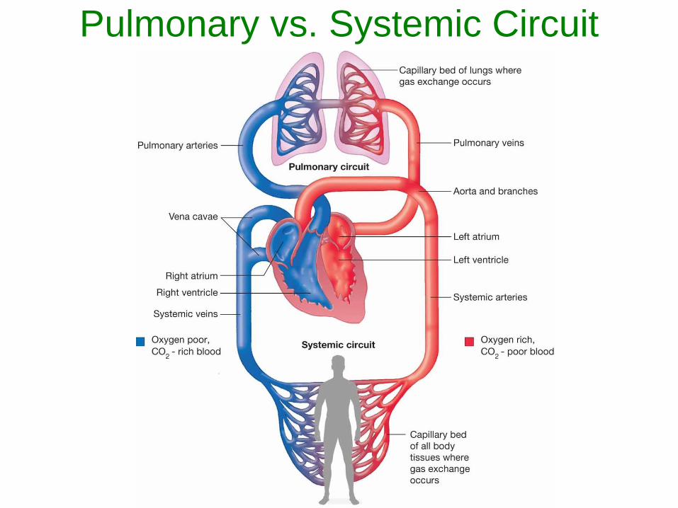
Pulmonary vs. Systemic Circuit
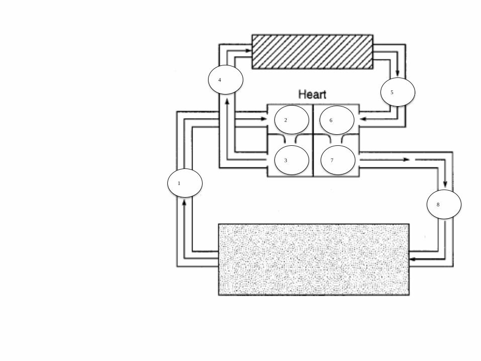
1
3 7
4
2 6
5
8
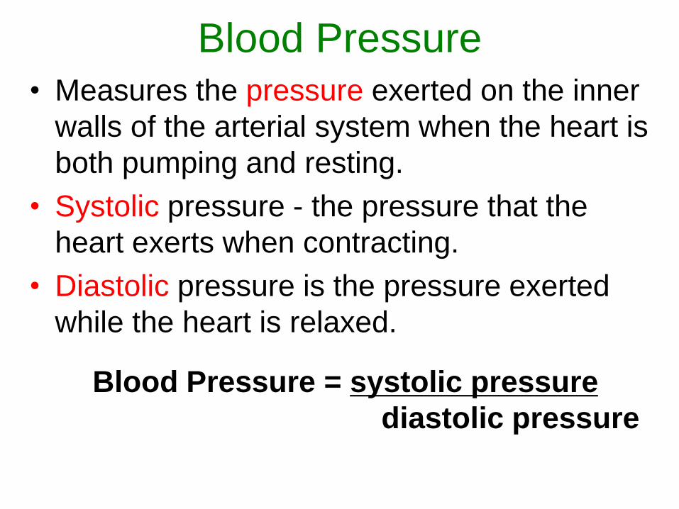
Blood Pressure• Measures the pressure exerted on the inner
walls of the arterial system when the heart is
both pumping and resting.
• Systolic pressure - the pressure that the
heart exerts when contracting.
• Diastolic pressure is the pressure exerted
while the heart is relaxed.
Blood Pressure = systolic pressure
diastolic pressure
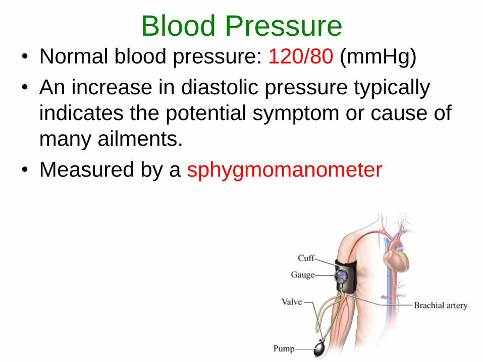
Blood Pressure• Normal blood pressure: 120/80 (mmHg)
• An increase in diastolic pressure typically
indicates the potential symptom or cause of
many ailments.
• Measured by a sphygmomanometer
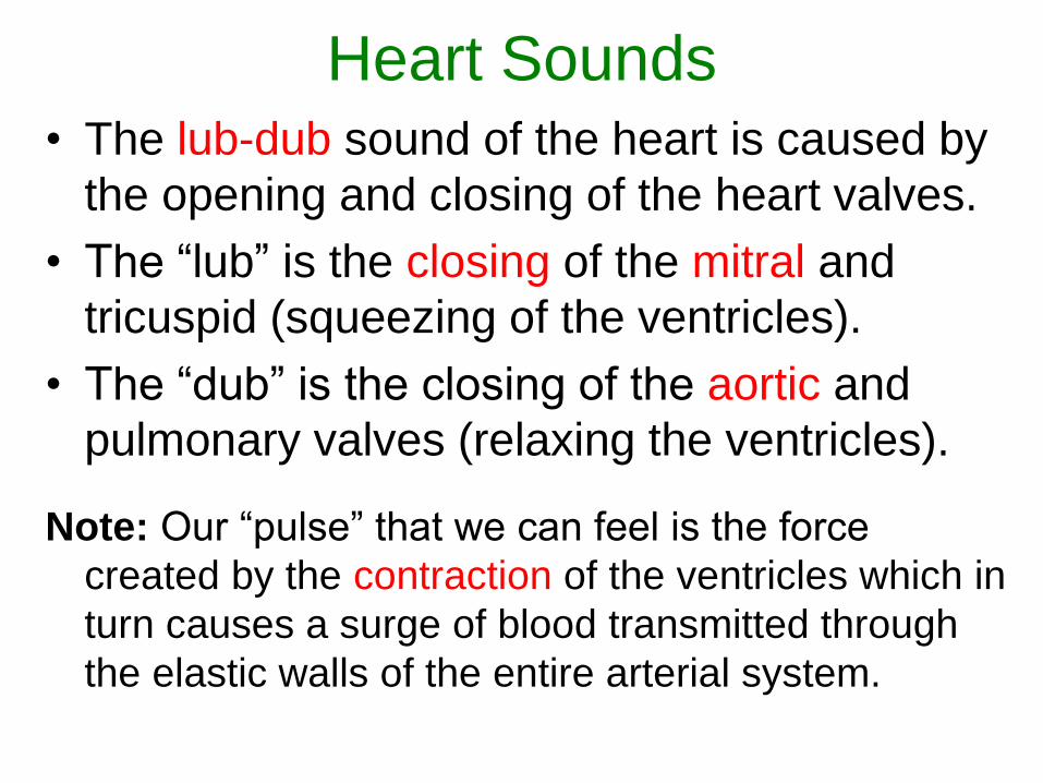
Heart Sounds• The lub-dub sound of the heart is caused by
the opening and closing of the heart valves.
• The “lub” is the closing of the mitral and
tricuspid (squeezing of the ventricles).
• The “dub” is the closing of the aortic and
pulmonary valves (relaxing the ventricles).
Note: Our “pulse” that we can feel is the force
created by the contraction of the ventricles which in
turn causes a surge of blood transmitted through
the elastic walls of the entire arterial system.
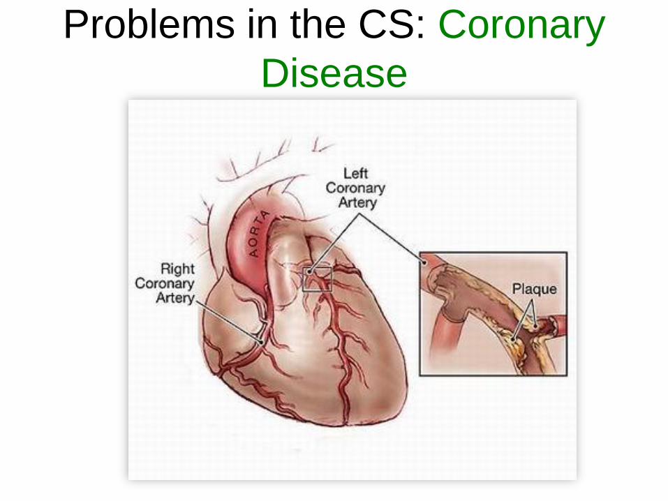
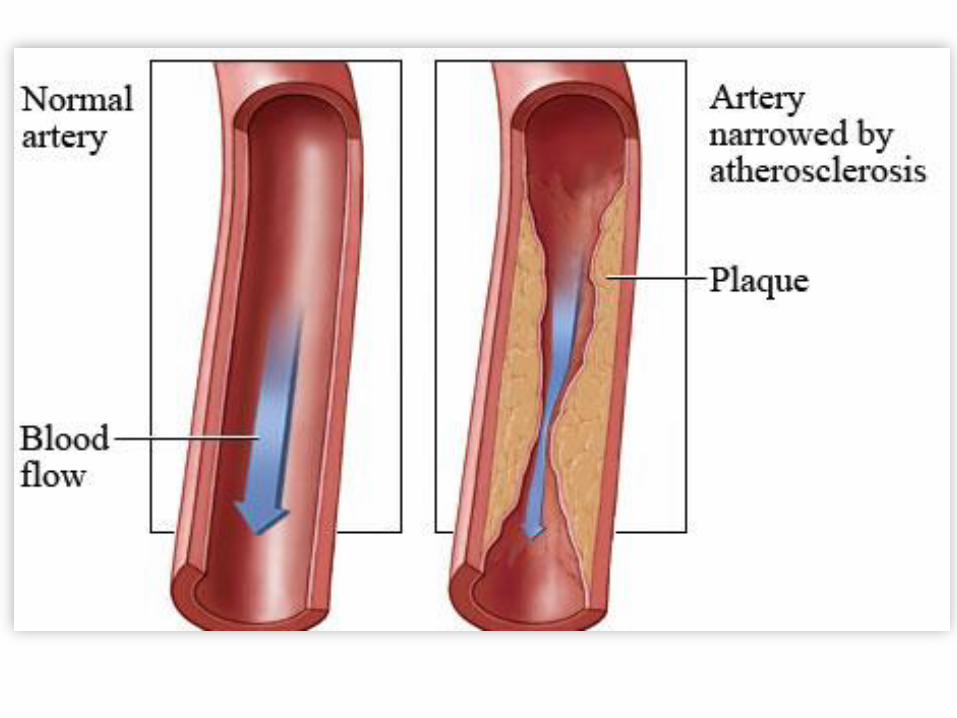
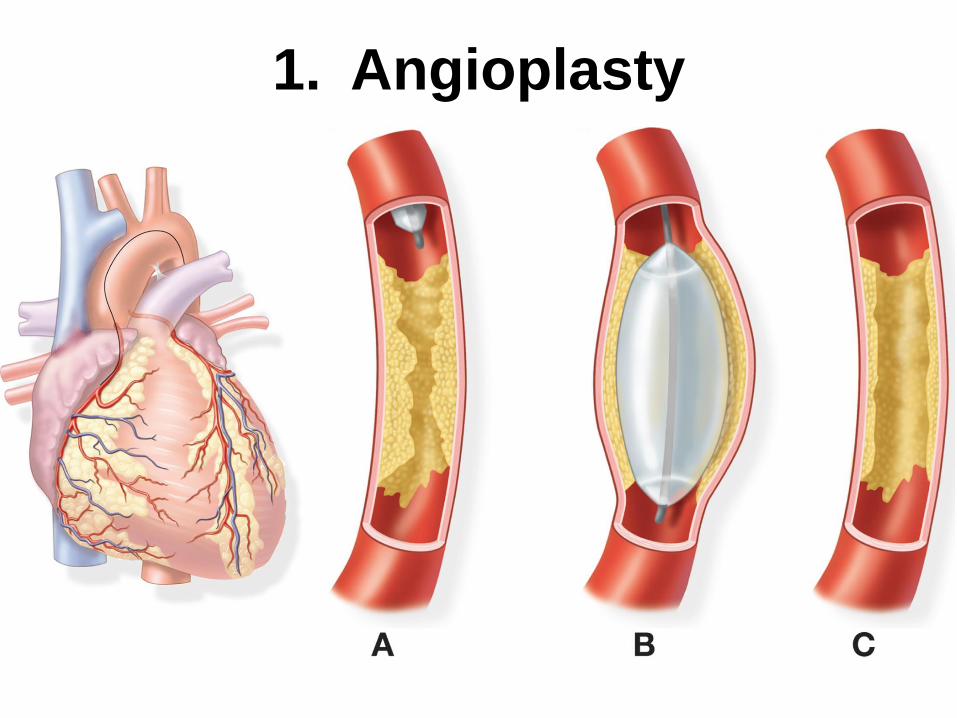
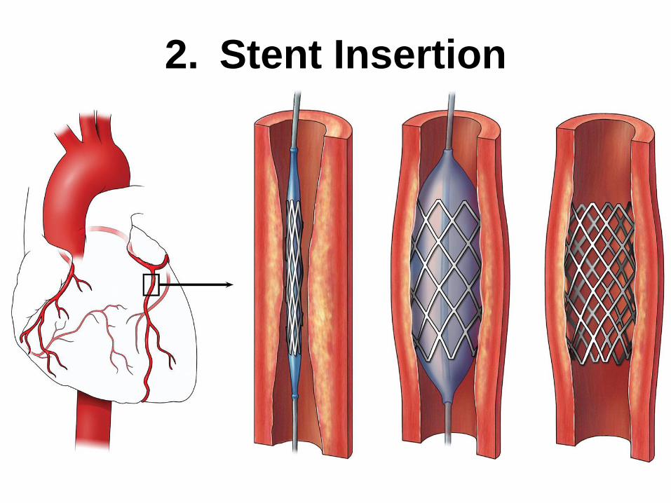
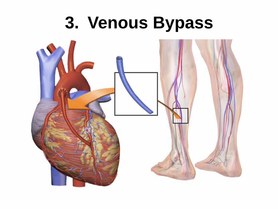
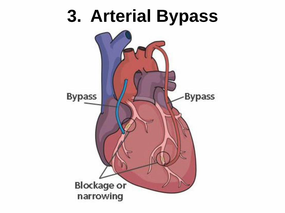
Problems in the CS: Coronary
Disease
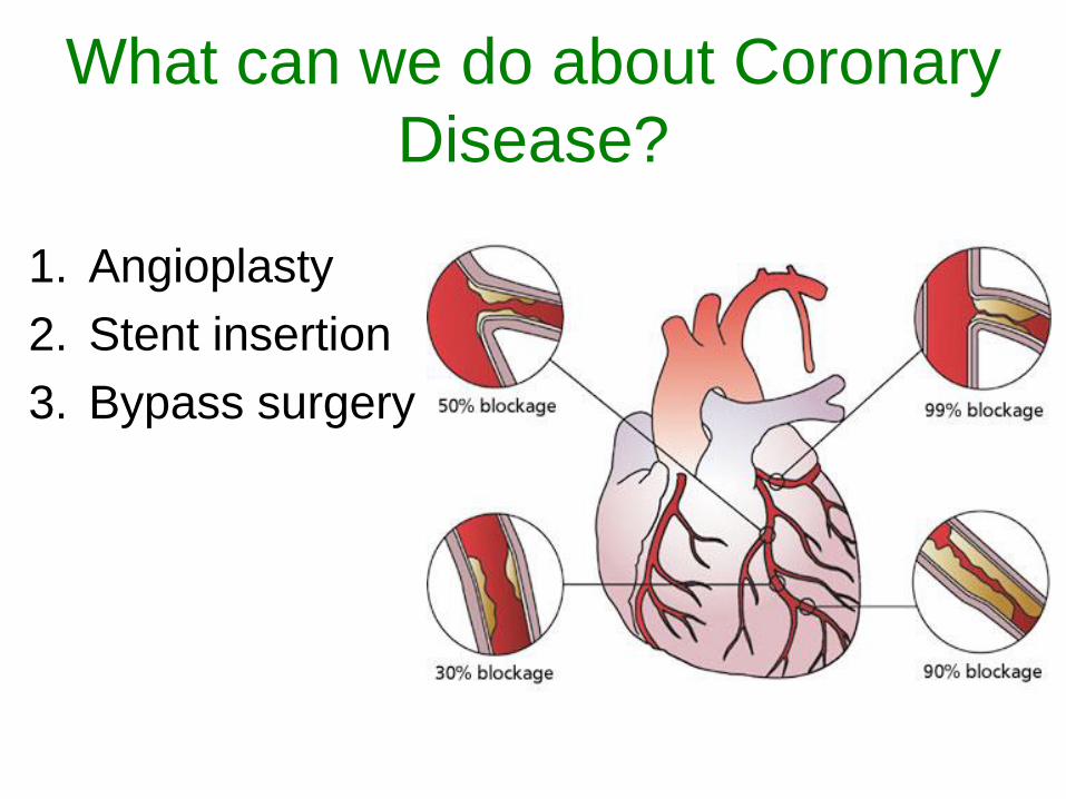
What can we do about Coronary
Disease?
1. Angioplasty
2. Stent insertion
3. Bypass surgery
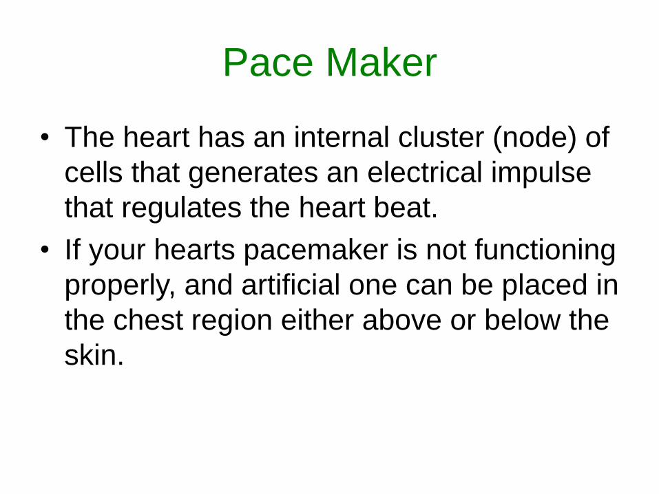
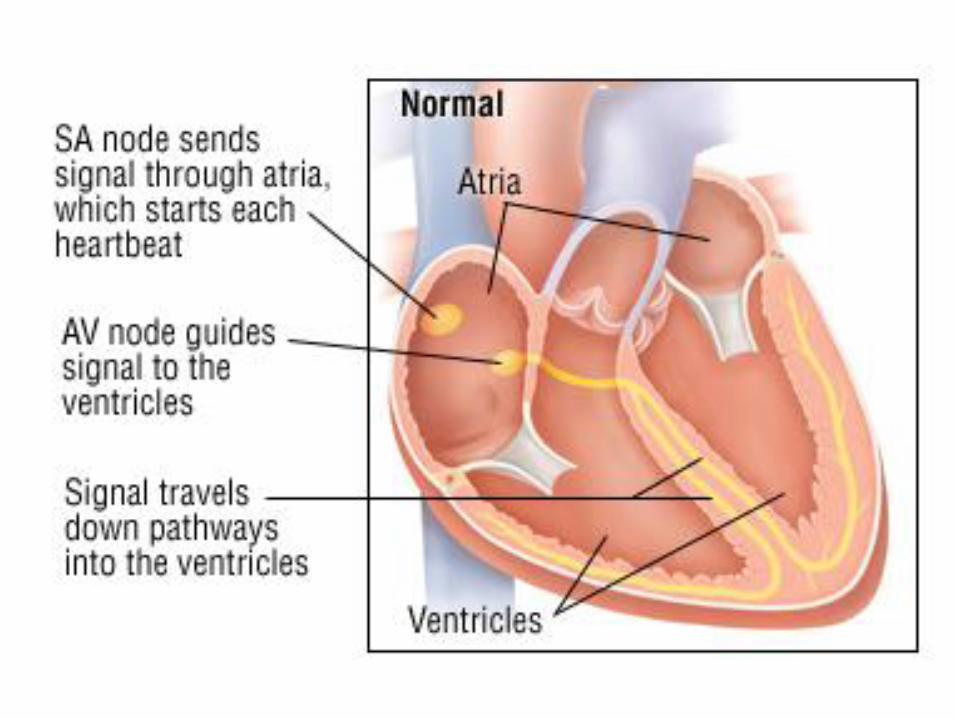
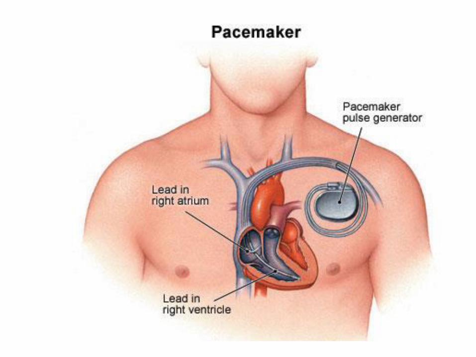
Pace Maker
• The heart has an internal cluster (node) of
cells that generates an electrical impulse
that regulates the heart beat.
• If your hearts pacemaker is not functioning
properly, and artificial one can be placed in
the chest region either above or below the
skin.
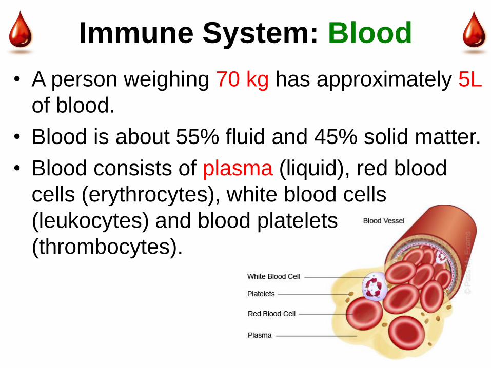
Immune System: Blood
• A person weighing 70 kg has approximately 5L
of blood.
• Blood is about 55% fluid and 45% solid matter.
• Blood consists of plasma (liquid), red blood
cells (erythrocytes), white blood cells
(leukocytes) and blood platelets
(thrombocytes).
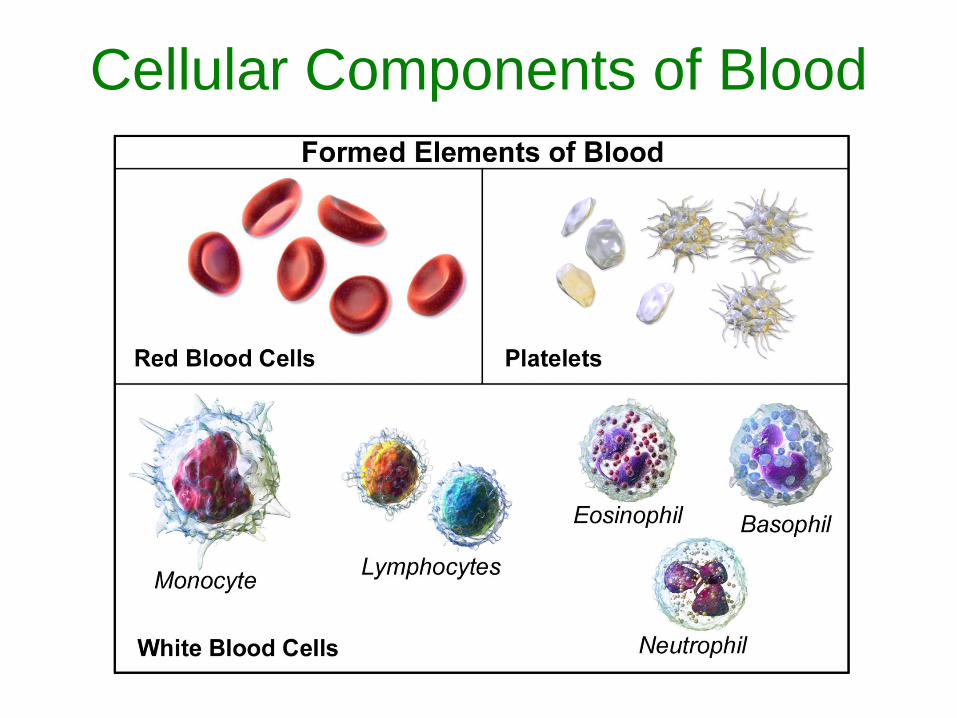
Cellular Components of Blood
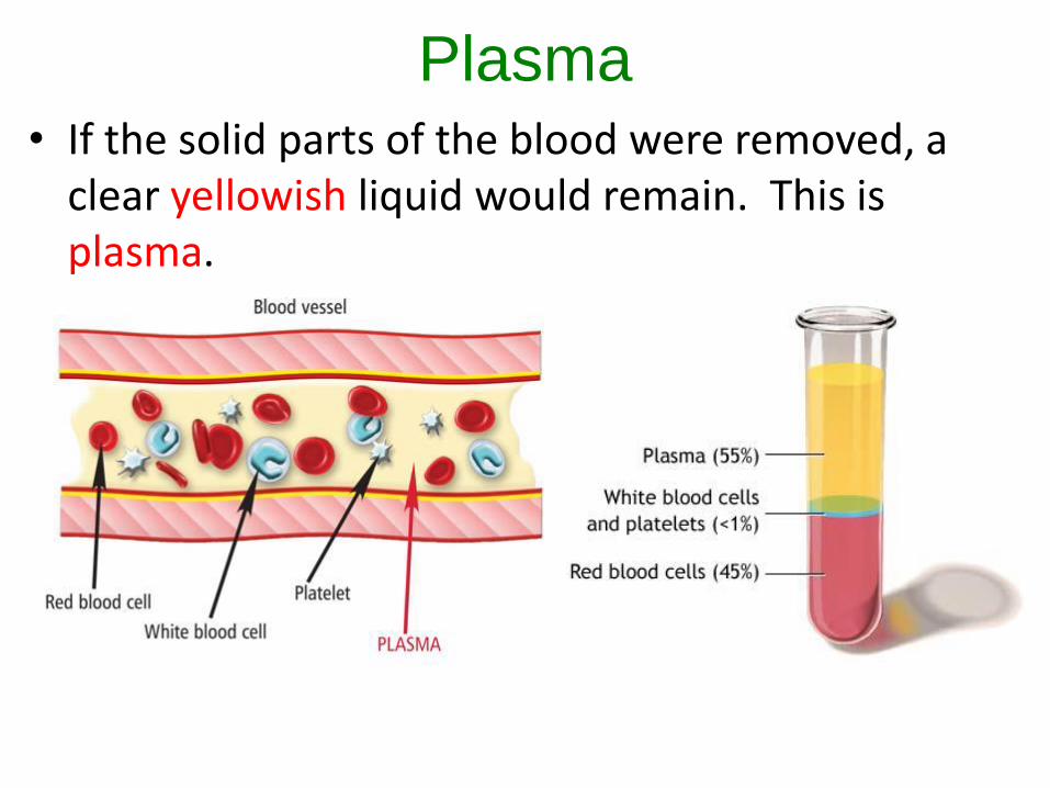
Plasma• If the solid parts of the blood were removed, a
clear yellowish liquid would remain. This is plasma.
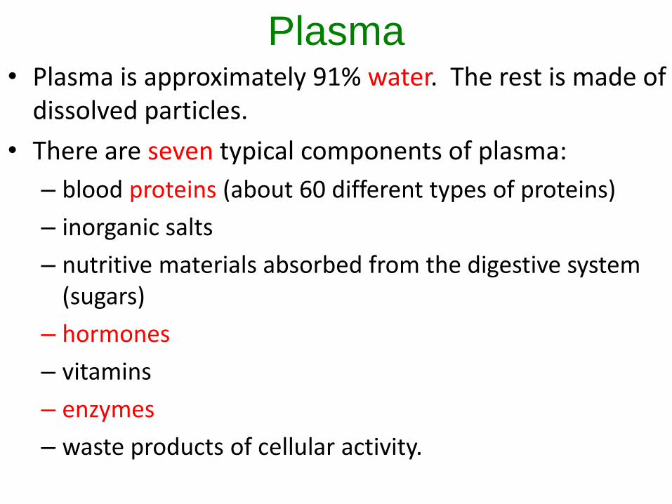
Plasma• Plasma is approximately 91% water. The rest is made of
dissolved particles.
• There are seven typical components of plasma:
– blood proteins (about 60 different types of proteins)
– inorganic salts
– nutritive materials absorbed from the digestive system (sugars)
– hormones
– vitamins
– enzymes
– waste products of cellular activity.
Proteins Dissolved in Blood
There are three groups of blood proteins found
in blood:
1. Albumins – establish osmotic pressure that
draws water back into capillaries which helps
maintain body fluid levels.
2. Globulins – produce antibodies that provide
protection against invading microbes.
3. Fibrinogens – important in blood clotting.
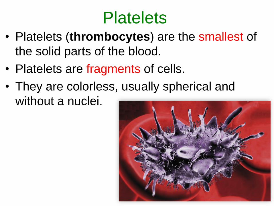
Platelets• Platelets (thrombocytes) are the smallest of
the solid parts of the blood.
• Platelets are fragments of cells.
• They are colorless, usually spherical and
without a nuclei.
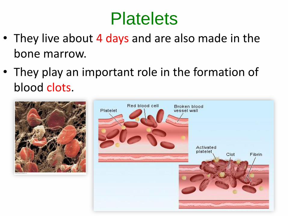
Platelets• They live about 4 days and are also made in the
bone marrow.
• They play an important role in the formation of blood clots.

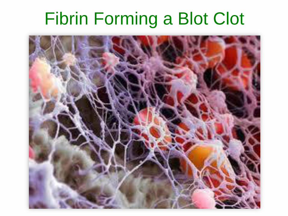
Clotting• When rough edges of a cut or torn vessel are encountered,
platelets stick to the rough edges and break up.
• This causes the release of thromboplastin. When calcium is present in the blood, thromboplastin acts as an enzyme to activate prothrombin, a plasma protein.
• Prothrombin changes to thrombin.
• Thrombin acts as an enzyme to convert fibrinogen into fibrin, an insoluble substance that forms threads within the plasma.
• Blood cells are trapped in the network of fibrin threads, building a clot.
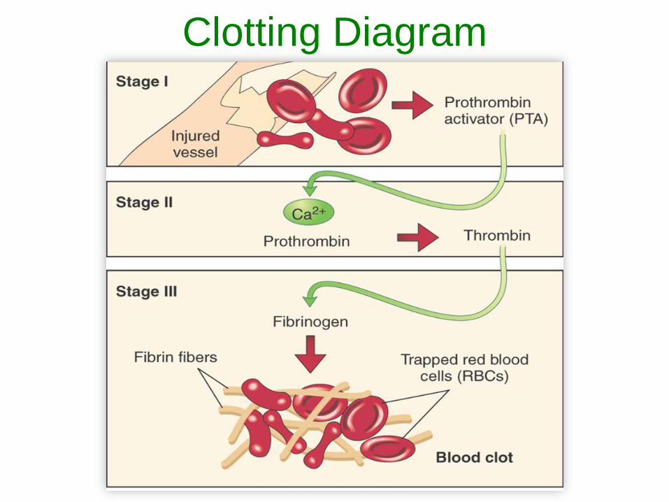
Clotting Diagram
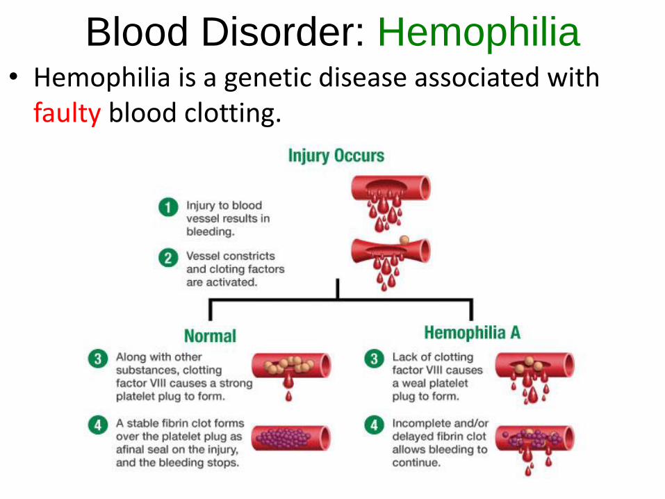
Blood Disorder: Hemophilia• Hemophilia is a genetic disease associated with
faulty blood clotting.
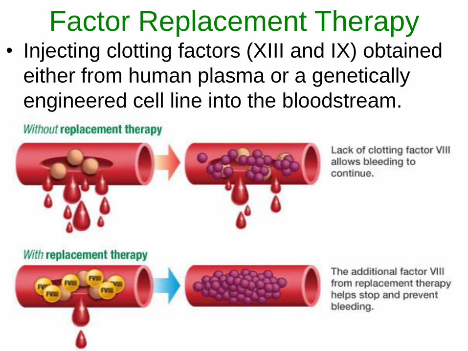
Factor Replacement Therapy• Injecting clotting factors (XIII and IX) obtained
either from human plasma or a genetically
engineered cell line into the bloodstream.
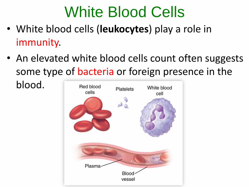
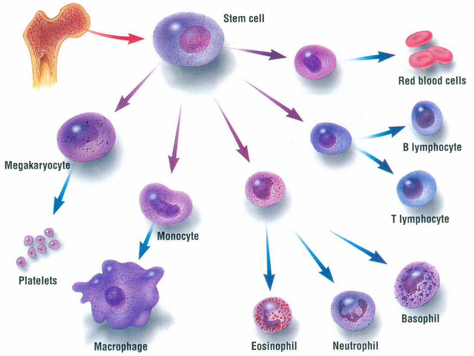
White Blood Cells• White blood cells (leukocytes) play a role in
immunity.
• An elevated white blood cells count often suggests some type of bacteria or foreign presence in the blood.
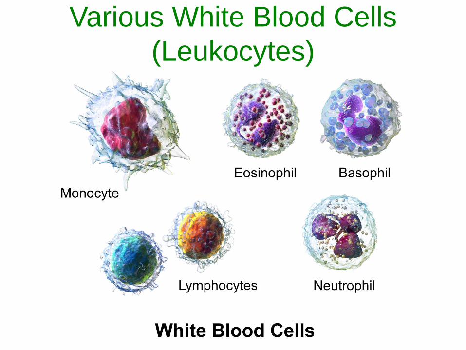
Various White Blood Cells
(Leukocytes)
White Blood Cells
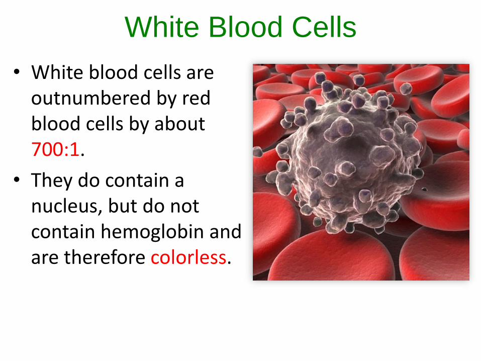
• White blood cells are outnumbered by red blood cells by about 700:1.
• They do contain a nucleus, but do not contain hemoglobin and are therefore colorless.
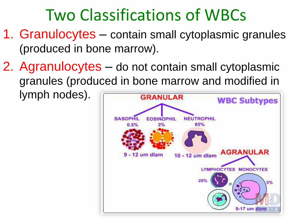
Two Classifications of WBCs1. Granulocytes – contain small cytoplasmic granules
(produced in bone marrow).
2. Agranulocytes – do not contain small cytoplasmic
granules (produced in bone marrow and modified in
lymph nodes).
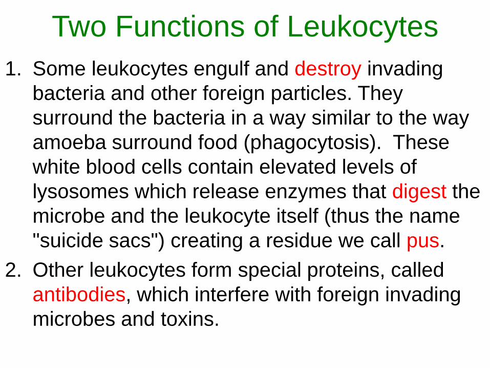
Two Functions of Leukocytes
1. Some leukocytes engulf and destroy invading
bacteria and other foreign particles. They
surround the bacteria in a way similar to the way
amoeba surround food (phagocytosis). These
white blood cells contain elevated levels of
lysosomes which release enzymes that digest the
microbe and the leukocyte itself (thus the name
"suicide sacs") creating a residue we call pus.
2. Other leukocytes form special proteins, called
antibodies, which interfere with foreign invading
microbes and toxins.
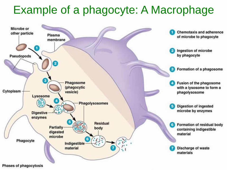
Example of a phagocyte: A Macrophage
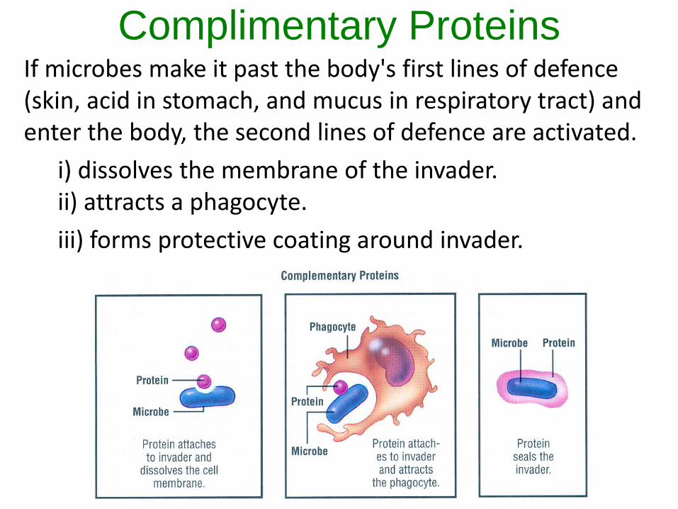
Complimentary ProteinsIf microbes make it past the body's first lines of defence (skin, acid in stomach, and mucus in respiratory tract) and enter the body, the second lines of defence are activated.
i) dissolves the membrane of the invader.ii) attracts a phagocyte.
iii) forms protective coating around invader.
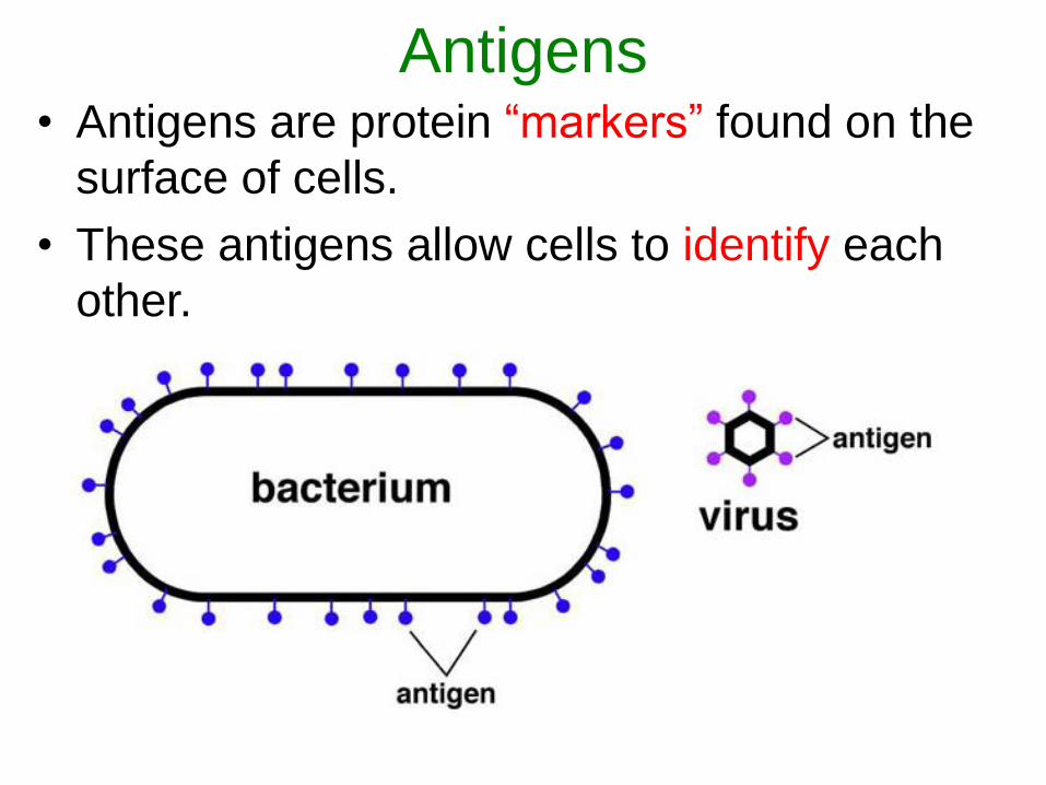
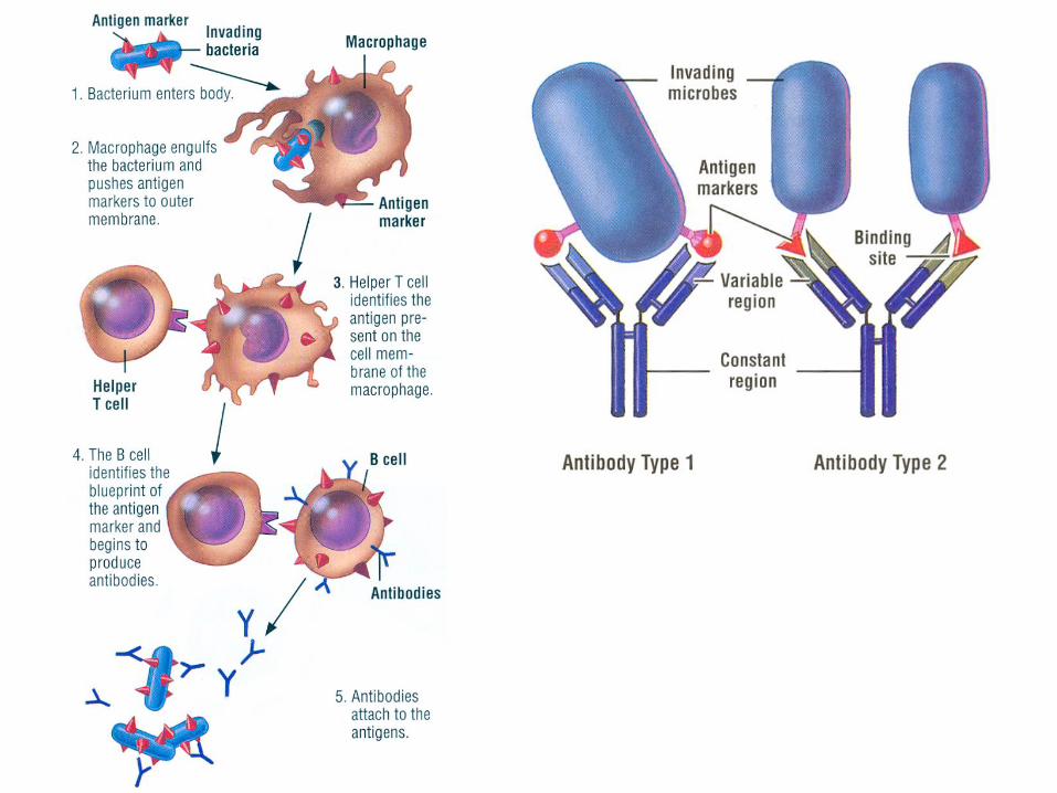
Antigens• Antigens are protein “markers” found on the
surface of cells.
• These antigens allow cells to identify each
other.
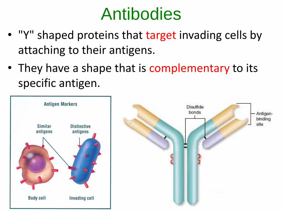
Antibodies• "Y" shaped proteins that target invading cells by
attaching to their antigens.
• They have a shape that is complementary to its specific antigen.
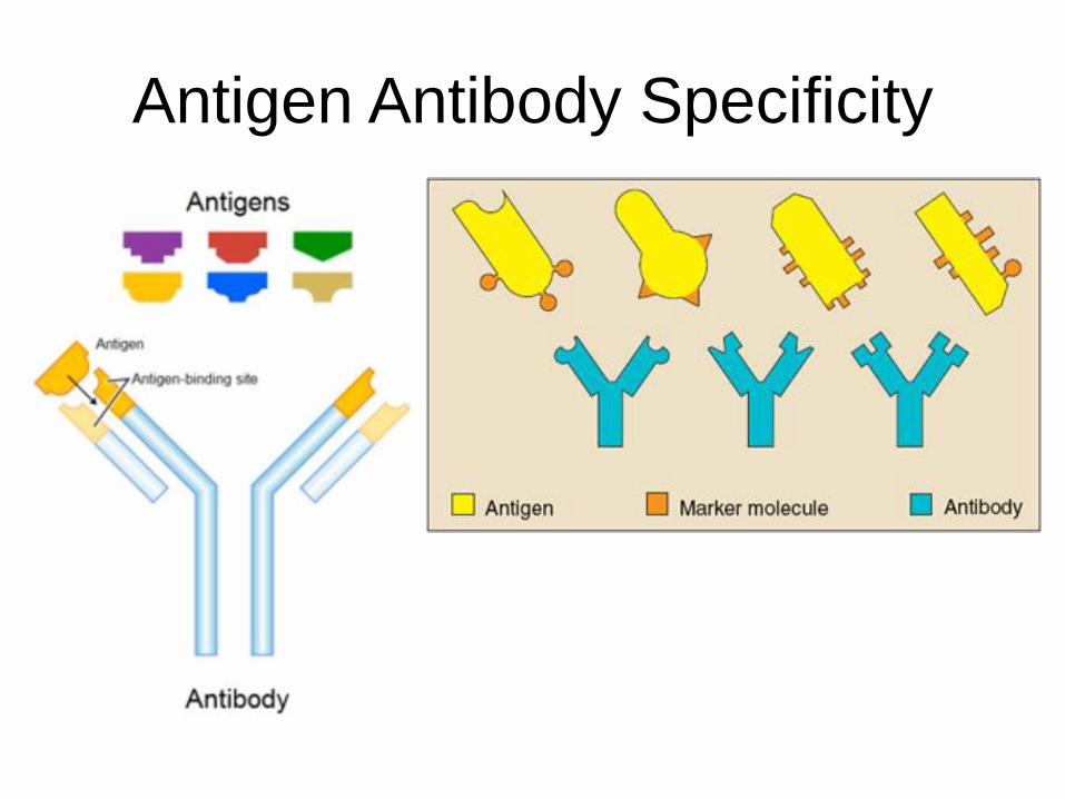
Antigen Antibody Specificity
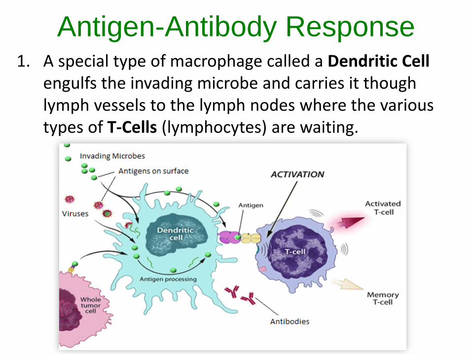
Antigen-Antibody Response1. A special type of macrophage called a Dendritic Cell
engulfs the invading microbe and carries it though lymph vessels to the lymph nodes where the various types of T-Cells (lymphocytes) are waiting.
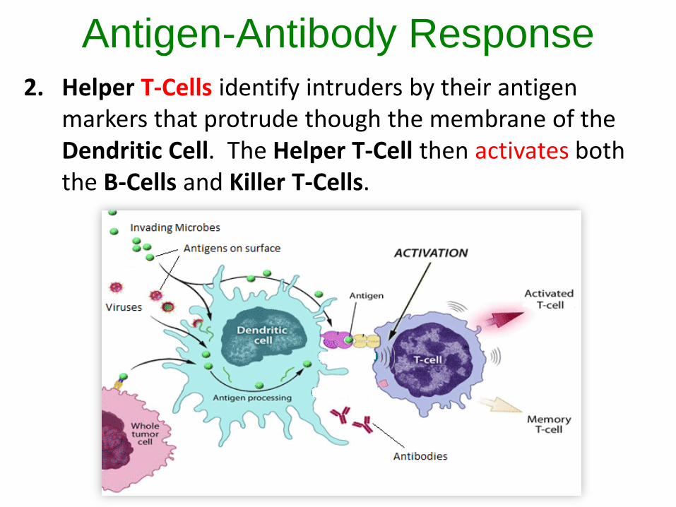
Antigen-Antibody Response2. Helper T-Cells identify intruders by their antigen
markers that protrude though the membrane of the Dendritic Cell. The Helper T-Cell then activates both the B-Cells and Killer T-Cells.

Antigen-Antibody Response3. The B-Cells use the antigen marker information to
produce antibodies. The antibodies head to the
“battle field” to latch on to invading microbes
antigens. They are then engulfed by a macrophage.
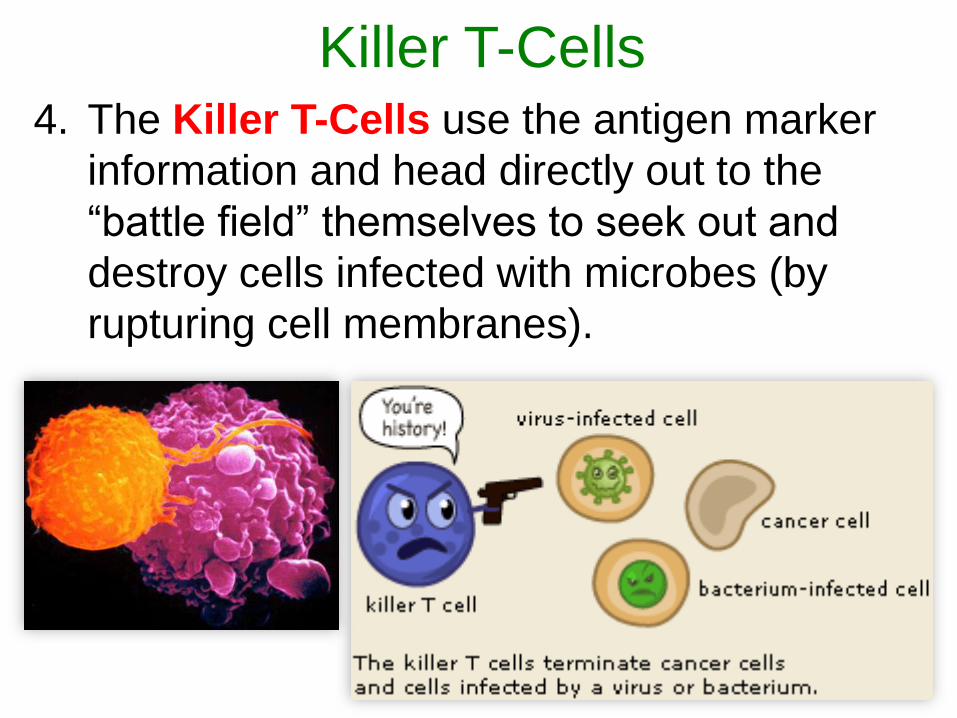
Killer T-Cells4. The Killer T-Cells use the antigen marker
information and head directly out to the
“battle field” themselves to seek out and
destroy cells infected with microbes (by
rupturing cell membranes).
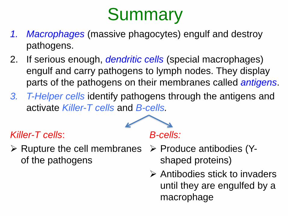
Summary1. Macrophages (massive phagocytes) engulf and destroy
pathogens.
2. If serious enough, dendritic cells (special macrophages)
engulf and carry pathogens to lymph nodes. They display
parts of the pathogens on their membranes called antigens.
3. T-Helper cells identify pathogens through the antigens and
activate Killer-T cells and B-cells.
Killer-T cells:
Rupture the cell membranes
of the pathogens
B-cells:
Produce antibodies (Y-
shaped proteins)
Antibodies stick to invaders
until they are engulfed by a
macrophage
Red Blood Cells
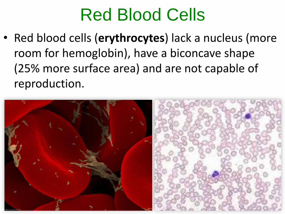
• Red blood cells (erythrocytes) lack a nucleus (more room for hemoglobin), have a biconcave shape (25% more surface area) and are not capable of reproduction.
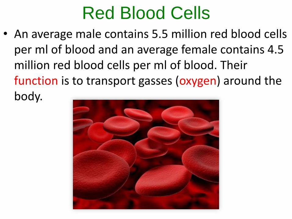
Red Blood Cells• An average male contains 5.5 million red blood cells
per ml of blood and an average female contains 4.5 million red blood cells per ml of blood. Their function is to transport gasses (oxygen) around the body.
Red Blood Cells
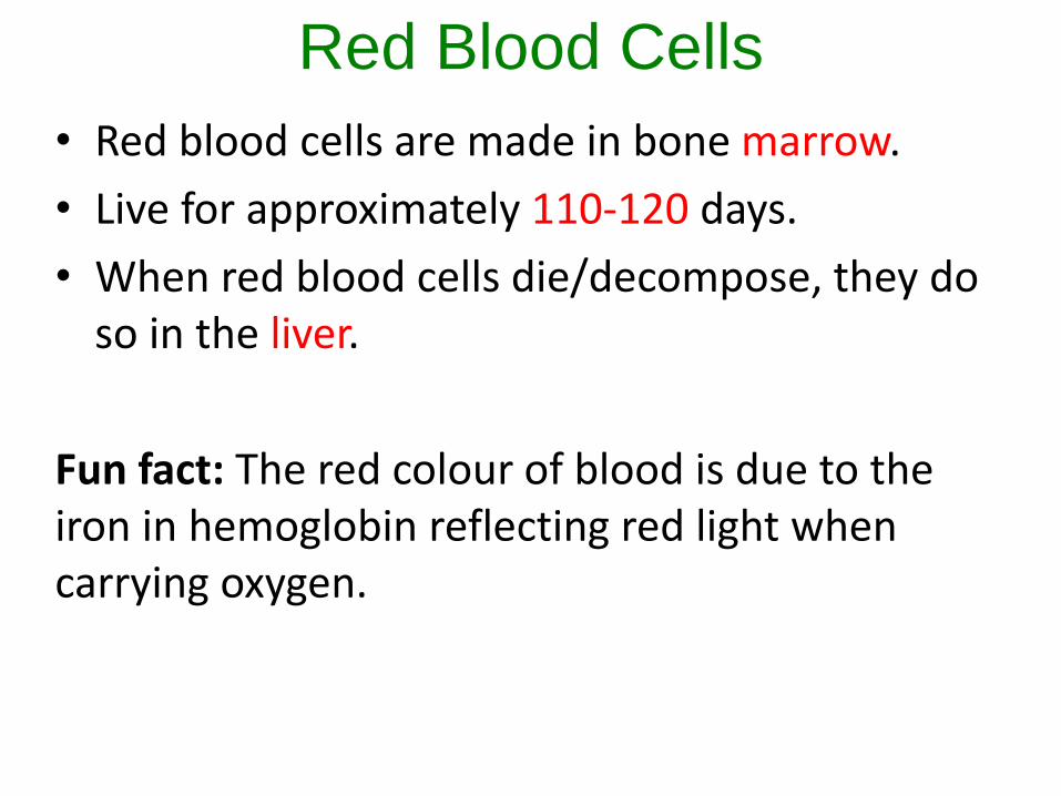
• Red blood cells are made in bone marrow.
• Live for approximately 110-120 days.
• When red blood cells die/decompose, they do so in the liver.
Fun fact: The red colour of blood is due to the iron in hemoglobin reflecting red light when carrying oxygen.
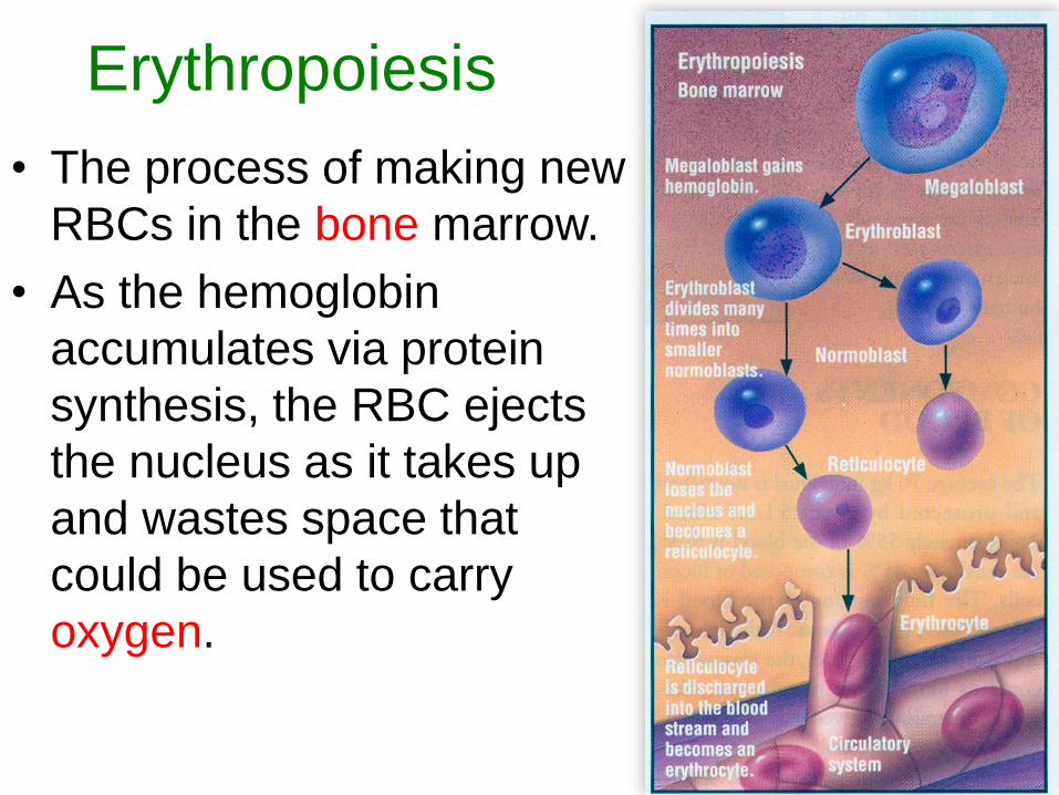
Erythropoiesis
• The process of making new
RBCs in the bone marrow.
• As the hemoglobin
accumulates via protein
synthesis, the RBC ejects
the nucleus as it takes up
and wastes space that
could be used to carry
oxygen.
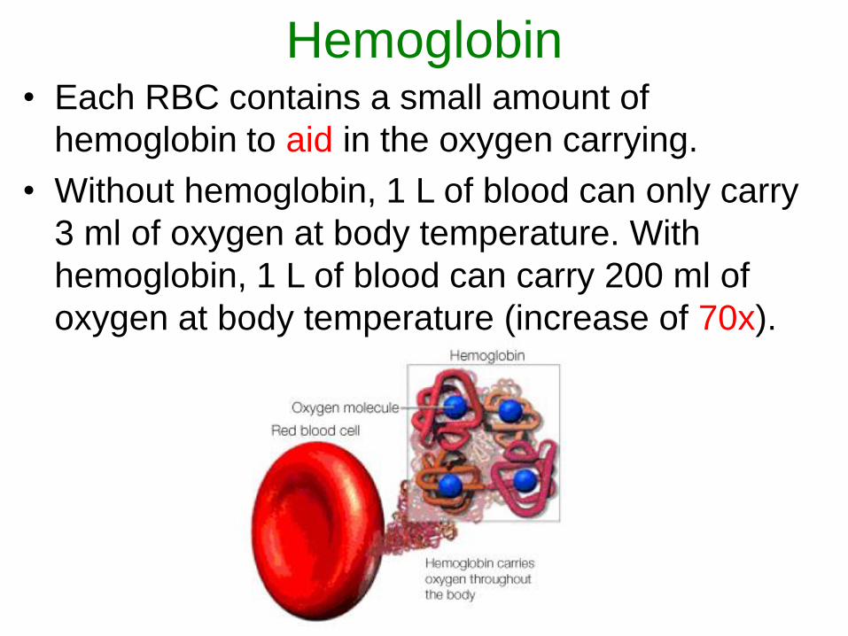
Hemoglobin• Each RBC contains a small amount of
hemoglobin to aid in the oxygen carrying.
• Without hemoglobin, 1 L of blood can only carry
3 ml of oxygen at body temperature. With
hemoglobin, 1 L of blood can carry 200 ml of
oxygen at body temperature (increase of 70x).
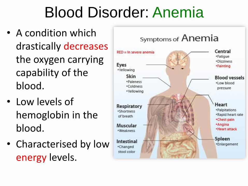
Blood Disorder: Anemia
• A condition which drastically decreasesthe oxygen carrying capability of the blood.
• Low levels of hemoglobin in the blood.
• Characterised by low energy levels.
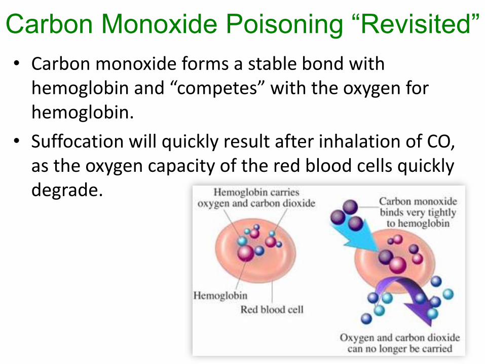
Carbon Monoxide Poisoning “Revisited”
• Carbon monoxide forms a stable bond with hemoglobin and “competes” with the oxygen for hemoglobin.
• Suffocation will quickly result after inhalation of CO, as the oxygen capacity of the red blood cells quickly degrade.
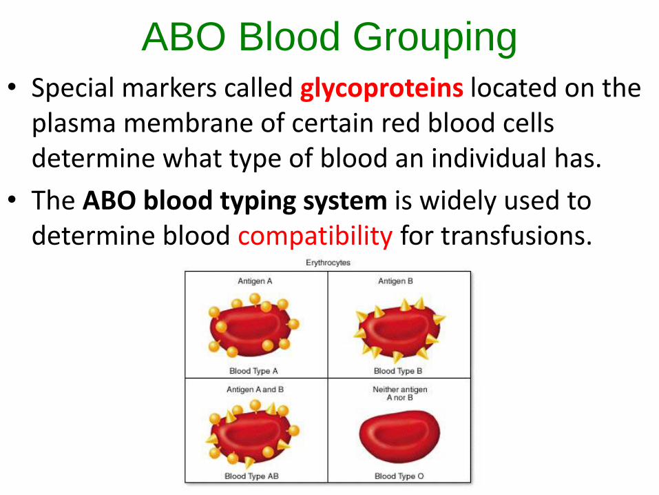
ABO Blood Grouping• Special markers called glycoproteins located on the
plasma membrane of certain red blood cells determine what type of blood an individual has.
• The ABO blood typing system is widely used to determine blood compatibility for transfusions.
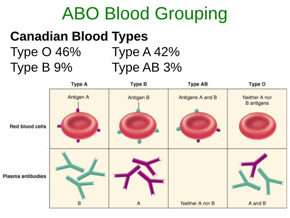
ABO Blood Grouping
Canadian Blood Types
Type O 46% Type A 42%
Type B 9% Type AB 3%
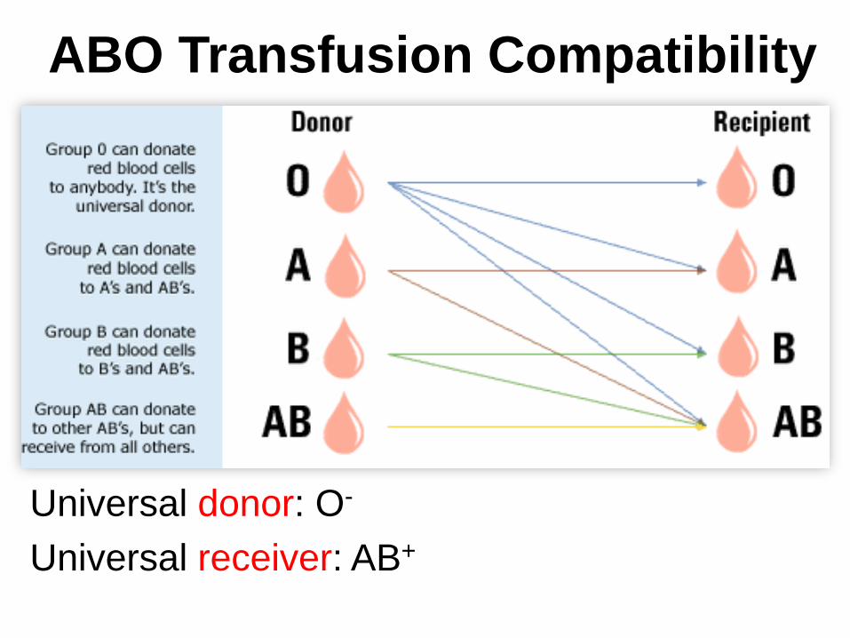
ABO Transfusion Compatibility
Universal donor: O-
Universal receiver: AB+
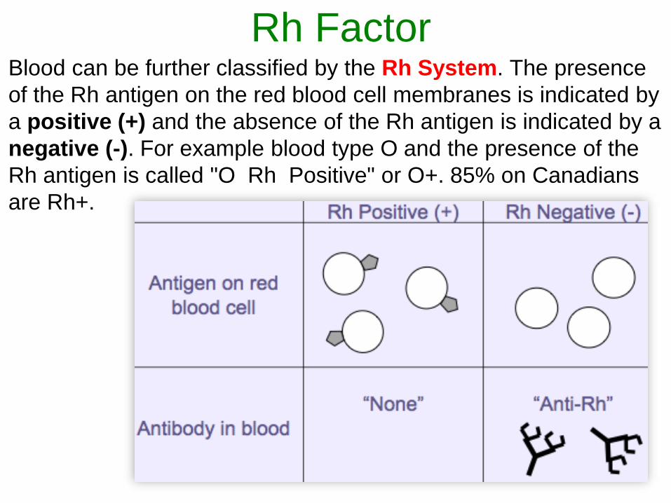
Rh FactorBlood can be further classified by the Rh System. The presence
of the Rh antigen on the red blood cell membranes is indicated by
a positive (+) and the absence of the Rh antigen is indicated by a
negative (-). For example blood type O and the presence of the
Rh antigen is called "O Rh Positive" or O+. 85% on Canadians
are Rh+.
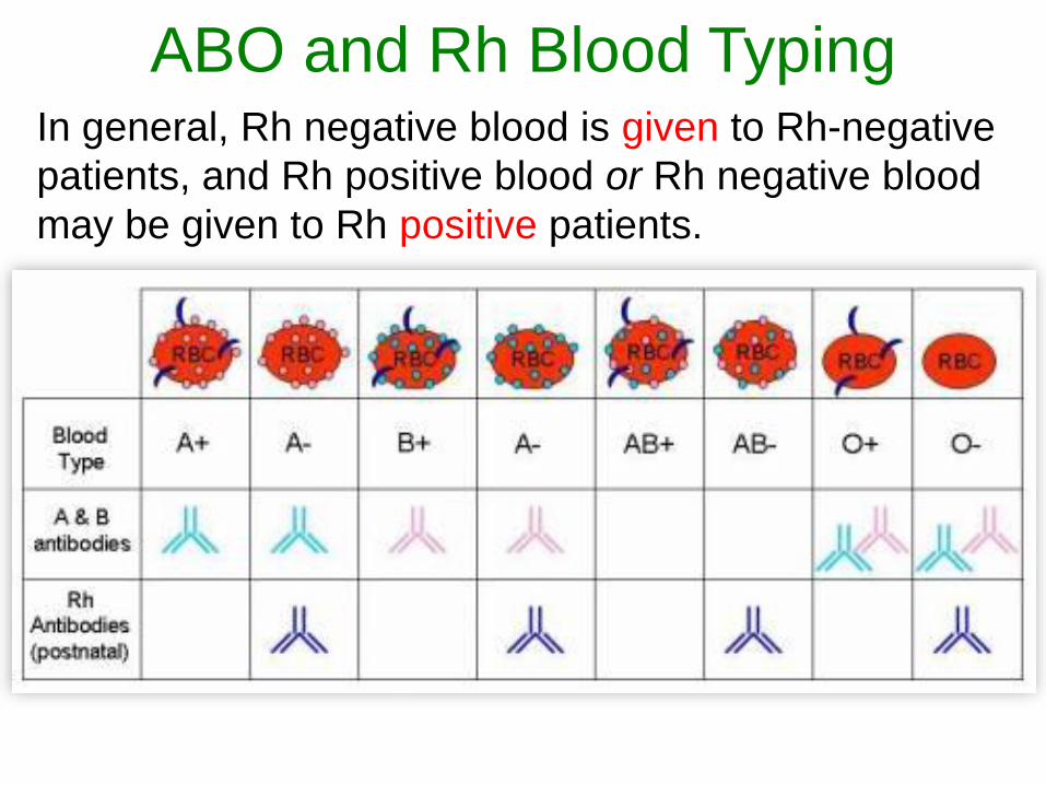
ABO and Rh Blood TypingIn general, Rh negative blood is given to Rh-negative
patients, and Rh positive blood or Rh negative blood
may be given to Rh positive patients.
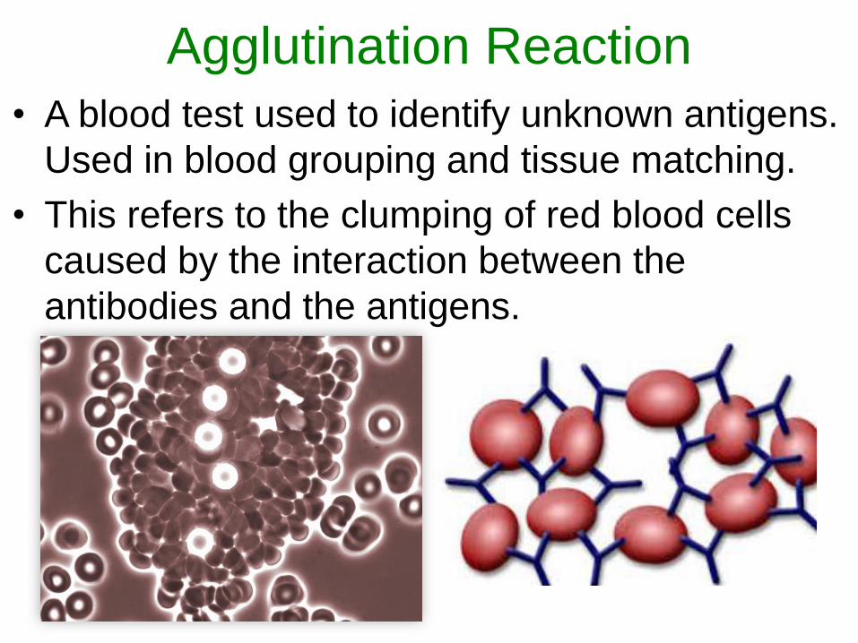
Agglutination Reaction
• A blood test used to identify unknown antigens.
Used in blood grouping and tissue matching.
• This refers to the clumping of red blood cells
caused by the interaction between the
antibodies and the antigens.
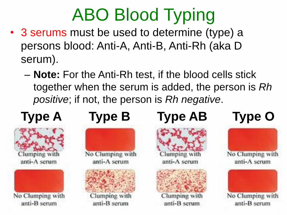
ABO Blood Typing• 3 serums must be used to determine (type) a
persons blood: Anti-A, Anti-B, Anti-Rh (aka D
serum).
– Note: For the Anti-Rh test, if the blood cells stick
together when the serum is added, the person is Rh
positive; if not, the person is Rh negative.
Type A Type B Type AB Type O
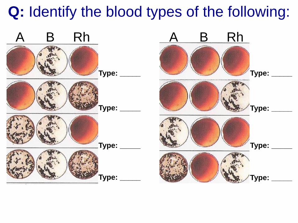
Q: Identify the blood types of the following:
A B Rh
Type: _____
Type: _____
Type: _____
Type: _____
Type: _____
Type: _____
Type: _____
Type: _____
A B Rh
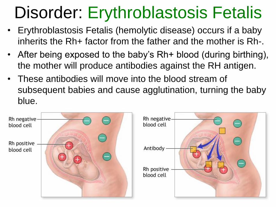
Disorder: Erythroblastosis Fetalis• Erythroblastosis Fetalis (hemolytic disease) occurs if a baby
inherits the Rh+ factor from the father and the mother is Rh-.
• After being exposed to the baby’s Rh+ blood (during birthing),
the mother will produce antibodies against the RH antigen.
• These antibodies will move into the blood stream of
subsequent babies and cause agglutination, turning the baby
blue.
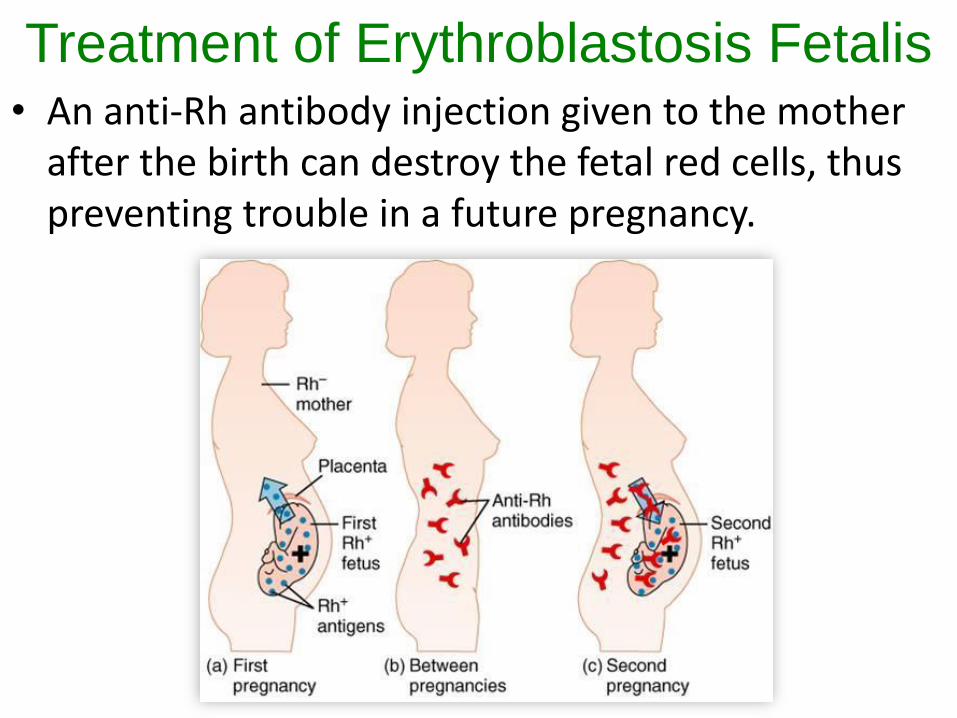
Treatment of Erythroblastosis Fetalis• An anti-Rh antibody injection given to the mother
after the birth can destroy the fetal red cells, thus preventing trouble in a future pregnancy.
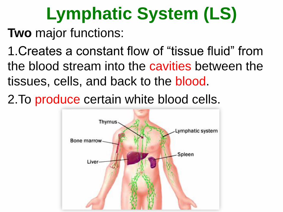
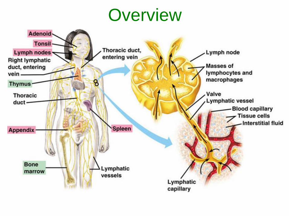
Lymphatic System (LS)Two major functions:
1.Creates a constant flow of “tissue fluid” from
the blood stream into the cavities between the
tissues, cells, and back to the blood.
2.To produce certain white blood cells.
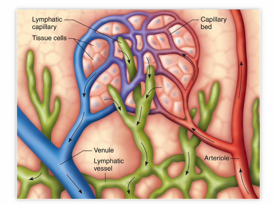
Lymphatic System (LS)• A network of vessels that carry lymph from the
intracellular spaces of the body towards the
heart.
• Lymph is “tissue fluid”, which is similar to
blood plasma. It is a colourless fluid
containing WBCs.
• Lymph acts to remove bacteria from the
tissues and supply mature lymphocytes (a
form of small WBC) to the blood.
• The LS is not a closed system.
Lymphatic System• The LS absorbs approximately 3 L of lymph per
day back into circulatory system.
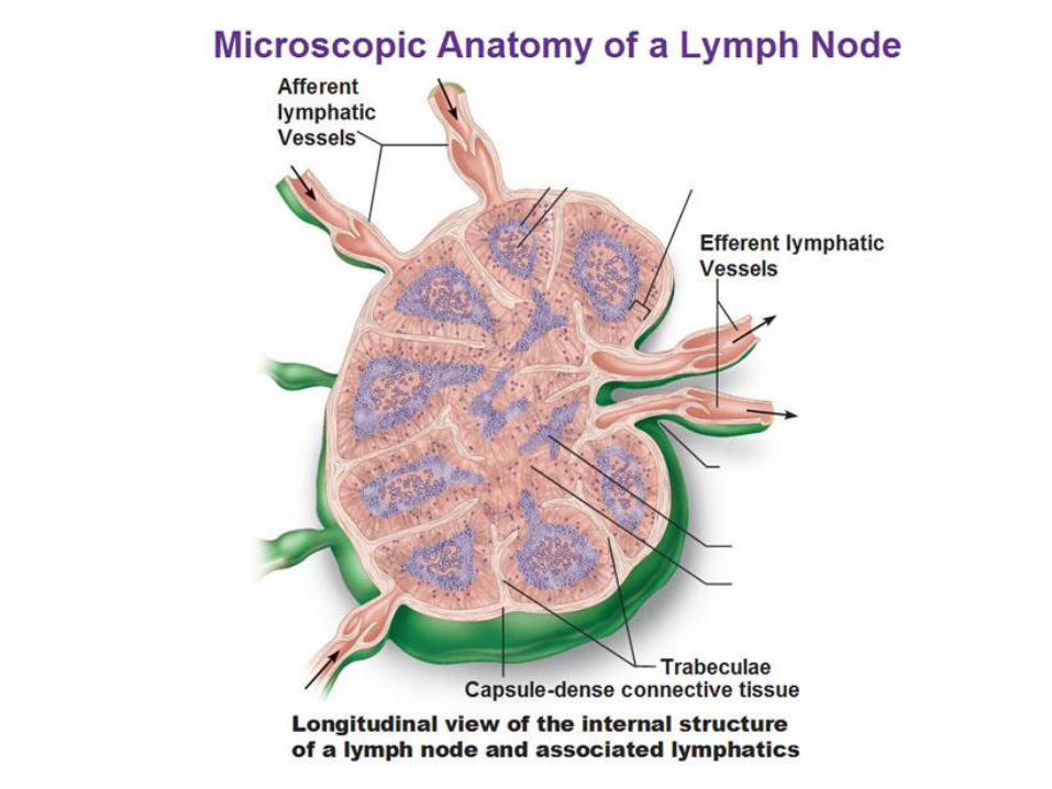
• Includes lymph nodes which house many of the T-
cells and B-cells of the immune system.
• Lymph nodes filter out and digest bacteria and
other fragments of foreign material picked up by
the lymph when it is between the tissue.
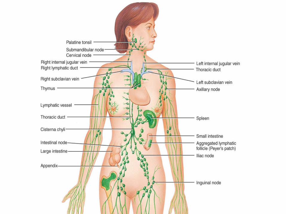
• Lymph nodes are found near the body’s major
organs – in the neck, under the arms, and in the
limbs.
• Returns fluid to the circulatory system through the
right lymphatic duct and the thoractic duct.
An Aside: The Spleen & AppendixFunctions:
• Spleen: Works to protect the body, clearing
worn out red blood cells and other foreign
bodies from the bloodstream to help fight off
infection.
• Appendix: The presence of lymphoid tissue
suggests that the appendix may play a role
in the immune/lymphatic system.
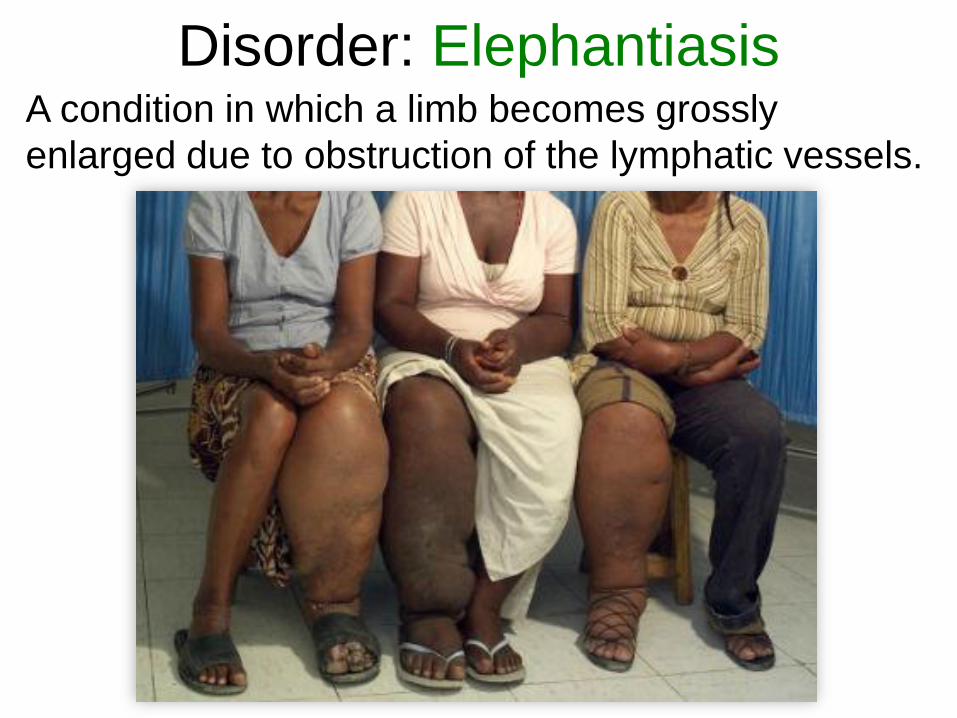
Disorder: ElephantiasisA condition in which a limb becomes grossly
enlarged due to obstruction of the lymphatic vessels.
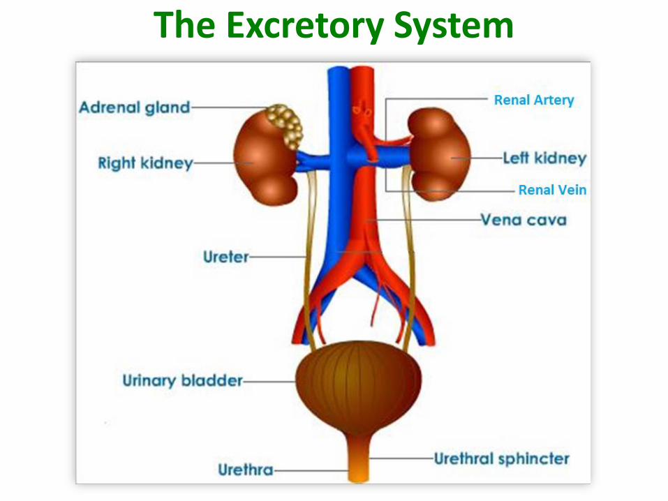
The Excretory System
The Excretory System• The excretory system is responsible for removing
wastes from the body.
• These wastes include water, salts, urine, and other metabolic waste.
• About 1/1000 to 2/1000 of blood that flows through our bodies becomes waste after filtration.
• The primary organs in the excretory system are the kidneys.
• The primary function of the kidneys is to eliminate waste from the bloodstream by the production of urine.
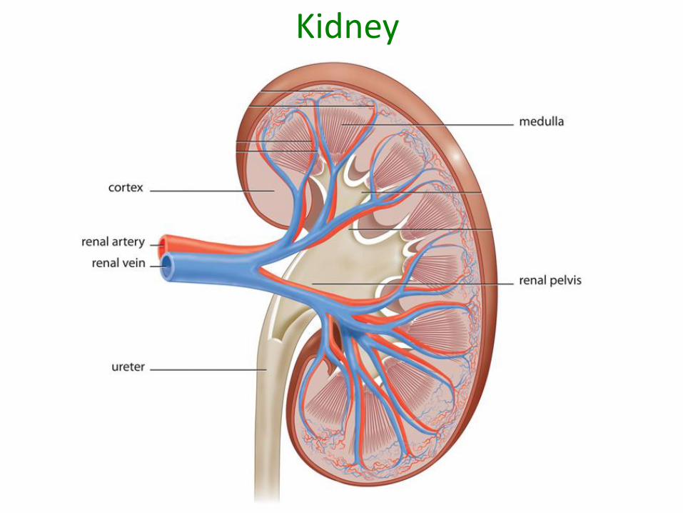
Kidney
Key Functions of the Kidney
• Maintain volume of extracellular fluid.
• Maintain ionic balance in extracellular fluid.
• Maintain pH and osmotic concentration of the extracellular fluid.
• Excrete toxic metabolic by-products such as urea, ammonia, and uric acid.
Other Facts about Kidneys
• ~ 700-800 L of blood pass through the
kidneys each day.
• 99% of the water that filters out of the
blood into the kidneys is reabsorbed back
into the blood.
• To replenish water lost to urine each day,
we must drink 1.4 – 1.8 L of water each
day.

Excretion vs. Secretion vs. Elimination
• Excretion - the removal of wastes from the
cells to the blood. Ex: salts, minerals, and
water.
• Secretion - the removal of useful substances
from cells. Ex: digestive enzymes and
hormones.
• Elimination - Elimination is the removal of
wastes from the body; i.e. getting rid of waste
substances from the organism completely.
Ex: CO2, food waste, water, and urea.
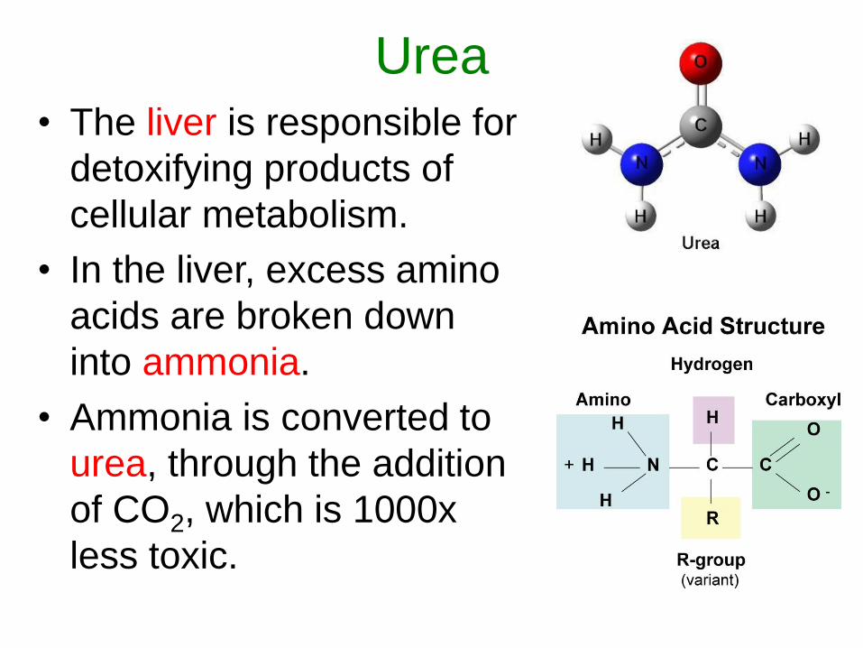
Urea• The liver is responsible for
detoxifying products of
cellular metabolism.
• In the liver, excess amino
acids are broken down
into ammonia.
• Ammonia is converted to
urea, through the addition
of CO2, which is 1000x
less toxic.
Urea
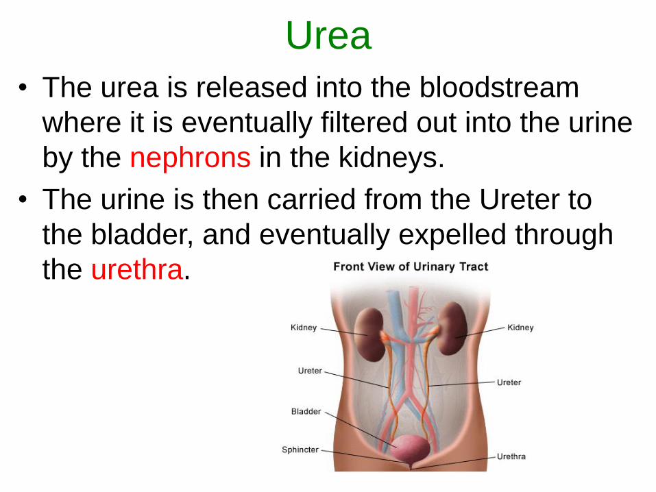
• The urea is released into the bloodstream
where it is eventually filtered out into the urine
by the nephrons in the kidneys.
• The urine is then carried from the Ureter to
the bladder, and eventually expelled through
the urethra.
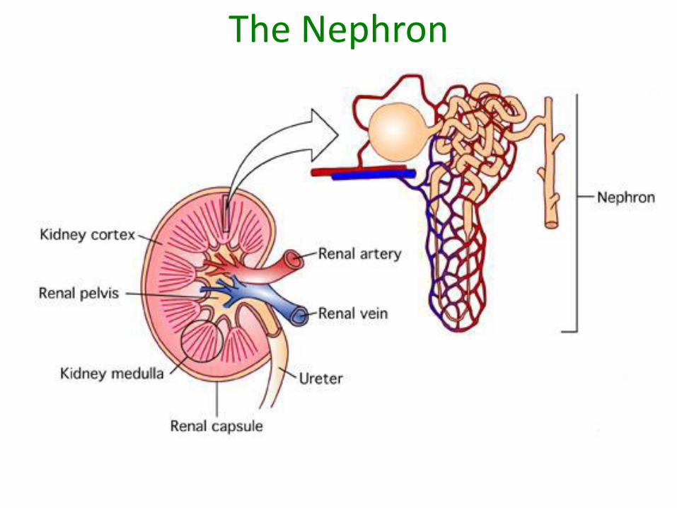
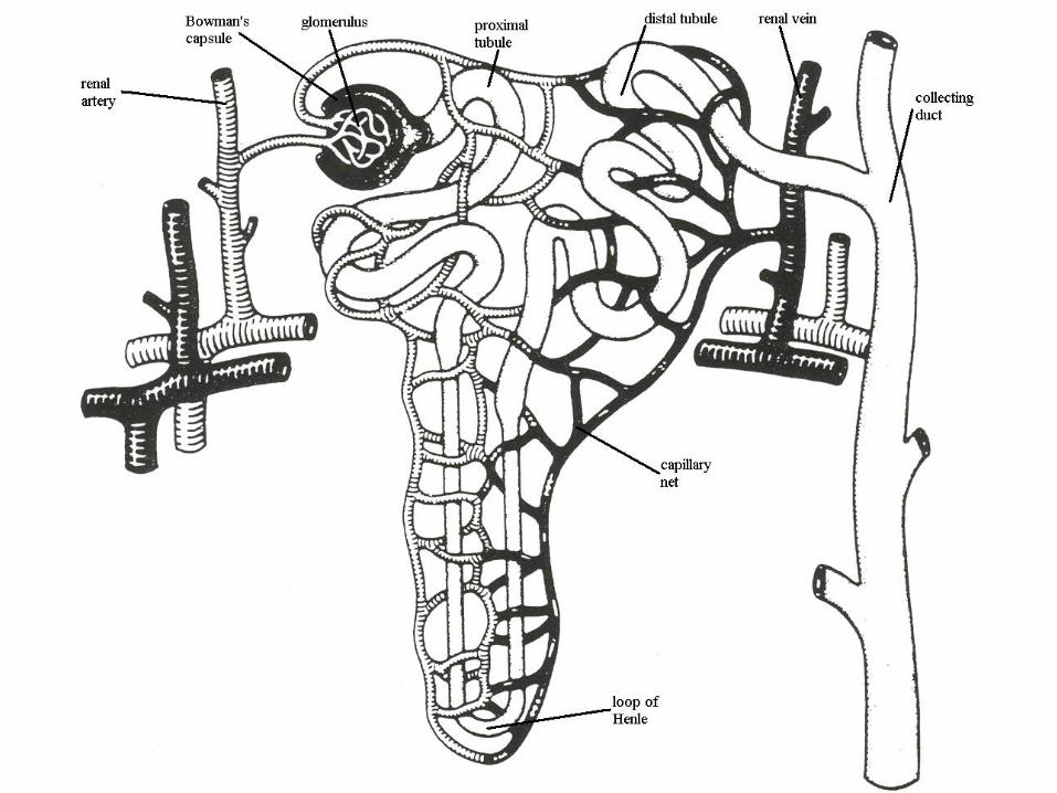
The Nephron
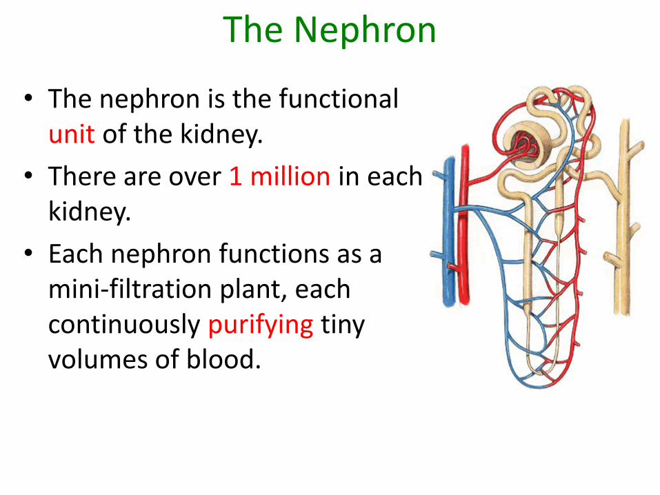
The Nephron
• The nephron is the functional unit of the kidney.
• There are over 1 million in each kidney.
• Each nephron functions as a mini-filtration plant, each continuously purifying tiny volumes of blood.

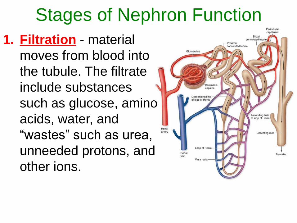
Stages of Nephron Function
1. Filtration - material
moves from blood into
the tubule. The filtrate
include substances
such as glucose, amino
acids, water, and
“wastes” such as urea,
unneeded protons, and
other ions.
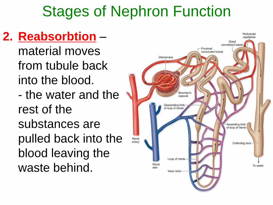
Stages of Nephron Function
2. Reabsorbtion –
material moves
from tubule back
into the blood.
- the water and the
rest of the
substances are
pulled back into the
blood leaving the
waste behind.
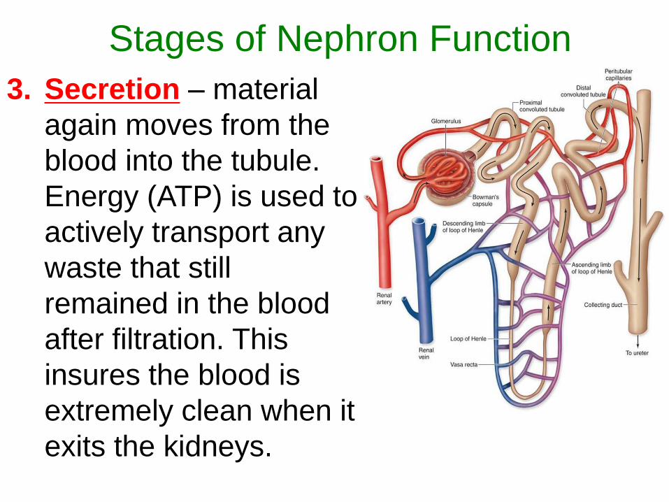
Stages of Nephron Function
3. Secretion – material
again moves from the
blood into the tubule.
Energy (ATP) is used to
actively transport any
waste that still
remained in the blood
after filtration. This
insures the blood is
extremely clean when it
exits the kidneys.
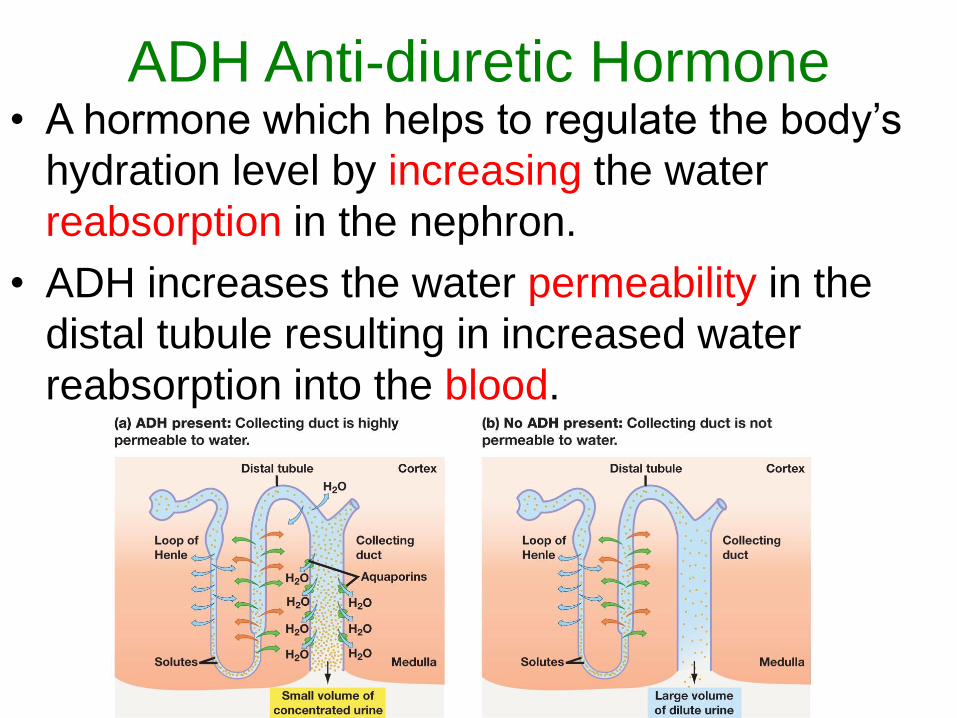
ADH Anti-diuretic Hormone• A hormone which helps to regulate the body’s
hydration level by increasing the water
reabsorption in the nephron.
• ADH increases the water permeability in the
distal tubule resulting in increased water
reabsorption into the blood.
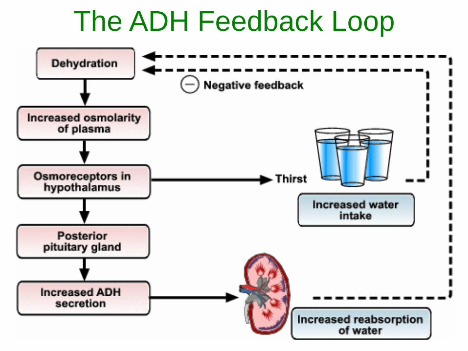
The ADH Feedback Loop
ADH Anti-diuretic Hormone
• When do your levels of ADH go up?
When you become dehydrated.
• What sort of things cause dehydration?
Not drinking enough water
Excess heat and sweating
Vomiting
Diarrhea
ADH Anti-diuretic Hormone• What does increased levels of ADH do to your
volume of urine?
You expel less urine.
• What does increased levels of ADH do to your
concentration of urine?
Your urine becomes MORE concentrated.
• What does decreased levels of ADH do to your
volume of urine?
You expel more urine.
• What does decreased levels of ADH do to your
concentration of urine?
Your urine becomes less concentrated.