Embed Size (px)
Citation preview
1
How to Bend the Uranyl Cation via Crystal Engineering
Korey P. Carter§, Mark Kalaj§, Andrew Kerridge†, J. August Ridenour§, and Christopher
L. Cahill*§
§ Department of Chemistry, The George Washington University, 800 22nd Street, NW,
Washington, D.C. 20052, United States
† Department of Chemistry, Lancaster University, Bailrigg, Lancaster LA1 4YB, United
Kingdom
Abstract
Bending the linear uranyl (UO22+) cation represents both a significant challenge and
opportunity within the field of actinide hybrid materials. As part of related efforts to
engage the nominally terminal oxo atoms of uranyl cation in non-covalent interactions,
we synthesized a new uranyl complex, [UO2(C12H8N2)2(C7H2Cl3O2)2]•2H2O (complex 2),
that featured both deviations from equatorial planarity and uranyl linearity from simple
hydrothermal conditions. Based on this complex, we developed an approach to probe the
nature and origin of uranyl bending within a family of hybrid materials, which was done
via the synthesis of complexes 1-3 that display significant deviations from equatorial
planarity and uranyl linearity (O-U-O bond angles between 162-164º) featuring 2,4,6-
trihalobenzoic acid ligands (where X=F, Cl, and Br) and 1,10-phenanthroline, along with
nine additional ‘non-bent’ hybrid materials that either co-formed with the ‘bent’
complexes (4-6) or were prepared as part of complementary efforts to understand the
mechanism(s) of uranyl bending (7-12). Complexes were characterized via single crystal
X-ray diffraction, Raman, Infrared (IR), and luminescence spectroscopy, as well as via
quantum chemical calculations and density-based quantum theory of atoms in molecules
(QTAIM) analysis. Looking comprehensively, these results are compared with the small
library of ‘bent’ uranyl complexes in the literature, and herein we computationally
demonstrate the origin of uranyl bending and delineate the energetics behind this process.
2
Introduction
Crystal engineering with the uranyl cation is an area of sustained interest within
5f hybrid materials as it presents a route to access unique structure types and unexpected
properties across a range of dimensionalities (i.e. molecular complexes, coordination
polymers (CPs), Metal-organic frameworks (MOFs), etc.) that are otherwise inaccessible
via traditional coordination chemistry.1-13 Within the crystal engineering umbrella is
supramolecular assembly, and use of this approach for producing uranyl hybrid materials
necessitates a cognizance of the relationship between intra- and intermolecular
interactions and resulting global structures.14-16 Judicious selection of uranyl acceptor-
donor pairings allows for exercising some control over the nature and directionality of
non-covalent interactions within uranyl hybrid materials, which is particularly attractive
as it allows for tectons and synthons to be selected for, thereby avoiding unpredictable
uranyl hydrolysis products. Our group has recently focused on this problem via the
hydrothermal synthesis of discrete, reproducible tectons featuring polypyridyl N-donor
capping ligands in the first coordination sphere, which promote a single uranyl species
with a specific coordination geometry.17-21 As part of this strategy, N-donor capping
ligands are paired with halogen functionalized benzoic acids such that tecton assembly
occurs by way of halogen or hydrogen bonding interactions, and moreover, this has
proven valuable for systematically engaging the nominally terminal uranyl oxo groups.19-
20
Engaging the oxo atoms of the uranyl cation is of interest within uranyl chemistry
as the linear, triatomic uranyl cation (UO22+) is known for its rigid trans-dioxo
stereochemistry with O=U=O angles that infrequently deviate from linearity (180º). This
3
is likely a result of appreciable 5fz3 and 6pz character within the -bonding framework of
the uranyl unit,22 although theoretical investigations have shown that, in the absence of
polydentate equatorial ligands, the cis isomer of the uranyl cation may lay as low as 18
kcal/mol above the stable trans isomer.23 The terminal nature of uranyl oxo atoms
generally precludes binding in this dimension, with further coordination constrained to
the equatorial plane,24 and the robust nature of the trans uranyl unit, in contrast with its
unknown cis analogue, suppresses reactivity of the uranyl cation. Multiple approaches
have shown particular promise for affecting uranyl coordination chemistry and reactivity
including oxo functionalization,25-28 distorting equatorial planarity,29-32 and breaking the
linearity of the uranyl unit,33-37 yet all of these strategies typically require sophisticated
synthetic processes and the presence of sterically bulky, complex ligands.38-39 The ‘state
of the science’ on breaking the linearity of the uranyl unit was reviewed very recently by
Hayton,40 wherein he demonstrate the nascent nature of this area of research.
Complimenting this recent review are the efforts described herein, which grew out of our
own group efforts to engage the oxo atoms of the uranyl cation via supramolecular
interactions. During the course of a related study,18 we prepared a complex that featured
both deviations from equatorial planarity and uranyl linearity from simple hydrothermal
conditions. This complex is similar to the [UO2(phen)2Cl2] material recently reported by
Ikeda-Ohno and colleagues,41 yet we report herein a strategy and the building blocks for
a general approach to the manipulation of the O=U=O bond angle via a combination of
coordination chemistry and promoted supramolecular interactions to produce a family of
materials featuring significant deviations from equatorial planarity and uranyl linearity.
4
As a consequence, we describe the synthesis and characterization of three ‘bent’
complexes (1-3) featuring 2,4,6-trihalobenzoic acid ligands (where Hal=F, Cl, and Br)
and 1,10-phenanthroline (phen), along with nine additional ‘non-bent’ hybrid materials
that either co-formed with the ‘bent’ complexes (4-6) or were prepared as part of
complementary efforts to understand the mechanism(s) of uranyl bending (7-12).
Changes in the size of halogen on the benzoic acid ligands are found to increase the
extent of uranyl bending and this observation has been probed comprehensively via
structural, computational, and spectroscopic means. Moreover, we performed quantum
chemical calculations at the density functional (DFT) level of theory along with density-
based quantum theory of atoms in molecules (QTAIM) analysis, which ultimately
showed, using the 2,4,6-trifluorobenzoic acid-phen complex (1) as a representative
example, that the bending of the uranyl unit has electronic origins and is energetically
allowed until O-U-O angles reach approximately 162º. Additionally, the vibrational and
luminescence spectra of complexes 1-9 were collected and demonstrated that whereas
spectra are indeed affected by uranyl bending, the nuclearity of the complex and the
identity of the equatorial ligands also contribute to the observed evolution in Raman, IR,
and luminescence peak values.
Experimental Methods
Synthesis
All complexes discussed herein were synthesized via hydrothermal methods at
autogenous pressure in a 23 mL Teflon-lined Parr bomb at varying oven temperatures.
Complete synthetic details which yielded X-ray quality crystalline materials for ‘bent’
and ‘non-bent’ complexes 1-9 are included in the Supporting Information.
5
X-Ray Structure Determination
Single crystals from the bulk sample of each bent phase were isolated based on
crystal color and luminescence under a UV lamp (Figure S1, Supporting Information),
and then mounted on MiTeGen micromounts. Structure determination for each of the
single crystals was achieved by collecting reflections using 0.5˚ scans on a Bruker
SMART diffractometer equipped with an APEX II CCD detector using MoKα
(=0.71073 Å) radiation at both 100(2) and 293(2) K. The data were integrated using the
SAINT software package42 contained within the APEX II software suite43 and absorption
corrections were applied using SADABS.44 Complexes 1-3 were solved via direct methods
using SIR9245 and all three complexes were refined using SHELXL-201446 contained
within the WinGX software suite.47 In each structure, all non-hydrogen atoms were
located via difference Fourier maps and refined anisotropically. Aromatic hydrogen
atoms were located via difference Fourier maps, yet were placed at their idealized
positions and allowed to ride on the coordinates of their parent carbon atom ((Uiso) fixed
at 1.2Ueq). Positional disorder in the planar phen moiety of 3 (C4, C12) was restrained via
the ISOR command with uncertainty values of 0.005 and 0.01, respectively. All figures
were prepared with Crystal Maker.48 Data collection and refinement details for low
temperature and room temperature collections of 1-3 are included in Tables S1 (LT) and
S2 (RT) (Electronic Supporting Information, ESI), respectively.
Single crystals from non-bent phases were also isolated and mounted on
MiTeGen micromounts. Similar procedures as described above were used for structure
determination of 4-12 with data for 4-7, 10, and 12 collected at 293(2) K and for 8, 9, and
11 collected at 100(2) K. Structures for complexes 4-7 and 9-12 were solved via direct
6
methods using SIR9245 and via the Patterson Method46 for complex 8 and all nine
complexes were refined using SHELXL-201446 contained within the WinGX software
suite.47 Similar to 1-3, aromatic hydrogen atoms for 4-12 were located via difference
Fourier maps, yet were placed at their idealized positions and allowed to ride on the
coordinates of their parent carbon atom. Complexes 4, 6, and 8 feature bridging
hydroxide groups, confirmed via bond-valence summations (Tables S15-S17, ESI), and
the hydrogen atoms on the hydroxide moieties in 4 and 6 were located and refined with
DFIX restraints. Methyl hydrogen atoms on the bridging acetate groups in 8 were placed
at their idealized positions with torsion angles based on electron density. Data collection
and refinement details for 4-12 are included in Table S3 (ESI).
Powder X-ray Diffraction
Powder X-ray diffraction (PXRD) data on the bulk reaction product of complexes
1-9 (Figures S20-S28, ESI) were used to examine the purity of each sample. All data
were collected on a Rigaku Miniflex (Cu Kα, 2θ=3-60˚) and were analyzed using the
Match software program.49 Initially, the bulk products of complexes 1-3, 6, 8 and 9 were
found to contain multiple solid-state phases. Complex 1 was found to primarily co-form
with 4 (Figure S20, Supporting Information) and complexes 2 and 3 was found to co-
form with 5 and 6, respectively (Figures S21 and S22, Supporting Information). Attempts
to isolate complex 6 as a single phase also yielded complex 7 and a small amount of
complex 8 (Figure S25, Supporting Information), the former of which could be isolated
as a pure phase (Figure S26, Supporting Information). Regarding the impurities in the
bulk products of 8 and 9, multiple attempts were made to identify and/or remove these
phases, yet they persisted and were not identified.
7
Spectroscopic Characterization
Raman and luminescence spectra for single crystals of 1-9 were collected on a
Horiba LabRAM HR Evolution Spectrometer. For Raman spectra, data were collected for
five seconds with ten signal accumulations over the range 600-1200 cm-1 using a 532 nm
laser, whereas for luminescence spectra, data were collected using a 405 nm excitation
laser over the 450-650 nm range.
Infrared (IR) spectra for single crystals of 1-9 were collected on a Nicolet 6700
FTIR coupled with a diamond coated ATR and MCT-A detector. Data were collected
over the range 650-4000 cm-1, and 512 scans were collected for each spectrum to enhance
the signal-to-noise ratio and minimize background effects.
Computational Details
Density functional theory (DFT) calculations have been performed on individual
molecules using version 6.4 of the TURBOMOLE quantum chemistry software
package.50 Alrichs def2-TZVP basis sets of triple-quality have been used for the C, H, O,
and N atoms,51 whereas the Alrichs def-TZVP basis set of triple-zeta quality, which
incorporates a relativistic ECP comprising 60 core electrons has been used for the U
atoms.52 Hereafter this basis set will be referred to as def(2)-TZVP. All simulations were
performed using the B3LYP hybrid-GGA exchange-correlation functional, which has
been to shown to reproduce experimental parameters of uranyl complexes with high
accuracy.53-54 Analysis of resultant electron densities was performed using Bader’s
Quantum Theory of Atoms in Molecules (QTAIM) approach55 via version 13.11.04 of
the AIMA11 software suite.56
Results
8
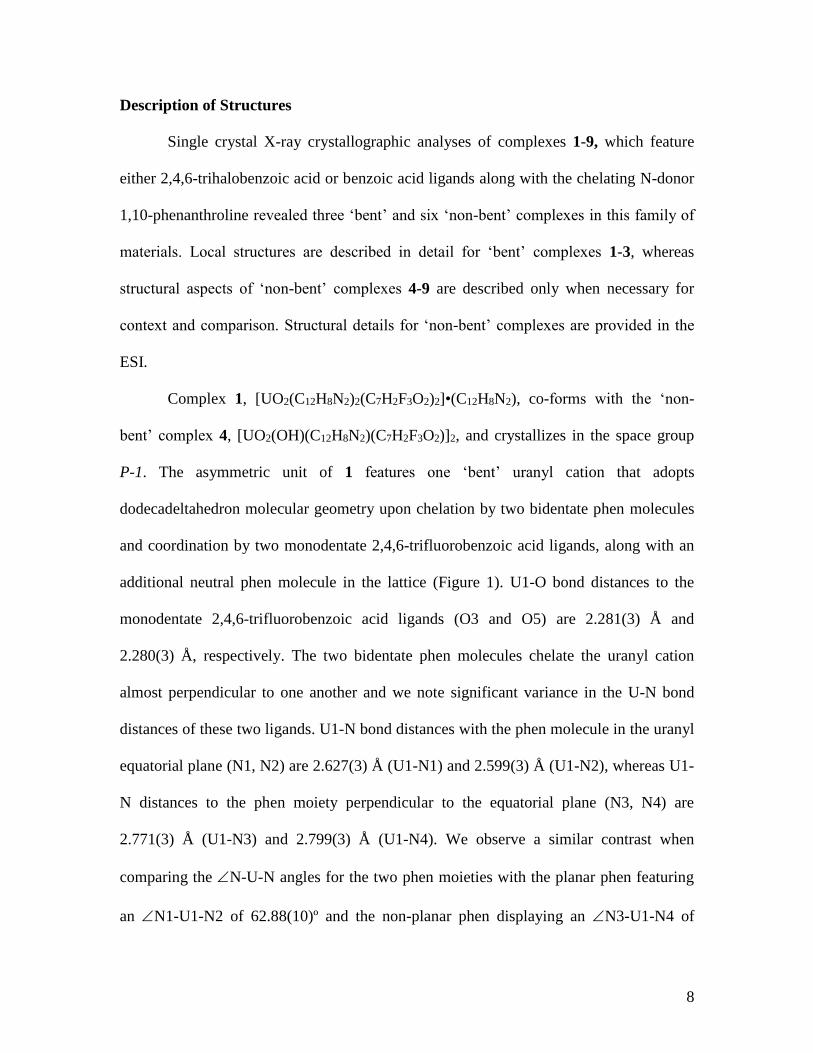
Description of Structures
Single crystal X-ray crystallographic analyses of complexes 1-9, which feature
either 2,4,6-trihalobenzoic acid or benzoic acid ligands along with the chelating N-donor
1,10-phenanthroline revealed three ‘bent’ and six ‘non-bent’ complexes in this family of
materials. Local structures are described in detail for ‘bent’ complexes 1-3, whereas
structural aspects of ‘non-bent’ complexes 4-9 are described only when necessary for
context and comparison. Structural details for ‘non-bent’ complexes are provided in the
ESI.
Complex 1, [UO2(C12H8N2)2(C7H2F3O2)2]•(C12H8N2), co-forms with the ‘non-
bent’ complex 4, [UO2(OH)(C12H8N2)(C7H2F3O2)]2, and crystallizes in the space group
P-1. The asymmetric unit of 1 features one ‘bent’ uranyl cation that adopts
dodecadeltahedron molecular geometry upon chelation by two bidentate phen molecules
and coordination by two monodentate 2,4,6-trifluorobenzoic acid ligands, along with an
additional neutral phen molecule in the lattice (Figure 1). U1-O bond distances to the
monodentate 2,4,6-trifluorobenzoic acid ligands (O3 and O5) are 2.281(3) Å and
2.280(3) Å, respectively. The two bidentate phen molecules chelate the uranyl cation
almost perpendicular to one another and we note significant variance in the U-N bond
distances of these two ligands. U1-N bond distances with the phen molecule in the uranyl
equatorial plane (N1, N2) are 2.627(3) Å (U1-N1) and 2.599(3) Å (U1-N2), whereas U1-
N distances to the phen moiety perpendicular to the equatorial plane (N3, N4) are
2.771(3) Å (U1-N3) and 2.799(3) Å (U1-N4). We observe a similar contrast when
comparing the N-U-N angles for the two phen moieties with the planar phen featuring
an N1-U1-N2 of 62.88(10)º and the non-planar phen displaying an N3-U1-N4 of

9
57.98(10)º. The U-N and N-U-N distances and angles observed in 1 represent the
longest U-N bonds and smallest N-U-N angle ever observed in a uranyl hybrid material
according to a search of the Cambridge Structural Database (v 5.38, Nov. 2016),57 and
likely drive the unusual behavior of the uranyl cation described below.
Figure 1 Polyhedral representation of local coordination geometry of 1. Yellow
polyhedra represent uranium metal centers, whereas green, red, and blue spheres
represent fluorine, oxygen, and nitrogen atoms, respectively. All H atoms have been
omitted for clarity.
The breaking of equatorial planarity by one the phen molecules (N3 and N4) does
not affect U1-O bond distances to oxo atoms O1 and O2, which are characteristic of
uranyl materials at 1.778(3) Å and 1.785(3) Å,24 yet we do see a manifestation of the
deviation from planarity in the O1-U1-O2 angle, which is considerably bent away from
linear at 164.93(12)º. A recent study from Hayton et al. surveyed uranyl bending in
hybrid materials and reported that the smallest observed O-U-O angles for the uranyl
unit were between 166-168º.37 In their study, they highlighted three compounds made
with the uranyl cation and a 12-membered macrocycle with O-U-O angles between
10
161.7(5)º and 164.1(3)º, thereby setting a new mark for uranyl bending, which has
recently been matched by Ikeda-Ohno et al.41 The bending of the uranyl cation in 1
coupled with the concomitant deviation from equatorial planarity also observed is a rare
combination in uranyl hybrid materials, with the [UO2Cl2(phen)2] complex characterized
recently by Ikeda-Ohno et al. the only analogue to 1 found in the literature.41
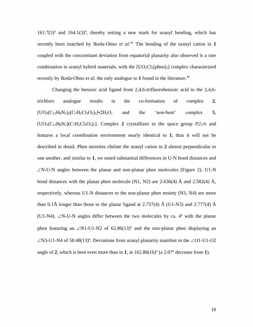
Changing the benzoic acid ligand from 2,4,6-trifluorobenzoic acid to the 2,4,6-
trichloro analogue results in the co-formation of complex 2,
[UO2(C12H8N2)2(C7H2Cl3O2)2]•2H2O, and the ‘non-bent’ complex 5,
[UO2(C12H8N2)(C7H2Cl3O2)2]. Complex 2 crystallizes in the space group P21/n and
features a local coordination environment nearly identical to 1, thus it will not be
described in detail. Phen moieties chelate the uranyl cation in 2 almost perpendicular to
one another, and similar to 1, we noted substantial differences in U-N bond distances and
N-U-N angles between the planar and non-planar phen molecules (Figure 2). U1-N
bond distances with the planar phen molecule (N1, N2) are 2.636(4) Å and 2.582(4) Å,
respectively, whereas U1-N distances to the non-planar phen moiety (N3, N4) are more
than 0.1Å longer than those to the planar ligand at 2.757(4) Å (U1-N3) and 2.777(4) Å
(U1-N4). N-U-N angles differ between the two molecules by ca. 4º with the planar
phen featuring an N1-U1-N2 of 62.86(13)º and the non-planar phen displaying an
N3-U1-N4 of 58.48(13)º. Deviations from uranyl planarity manifest in the O1-U1-O2
angle of 2, which is bent even more than in 1, at 162.86(16)º (a 2.07º decrease from 1).
11
Figure 2 Polyhedral representation of local coordination geometry of 2. Lime green
spheres represent chlorine atoms. Lattice water molecules have been omitted for clarity.
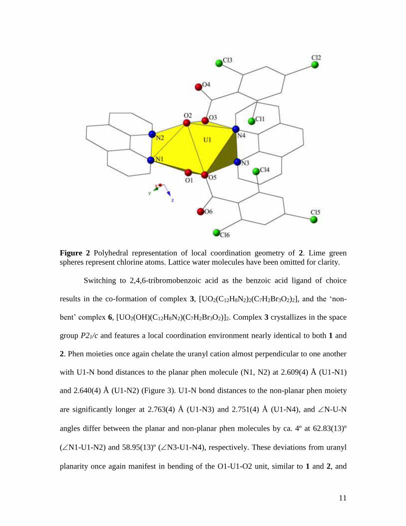
Switching to 2,4,6-tribromobenzoic acid as the benzoic acid ligand of choice
results in the co-formation of complex 3, [UO2(C12H8N2)2(C7H2Br3O2)2], and the ‘non-
bent’ complex 6, [UO2(OH)(C12H8N2)(C7H2Br3O2)]2. Complex 3 crystallizes in the space
group P21/c and features a local coordination environment nearly identical to both 1 and
2. Phen moieties once again chelate the uranyl cation almost perpendicular to one another
with U1-N bond distances to the planar phen molecule (N1, N2) at 2.609(4) Å (U1-N1)
and 2.640(4) Å (U1-N2) (Figure 3). U1-N bond distances to the non-planar phen moiety
are significantly longer at 2.763(4) Å (U1-N3) and 2.751(4) Å (U1-N4), and N-U-N
angles differ between the planar and non-planar phen molecules by ca. 4º at 62.83(13)º
(N1-U1-N2) and 58.95(13)º (N3-U1-N4), respectively. These deviations from uranyl
planarity once again manifest in bending of the O1-U1-O2 unit, similar to 1 and 2, and
12
we note an additional deviation from linearity of the uranyl unit in 3 as we switch to
benzoic acid units featuring larger halogen atoms with the O1-U1-O2 angle at
162.18(16)º, a 2.75º decrease from 1 and a 0.68º decrease from 2. The O-U-O angle in 3
is comparable to the smallest values reported in the literature by Hayton37 and Ikeda-
Ohno,41 and represents the most significant uranyl bending we observe in this family of
complexes.
Figure 3 Polyhedral representation of local coordination geometry of 3. Brown spheres
represent bromine atoms.
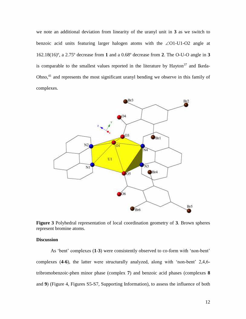
Discussion
As ‘bent’ complexes (1-3) were consistently observed to co-form with ‘non-bent’
complexes (4-6), the latter were structurally analyzed, along with ‘non-bent’ 2,4,6-
tribromobenzoic-phen minor phase (complex 7) and benzoic acid phases (complexes 8
and 9) (Figure 4, Figures S5-S7, Supporting Information), to assess the influence of both
13
benzoic acid and phen ligands, along with intramolecular interaction strength, on driving
deviations from equatorial planarity and uranyl linearity.
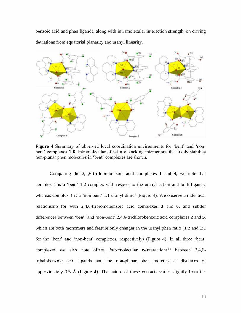
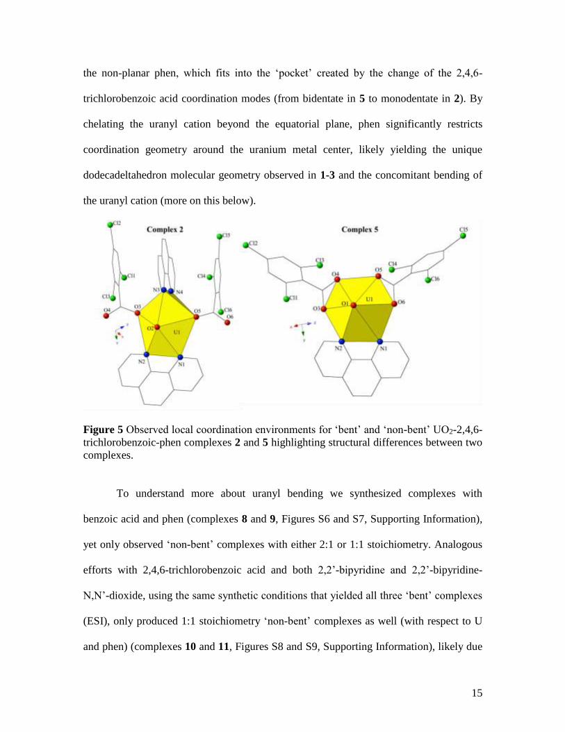
Figure 4 Summary of observed local coordination environments for ‘bent’ and ‘non-
bent’ complexes 1-6. Intramolecular offset π-π stacking interactions that likely stabilize
non-planar phen molecules in ‘bent’ complexes are shown.
Comparing the 2,4,6-trifluorobenzoic acid complexes 1 and 4, we note that
complex 1 is a ‘bent’ 1:2 complex with respect to the uranyl cation and both ligands,
whereas complex 4 is a ‘non-bent’ 1:1 uranyl dimer (Figure 4). We observe an identical
relationship for with 2,4,6-tribromobenzoic acid complexes 3 and 6, and subtler
differences between ‘bent’ and ‘non-bent’ 2,4,6-trichlorobenzoic acid complexes 2 and 5,
which are both monomers and feature only changes in the uranyl:phen ratio (1:2 and 1:1
for the ‘bent’ and ‘non-bent’ complexes, respectively) (Figure 4). In all three ‘bent’
complexes we also note offset, intramolecular π-interactions58 between 2,4,6-
trihalobenzoic acid ligands and the non-planar phen moieties at distances of
approximately 3.5 Å (Figure 4). The nature of these contacts varies slightly from the
14
intermolecular π-interactions described by Ikeda-Ohno et al. in their ‘bent’
[UO2Cl2(phen)2] uranyl complex, which features two unique sets of π-interactions, one
between planar phen molecules and the other between non-planar phen ligands.41 The
role of the intramolecular π-interactions in 1-3 is likely to stabilize the non-planar phen
molecules, and subsequently the overall ‘bent’ complexes. Additional stabilization of the
non-planar phen molecules is likely necessary as the U-N distances and N-U-N angles
for the non-planar phen moieties in 1-3 are significantly longer and smaller (respectively)
than have previously been observed in any uranyl hybrid material featuring phen.11, 17 In
fact, there are only two previous examples of uranyl-phen complexes displaying a U:phen
ratio other than 1:1: the rhombohedral [UO2(phen)3][OTf]2 from Berthet and colleagues30
and the recently synthesized [UO2Cl2(phen)2] from Ikeda-Ohno et al.41
A closer look at the synthetic conditions that produced ‘bent’ phases with 2,4,6-
trifluoro- and tribromobenzoic acids (complexes 1 and 3), indicates that single crystals of
the ‘bent’ phases were only found when the uranyl to phen molar ratio was increased to at
least 1:3 (at molar ratios of <1:3 only ‘non-bent’ phases (complexes 4 and 6) were
produced). In contrast, with 2,4,6-trichlorobenzoic acid the ‘bent’ phase was found to
only be uranyl starting salt dependent. From a structural and synthetic perspective, these
observations suggest that the benzoic acid ligands featured in 1-3 play an ancillary role in
driving uranyl bending as deviations from linearity are noted for varied conditions,
wherein the uranyl-benzoic acid ligand molar ratio is kept constant. The role of phen in
driving uranyl bending and breaking equatorial planarity however, is most clearly
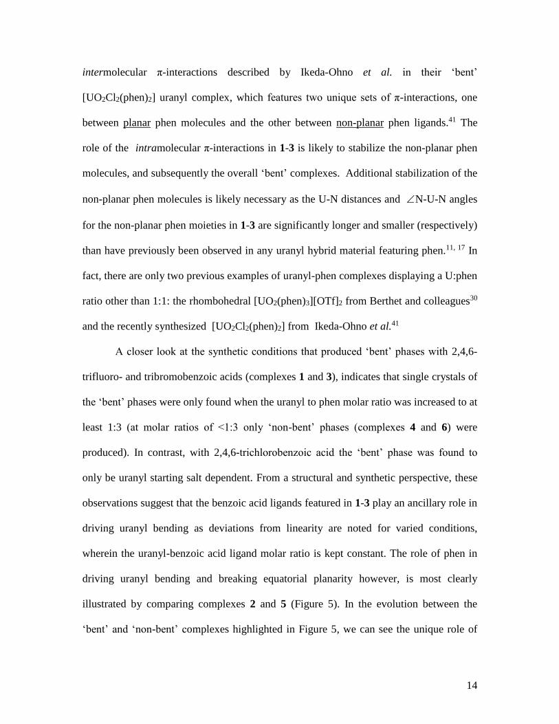
illustrated by comparing complexes 2 and 5 (Figure 5). In the evolution between the
‘bent’ and ‘non-bent’ complexes highlighted in Figure 5, we can see the unique role of
15
the non-planar phen, which fits into the ‘pocket’ created by the change of the 2,4,6-
trichlorobenzoic acid coordination modes (from bidentate in 5 to monodentate in 2). By
chelating the uranyl cation beyond the equatorial plane, phen significantly restricts
coordination geometry around the uranium metal center, likely yielding the unique
dodecadeltahedron molecular geometry observed in 1-3 and the concomitant bending of
the uranyl cation (more on this below).
Figure 5 Observed local coordination environments for ‘bent’ and ‘non-bent’ UO2-2,4,6-
trichlorobenzoic-phen complexes 2 and 5 highlighting structural differences between two
complexes.
To understand more about uranyl bending we synthesized complexes with
benzoic acid and phen (complexes 8 and 9, Figures S6 and S7, Supporting Information),
yet only observed ‘non-bent’ complexes with either 2:1 or 1:1 stoichiometry. Analogous
efforts with 2,4,6-trichlorobenzoic acid and both 2,2’-bipyridine and 2,2’-bipyridine-
N,N’-dioxide, using the same synthetic conditions that yielded all three ‘bent’ complexes
(ESI), only produced 1:1 stoichiometry ‘non-bent’ complexes as well (with respect to U
and phen) (complexes 10 and 11, Figures S8 and S9, Supporting Information), likely due
16
to limits in observed N-U-N angles for both chelating ligands. The importance of the
2,4,6-trihalobenzoic acids for yielding ‘bent’ complexes was further explored via
synthesis with 2,3,5-trichlorobenzoic acid and phen. Even at molar ratios of 1:2:5
(UO2:235triClBA:phen), only a ‘non-bent’ 1:1 stoichiometry complex was observed
(Figure S10, Supporting Information), and this observation hints at the importance of the
‘π-pocket’ of 1-3 highlighted in Figure 4. The intramolecular π-interactions between
2,4,6-trihalobenzoic acid ligands and non-planar phen molecules in 1-3 likely facilitate
the formation of these ‘bent’ complexes, and this supramolecular motif cannot be
repeated upon modification of the halogen positions on the benzoic acid ligands, perhaps
due to a change in electron density distribution or sterics.
Computational Results
In an effort to understand the deviations from equatorial planarity and uranyl
linearity highlighted thus far, we turned to density functional theory (DFT) calculations
and quantum theory of atoms in molecules (QTAIM) analysis to probe the results
detailed above. Initial DFT calculations on individual molecules representing complexes
1-3 and the simulated 2,4,6-triiodobenzoic acid analogue showed pronounced bending of
the uranyl unit, yet deviations from linearity were found to be relatively independent of
the halide species, in contrast to experiment (Table S4, Supporting Information). The
B3LYP/def(2)TZVP model chemistry was found to simulate U-oxo and U-O(equatorial)
bond lengths extremely well, particularly for X = F and Cl, but O-H bond lengths were
significantly underestimated and U-N bond lengths were significantly overestimated
(Table S4, Supporting Information). U-N bond lengths were then constrained at
experimental values and this led to a better reproduction of the uranyl bend, and its

17
observed halide dependence (Table 1). Whereas calculations using this model chemistry
do underestimate uranyl bending when X = F, there is a clear trend as the halide group is
descended (Table 1). A consequence of this constraint, however, is that other structural
metrics compare less favorably to experiment.
Table 1 Selected structural parameters of complexes 1-3. All calculations performed
using the B3LYO/def(2)TZVP mode chemistry. * UN bonds were constrained to the
experimental values.
X U-oxo
(Å)
OUO bend
()
O-H (Å) U-Oeq (Å) U-N (Å)
F Exp 1.781 164.94 2.401 2.281 2.613, 2.780
Calc 1.780 165.62 2.286 2.279 2.695, 2.907
Calc* 1.786 164.11 2.239 2.292 n/a
Cl Exp 1.774 162.87 2.360 2.282 2.665, 2.767
Calc 1.779 165.26 2.283 2.287 2.700, 2.897
Calc* 1.785 163.02 2.213 2.309 n/a
Br Exp 1.778 162.18 2.317 2.272 2.625, 2.758
Calc 1.778 165.25 2.278 2.293 2.703, 2.890
Calc* 1.784 162.24 2.203 2.315 n/a
QTAIM analysis on 1-3 using B3LYP/def(2)-TZVP derived densities indicate
that values of the electron density at the U-oxo bond critical point, BCP, are significantly
reduced in comparison to the free uranyl cation, suggestive of a weakening of the
covalent character of the U-oxo bonds comparable in magnitude to that associated with
cyano/isocyanate complexation,59-61 with energy densities (H) exhibiting the same
behavior (Table S5, Supporting Information). An interesting, albeit weak, trend can be
observed, namely an increase in the magnitudes of BCP, H, and the bond ellipticity, , as
the halide becomes larger (Table S5, Supporting Information). This is indicative of
increasing U-oxo bond covalency as the halide group is descended, along with a slight
deviation from pure triple bond character (evidenced by > 0). As an increase in
18
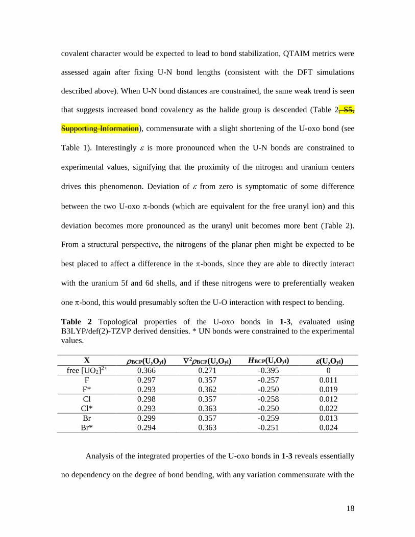
covalent character would be expected to lead to bond stabilization, QTAIM metrics were
assessed again after fixing U-N bond lengths (consistent with the DFT simulations
described above). When U-N bond distances are constrained, the same weak trend is seen
that suggests increased bond covalency as the halide group is descended (Table 2, S5,
Supporting Information), commensurate with a slight shortening of the U-oxo bond (see
Table 1). Interestingly is more pronounced when the U-N bonds are constrained to
experimental values, signifying that the proximity of the nitrogen and uranium centers
drives this phenomenon. Deviation of from zero is symptomatic of some difference
between the two U-oxo -bonds (which are equivalent for the free uranyl ion) and this
deviation becomes more pronounced as the uranyl unit becomes more bent (Table 2).
From a structural perspective, the nitrogens of the planar phen might be expected to be
best placed to affect a difference in the -bonds, since they are able to directly interact
with the uranium 5f and 6d shells, and if these nitrogens were to preferentially weaken
one -bond, this would presumably soften the U-O interaction with respect to bending.
Table 2 Topological properties of the U-oxo bonds in 1-3, evaluated using
B3LYP/def(2)-TZVP derived densities. * UN bonds were constrained to the experimental
values.
X BCP(U,Oyl) 2BCP(U,Oyl) HBCP(U,Oyl) (U,Oyl)
free [UO2]2+ 0.366 0.271 -0.395 0
F 0.297 0.357 -0.257 0.011
F* 0.293 0.362 -0.250 0.019
Cl 0.298 0.357 -0.258 0.012
Cl* 0.293 0.363 -0.250 0.022
Br 0.299 0.357 -0.259 0.013
Br* 0.294 0.363 -0.251 0.024
Analysis of the integrated properties of the U-oxo bonds in 1-3 reveals essentially
no dependency on the degree of bond bending, with any variation commensurate with the
19
slight elongation of the bond when the UN bonds are constrained, which makes a
mechanism based on chemical interactions with the nitrogens of the planar phen (as
proposed above) unlikely (Table S6, Supporting Information). In fact, topological
analysis of the UN interaction data did not yield any variation which might account for
the observed uranyl bending (Table S7, Supporting Information), suggesting that perhaps
the nitrogens of the non-planar phen impact the U-oxo bond electrostatically. If this were
the case, the effect of those nitrogens would be to redistribute electronic charge from one
U-oxo -bond onto the far side of the uranyl unit, enhancing the bonding interaction on
the far side of the uranyl while simultaneously depleting the interaction on the near side,
leading to the bending observed experimentally, and nitrogen charges for 1-3 are
highlighted in Table 3. For the non-planar nitrogens, these can be compared to the U-N
separations (see Table S8, Supporting Information), showing that substantial electrostatic
repulsion would be expected between the non-planar nitrogens and the electron charge
accumulated in the U-oxo bond. This repulsion increases as the halides are descended,
commensurate with an increase in bond ellipticity and the increased bending.
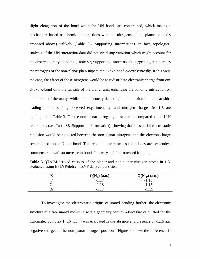
Table 3 QTAIM-derived charges of the planar and non-planar nitrogen atoms in 1-3,
evaluated using B3LYP/def(2)-TZVP derived densities.
X Q(Np) (a.u.) Q(Nnp) (a.u.)
F -1.17 -1.15
Cl -1.18 -1.15
Br -1.17 -1.15
To investigate the electrostatic origins of uranyl bending further, the electronic
structure of a free uranyl molecule with a geometry bent to reflect that calculated for the
fluorinated complex 1 (164.11 ) was evaluated in the absence and presence of -1.15 a.u.
negative charges at the non-planar nitrogen positions. Figure 6 shows the difference in
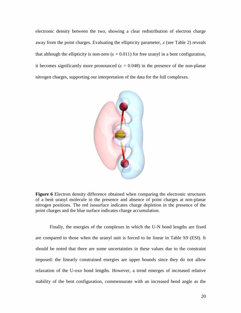
20
electronic density between the two, showing a clear redistribution of electron charge
away from the point charges. Evaluating the ellipticity parameter, (see Table 2) reveals
that although the ellipticity is non-zero ( = 0.011) for free uranyl in a bent configuration,
it becomes significantly more pronounced ( = 0.048) in the presence of the non-planar
nitrogen charges, supporting our interpretation of the data for the full complexes.
Figure 6 Electron density difference obtained when comparing the electronic structures
of a bent uranyl molecule in the presence and absence of point charges at non-planar
nitrogen positions. The red isosurface indicates charge depletion in the presence of the
point charges and the blue surface indicates charge accumulation.
Finally, the energies of the complexes in which the U-N bond lengths are fixed
are compared to those when the uranyl unit is forced to be linear in Table S9 (ESI). It
should be noted that there are some uncertainties in these values due to the constraint
imposed: the linearly constrained energies are upper bounds since they do not allow
relaxation of the U-oxo bond lengths. However, a trend emerges of increased relative
stability of the bent configuration, commensurate with an increased bend angle as the
21
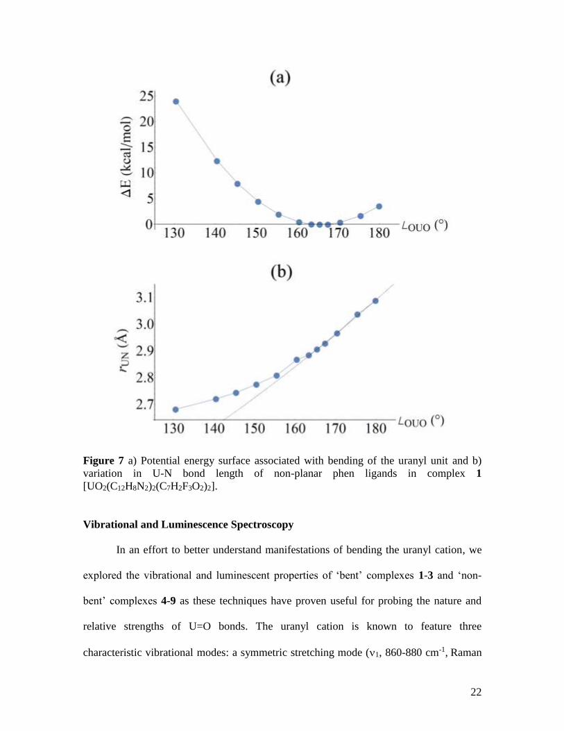
halides are descended. Using complex 1 as an example (X=F), the potential energy
surface as a function of uranyl bend angle was calculated (Figure 7). The potential energy
surface is quite flat around the minima, which may be due to the lack of crystal packing
effects in the simulations, yet we have found both experimentally and computationally
that as the uranyl unit deviates from linearity, the axially oriented non-planar phen ligand
is able to more closely coordinate the uranium center, thereby stabilizing the complex.
This phenomenon applies until ca. 162, wherein variation in U-N bond lengths reduces
and further bending of the uranyl unit becomes increasingly energetically unfavorable,
thereby indicating that there is likely a limit to uranyl bending via coordination chemistry
routes (Figure 7).
22
Figure 7 a) Potential energy surface associated with bending of the uranyl unit and b)
variation in U-N bond length of non-planar phen ligands in complex 1
[UO2(C12H8N2)2(C7H2F3O2)2].
Vibrational and Luminescence Spectroscopy
In an effort to better understand manifestations of bending the uranyl cation, we
explored the vibrational and luminescent properties of ‘bent’ complexes 1-3 and ‘non-
bent’ complexes 4-9 as these techniques have proven useful for probing the nature and
relative strengths of U=O bonds. The uranyl cation is known to feature three
characteristic vibrational modes: a symmetric stretching mode (1, 860-880 cm-1, Raman
23
active), a bending mode (2, 200-210 cm-1, infrared active), and an asymmetric stretching
mode (3, 930-960 cm-1, infrared active),62-64 and as 2 stretches fall well below the
detection limits of most instrumentation, we focus on the 1 and 3 characteristic stretches
of the uranyl cation.
Looking first at the Raman spectra of ‘bent’ complexes 1-3, the 1 symmetric
stretch is the most prominent signal in each spectrum at 816 cm-1, 844 cm-1, and 839 cm-
1, respectively (Table 4, Figure S11, Supporting Information). These results may seem
counterintuitive (at first) as 2,4,6-trifluorobenzoic acid is an electron withdrawing
benzoic acid ligand, and 1 features the least ‘bent’ uranyl unit of complexes 1-3, yet 1
does feature the longest U-oxo bond distances, which are indicators of ‘weaker’ U=O
bonds. Further, the electron withdrawing nature of 2,4,6-trifluorobenzoic acid, in contrast
to the (weakly) electron donating character of 2,4,6-trichlorobenzoic and 2,4,6-
tribromobenzoic acid, does not counteract the U=O bond effects of the non-planar phen
molecules, which likely increase the ionic interaction between the uranium center and the
oxo atoms by increasing electron density transferred from non-planar ligands into the π*-
antibonding orbitals of the uranyl cation.22, 53-54, 65 Comparing the 1 symmetric stretch
frequencies of 1-3 with their non-bent analogues 4-7, we note redshifts between ‘bent’
and ‘non-bent’ phases, independent of 2,4,6-trihalobenzoic acid ligand (Table 4, Figures
S12 and S13, Supporting Information). The magnitude of the redshifts between ‘bent’
and ‘non-bent’ complexes are mostly small (<5 cm-1), with the exception of 2,4,6-
trifluorobenzoic acid complexes 1 and 4 and 2,4,6-tribromobenzoic acid complexes 3 and
7. Based on only structural changes from ‘bent’ to ‘non-bent’ phases one would
anticipate that redshift magnitudes would be similar for 2,4,6-trifluorobenzoic acid
24
complexes 1 and 4 and 2,4,6-tribromobenzoic acid complexes 3 and 6 (‘bent’ complexes
and ‘non-bent’ uranyl dimers), and comparable for 2,4,6-trichlorobenzoic acid complexes
2 and 5 and 2,4,6-tribromobenzoic acid complexes 3 and 7 (‘bent’ complexes and ‘non-
bent’ uranyl monomers), yet the opposite is shown to be true (Table 4). These
observations confirm findings from our group and Hayton et al.,18, 21, 37 which have
demonstrated that the identity of equatorial ligands has a greater effect on Raman
frequencies, than modifying the O-U-O angle of the uranyl cation or changing uranyl
nuclearity.
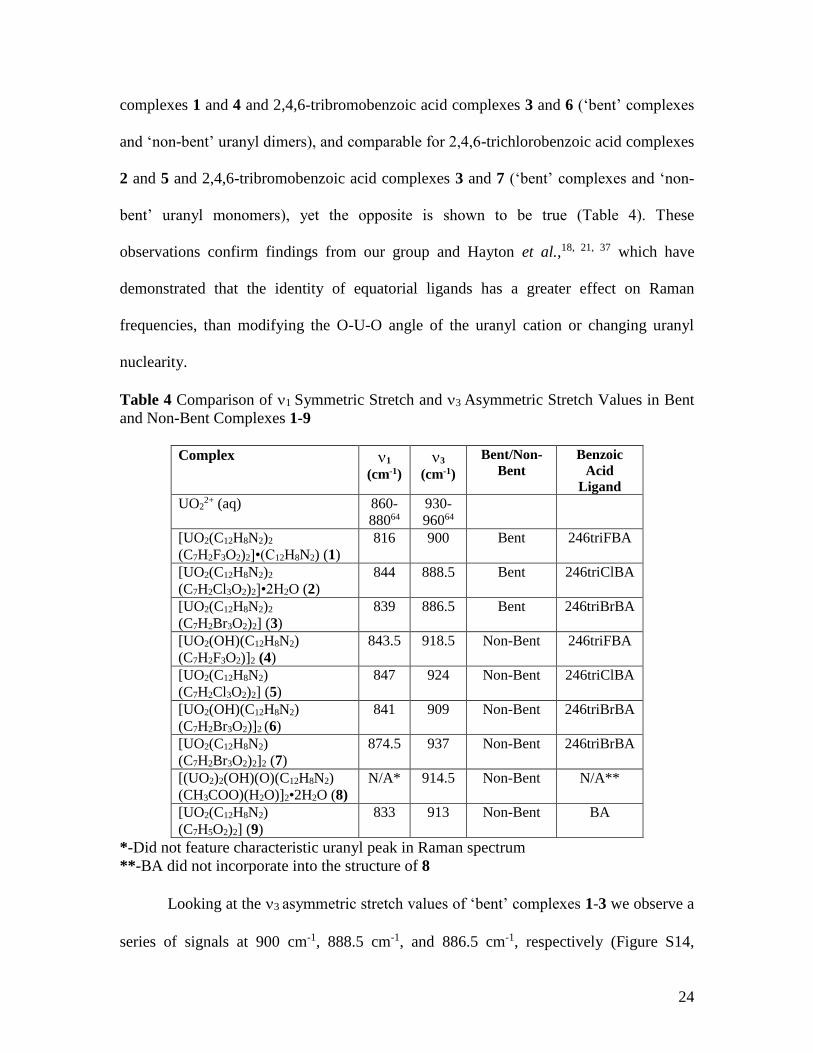
Table 4 Comparison of 1 Symmetric Stretch and 3 Asymmetric Stretch Values in Bent
and Non-Bent Complexes 1-9
Complex 1
(cm-1)
3
(cm-1)
Bent/Non-
Bent
Benzoic
Acid
Ligand
UO22+ (aq) 860-
88064
930-
96064
[UO2(C12H8N2)2
(C7H2F3O2)2]•(C12H8N2) (1)
816 900 Bent 246triFBA
[UO2(C12H8N2)2
(C7H2Cl3O2)2]•2H2O (2)
844 888.5 Bent 246triClBA
[UO2(C12H8N2)2
(C7H2Br3O2)2] (3)
839 886.5 Bent 246triBrBA
[UO2(OH)(C12H8N2)
(C7H2F3O2)]2 (4)
843.5 918.5 Non-Bent 246triFBA
[UO2(C12H8N2)
(C7H2Cl3O2)2] (5)
847 924 Non-Bent 246triClBA
[UO2(OH)(C12H8N2)
(C7H2Br3O2)]2 (6)
841 909 Non-Bent 246triBrBA
[UO2(C12H8N2)
(C7H2Br3O2)2]2 (7)
874.5 937 Non-Bent 246triBrBA
[(UO2)2(OH)(O)(C12H8N2)
(CH3COO)(H2O)]2•2H2O (8)
N/A* 914.5 Non-Bent N/A**
[UO2(C12H8N2)
(C7H5O2)2] (9)
833 913 Non-Bent BA
*-Did not feature characteristic uranyl peak in Raman spectrum
**-BA did not incorporate into the structure of 8
Looking at the 3 asymmetric stretch values of ‘bent’ complexes 1-3 we observe a
series of signals at 900 cm-1, 888.5 cm-1, and 886.5 cm-1, respectively (Figure S14,
25
Supporting Information, Table 4). The trend of increasing redshifts with greater bending
contrasts with the Raman frequencies of 1-3 (detailed above), and additionally, the
asymmetric frequencies of 1-3 are redshifted further from the free uranyl cation (Table
4), suggesting that deviations from linearity may have a greater impact on uranyl
asymmetric stretches. Comparing the 3 asymmetric stretch frequencies of 1-3 with their
non-bent analogues 4-7, we once again note redshifts between ‘bent’ and ‘non-bent’
phases, independent of 2,4,6-trihalobenzoic acid ligand (Table 4, Figures S15 and S16,
Supporting Information). The magnitude of the redshifts between ‘bent’ and ‘non-bent’
complexes are notably larger in the IR (>15 cm-1), which also suggests that uranyl
bending may exert a greater influence on asymmetric stretching frequencies (as compared
to symmetric stretches in Raman spectra).
Finally, room temperature solid-state luminescence studies were carried out on
several single crystals from the bulk phases of 1-9. Uranyl materials are known to exhibit
a characteristic green emission profile that results from ligand-to-metal charge transfer
transitions between uranyl bonding (3u, 3g, 2πu, and 1πg) and non-bonding (5f u and
u) molecular orbitals,22, 66 and for 2-7, characteristic emission (four to five major
vibronic peaks) was observed upon excitation at 420 nm (Figures S17-S19, Supporting
Information). The average vibronic progression of the emission bands are coupled to the
Raman active vibrational modes, and for 2-7 these values were found to be in excellent
agreement with measured Raman frequencies detailed in Table 4. Emission for ‘bent’
complex 1 was not completely resolved at room temperature, thus a similar comment
cannot be made for this material. The redshifts observed when comparing ‘bent’ and
‘non-bent’ complexes in Raman and IR spectra (Table 4, Figures S11-S16, Supporting
26
Information) are also noted in luminescence spectra with the largest shift between ‘bent’
and ‘non-bent’ 2,4,6-trichlorobenzoic acid complexes 2 and 5.
Conclusions
The syntheses and crystal structures of three ‘bent’ (1-3) and four ‘non-bent’ (4-7)
uranyl hybrid materials containing 2,4,6-trihalobenzoic acid ligands and 1,10-
phenanthroline are reported along with the structures of five additional uranyl complexes
that were made in the process of probing the mechanism of uranyl bending. Two of these
additional complexes feature benzoic acid and phen ligands (8 and 9), whereas the other
three complexes include 2,4,6-trichlorobenzoic acid and either 2,2,-bipyridine and 2,2’-
bipyridine-N,N’-dioxide (10 and 11) or 2,3,5-trichlorobenzoic acid and phen (12). The
deviations from uranyl linearity displayed by complexes 1-3 have been compared to the
small library of bent uranyl hybrid materials in the literature, and all three O-U-O
angles are less than 165º, which represents some of the most significant bending of the
uranyl cation that has been observed to date.37, 41 These results are particularly exciting as
they demonstrate that distorting equatorial planarity and breaking uranyl linearity can be
achieved without the use of complex, bulky ligands. Rather via a dual ligand strategy that
combines coordination and supramolecular chemistry, facilitated by the flexibility of
1,10-phenanthroline molecules and stabilized via the creation of a ‘π-pocket’ by the
2,4,6-trihalobenzoic acid ligands, we were able to systematically explore structural
aspects of uranyl bending. These findings were compared to results from density
functional calculations and QTAIM analysis, which indicated that the bending of the
uranyl unit has electrostatic origins and is energetically favorable until O-U-O angles
reach approximately 162º, thus the uranyl bending described herein and in the recent
27
examples from Hayton et al.37 and Ikeda-Ohno and colleagues41 likely represent the
upper limit of uranyl bending that can be achieved via coordination chemistry pathways.
Follow up studies further exploring the effects of bending the uranyl cation while
simultaneously engaging the uranyl oxo atoms in non-covalent assembly are in progress
and will be published in the near future. Additionally, solution state NMR studies of
‘bent’ species are in development.
Supporting Information Available
X-ray crystallographic files in CIF format, ORTEP figures, PXRD spectra,
detailed synthetic information, Raman, IR, and luminescence spectra for complexes 1-9,
single crystal XRD data for complexes 1-12, tables of selected bond lengths and bond
valence summations are all available. CIFs have also been deposited at the Cambridge
Crystallographic Database Centre and may be obtained from http://www.ccdc.cam.ac.uk
by citing reference numbers 1582942-1582956 for complexes 1-3 (RT and LT) and 4-12,
respectively.
Author Information
Corresponding Author
*E-mail: [email protected]
Phone: (202) 994-6959
Notes
The authors declare no competing financial interest.
Acknowledgements
This study was supported by the U.S. Department of Energy (DOE)—Chemical Sciences,
Geosciences and Biosciences Division, Office of Basic Sciences, Office of Science,
28
Heavy Elements Program, under grant number DE-FG02-05ER15736. K. P. C. would
also like to acknowledge George Washington University for a Presidential Merit
Fellowship award. The authors would also like to thank Dr. Karah Knope and Ms. Alyssa
Adcock of Georgetown University for providing Raman microscope time and Ms.
Jennifer Szymanowski for assistance with IR measurements, which were collected at the
Notre Dame Energy Center.
References
1. Deifel, N. P.; Cahill, C. L., The uranyl tetrachloride anion as a tecton in the
assembly of U(VI) hybrid materials. CrystEngComm 2009, 11 (12), 2739-2744.
2. Wang, K.-X.; Chen, J.-S., Extended Structures and Physicochemical Properties of
Uranyl–Organic Compounds. Accounts of Chemical Research 2011, 44 (7), 531-540.
3. Baker, R. J., New Reactivity of the Uranyl(VI) Ion. Chemistry – A European
Journal 2012, 18 (51), 16258-16271.
4. Thuéry, P., 2,2′-Bipyridine and 1,10-Phenanthroline as Coligands or Structure-
Directing Agents in Uranyl–Organic Assemblies with Polycarboxylic Acids. European
Journal of Inorganic Chemistry 2013, 2013 (26), 4563-4573.
5. Unruh, D. K.; Gojdas, K.; Libo, A.; Forbes, T. Z., Development of Metal–Organic
Nanotubes Exhibiting Low-Temperature, Reversible Exchange of Confined “Ice
Channels”. Journal of the American Chemical Society 2013, 135 (20), 7398-7401.
6. Andrews, M. B.; Cahill, C. L., Uranyl Bearing Hybrid Materials: Synthesis,
Speciation, and Solid-State Structures. Chemical Reviews 2013, 113 (2), 1121-1136.
7. Loiseau, T.; Mihalcea, I.; Henry, N.; Volkringer, C., The crystal chemistry of
uranium carboxylates. Coordination Chemistry Reviews 2014, 266–267, 69-109.
8. Yang, W.; Parker, T. G.; Sun, Z.-M., Structural chemistry of uranium
phosphonates. Coordination Chemistry Reviews 2015, 303, 86-109.
9. Surbella III, R. G.; Cahill, C. L., Hybrid Materials of the f-Elements Part II: The
Uranyl Cation. In Handbook on the Physics and Chemistry of Rare Earths, Bünzli, J.-C.
G.; Pecharsky, V. K., Eds. Elsevier: Amsterdam, 2015; Vol. 48, pp 163-285.
10. Mei, L.; Wang, C.-Z.; Wang, L.; Zhao, Y.-L.; Chai, Z.-F.; Shi, W.-Q., Halogen
Bonded Three-Dimensional Uranyl–Organic Compounds with Unprecedented Halogen–
Halogen Interactions and Structure Diversity upon Variation of Halogen Substitution.
Crystal Growth & Design 2015, 15 (3), 1395-1406.
11. Thuery, P.; Harrowfield, J., Anchoring flexible uranyl dicarboxylate chains
through stacking interactions of ancillary ligands on chiral U(VI) centres.
CrystEngComm 2016, 18 (21), 3905-3918.
12. Kalaj, M.; Carter, K. P.; Cahill, C. L., Utilizing bifurcated halogen-bonding
interactions with the uranyl oxo group in the assembly of a UO2-3-bromo-5-iodobenzoic
acid coordination polymer. Acta Crystallographica Section B 2017, 73 (2), 234-239.
29
13. Kalaj, M.; Carter, K. P.; Savchenkov, A. V.; Pyrch, M. M.; Cahill, C. L.,
Syntheses, Structures, and Comparisons of Heterometallic Uranyl Iodobenzoates with
Monovalent Cations. Inorganic Chemistry 2017, 56 (15), 9156-9168.
14. Aakeröy, C. B.; Baldrighi, M.; Desper, J.; Metrangolo, P.; Resnati, G.,
Supramolecular Hierarchy among Halogen-Bond Donors. Chemistry – A European
Journal 2013, 19 (48), 16240-16247.
15. Surbella III, R. G.; Andrews, M. B.; Cahill, C. L., Self-assembly of [UO2X4]2−
(X=Cl, Br) dianions with γ substituted pyridinium cations: Structural systematics and
fluorescence properties. Journal of Solid State Chemistry 2016, 236, 257-271.
16. Surbella III, R. G.; Ducati, L. C.; Pellegrini, K. L.; McNamara, B. K.;
Autschbach, J.; Schwantes, J. M.; Cahill, C. L., Transuranic Hybrid Materials:
Crystallographic and Computational Metrics of Supramolecular Assembly. Journal of the
American Chemical Society 2017, 139 (31), 10843-10855.
17. Carter, K. P.; Cahill, C. L., Combining coordination and supramolecular
chemistry to explore uranyl assembly in the solid state. Inorganic Chemistry Frontiers
2015, 2 (2), 141-156.
18. Carter, K. P.; Kalaj, M.; Cahill, C. L., Probing the Influence of N-Donor Capping
Ligands on Supramolecular Assembly in Molecular Uranyl Materials. European Journal
of Inorganic Chemistry 2016, 2016 (1), 126-137.
19. Carter, K. P.; Kalaj, M.; Cahill, C. L., Harnessing uranyl oxo atoms via halogen
bonding interactions in molecular uranyl materials featuring 2,5-diiodobenzoic acid and
N-donor capping ligands. Inorganic Chemistry Frontiers 2017, 4 (1), 65-78.
20. Carter, K. P.; Kalaj, M.; III, R. G. S.; Ducati, L. C.; Autschbach, J.; Cahill, C. L.,
Engaging the terminal: promoting halogen bonding interactions with uranyl oxo atoms.
Chemistry – A European Journal 2017, 23, 15355-15369.
21. Kalaj, M.; Carter, K. P.; Cahill, C. L., Isolating Equatorial and Oxo Based
Influences on Uranyl Vibrational Spectroscopy in a Family of Hybrid Materials Featuring
Halogen Bonding Interactions with Uranyl Oxo Atoms. European Journal of Inorganic
Chemistry 2017, 2017, 4702-4713.
22. Denning, R. G., Electronic Structure and Bonding in Actinyl Ions and their
Analogs. The Journal of Physical Chemistry A 2007, 111 (20), 4125-4143.
23. Schreckenbach, G.; Hay, P. J.; Martin, R. L., Theoretical Study of Stable Trans
and Cis Isomers in [UO2(OH)4]2- Using Relativistic Density Functional Theory.
Inorganic Chemistry 1998, 37 (17), 4442-4451.
24. Burns, P. C., U6+ Minerals and Inorganic Compounds: Insights Into an Expanded
Structural Hierarchy of Crystal Structures. The Canadian Mineralogist 2005, 43 (6),
1839-1894.
25. Fortier, S.; Hayton, T. W., Oxo ligand functionalization in the uranyl ion (UO22+).
Coordination Chemistry Reviews 2010, 254 (3–4), 197-214.
26. Arnold, P. L.; Pécharman, A.-F.; Hollis, E.; Yahia, A.; Maron, L.; Parsons, S.;
Love, J. B., Uranyl oxo activation and functionalization by metal cation coordination.
Nature Chemistry 2010, 2 (12), 1056-1061.
27. Arnold, P. L.; Pécharman, A.-F.; Lord, R. M.; Jones, G. M.; Hollis, E.; Nichol, G.
S.; Maron, L.; Fang, J.; Davin, T.; Love, J. B., Control of Oxo-Group Functionalization
and Reduction of the Uranyl Ion. Inorganic Chemistry 2015, 54 (7), 3702-3710.
30
28. Seaman, L. A.; Pedrick, E. A.; Wu, G.; Hayton, T. W., Promoting oxo
functionalization in the uranyl ion by ligation to ketimides. Journal of Organometallic
Chemistry 2017 https://doi.org/10.1016/j.jorganchem.2017.08.007.
29. Sessler, J. L.; Seidel, D.; Vivian, A. E.; Lynch, V.; Scott, B. L.; Keogh, D. W.,
Hexaphyrin(1.0.1.0.0.0): An Expanded Porphyrin Ligand for the Actinide Cations Uranyl
(UO22+) and Neptunyl (NpO2
+). Angewandte Chemie International Edition 2001, 40 (3),
591-594.
30. Berthet, J.-C.; Nierlich, M.; Ephritikhine, M., A novel coordination geometry for
the uranyl ion. Rhombohedral uranium environment in [UO2(OTf)2(bpy)2] and
[UO2(phen)3][OTf]2. Chemical Communications 2003, (14), 1660-1661.
31. Copping, R.; Jeon, B.; Pemmaraju, C. D.; Wang, S.; Teat, S. J.; Janousch, M.;
Tyliszczak, T.; Canning, A.; Grønbech-Jensen, N.; Prendergast, D.; Shuh, D. K., Toward
Equatorial Planarity about Uranyl: Synthesis and Structure of Tridentate Nitrogen-Donor
{UO2}2+ Complexes. Inorganic Chemistry 2014, 53 (5), 2506-2515.
32. Pemmaraju, C. D.; Copping, R.; Smiles, D. E.; Shuh, D. K.; Grønbech-Jensen, N.;
Prendergast, D.; Canning, A., Coordination Characteristics of Uranyl BBP Complexes:
Insights from an Electronic Structure Analysis. ACS Omega 2017, 2 (3), 1055-1062.
33. Wilkerson, M. P.; Burns, C. J.; Morris, D. E.; Paine, R. T.; Scott, B. L., Steric
Control of Substituted Phenoxide Ligands on Product Structures of Uranyl Aryloxide
Complexes. Inorganic Chemistry 2002, 41 (12), 3110-3120.
34. Maynadie, J.; Berthet, J.-C.; Thuery, P.; Ephritikhine, M., The first
cyclopentadienyl complex of uranyl. Chemical Communications 2007, (5), 486-488.
35. Berthet, J.-C.; Thuéry, P.; Dognon, J.-P.; Guillaneux, D.; Ephritikhine, M.,
Sterically Congested Uranyl Complexes with Seven-Coordination of the UO2 Unit: the
Peculiar Ligation Mode of Nitrate in [UO2(NO3)2(Rbtp)] Complexes. Inorganic
Chemistry 2008, 47 (15), 6850-6862.
36. Szigethy, G.; Raymond, K. N., Hexadentate Terephthalamide(bis-
hydroxypyridinone) Ligands for Uranyl Chelation: Structural and Thermodynamic
Consequences of Ligand Variation. Journal of the American Chemical Society 2011, 133
(20), 7942-7956.
37. Pedrick, E. A.; Schultz, J. W.; Wu, G.; Mirica, L. M.; Hayton, T. W., Perturbation
of the O–U–O Angle in Uranyl by Coordination to a 12-Membered Macrocycle.
Inorganic Chemistry 2016, 55 (11), 5693-5701.
38. Kiernicki, J. J.; Cladis, D. P.; Fanwick, P. E.; Zeller, M.; Bart, S. C., Synthesis,
Characterization, and Stoichiometric U–O Bond Scission in Uranyl Species Supported by
Pyridine(diimine) Ligand Radicals. Journal of the American Chemical Society 2015, 137
(34), 11115-11125.
39. Anderson, N. H.; Xie, J.; Ray, D.; Zeller, M.; Gagliardi, L.; Bart, S. C.,
Elucidating bonding preferences in tetrakis(imido)uranate(VI) dianions. Nature
Chemistry 2017, 9 (9), 850-855.
40. Hayton, T. W., Understanding the origins of Oyl-U-Oyl bending in the uranyl
(UO22+) ion. Dalton Transactions 2018 10.1039/C7DT04123C.
41. Schöne, S.; Radoske, T.; März, J.; Stumpf, T.; Patzschke, M.; Ikeda-Ohno, A.,
[UO2Cl2(phen)2], a Simple Uranium(VI) Compound with a Significantly Bent Uranyl
Unit (phen=1,10-phenanthroline). Chemistry – A European Journal 2017, 23, 13574-
13578.
31
42. SAINT, Bruker AXS Inc.: Madison, Wisconsin, USA, 2007.
43. APEXII Software Suite, version 2.3; Bruker AXS Inc. : Madison, Wisconsin,
USA, 2008.
44. Krause, L.; Herbst-Irmer, R.; Sheldrick, G. M.; Stalke, D., Comparison of silver
and molybdenum microfocus X-ray sources for single-crystal structure determination.
Journal of Applied Crystallography 2015, 48 (1), 3-10.
45. Altomare, A.; Cascarano, G.; Giacovazzo, C.; Guagliardi, A.; Burla, M. C.;
Polidori, G.; Camalli, M., SIR92 – a program for automatic solution of crystal structures
by direct methods. Journal of Applied Crystallography 1994, 27 (3), 435-435.
46. Sheldrick, G., Crystal structure refinement with SHELXL. Acta
Crystallographica Section C 2015, 71 (1), 3-8.
47. Farrugia, L., WinGX and ORTEP for Windows: an update. Journal of Applied
Crystallography 2012, 45 (4), 849-854.
48. CrystalMaker, version 8.2.2; Crystal Maker Software Limited: Bicester, England,
2009.
49. Putz, H.; Brandenburg, K. Match! - Phase Identification from Powder Diffraction
Crystal Impact: Bonn, Germany, 2015.
50. Ahlrichs, R.; Bär, M.; Häser, M.; Horn, H.; Kölmel, C., Electronic structure
calculations on workstation computers: The program system turbomole. Chemical
Physics Letters 1989, 162 (3), 165-169.
51. Weigend, F.; Ahlrichs, R., Balanced basis sets of split valence, triple zeta valence
and quadruple zeta valence quality for H to Rn: Design and assessment of accuracy.
Physical Chemistry Chemical Physics 2005, 7 (18), 3297-3305.
52. Küchle, W.; Dolg, M.; Stoll, H.; Preuss, H., Energy‐adjusted pseudopotentials for
the actinides. Parameter sets and test calculations for thorium and thorium monoxide. The
Journal of Chemical Physics 1994, 100 (10), 7535-7542.
53. Di Pietro, P.; Kerridge, A., U–Oyl Stretching Vibrations as a Quantitative Measure
of the Equatorial Bond Covalency in Uranyl Complexes: A Quantum-Chemical
Investigation. Inorganic Chemistry 2016, 55 (2), 573-583.
54. Di Pietro, P.; Kerridge, A., Assessing covalency in equatorial U-N bonds: density
based measures of bonding in BTP and isoamethyrin complexes of uranyl. Physical
Chemistry Chemical Physics 2016, 18 (25), 16830-16839.
55. Bader, R. F. W., Atoms in Molecules: A Quantum Theory. Oxford University
Press: Oxford, UK, 1990.
56. Keith, T. A. AIMA11, version 13.11.04; TK Gristmall Software: Overland Park,
KS, 2014.
57. Groom, C. R.; Bruno, I. J.; Lightfoot, M. P.; Ward, S. C., The Cambridge
Structural Database. Acta Crystallographica Section B 2016, 72 (2), 171-179.
58. Janiak, C., A critical account on π-π stacking in metal complexes with aromatic
nitrogen-containing ligands. Journal of the Chemical Society, Dalton Transactions 2000,
(21), 3885-3896.
59. Sonnenberg, J. L.; Hay, P. J.; Martin, R. L.; Bursten, B. E., Theoretical
Investigations of Uranyl−Ligand Bonding: Four- and Five-Coordinate Uranyl Cyanide,
Isocyanide, Carbonyl, and Hydroxide Complexes. Inorganic Chemistry 2005, 44 (7),
2255-2262.
32
60. Iché-Tarrat, N.; Barros, N.; Marsden, C. J.; Maron, L., Linear Uranium
Complexes X2UL5 with L=Cyanide, Isocyanate: DFT Evidence for Similarities between
Uranyl (X=O) and Uranocene (X=Cp) Derivatives. Chemistry – A European Journal
2008, 14 (7), 2093-2099.
61. Fillaux, C.; Guillaumont, D.; Berthet, J.-C.; Copping, R.; Shuh, D. K.; Tyliszczak,
T.; Auwer, C. D., Investigating the electronic structure and bonding in uranyl compounds
by combining NEXAFS spectroscopy and quantum chemistry. Physical Chemistry
Chemical Physics 2010, 12 (42), 14253-14262.
62. Herzberg, G., Infrared and Raman Spectra of Polyatomic Molecules. D. Van
Nostrand Company, Inc.: New York, NY, 1946.
63. Nakamoto, K., Infrared and Raman Spectra of Inorganic and Coordination
Compounds Part A: Theory and Applications in Inorganic Chemistry. 5th ed.; John
Wiley & Sons, Inc. : New York, NY, 1997.
64. Jones, L. H.; Penneman, R. A., Infrared Spectra and Structure of Uranyl and
Transuranium (V) and (VI) Ions in Aqueous Perchloric Acid Solution. The Journal of
Chemical Physics 1953, 21 (3), 542-544.
65. Kerridge, A., Quantification of f-element covalency through analysis of the
electron density: insights from simulation. Chemical Communications 2017, 53 (50),
6685-6695.
66. Natrajan, L. S., Developments in the photophysics and photochemistry of actinide
ions and their coordination compounds. Coordination Chemistry Reviews 2012, 256 (15–
16), 1583-1603.
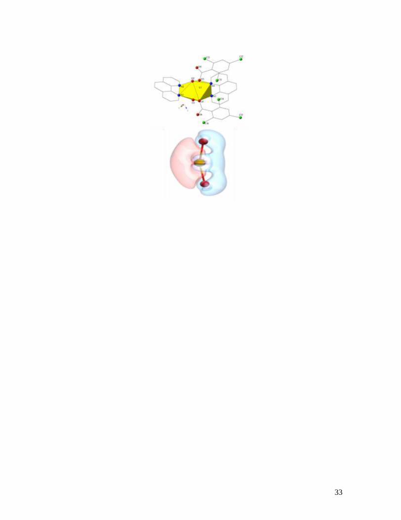
For Table of Contents Only
Three uranyl complexes featuring deviations from linearity of more than 15º have been
synthesized and their structural, supramolecular, spectroscopic, and computational
properties have been comprehensively explored. Additionally, these findings are put into
context via direct comparison with twelve ‘non-bent’ uranyl complexes that were also
prepared as part of this study. Presented is a strategy and general approach to the
manipulation of the O=U=O bond angle via a combination of simple coordination
chemistry and promoted supramolecular interactions.
33