Embed Size (px)
Citation preview

RESEARCH PAPER
How Full-Length FVIII Benefits from Its Heterogeneity – Insightsinto the Role of the B-Domain
Julia Anzengruber1 &Martin Feichtinger2 & Philipp Bärnthaler2 &Norbert Haider2 & Josenato Ilas1 &Nina Pruckner2 & Karima Benamara1 &
Friedrich Scheiflinger1 & Birgit M. Reipert1 & Mantas Malisauskas1
Received: 28 August 2018 /Accepted: 27 February 2019 /Published online: 1 April 2019# The Author(s) 2019
ABSTRACTPurpose To explore how the natural heterogeneity of hu-man coagulation factor VIII (FVIII) and the processing ofits B-domain specifically modulate protein aggregation.Methods Recombinant FVIII (rFVIII) molecular speciescontaining 70% or 20% B-domain, and B-domain-deleted rFVIII (BDD-rFVIII), were separated from full-length recombinant FVIII (FL-rFVIII). Purified humanplasma-derived FVIII (pdFVIII) was used as a comparator.Heterogeneity and aggregation of the various rFVIII mo-lecular species, FL-rFVIII and pdFVIII were analysed bySDS-PAGE, dynamic light scattering, high-performancesize-exclusion chromatography and flow cytometry-basedparticle analysis.Results FL-rFVIII and pdFVIII were heterogeneous in na-ture and demonstrated similar resistance to aggregationunder physical stress. Differences were observed betweenthese and among rFVIII molecular species. FVIII molecu-lar species exhibited diverging aggregation pathways de-pendent on B-domain content. The propensity to formaggregates increased with decreasing proportions of
B-domain, whereas the opposite was observed for oligomerformation. Development of cross-β sheet-containing aggre-gates in BDD-rFVIII induced effective homologousseeding and faster aggregation. Naturally heterogeneousFL-rFVIII and pdFVIII displayed the lowest propensityto aggregate in all experiments.Conclusions These results demonstrate that pdFVIII andFL-rFVIII have similar levels of molecular heterogeneity,and suggest that heterogeneity and the B-domain are in-volved in stabilising FVIII by modulating its aggregationpathway.
KEY WORDS blood proteins . factor VIII . haemophilia A .protein aggregates . protein stability
ABBREVIATIONSB20-rFVIII 20% B-domain recombinant factor VIIIB70-rFVIII 70% B-domain recombinant factor VIIIB100-rFVIII 100% B-domain recombinant factor VIIIBDD-rFVIII B-domain-deleted recombinant factor VIIICHO Chinese hamster ovaryDLS Dynamic light scatteringFL-rFVIII Full-length recombinant factor VIIIFVIII Factor VIIIHC Heavy chainHDX Hydrogen/deuterium exchangeHPLC High-performance liquid chromatographyLC Light chainMS Mass spectrometrypdFVIII Plasma-derived factor VIIIrFVIII Recombinant factor VIIISDS-PAGE Sodium dodecyl sulfate–polyacrylamide
gel electrophoresisSEC Size-exclusion chromatographyThT Thioflavin TVWF Von Willebrand factor
Electronic supplementary material The online version of this article(https://doi.org/10.1007/s11095-019-2599-2) contains supplementarymaterial, which is available to authorized users.
* Julia [email protected]
Birgit M. [email protected]
1 Research & DevelopmentBaxalta Innovations GmbH, a Takeda company, Vienna, Austria
2 Technical OperationsBaxalta Innovations GmbH, a Takeda company, Vienna, Austria
Pharm Res (2019) 36: 77https://doi.org/10.1007/s11095-019-2599-2
INTRODUCTION
Human factor VIII (FVIII) is an essential plasma glycopro-tein in the blood coagulation cascade, serving as a co-factorfor factor IXa in the conversion of factor X to factor Xa(1,2). A defect or deficiency in FVIII results in haemophiliaA, one of the most common severe bleeding disorders (3).
Recombinant protein technology has generated recom-binant FVIII (rFVIII) products to treat haemophilia A byprotein replacement. Products differ mainly in glycosyla-tion (4) and the presence or absence of the B-domain se-quence in the FVIII cDNA, commonly referred to as full-length (FL-) rFVIII and B-domain-deleted (BDD-) rFVIII(5–9).
All protein-based drugs, including FVIII, bear a certainrisk to aggregate during manufacturing and shelf storage,and a susceptibility to mishandling during treatment(10,11). In the clinical setting, the presence of aggregatesin protein therapeutics has induced unwanted immune re-sponses in some patients, which may affect the therapy’sefficacy (12–20).
FVIII is mainly produced by liver sinusoidal endothelialcells (21) as a large single-chain protein comprised of thedomain structure NH2-A1-a1-A2-a2-B-a3-A3-C1-C2-COOH. Different intra- and extracellular processing ofthe B-domain causes different heterodimeric molecularspecies to circulate in plasma. Thus, FVIII contains aconstant-sized light chain (LC) (a3-A3-C1-C2) and a heavychain (HC), minimally composed of the A1-a1-A2-a2 do-mains, but variable in size due to the presence of some orall of the adjacent B-domain (5) (Fig. 1a). HCs and LCs areassociated via a non-covalent linkage that requires a diva-lent metal ion (22,23).
The FVIII B-domain is heavily glycosylated and al-though dispensable for procoagulant activity (24), appearsto have functional roles throughout FVIII’s lifecycle (25).The B-domain may be involved in intracellular interac-tions that regulate quality control and secretion (26–28)and in regulation within plasma during activation andclearance (29–31). It has little effect on the overall FVIIIsecondary structure in solution (32).
We investigated the B-domain’s influence and thenatural heterogeneity originating from its presence inFL-rFVIII and human plasma-derived factor VIII(pdFVIII) on protein stability and aggregation. We studiedthe structural characteristics of the B-domain, and com-pared FL-rFVIII, pdFVIII and purified rFVIII molecularspecies with variable B-domain content with regard totheir aggregation behaviour upon physical stress. Basedon our observations, we built a schematic model ofFL-rFVIII and BDD-rFVIII aggregation and suggest anew role for the B-domain and molecular heterogeneityin ensuring the stability of the FVIII molecule.
MATERIALS AND METHODS
FVIII Samples and Chemicals
Human FL-rFVIII (0.23 mg/ml) used was an intermediatematerial from a production line for a commercial rFVIIIproduct expressed in Chinese hamster ovary (CHO) cellsand was provided by Baxalta Innovations GmbH, a Takedacompany, Vienna, Austria. Vials of historical lots of commer-cially available human rFVIII as well as commercially avail-able lyophilised human plasma FVIII product were providedby Baxalta Innovations GmbH, a Takeda company, Vienna,Austria. Chemicals were purchased from Sigma-Aldrich, St.Louis, MO, USA.
Purification of pdFVIII and rFVIII Molecular Species
VonWillebrand factor (VWF)-free pdFVIII was purified froma commercially available lyophilised plasma FVIII product,which was provided by Baxalta Innovations GmbH, aTakeda company, Vienna, Austria. Multiple vials of the com-mercially available lyophilised human pdFVIII product werereconstituted and pooled to achieve a homogeneous startingmaterial. FVIII was captured on an anti-FVIII affinity columnand further processed using strong cation exchange chroma-tography. Another purification step on a strong anion ex-change resin was performed for buffer exchange and to in-crease the FVIII concentration.
rFVIII molecular species with different degrees of B-domaintruncation were isolated from an intermediate material of acommercially available human FL-rFVIII product expressedin CHO cells, which was provided by Baxalta InnovationsGmbH, a Takeda company, Vienna, Austria. A high-resolution anion exchange chromatography step with a flat gra-dient was used to pre-separate the entities. Pools with enrichedsub-species were generated and further purified by preparativesize-exclusion chromatography (SEC), followed by concentra-tion and buffer exchange on a strong anion exchange resin.
Sodium Dodecyl Sulfate–Polyacrylamide GelElectrophoresis (SDS-PAGE)
SDS-PAGE was carried out using Novex NuPAGE (ThermoFisher Scientific, Waltham, MA, USA). Samples were mixedwith 0.5 M iodoacetamide and incubated for 30 min at 37°C.NuPAGE lithium dodecyl sulfate (LDS) sample buffer andNuPAGE reducing agent were added to the reaction mixtureand incubation was continued for 30 min at 37°C. Samplesand Precision Plus unstained protein standard (Bio-Rad,Hercules, CA, USA) were loaded onto a 7%Tris-acetate minigel. Electrophoresis was performed for 90 min at 150 V.Protein bands were visualised with SilverQuest silver stainingkit (Thermo Fisher Scientific).
77 Page 2 of 12 Pharm Res (2019) 36: 77
In Silico Protein Analysis
BioAnnotator from Vector NTI Advance 11 (Thermo FisherScientific, Waltham, MA, USA) was used to calculate theaverage hydropathicity of the B-domain.
Hydrogen/Deuterium Exchange Mass Spectrometry
Local amide hydrogen/deuterium exchange (HDX) kineticswere followed after 3, 10 and 30 s; 2, 10 and 60 min; 3 h; and3 days of incubation. All HDX reactions were performed at22°C, except for the 3-s reaction (6°C). Human rFVIII con-taining 70% B-domain (B70-rFVIII) was mixed with deuter-ated buffer (Tris, pH 6.7, containing CaCl2 and NaCl). Thereaction was stopped with ice-cold phosphate buffer, pH 2.3,containing 100 mM Tris(2-carboxyethyl)phosphine and3.3 M urea, and by subsequent snap freezing in liquid nitro-gen. Samples were digested using a high-performance liquidchromatography (HPLC) column (ACE, Aberdeen, UK)packed with pepsin-agarose from porcine gastric mucosa(Sigma-Aldrich) and desalted on a C18 pre-column (ACE).Peptic peptides were subjected to liquid chromatographycoupled to mass spectrometry (MS) using a HALO C18/1.8 μm column (Advanced Materials Technology,Wilmington, DE, USA). Peptides were eluted by an aceto-nitrile gradient and analysed on an Orbitrap XL MS(60,000 resolution at m/z 400; Thermo FisherScientific). Peptic peptides were identified by three in-dependent liquid chromatography-MS/MS analyses of anon-deuterated sample using the same procedure as for thedeuterated samples.
FVIII Chromogenic Activity
FVIII samples were dialysed against phosphate-buffered sa-line containing 0.9 mM CaCl2 and 0.5 mM MgCl2. FVIIIactivity was measured by chromogenic assay using commer-cially available reagents (Siemens Healthcare, Erlangen,Germany) on an automated coagulation analyser (BCS XP;Siemens Healthcare). The reference standard was commer-cially available FL-rFVIII (Baxalta Innovations GmbH, aTakeda company), calibrated against the World HealthOrganization international standard.
Preparation of FVIII Aggregates
All FVIII samples were dialysed against phosphate-bufferedsaline containing 0.9 mM CaCl2 and 0.5 mM MgCl2. Toensure reproducibility, all experiments were performed atleast twice.
Temperature-Dependent Aggregation
All FVIII samples with protein concentrations of either0.122 μM for high-performance size-exclusion chroma-tography (HPLC-SEC) or 0.61 μM for dynamic lightscattering (DLS) were incubated at 25, 30, 35, 40, 45or 50°C for 20 h in polystyrene microplates (CorningIncorporated – Life Sciences, Tewksbury, MA, USA)covered with plate sealers in a Synergy H4 HybridReader (BioTek, Winooski, VT, USA) with 20 s medi-um shaking every 10 min. Samples were subsequently frozenat −80°C until analysis.
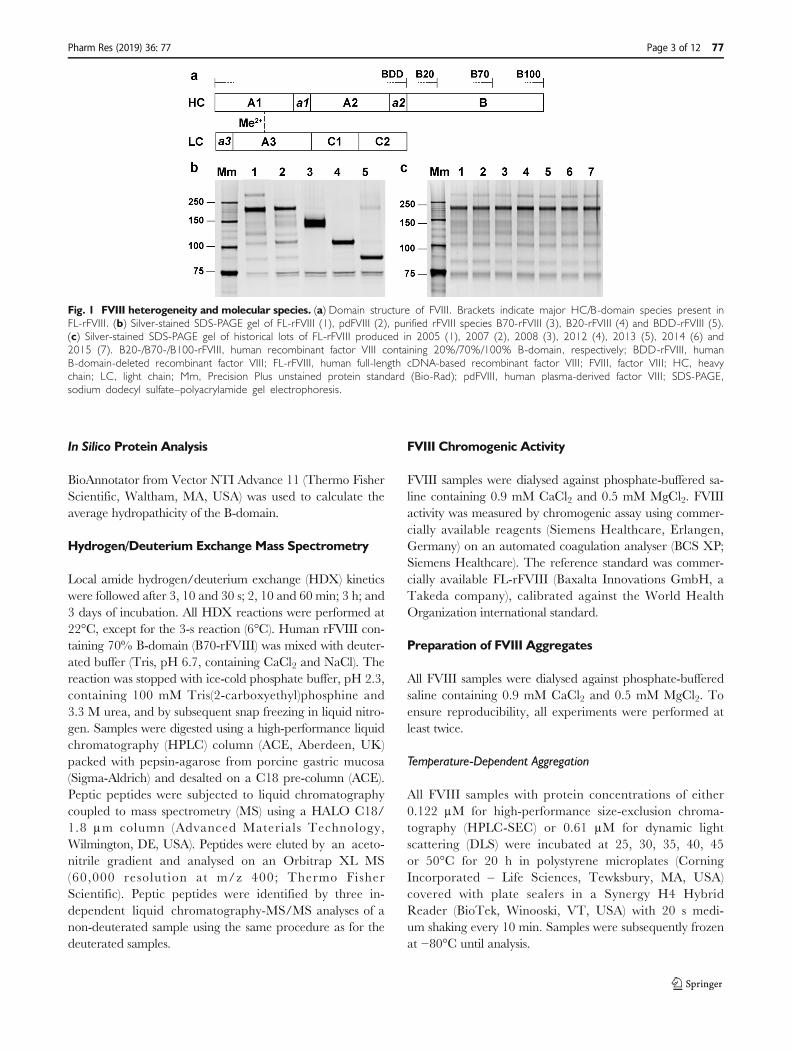
Fig. 1 FVIII heterogeneity and molecular species. (a) Domain structure of FVIII. Brackets indicate major HC/B-domain species present inFL-rFVIII. (b) Silver-stained SDS-PAGE gel of FL-rFVIII (1), pdFVIII (2), purified rFVIII species B70-rFVIII (3), B20-rFVIII (4) and BDD-rFVIII (5).(c) Silver-stained SDS-PAGE gel of historical lots of FL-rFVIII produced in 2005 (1), 2007 (2), 2008 (3), 2012 (4), 2013 (5), 2014 (6) and2015 (7). B20-/B70-/B100-rFVIII, human recombinant factor VIII containing 20%/70%/100% B-domain, respectively; BDD-rFVIII, humanB-domain-deleted recombinant factor VIII; FL-rFVIII, human full-length cDNA-based recombinant factor VIII; FVIII, factor VIII; HC, heavychain; LC, light chain; Mm, Precision Plus unstained protein standard (Bio-Rad); pdFVIII, human plasma-derived factor VIII; SDS-PAGE,sodium dodecyl sulfate–polyacrylamide gel electrophoresis.
Pharm Res (2019) 36: 77 Page 3 of 12 77

Time-Dependent Aggregation at 45°C
All FVIII samples (0.122 μM)were incubated at 45°C for 24 hin polystyrenemicroplates (Corning) coveredwith plate sealersin a plate thermo shaker (Biosan, Riga, Latvia). Samples werewithdrawn after various time intervals and immediately frozenat−80°C until HPLC-SEC analysis.
Homologous Seeding of FVIII Aggregation
To prepare seeds, FVIII samples (0.122 μM) were incubatedfor 2, 5, 8 or 18 h at 45°C in polystyrene microplates(Corning) covered with plate sealers in a plate thermo shaker(Biosan). Native FVIII samples (0.122 μM) were mixed 1:1with the corresponding seeds and time-dependent aggregationat 45°C was initiated. Samples were stored at −80°C untilHPLC-SEC analysis.
Agitation and Shear Stress-Induced Aggregation
Samples (0.244 μM) were hand agitated for 10 min in a dis-posable Omnifix syringe (Braun, Melsungen, Germany).Shear stress was induced by injecting the solution through‘winged infusion sets with needle protection’ (23G ×¾″;L = 35 cm, V = 0.25 ml) from Terumo Europe, Leuven,Belgium, after which all samples were stored at −80°C untilflow cytometry-based particle analysis.
DLS
DLS was performed using a Malvern NanoZetasizer ZSP(Malvern Instruments, Malvern, UK). Samples (0.244 μM)were centrifuged (Centrifuge 5415C; Eppendorf, Vienna,Austria) at 10,000 rpm for 5 min and filled into a ZEN0040disposable microcuvette. Operation temperature was set at25°C with an equilibration time of 2 min. The anglewas set to 173° backscatter to determine the hydrody-namic diameter of a protein and thus, the effective sizeof proteins. A minimum of three runs per sample were mea-sured to obtain an average result.
HPLC-SEC
HPLC-SEC was performed using a 7.8 × 300 mm TSKgelG4000SWxl column (Tosoh Bioscience, Tokyo, Japan) and a6 × 40mmTSK guard column (Tosoh Bioscience) coupled toan HPLC 1260 infinity system (Agilent Technologies, SantaClara, CA, USA). SEC was carried out under isocratic condi-tions at a flow rate of 0.3 ml/min using a buffer consisting of50 mM Tris-HCl, 5 mM CaCl2, 400 mM NaCl and 0.05%NaN3, pH 7.0. A sample volume of 100 μl (0.122 μM proteinconcentration) wasmixed with 3 μl Thioflavin T (ThT; 1mM)and subsequently loaded onto the column. To monitor the
elutionof theproteinwith fluorescencedetection, theexcitationand emission wavelengths were set to 280 nm and zero order,respectively. ThT fluorescence was monitored with 440-nmexcitation and zero-order emission. Peaks eluting with the voidvolume (retention time 18.0–21.2min), with retention times of21.2–27.0 min and 27.0–43.0 min, were designated as solubleprotein aggregates, oligomers and monomers, respectively.The amount of each was calculated as a percentage of the totalarea of all peaks in the chromatogram.ThTbindingwas calcu-lated as the ratio between ThT and intrinsic protein fluores-cence signals. The protein-based gel filtration standard (Bio-Rad) was analysed in between samples to monitor optimal col-umnperformance.All sampleswere analysed in randomorder.
Curve Fitting and Statistical Analysis
Curve fitting was computed by GraphPad Prism 6(GraphPad Software, La Jolla, CA, USA). Kinetic rate con-stants for oligomerisation (koligo [h−1]) were derived byfitting data to the one-phase association model accordingto: y = y0 + (plateau-y0)*(1-exp[−koligo*x]); y = oligomer amount(%); x = time (h); y0 = y value when x is zero; plateau= y value atinfinite times. Aggregation rates (kagg [h
−1]) were derived byfitting data to the Boltzmann sigmoidal model according to:y = ymin + (ymax-ymin)/(1 + exp[(x1/2-x)/(1/kagg)]); y = aggregateamount (%); ymin = y during lag phase; ymax = y after aggregationended; x = time (h); x1/2 = time at half-maximum y (33).
The statistical differences in Figure 6 were computed byGraphPad Prism 6 using unpaired t-test.
Flow Cytometry-Based Particle Analysis
A flow cytometry-based particle analysis method was used todetect sub-visible particles 0.75–70 μm in size, as previouslydescribed (34). A combination of size calibration beads(Fluoresbrite® YG Carboxylate Size Range beads;Polysciences Inc., Warrington, PA, USA), counting beads(CountBright™ Absolute Counting Beads; InvitrogenCorporation, Carlsbad, CA, USA) and fluorescent probeswas used to characterise sub-visible particles. To distinguishprotein and protein-containing particles from non-proteinsub-visible particles, samples were stained with the fluorescentdye 4,4′-Dianilino-1,1′-binaphthyl-5,5′-disulfonic aciddipotassium salt (Bis-ANS).
RESULTS
Similar Heterogeneity of pdFVIII and FL-rFVIII
Figure 1a provides a schematic overview of the multi-domain structure of FVIII. Brackets indicate molecular spe-cies resulting from complex post-translational processing
77 Page 4 of 12 Pharm Res (2019) 36: 77

within the B-domain. rFVIII molecular species containing100% (B100-), 70% (B70-), 20% (B20-) or 0% B-domainwere the main rFVIII species found in FL-rFVIII, withHC migration levels at 180, 150, 110 and 90 kDa, respec-tively (Fig. 1b). Percentages of B-domain content (100%,70% or 20%) in rFVIII species nomenclature were calcu-lated on the basis of the apparent molecular mass of theB-domain of the respective rFVIII HC derived from theSDS-PAGE gel migration levels. The B-domain of theB100-HC (180 kDa, lane 1) was calculated to have90 kDa and contain 100% of total B-domain; that of theB70-HC (150 kDa, lane 3) was 60 kDa and ~70% of totalB-domain, and that of the B20-HC (110 kDa, lane 4) was20 kDa and ~20% of total B-domain.
pdFVIII isolated from pooled human plasma was highlypurified, whereby VWF was depleted to 7.5 μg VWF/mgFVIII. pdFVIII and CHO-derived FL-rFVIII showed almostidentical heterogenic protein profiles on the silver-stainedSDS-PAGE gel (Fig. 1b, lanes 1–2). Both displayed the mostintense band at ~180 kDa, indicating glycosylated B100-HCspecies, and several truncated HC/B-domain species withlower molecular weight migrating at comparable levels. Thepresence of the main molecular FVIII species in FL-rFVIIIand pdFVIII was confirmed in SEC profiles (Fig. S1). In ad-dition, the heterogeneity in FL-rFVIII was consistent for lotsproduced between 2005 and 2015 (Fig. 1c).
Specific FVIII activity of FL-rFVIII and pdFVIII was4380 ± 868 and 4457 ± 493 IU/mg using chromogenicassay.
Purification and Characterisation of rFVIII MolecularSpecies
BDD-rFVIII, B20-rFVIII and B70-rFVIII were isolated to95%, 94% and 92% purity, respectively, based on C4HPLC analysis (data not shown). The HC of B70-rFVIII,B20-rFVIII and BDD-rFVIII exhibited apparent molecularweights of 150, 110 and 90 kDa on the SDS-PAGE gel, dueto varying amounts of B-domain (Fig. 1b, lanes 3–5). An ap-parent molecular weight band of 75 kDa was observed for theLC of each species. The integrity of the purified rFVIII specieswas confirmed by HPLC-SEC (Fig. S1). B100-FVIII couldnot be purified to sufficient quantity and was not used in thisstudy.
Specific FVIII activity was 5088 ± 403, 5202 ± 317and 6261 ± 263 IU/mg for BDD-rFVIII, B20-rFVIIIand B70-rFVIII, as measured by chromogenic assay.
B-Domain Structural Characteristics
The B-domain has an amino acid sequence with lowoverall hydrophobicity. The average hydropathicity cal-culated according to the Kyte-Doolittle method (35) was
−0.751 for the total sequence. Clusters of low hydropho-bicity were evenly distributed. B-domain sequences inB100-, B70- and B20-rFVIII exhibited similar averagehydropathicity values of −0.779, −0.741 and − 0.896, re-spectively. Low hydrophobicity characterises natively un-folded proteins (36).
B70-rFVIII was subjected to HDX-MS; the kinetics of 120peptides from the total protein sequence, including nine pep-tides from the B-domain sequence, were measured (Table S1).All peptides from sequences belonging to the B-domain dem-onstrated rapid kinetics of deuterium incorporation. Even atthe shortest incubation time (3 s), all peptides incorporated thesame amount of deuterium as the corresponding fully deuter-ated sample after 3 days.
TheHDX-MS data together with the amino acid sequencecharacteristics indicate that the B-domain of B70-rFVIII isintrinsically disordered and flexible. Based on the in silico anal-ysis, similar solvent exposure and flexibility is expected for thetotal B-domain and several B-domain truncations present inFL-rFVIII and pdFVIII.
Aggregation Behaviour of FL-rFVIII and rFVIIIMolecular Species at Elevated Temperatures
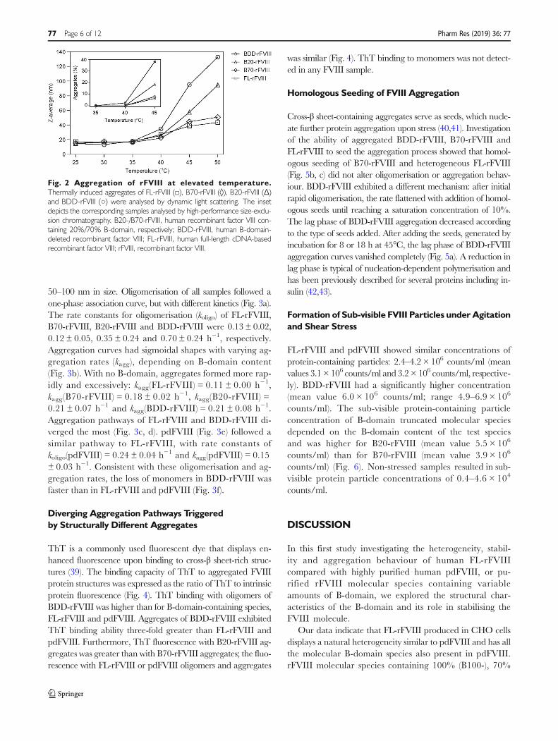
FL-rFVIII, B70-rFVIII, B20-rFVIII and BDD-rFVIII wereexposed to temperatures of 25–50°C and analysed by DLSand HPLC-SEC (Fig. 2). Starting at 40°C, an increase inZ-average, describing the intensity weighted mean hydrody-namic diameter of protein aggregates, was observed for allitems, with clear differences between rFVIII samples. Themean aggregate size increased with decreasing B-domaincontent (aggregation propensity order BDD-rFVIII >B20-rFVIII > B70-rFVIII > FL-rFVIII). FL-rFVIII, beinga heterogenic mixture, exhibited the lowest propensity toaggregate. The fold increase of the aggregate average sizewas 9.2 for BDD-rFVIII and 6.4 for B20-rFVIII, but only3.4 for B70-rFVIII and 2.5 for FL-rFVIII. The same trendin aggregation was shown by HPLC-SEC analysis (Fig. 2inset). Furthermore, SDS-PAGE showed that FL-rFVIII ag-gregates, separated from FL-rFVIII monomers byHPLC-SEC, contained each rFVIII molecular species in asimilar ratio to native FL-rFVIII (Fig. S2).
FVIII Aggregation Pathways Depend on MolecularHeterogeneity and B-Domain Content
Detailed time-dependent aggregation analysis was performed at45°C, the temperature at which conformational changes in theFVIII molecule are initiated (37, 38). Aggregation kinetics werefollowed by HPLC-SEC and showed clear differences in aggre-gation and oligomerisation pathways between rFVIII molecularspecies and FL-rFVIII (Fig. 3). Based on the exclusion limit of theanalysis column, oligomers were 10–50 nm and aggregates
Pharm Res (2019) 36: 77 Page 5 of 12 77
50–100 nm in size. Oligomerisation of all samples followed aone-phase association curve, but with different kinetics (Fig. 3a).The rate constants for oligomerisation (koligo) of FL-rFVIII,B70-rFVIII, B20-rFVIII and BDD-rFVIII were 0.13 ± 0.02,0.12 ± 0.05, 0.35 ± 0.24 and 0.70 ± 0.24 h−1, respectively.Aggregation curves had sigmoidal shapes with varying ag-gregation rates (kagg), depending on B-domain content(Fig. 3b). With no B-domain, aggregates formed more rap-idly and excessively: kagg(FL-rFVIII) = 0.11 ± 0.00 h−1,kagg(B70-rFVIII) = 0.18 ± 0.02 h−1, kagg(B20-rFVIII) =0.21 ± 0.07 h−1 and kagg(BDD-rFVIII) = 0.21 ± 0.08 h−1.Aggregation pathways of FL-rFVIII and BDD-rFVIII di-verged the most (Fig. 3c, d). pdFVIII (Fig. 3e) followed asimilar pathway to FL-rFVIII, with rate constants ofkoligo(pdFVIII) = 0.24 ± 0.04 h−1 and kagg(pdFVIII) = 0.15± 0.03 h−1. Consistent with these oligomerisation and ag-gregation rates, the loss of monomers in BDD-rFVIII wasfaster than in FL-rFVIII and pdFVIII (Fig. 3f).
Diverging Aggregation Pathways Triggeredby Structurally Different Aggregates
ThT is a commonly used fluorescent dye that displays en-hanced fluorescence upon binding to cross-β sheet-rich struc-tures (39). The binding capacity of ThT to aggregated FVIIIprotein structures was expressed as the ratio of ThT to intrinsicprotein fluorescence (Fig. 4). ThT binding with oligomers ofBDD-rFVIII was higher than for B-domain-containing species,FL-rFVIII and pdFVIII. Aggregates of BDD-rFVIII exhibitedThT binding ability three-fold greater than FL-rFVIII andpdFVIII. Furthermore, ThT fluorescence with B20-rFVIII ag-gregates was greater than with B70-rFVIII aggregates; the fluo-rescence with FL-rFVIII or pdFVIII oligomers and aggregates
was similar (Fig. 4). ThT binding to monomers was not detect-ed in any FVIII sample.
Homologous Seeding of FVIII Aggregation
Cross-β sheet-containing aggregates serve as seeds, which nucle-ate further protein aggregation upon stress (40,41). Investigationof the ability of aggregated BDD-rFVIII, B70-rFVIII andFL-rFVIII to seed the aggregation process showed that homol-ogous seeding of B70-rFVIII and heterogeneous FL-rFVIII(Fig. 5b, c) did not alter oligomerisation or aggregation behav-iour. BDD-rFVIII exhibited a different mechanism: after initialrapid oligomerisation, the rate flattened with addition of homol-ogous seeds until reaching a saturation concentration of 10%.The lag phase of BDD-rFVIII aggregation decreased accordingto the type of seeds added. After adding the seeds, generated byincubation for 8 or 18 h at 45°C, the lag phase of BDD-rFVIIIaggregation curves vanished completely (Fig. 5a). A reduction inlag phase is typical of nucleation-dependent polymerisation andhas been previously described for several proteins including in-sulin (42,43).
Formation of Sub-visible FVIII Particles under Agitationand Shear Stress
FL-rFVIII and pdFVIII showed similar concentrations ofprotein-containing particles: 2.4–4.2 × 10⁶ counts/ml (meanvalues 3.1 × 10⁶ counts/ml and 3.2× 10⁶ counts/ml, respective-ly). BDD-rFVIII had a significantly higher concentration(mean value 6.0 × 10⁶ counts/ml; range 4.9–6.9 × 10⁶
counts/ml). The sub-visible protein-containing particleconcentration of B-domain truncated molecular speciesdepended on the B-domain content of the test speciesand was higher for B20-rFVIII (mean value 5.5 × 10⁶
counts/ml) than for B70-rFVIII (mean value 3.9 × 10⁶
counts/ml) (Fig. 6). Non-stressed samples resulted in sub-visible protein particle concentrations of 0.4–4.6 × 10⁴
counts/ml.
DISCUSSION
In this first study investigating the heterogeneity, stabil-ity and aggregation behaviour of human FL-rFVIIIcompared with highly purified human pdFVIII, or pu-rified rFVIII molecular species containing variableamounts of B-domain, we explored the structural char-acteristics of the B-domain and its role in stabilising theFVIII molecule.
Our data indicate that FL-rFVIII produced in CHO cellsdisplays a natural heterogeneity similar to pdFVIII and has allthe molecular B-domain species also present in pdFVIII.rFVIII molecular species containing 100% (B100-), 70%
Fig. 2 Aggregation of rFVIII at elevated temperature.Thermally induced aggregates of FL-rFVIII (□), B70-rFVIII (◊), B20-rFVIII (Δ)and BDD-rFVIII (○) were analysed by dynamic light scattering. The insetdepicts the corresponding samples analysed by high-performance size-exclu-sion chromatography. B20-/B70-rFVIII, human recombinant factor VIII con-taining 20%/70% B-domain, respectively; BDD-rFVIII, human B-domain-deleted recombinant factor VIII; FL-rFVIII, human full-length cDNA-basedrecombinant factor VIII; rFVIII, recombinant factor VIII.
77 Page 6 of 12 Pharm Res (2019) 36: 77

(B70-), 20% (B20-) or 0% B-domain were the main rFVIIIspecies found in FL-rFVIII and pdFVIII in the present study.Jankowski et al. previously described the terminating aminoacid positions of the B-domain truncations of the mainrFVIII species present in FL-rFVIII as Arg1313, Arg1115,Ser817 and Ser740, which likely correspond to the terminatingamino acids of B100-, B70-, B20- and BDD-rFVIII HCs pre-sented in this study. The B-domain part spanning fromArg1313 to Arg1648 is most likely completely removed aftercleavage as it was not detected among the secreted FVIIIHC species (5).
Interestingly, FL-rFVIII produced in a baby hamster kidneycell line exhibits a slightly different protein profile regardingband intensity and migration level than CHO cell-derivedFL-rFVIII and pdFVIII (5). In general, FVIII’s heterogeneity,even with minor differences in the HC/B-domain truncationlength, is species independent. This was true for humanpdFVIII and FL-rFVIII in this and previous work (5) and alsofor porcine pdFVIII (44). In contrast, the purified BDD-rFVIIIspecies used in this study, and marketed BDD-rFVIII products,exhibit a monogenic protein pattern and thus large differencesfrom heterogenic pdFVIII (6–8).
Fig. 3 Pathways of rFVIIIoligomer and aggregateformation. FL-rFVIII (□, d),B70-rFVIII (◊), B20-rFVIII (Δ),BDD-rFVIII (○, c) and pdFVIII (*, e)were incubated at 45°C for 24 h.The amount of oligomers (green,a), aggregates (red, b) andmonomers (blue, f) wascontinuously analysed by high-performance size-exclusionchromatography and plotted againsttime of incubation. B20-/B70-rFVIII,human recombinant factor VIIIcontaining 20%/70% B-domain,respectively; BDD-rFVIII, human B-domain-deleted recombinant factorVIII; FL-rFVIII, human full-lengthcDNA-based recombinant factorVIII; pdFVIII, human plasma-derivedfactor VIII; rFVIII, recombinant factorVIII.
Fig. 4 Binding of the fluorescent dye ThT to oligomers and aggre-gates of FVIII. The binding capacity of ThT to protein oligomers and ag-gregates is expressed as the ratio of the fluorescent signals at 440- and 280-nmexcitation after 24 h of incubation at 45°C. n=2–4, error bars indicate SDvalues. B20-/B70-rFVIII, human recombinant factor VIII containing 20%/70%B-domain, respectively; BDD-rFVIII, human B-domain-deleted recombinantfactor VIII; FL-rFVIII, human full-length cDNA-based recombinant factor VIII;FVIII, factor VIII; pdFVIII, human plasma-derived factor VIII; ThT, Thioflavin T.
Pharm Res (2019) 36: 77 Page 7 of 12 77

FL-rFVIII and pdFVIII demonstrated the lowest propen-sity to form aggregates. The aggregation tendency increasedwith decreasing B-domain content of the rFVIII molecularspecies. Detailed time-dependent analysis of oligomerisationand aggregation under thermal stress revealed diverging path-ways for different FVIII samples. The slow oligomerisation ofthermally stressed FL-rFVIII and pdFVIII nearly inhibitedaggregation. The kinetics of oligomer and aggregate forma-tion were more rapid with BDD-rFVIII. ThT-positive cross-βsheet-rich structures were detected in thermally inducedBDD-rFVIII oligomers and aggregates, but less so or not atall in B-domain-containing FVIII. It is likely that cross-β sheetsin BDD-rFVIII oligomers trigger extensive aggregation andfacilitate homologous seeding of BDD-rFVIII aggregation,but not in the other FVIII samples.
Based on these observations, we built a schematic model de-scribing the diverging pathways of BDD-rFVIII and FL-rFVIIIoligomer and aggregate formation (Fig. 7). While the startingmaterial in FL-rFVIII is a heterogenic mixture of all rFVIII
species, BDD-rFVIII exists only as one species. Arrow lengthsin the model indicate oligomerisation and aggregation rates,both of which are much faster for BDD-rFVIII than forFL-rFVIII. While BDD-rFVIII forms ordered, large cross-βsheet-rich aggregates, FL-rFVIII aggregates lack this repetitivenature and are smaller in size. The assembly of soluble proteinsinto ordered cross-β sheet-containing structures is seen in manyhuman neurodegenerative diseases, such as Alzheimer’s diseaseand Parkinson’s disease, and in spongiform encephalopathies(45). Development of such disorders has been associated withthe seeding ability of respective accumulated protein aggregates(41).
Supported by a previous study in which the B-domain hadlittle to no effect on the overall FVIII secondary structure insolution as measured by far UV circular dichroism spectros-copy (32), the present study demonstrates that the B-domain issolvent exposed, disordered and flexible.We thus propose thatthe B-domain has an aggregation-protective function forFVIII, similar to that in other proteins with significantly
Fig. 5 Homologous seeding ofrFVIII aggregation. Seeds wereprepared by incubation of BDD-rFVIII, B70-rFVIII and FL-rFVIII for 2,5, 8 or 18 h at 45°C. BDD-rFVIII(a), B70-rFVIII (b) and FL-rFVIII (c)samples were mixed 1:1 withrespective pre-formed seeds andincubated at 45°C for 24 h. Theamount of oligomers (green) andaggregates (red) was continuouslyanalysed by high-performancesize-exclusion chromatography andplotted against incubation time.B70-rFVIII, human recombinantfactor VIII containing 70% B-domain; BDD-rFVIII, human B-domain-deleted recombinant factorVIII; FL-rFVIII, human full-lengthcDNA-based recombinant factorVIII; rFVIII, recombinant factor VIII.
77 Page 8 of 12 Pharm Res (2019) 36: 77

disordered segments, such as α-synuclein. The natively un-folded C-terminal region of α-synuclein was shown to be es-sential in stabilising the protein. Aggregation of α-synucleinwas clearly dependent on the length of the C-terminal regionand decreased with increasing content of the disordered re-gion (46–48).
Heterogeneity, as shown in FL-rFVIII and pdFVIII, causeslower sequence similarity between FVIII species. Sequence di-versity in proteins has already been demonstrated in previousstudies as being essential in reducing aggregation susceptibilityand seeding processes. Wright and co-workers’ findings on the
multi-domain protein titin showed that the co-aggregation effi-ciency among different domains decreases markedly with de-creasing sequence identity. Furthermore, they claimed thatmaintaining a low sequence identity between proteins is animportant evolutionary characteristic that strongly inhibits ag-gregation in the crowded environment of a living system (49).Similarly, the efficiency of seeding in the fibril formation oflysozymes was shown to strongly depend on the similarity oftheir sequences (50).
Using agitation and shear stress to mimic mishandling ofFVIII therapeutic products, and facilitation of the interactionof proteins with silicone oil, which is often present on the interiorsurfaces of syringes (34,51–53), showed that sub-visible protein-containing particle formation is clearly dependent on B-domaincontent. Particle concentrations in FL-rFVIII and pdFVIIIreached similar levels, whereas significantly more particles weredetected in BDD-rFVIII.
Previously published work demonstrated that glycosylationsignificantly influences the stability of FL-rFVIII, as shown byreduced aggregation resistance of de-glycosylated FL-rFVIII(54). Given that ~80% of the N-glycosylation sites are distrib-uted within the B-domain (23), deletion of this domain is likelyto render the protein more susceptible to aggregation.Manufacturing and formulation specifications further influ-ence aggregation of FVIII and protein therapeutics (55).Large differences in quality attributes, such as aggregate andsub-visible particle concentrations, were observed formarketed rFVIII products that differ mainly in B-domainstructure and manufacturing processes (56).
Protein aggregates not only influence protein drugs’ stabil-ity and shelf-life, but also increase their immunogenicity (55).The repetitive nature of protein aggregates can be detected bypattern recognition receptors or cross-link antigen receptorson immune cells. Anti-drug antibodies may have a
Fig. 7 Schematic model of oligomer and aggregate formation after exposure of rFVIII to thermal stress. FL-rFVIII is depicted as a heteroge-neous mixture of rFVIII species, but does not reflect the actual ratio of species. The length of arrows indicates the differences in oligomerisation and aggregation rates ofFL-rFVIII and BDD-rFVIII. B20-/B70-/B100-rFVIII, human recombinant factor VIII containing 20%/70%/100% B-domain, respectively; BDD-rFVIII, humanB-domain-deleted recombinant factor VIII; FL-rFVIII, human full-length cDNA-based recombinant factor VIII; rFVIII, recombinant factor VIII; ThT, Thioflavin T.
Fig. 6 Formation of protein-containing sub-visible particles. FVIIIsamples (0.244 μM)were exposed to agitation and shear stress and subjectedto flow cytometry-based particle analysis. Statistical differences were shown byusing unpaired t-test. Protein particle concentrations were significantly differentbetween BDD-rFVIII and FL-rFVIII (P=0.0002), BDD-rFVIII and pdFVIII(P= 0.0010), B20-rFVIII and FL-rFVIII (P< 0.0001) and B20-rFVIII andpdFVIII (P<0.0001). n=4–6, error bars indicate SD values. B20-/B70-rFVIII, human recombinant factor VIII containing 20%/70% B-domain,respectively; BDD-rFVIII, human B-domain-deleted recombinant factorVIII; FL-rFVIII, human full-length cDNA-based recombinant factor VIII;FVIII, factor VIII; pdFVIII, human plasma-derived factor VIII.
Pharm Res (2019) 36: 77 Page 9 of 12 77

neutralising effect on the protein, which may affect its potencyor pharmacokinetics, and especially in therapeutics related toan endogenous protein, pose patient safety risks (12).Inhibitory antibodies are formed in approximately one-fifthof patients with haemophilia A treated with FVIII (57,58).In vivo studies in haemophilia A mice showed that proteinaggregates modulate FVIII immunogenicity differently, de-pending on the nature of the aggregates and how they wereformed (59). However, to date no data are available on howprotein aggregates in FVIII products modulate their immuno-genicity in humans or on the immunogenic properties of theoligomers and aggregates characterised in this study.
CONCLUSION
In summary, our data demonstrate similar levels of protein het-erogeneity in FL-rFVIII and pdFVIII, and suggest a beneficialeffect thereof in reducing the protein aggregation susceptibilityupon exposure to physical stress. The B-domain was shown to beinvolved in ensuring the stability of the FVIII molecule by mod-ulating the protein aggregation pathway. These findings shouldbe considered in the design of future FVIII therapeutics to im-prove their stability, shelf-life and, most importantly, their safety.
ACKNOWLEDGMENTS AND DISCLOSURES
The authors thank B. Dass, A. Gringeri, K. Steinitz-Trost andG.D. Spotts for carefully reviewing the manuscript andA. Öhman and HDXperts for HDX-MS analysis.J. Anzengruber, N. Haider, N. Pruckner, F. Scheiflinger andB.M. Reipert are employees of Baxalta Innovations GmbH, aTakeda company. M. Feichtinger, P. Bärnthaler, J. Ilas,K. Benamara and M. Malisauskas were employees of BaxaltaInnovations GmbH, a Takeda company, at the time of the pres-ent study. P. Bärnthaler, F. Scheiflinger, B.M. Reipert andM. Malisauskas own stock in Takeda. J. Anzengruber,M. Feichtinger, P. Bärnthaler, B.M. Reipert andM. Malisauskas have a patent pending relevant to the work:‘Purification of Factor VIII subspecies,’ priority application filedin June 2017.
AUTHOR CONTRIBUTIONS
J. Anzengruber designed the research, performed experiments,analysed and interpreted data and wrote the manuscript.M. Feichtinger, P. Bärnthaler, N. Haider, J. Ilas andN. Pruckner designed and performed experiments.K. Benamara, F. Scheiflinger and B.M. Reipert revised the in-tellectual content. M. Malisauskas designed the research conceptand interpreted results and all authors approved the final manu-script.
OpenAccessThis article is distributed under the terms of theCreative Commons Attribution 4.0 International License(http://creativecommons.org/licenses/by/4.0/), which per-mits unrestricted use, distribution, and reproduction in anymedium, provided you give appropriate credit to the originalauthor(s) and the source, provide a link to the CreativeCommons license, and indicate if changes were made.
REFERENCES
1. Toole JJ, Knopf JL, Wozney JM, Sultzman LA, Buecker JL,Pittman DD, et al. Molecular cloning of a cDNA encoding humanantihaemophilic factor. Nature. 1984;312(5992):342–7.
2. Vehar GA, Keyt B, Eaton D, Rodriguez H, O'Brien DP, Rotblat F,et al. Structure of human factor VIII. Nature. 1984;312(5992):337–42.
3. Mannucci PM, Duga S, Peyvandi F. Recessively inherited coagu-lation disorders. Blood. 2004;104(5):1243–52.
4. Malisauskas M, Anzengruber J, Bärnthaler P, Feichtinger M,Scheiflinger F, Reipert BM. Different N-glycosylation of factorVIII: similarities and differences of plasma derived and recombi-nant factor VIII products. Abstracts of the XXVI congress of theInternational Society on Thrombosis and Haemostasis, July 8–13,2017. Res Pract Thromb Haemost. 2017;1(S1):1–15.
5. Jankowski MA, Patel H, Rouse JC, Marzilli LA, Weston SB,Sharpe PJ. Defining 'full-length' recombinant factor VIII: a com-parative structural analysis. Haemophilia. 2007;13(1):30–7.
6. D'Amici GM, Timperio AM, Gevi F, Grazzini G, Zolla L.Recombinant clotting factor VIII concentrates: heterogeneity andhigh-purity evaluation. Electrophoresis. 2010;31(16):2730–9.
7. Thim L, Vandahl B, Karlsson J, Klausen NK, Pedersen J, KroghTN, et al. Purification and characterization of a new recombinantfactor VIII (N8). Haemophilia. 2010;16(2):349–59.
8. Peters RT, Toby G, Lu Q, Liu T, Kulman JD, Low SC, et al.Biochemical and functional characterization of a recombinant mo-nomeric factor VIII-fc fusion protein. J Thromb Haemost.2013;11(1):132–41.
9. Kannicht C, RamstromM,Kohla G, TiemeyerM,Casademunt E,Walter O, et al. Characterisation of the post-translational modifi-cations of a novel, human cell line-derived recombinant humanfactor VIII. Thromb Res. 2013;131(1):78–88.
10. Joubert MK, Luo Q, Nashed-Samuel Y, Wypych J, Narhi LO.Classification and characterization of therapeutic antibody aggre-gates. J Biol Chem. 2011;286(28):25118–33.
11. Roberts CJ. Therapeutic protein aggregation: mechanisms, design,and control. Trends Biotechnol. 2014;32(7):372–80.
12. Moussa EM, Panchal JP, Moorthy BS, Blum JS, Joubert MK,Narhi LO, et al. Immunogenicity of therapeutic protein aggregates.J Pharm Sci. 2016;105(2):417–30.
Funding
This work was financially supported by Baxalta InnovationsGmbH, a Takeda company, Vienna, Austria. Under the directionof the authors, editorial support for this manuscript was providedby Margit Rezabek, DVM, PhD, employee of Excel Medical Affairs(Southport, CT, USA), and was funded by Baxalta US Inc., aTakeda company, Lexington, MA, USA.
77 Page 10 of 12 Pharm Res (2019) 36: 77

13. Hermeling S, Schellekens H, Crommelin DJ, Jiskoot W. Micelle-associated protein in epoetin formulations: a risk factor for immu-nogenicity? Pharm Res. 2003;20(12):1903–7.
14. van BeersMM, JiskootW, Schellekens H. On the role of aggregatesin the immunogenicity of recombinant human interferon beta inpatients with multiple sclerosis. J Interferon Cytokine Res.2010;30(10):767–75.
15. Barnard JG, BabcockK, Carpenter JF. Characterization and quan-titation of aggregates and particles in interferon-beta products: po-tential links between product quality attributes and immunogenic-ity. J Pharm Sci. 2013;102(3):915–28.
16. Robbins DC, Cooper SM, Fineberg SE, Mead PM. Antibodies tocovalent aggregates of insulin in blood of insulin-using diabeticpatients. Diabetes. 1987;36(7):838–41.
17. Robbins DC, Mead PM. Free covalent aggregates of therapeuticinsulin in blood of insulin-dependent diabetes. Diabetes.1987;36(2):147–51.
18. Maislos M, Mead PM, Gaynor DH, Robbins DC. The source ofthe circulating aggregate of insulin in type I diabetic patients istherapeutic insulin. J Clin Invest. 1986;77(3):717–23.
19. Ahmadi M, Bryson CJ, Cloake EA, Welch K, Filipe V, Romeijn S,et al. Small amounts of sub-visible aggregates enhance the immu-nogenic potential of monoclonal antibody therapeutics. PharmRes.2015;32(4):1383–94.
20. Joubert MK, Hokom M, Eakin C, Zhou L, Deshpande M, BakerMP, et al. Highly aggregated antibody therapeutics can enhancethe in vitro innate and late-stage T-cell immune responses. J BiolChem. 2012;287(30):25266–79.
21. Do H, Healey JF, Waller EK, Lollar P. Expression of factor VIII bymurine liver sinusoidal endothelial cells. J Biol Chem. 1999;274(28):19587–92.
22. Kaufman RJ, Wasley LC, Dorner AJ. Synthesis, processing, andsecretion of recombinant human factor VIII expressed in mamma-lian cells. J Biol Chem. 1988;263(13):6352–62.
23. Fay PJ. Factor VIII structure and function. Int J Hematol.2006;83(2):103–8.
24. Toole JJ, Pittman DD, Orr EC, Murtha P, Wasley LC, KaufmanRJ. A large region (approximately equal to 95 kDa) of human factorVIII is dispensable for in vitro procoagulant activity. Proc NatlAcad Sci U S A. 1986;83(16):5939–42.
25. Pipe SW. Functional roles of the factor VIII B domain.Haemophilia. 2009;15(6):1187–96.
26. Pipe SW, Morris JA, Shah J, Kaufman RJ. Differential interactionof coagulation factor VIII and factor V with protein chaperonescalnexin and calreticulin. J Biol Chem. 1998;273(14):8537–44.
27. Pipe SW, Miao HZ, Kucab PF, McVey JH, Kaufman RJ. Thesecretion efficiency of factor VIII can be regulated by the size andoligosaccharide content of the B domain. Blood. 2005;106(11):Abstract 687.
28. Zhang B, Kaufman RJ, Ginsburg D. LMAN1 and MCFD2form a cargo receptor complex and interact with coagulationfactor VIII in the early secretory pathway. J Biol Chem.2005;280(27):25881–6.
29. Khrenov AV, Ananyeva NM, Saenko EL. Role of the B domain inproteolytic inactivation of activated coagulation factor VIII by ac-tivated proteinC and activated factor X. Blood Coagul Fibrinolysis.2006;17(5):379–88.
30. Li X, Gabriel DA. The physical exchange of factor VIII (FVIII)between vonWillebrand factor and activated platelets and the effectof the FVIII B-domain on platelet binding. Biochemistry.1997;36(35):10760–7.
31. Bonazza K, Rottensteiner H, Schrenk G, Fiedler C, Scheiflinger F,Allmaier G, et al. Ca2+ concentration-dependent conformationalchange of FVIII B-domain observed by atomic force microscopy.Anal Bioanal Chem. 2015;407(20):6051–6.
32. Grushin K, Miller J, DalmD, Parker ET, Healey JF, Lollar P, et al.Lack of recombinant factor VIII B-domain induces phospholipidvesicle aggregation: implications for the immunogenicity of factorVIII. Haemophilia. 2014;20(5):723–31.
33. Uversky VN, Li J, Fink AL. Evidence for a partially folded inter-mediate in alpha-synuclein fibril formation. J Biol Chem.2001;276(14):10737–44.
34. Lubich C, Malisauskas M, Prenninger T, Wurz T, Matthiessen P,Turecek PL, et al. A flow-cytometry-based approach to facilitatequantification, size estimation and characterization of sub-visibleparticles in protein solutions. Pharm Res. 2015;32(9):2863–76.
35. Kyte J, Doolittle RF. A simple method for displaying the hydro-pathic character of a protein. J Mol Biol. 1982;157(1):105–32.
36. Uversky VN. What does it mean to be natively unfolded? Eur JBiochem. 2002;269(1):2–12.
37. Grillo AO, Edwards KL, Kashi RS, Shipley KM, Hu L, BesmanMJ, et al. Conformational origin of the aggregation of recombinanthuman factor VIII. Biochemistry. 2001;40(2):586–95.
38. Ramani K, Purohit VS, Miclea RD, Middaugh CR,Balasubramanian SV. Lipid binding region (2303-2332) isinvolved in aggregation of recombinant human FVIII(rFVIII). J Pharm Sci. 2005;94(6):1288–99.
39. Biancalana M, Koide S. Molecular mechanism of thioflavin-Tbinding to amyloid fibrils. Biochim Biophys Acta. 2010;1804(7):1405–12.
40. Gsponer J, Vendruscolo M. Theoretical approaches to protein ag-gregation. Protein Pept Lett. 2006;13(3):287–93.
41. Jarrett JT, Lansbury PT Jr. Seeding "one-dimensional crystalliza-tion" of amyloid: a pathogenic mechanism in Alzheimer's diseaseand scrapie? Cell. 1993;73(6):1055–8.
42. Arosio P, Knowles TP, Linse S. On the lag phase in amyloid fibrilformation. Phys Chem Chem Phys. 2015;17(12):7606–18.
43. Surmacz-Chwedoruk W, Malka I, Bozycki L, Nieznanska H,Dzwolak W. On the heat stability of amyloid-based biological ac-tivity: insights from thermal degradation of insulin fibrils. PLoSOne. 2014;9(1):e86320.
44. Lollar P, Hill-Eubanks DC, Parker CG. Association of the factorVIII light chain with von Willebrand factor. J Biol Chem.1988;263(21):10451–5.
45. Chiti F, Dobson CM. Protein misfolding, functional amyloid, andhuman disease. Annu Rev Biochem. 2006;75:333–66.
46. Murray IV, Giasson BI, Quinn SM, Koppaka V, Axelsen PH,Ischiropoulos H, et al. Role of alpha-synuclein carboxy-terminuson fibril formation in vitro. Biochemistry. 2003;42(28):8530–40.
47. Serpell LC, Berriman J, Jakes R, Goedert M, Crowther RA. Fiberdiffraction of synthetic alpha-synuclein filaments shows amyloid-likecross-beta conformation. Proc Natl Acad Sci U S A. 2000;97(9):4897–902.
48. Hoyer W, Cherny D, Subramaniam V, Jovin TM. Impact of theacidic C-terminal region comprising amino acids 109-140 onalpha-synuclein aggregation in vitro. Biochemistry. 2004;43(51):16233–42.
49. Wright CF, Teichmann SA, Clarke J, Dobson CM. The impor-tance of sequence diversity in the aggregation and evolution ofproteins. Nature. 2005;438(7069):878–81.
50. Krebs MR, Morozova-Roche LA, Daniel K, Robinson CV,Dobson CM. Observation of sequence specificity in the seeding ofprotein amyloid fibrils. Protein Sci. 2004;13(7):1933–8.
51. Gerhardt A, McGraw NR, Schwartz DK, Bee JS, Carpenter JF,Randolph TW. Protein aggregation and particle formation in pre-filled glass syringes. J Pharm Sci. 2014;103(6):1601–12.
52. Thirumangalathu R, Krishnan S, Ricci MS, Brems DN, RandolphTW, Carpenter JF. Silicone oil- and agitation-induced aggregationof a monoclonal antibody in aqueous solution. J Pharm Sci.2009;98(9):3167–81.
Pharm Res (2019) 36: 77 Page 11 of 12 77
53. den Engelsman J, Garidel P, Smulders R, Koll H, Smith B,Bassarab S, et al. Strategies for the assessment of protein aggregatesin pharmaceutical biotech product development. Pharm Res.2011;28(4):920–33.
54. Kosloski MP, Miclea RD, Balu-Iyer SV. Role of glycosylation inconformational stability, activity, macromolecular interaction andimmunogenicity of recombinant human factor VIII. AAPS J.2009;11(3):424–31.
55. Eon-Duval A, Broly H, Gleixner R. Quality attributes of recombi-nant therapeutic proteins: an assessment of impact on safety andefficacy as part of a quality by design development approach.Biotechnol Prog. 2012;28(3):608–22.
56. Malisauskas M, Lubich C, Prenninger T, Gringeri A, ScheiflingerF, Reipert BM. Are all marketed FVIII products the same?Comparative analysis of important quality parameters of FVIIIproducts. WFH 2016 World Congress abstracts, Orlando,Florida, USA, July 24–28, 2016. Haemophilia. 2016;22(S4):3–138.
57. Gouw SC, van der Bom JG, Ljung R, Escuriola C, Cid AR,Claeyssens-Donadel S, et al; PedNet and RODIN Study Group.Factor VIII products and inhibitor development in severe hemo-philia A. N Engl J Med. 2013;368(3):231–9.
58. Hay CR. Factor VIII inhibitors in mild and moderate-severityhaemophilia A. Haemophilia. 1998;4(4):558–63.
59. Pisal DS, Kosloski MP, Middaugh CR, Bankert RB, Balu-Iyer SV.Native-like aggregates of factor VIII are immunogenic in vonWillebrand factor deficient and hemophilia a mice. J Pharm Sci.2012;101(6):2055–65.
Publisher’s Note Springer Nature remains neutral with regard to juris-dictional claims in published maps and institutional affiliations.
77 Page 12 of 12 Pharm Res (2019) 36: 77