Embed Size (px)
Citation preview
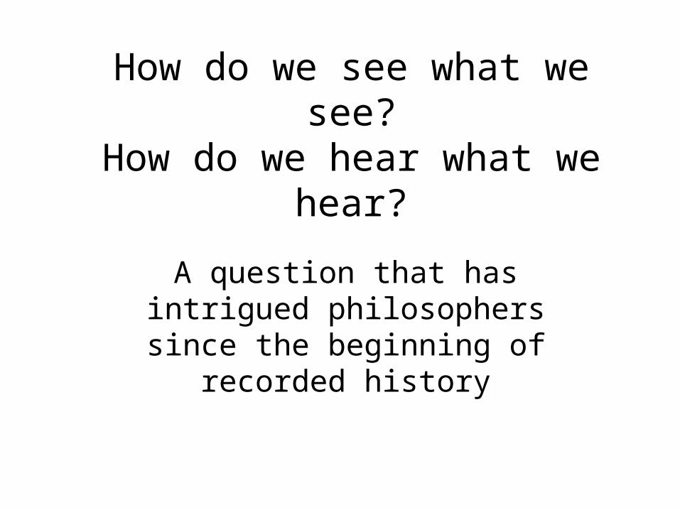
How do we see what we see?How do we hear what we hear?
A question that has intrigued philosophers since the beginning of
recorded history
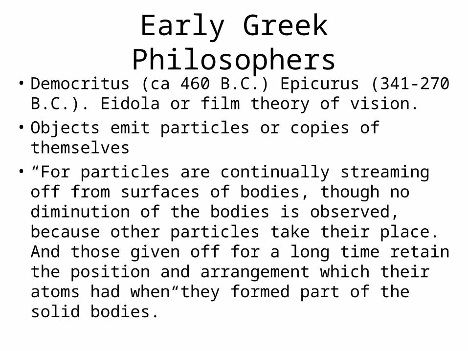
Early Greek Philosophers• Democritus (ca 460 B.C.) Epicurus (341-270
B.C.). Eidola or film theory of vision.
• Objects emit particles or copies of themselves
• “For particles are continually streaming off from surfaces of bodies, though no diminution of the bodies is observed, because other particles take their place. And those given off for a long time retain the position and arrangement which their atoms had when they formed part of the solid bodies.”
Early Greek Philosophers Problems with Eidola theory
• Why isn’t matter used up?
• How does the image get in the eye?
• Why can’t we see in the dark?
• Are copies given off in all directions?
• These problems led to other theories being proposed.

Early Greek PhilosophersScientific progress: Problems lead to new theories
• Extramission or “visual touch” theories.
• Plato (ca 427-347 B.C). Eyes emit “visual fire” which coalesces with daylight to contact objects and returns an impression of the object.
• Problems with “visual touch” theories.
• Exactly what is emitted?
• How does it return impressions?
• Why can’t you see in the dark?
Early Greek PhilosophersExtramission theory: Theoretical development
• Euclid’s (300 B.C.) geometric theory of vision. Assume
• That the rectilinear rays proceeding from the eye diverge indefinitely;
• That the figure contained by a set of visual rays is a cone of which the vertex is at the eye and the base at the surface of the object seen;
• That those things are seen upon which visual rays fall and those things are not seen upon which visual rays do not fall.
Early Greek PhilosophersExtramission theory: Theoretical development
• That things seen under a larger angle appear larger, those under a smaller angle appear smaller, and those under equal angles appear equal;
• That things seen by higher visual rays appear higher, and things seen by lower rays appear lower;
• Etc.
Extramission theory: Problems led to modifications
• Galen’s answer (ca. 129-199 A.D.) as to why light is needed.
• “When it (the air) has been illuminated by the sun, it is already an instrument of vision of the same description as the pneuma arriving from the brain; but until it is illuminated it does not turn into a sympathetic instrument in accordance with the change effected by the outflow of pneuma into it.
After the fall of the Roman Empire
• No further developments that we know of until;
• The work of the Islamic scholars
• Al-Kinde (9th century) took elements of extramission theory and Euclid’s geometry and fused them into a coherent theory of vision.
• Alhazen (969-1039 A.D.), however, took visual theory to a whole new level.
The Islamic Scholars: Alhazen
• Proposed a new intromission theory.
• Took Euclid and turned it around.
• Points on a body radiate light in all directions.
• “From each point of every colored body, illuminated by any light, issue light and color along every straight line that can be drawn from that point.
The Islamic Scholars: Alhazen
• Only those rays that enter the pupil of the eye produce a visual impression.
• One problem.
• Superfluity of rays problem.
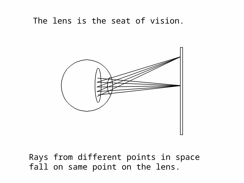
The lens is the seat of vision.
Rays from different points in space fall on same point on the lens.
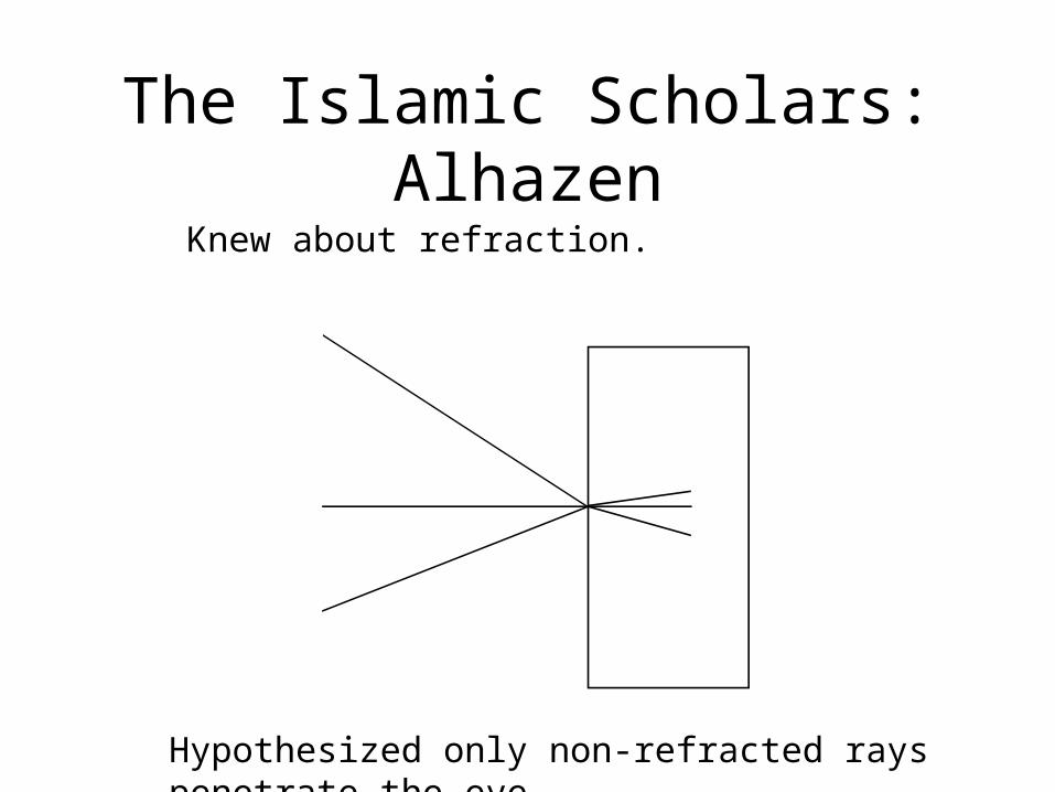
The Islamic Scholars: Alhazen
Knew about refraction.
Hypothesized only non-refracted rays penetrate the eye
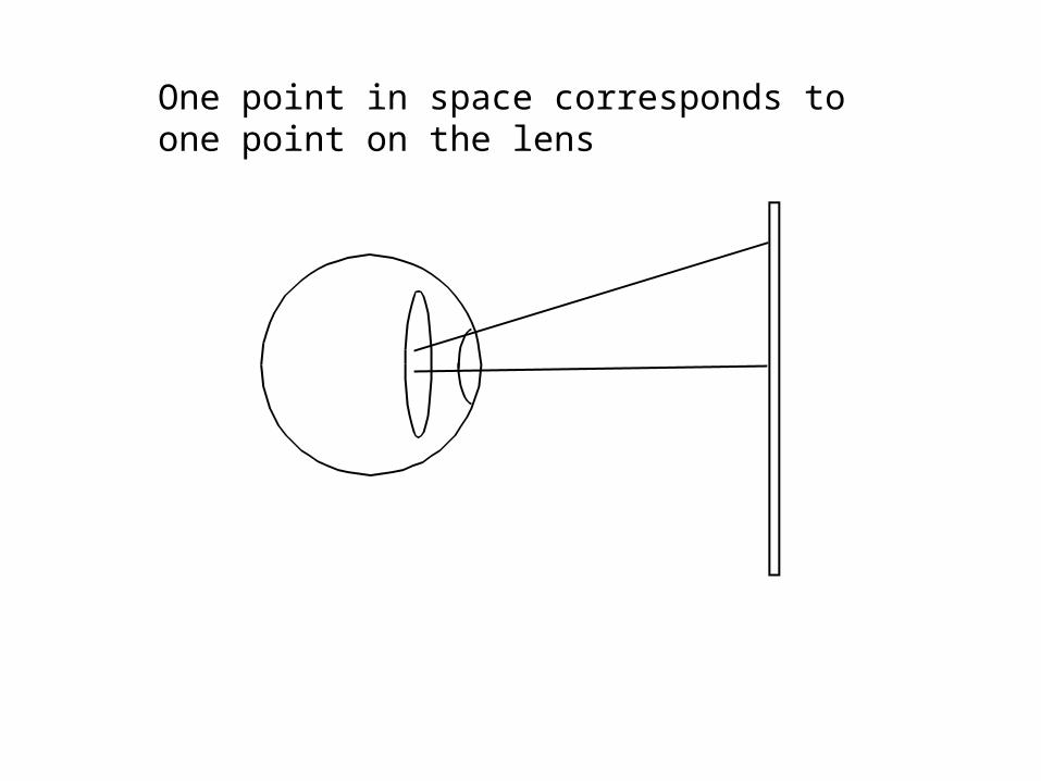
One point in space corresponds to one point on the lens
Alhazen theory was not overturned until the work of Kepler
• Kepler (1571-1630) worked out the geometric optics of the eye.
• Light is reflected in all directions from each point on a non-mirror surface.
• He traced the rays and proved that an upside-down and right-left reversed image should appear on the retina.
Thus, some 20 centuries later we finally solved how images were
sensed by the eye
• But now we had new problems to solve.
• Images on the retina change with distance
• Images on the retina change with orientation
• Images on the retina change with angle of view
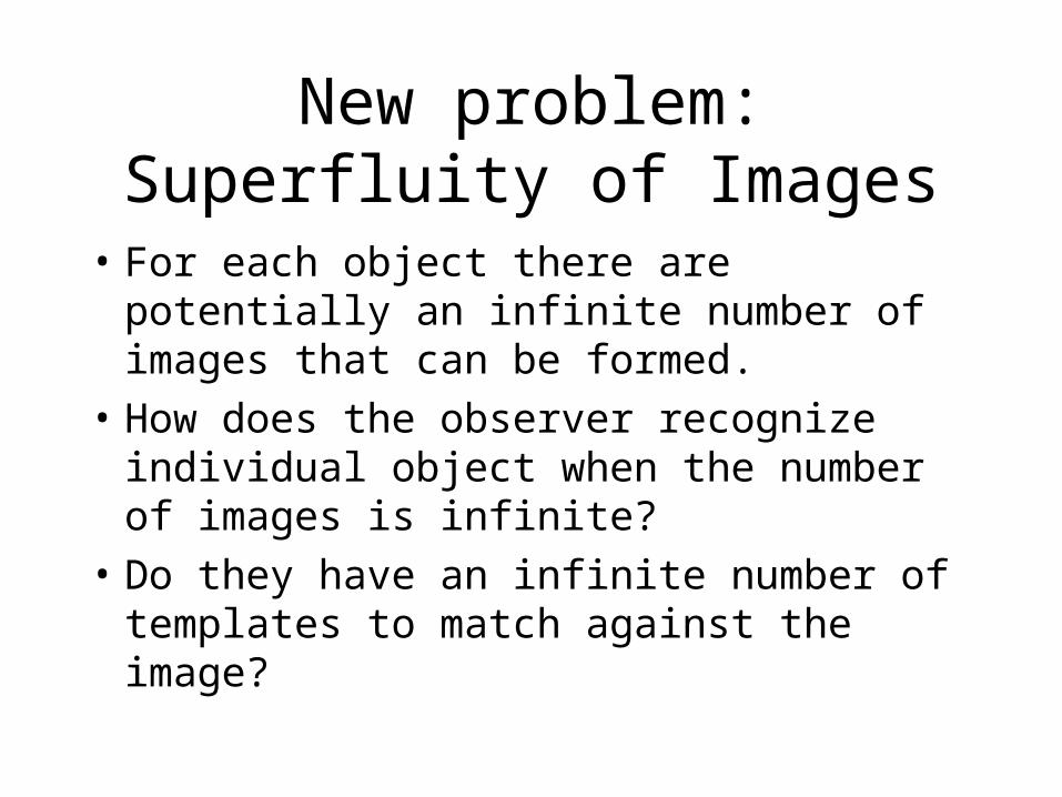
New problem: Superfluity of Images
• For each object there are potentially an infinite number of images that can be formed.
• How does the observer recognize individual object when the number of images is infinite?
• Do they have an infinite number of templates to match against the image?
To answer this problem we need to consider how signals are
processed• How do the eye and brain solve the
superfluity of images problem?.
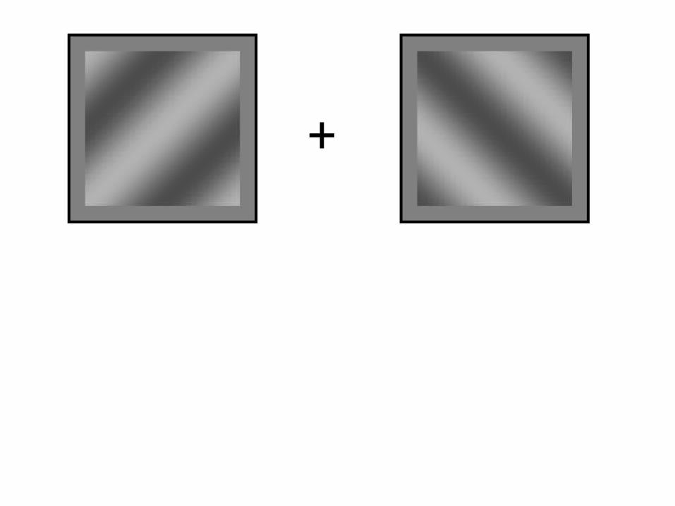
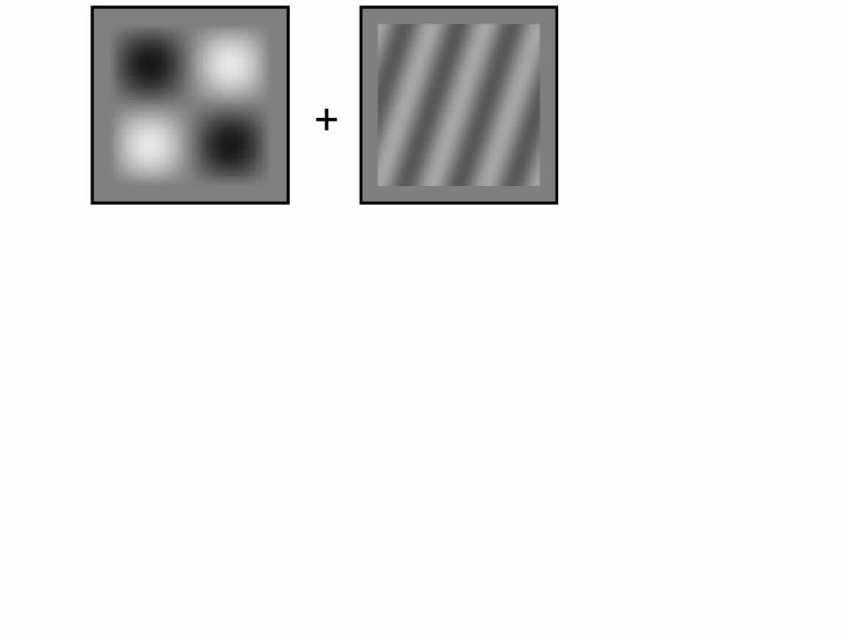
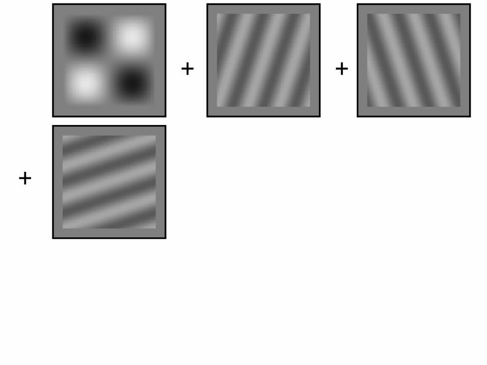
• Question: How much information do we need to characterize an image?

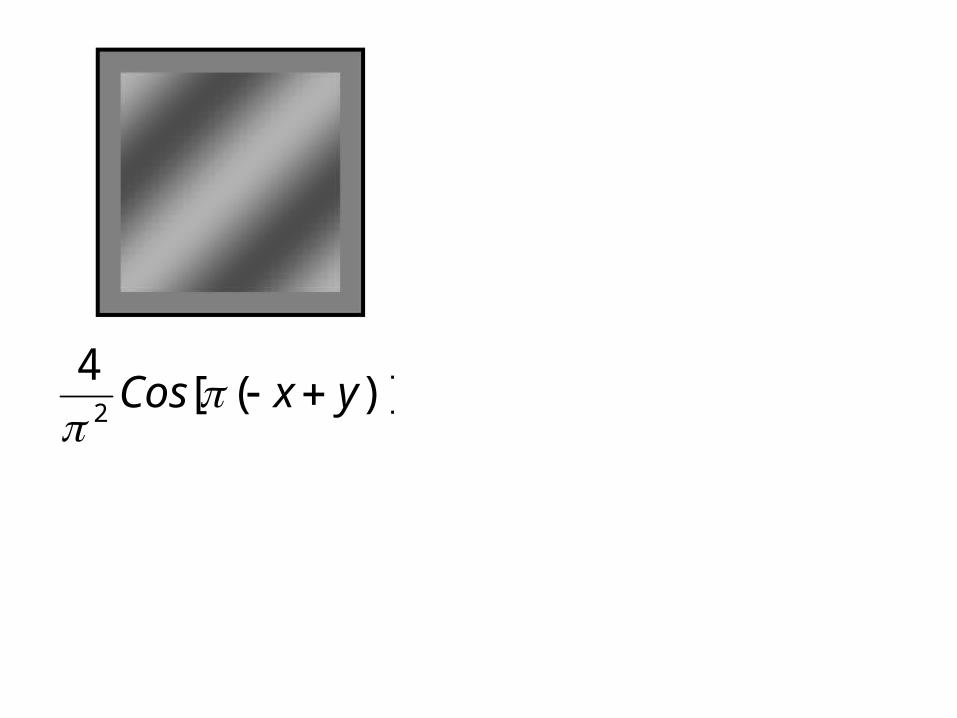
)]([4
2yxCos
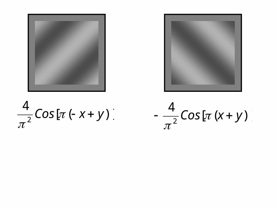
)]([4
2yxCos
)]([
42
yxCos

+
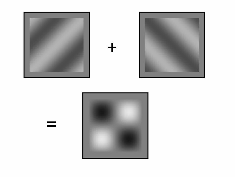
+
=
+
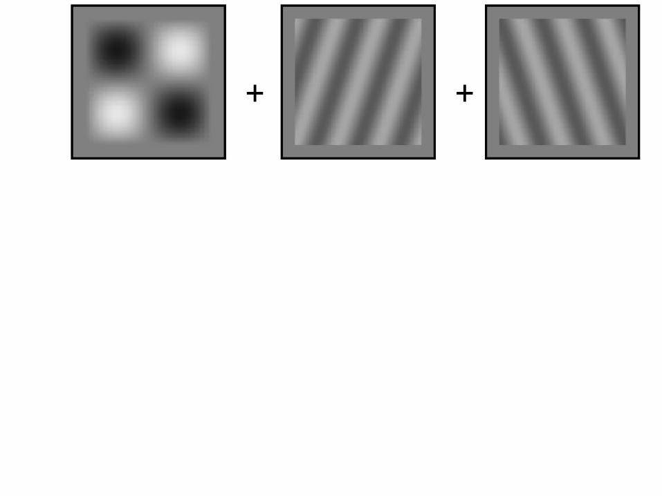
+ +
+ +
+
+ +
++
+ +
++ +
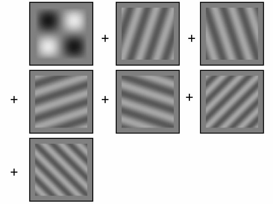
+ +
++ +
+
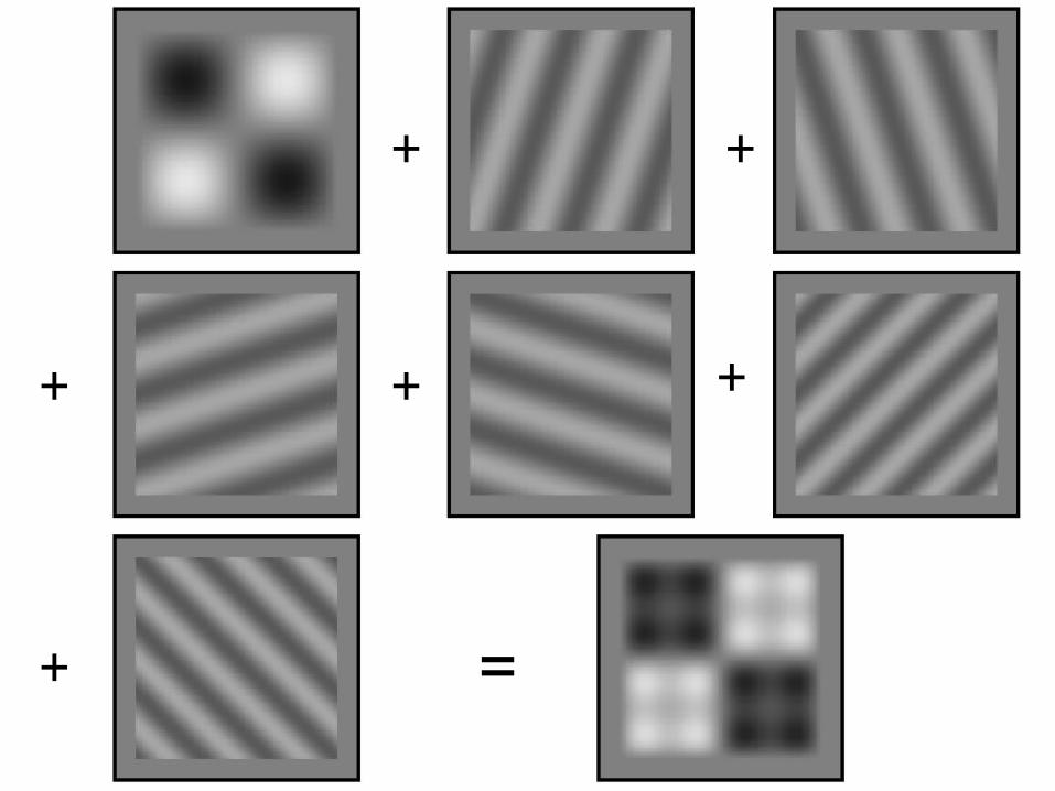
+ +
++ +
+ =
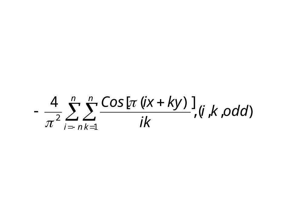
),,(,)]([4
12 oddki
ik
kyixCosn
ni
n
k
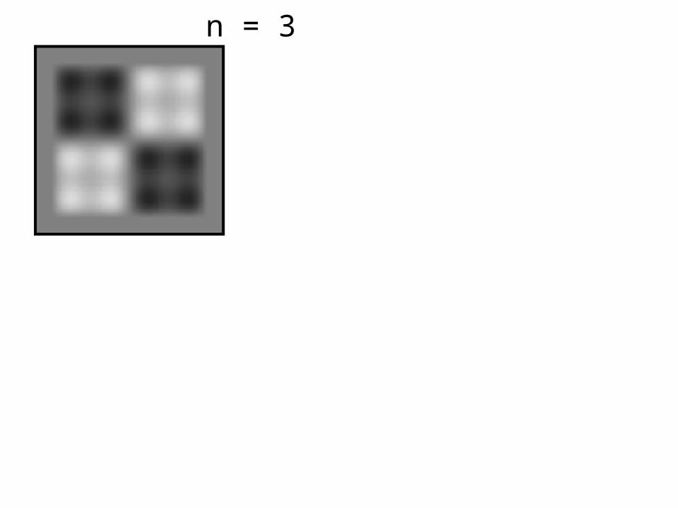
n = 3
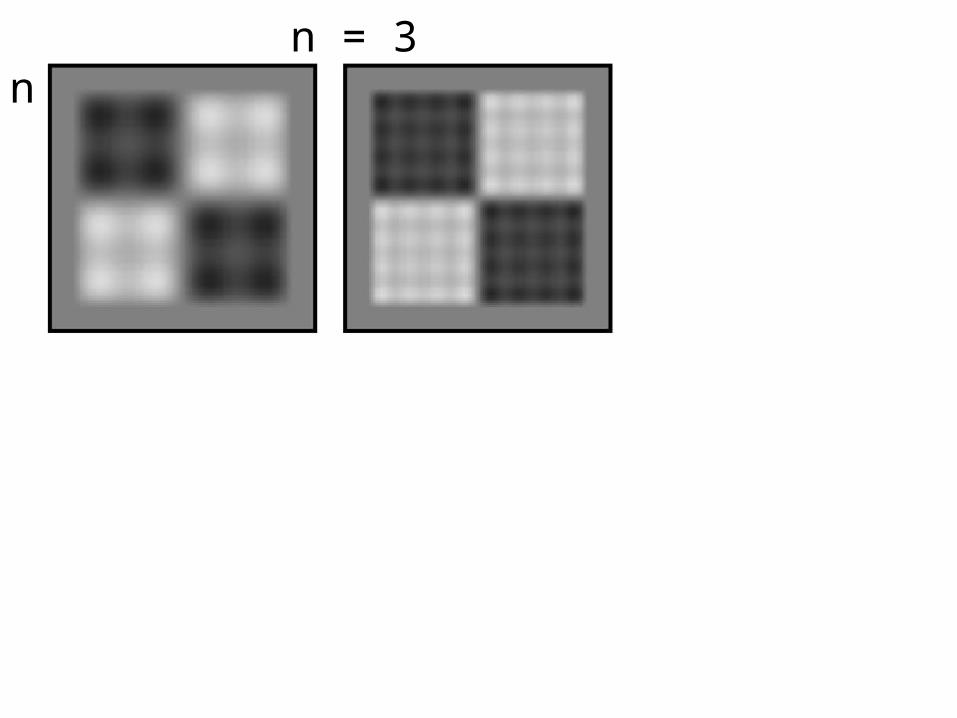
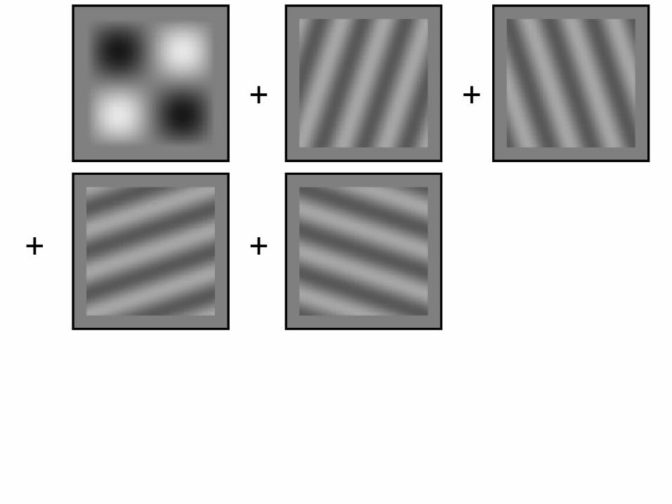
n = 3 n = 7
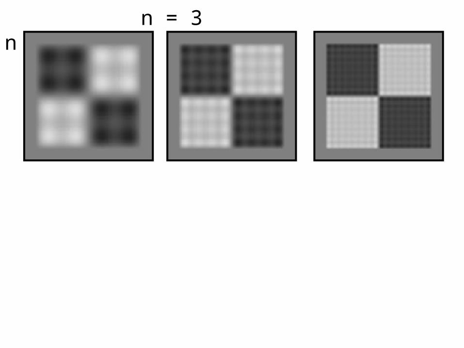
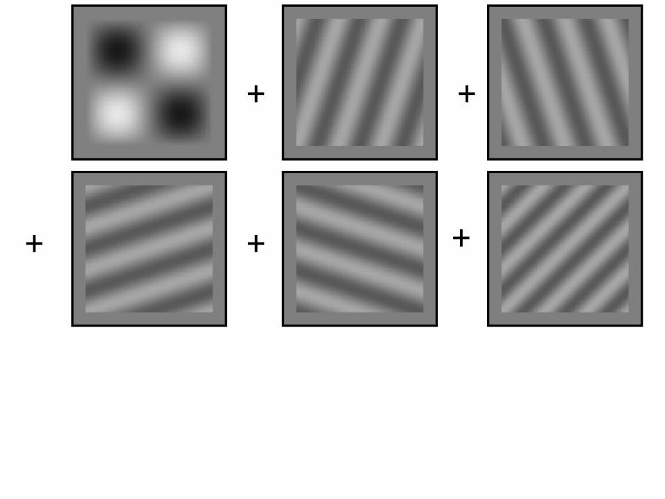
n = 3 n = 7 n = 15
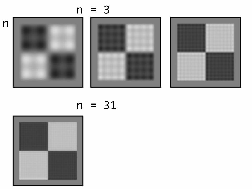
n = 3 n = 7 n = 15
n = 31
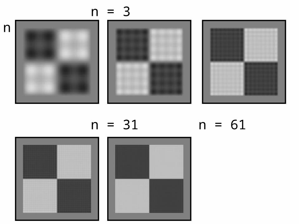
n = 3 n = 7 n = 15
n = 31 n = 61
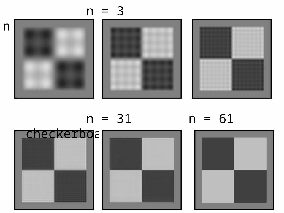
n = 3 n = 7 n = 15
n = 31 n = 61 checkerboard
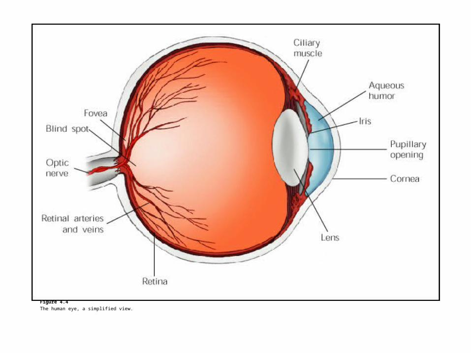
Figure 4.4
The human eye, a simplified view.
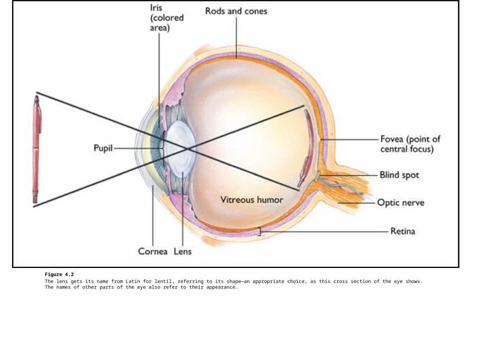
Figure 4.2
The lens gets its name from Latin for lentil, referring to its shape—an appropriate choice, as this cross section of the eye shows. The names of other parts of the eye also refer to their appearance.
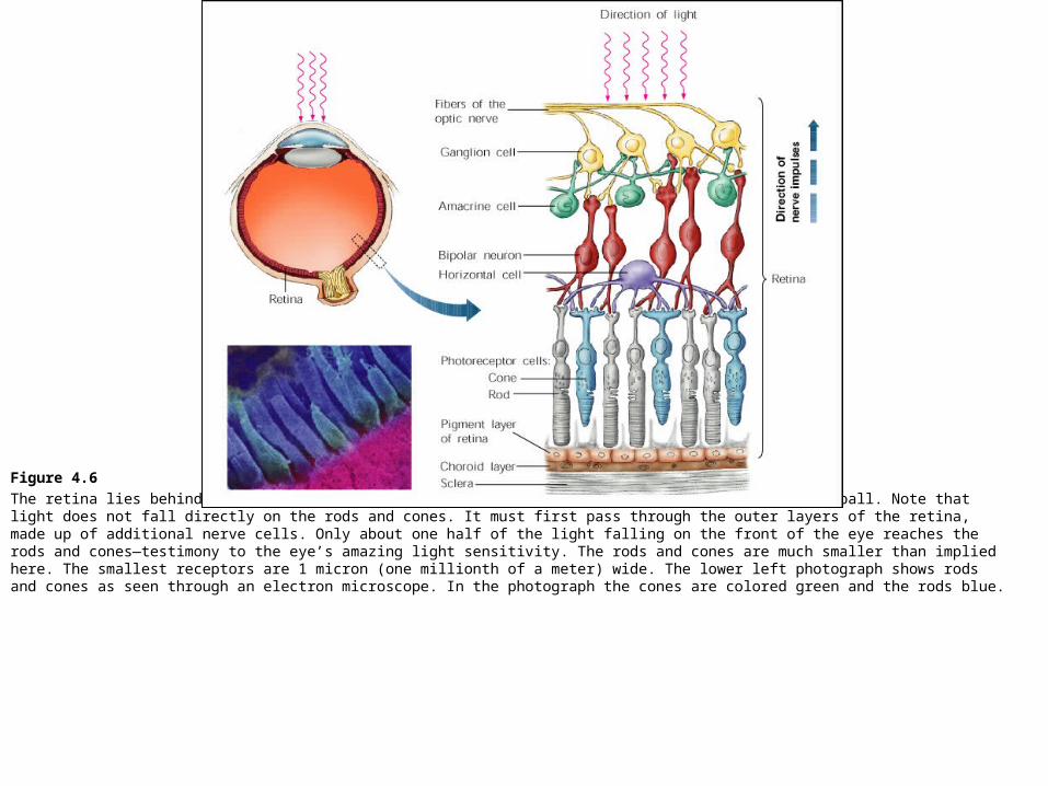
Figure 4.6
The retina lies behind the vitreous humor, which is the jelly-like substance that fills the eyeball. Note that light does not fall directly on the rods and cones. It must first pass through the outer layers of the retina, made up of additional nerve cells. Only about one half of the light falling on the front of the eye reaches the rods and cones—testimony to the eye’s amazing light sensitivity. The rods and cones are much smaller than implied here. The smallest receptors are 1 micron (one millionth of a meter) wide. The lower left photograph shows rods and cones as seen through an electron microscope. In the photograph the cones are colored green and the rods blue.
![Our first isms HUMANISM see p. 55 Idealism [see glossary] – a perfect world lies behind/beyond/within/separate-from this world we sense (see, touch, hear,](https://img.pdfslide.us/doc/110x75/56649e755503460f94b75ccd/our-first-isms-humanism-see-p-55-idealism-see-glossary-a-perfect-world.jpg)