Embed Size (px)
Citation preview

How do we know about the brain?
Lesion: natural or experimentally damaged tissue of the brain used to study portions of the brain.
Old Way:
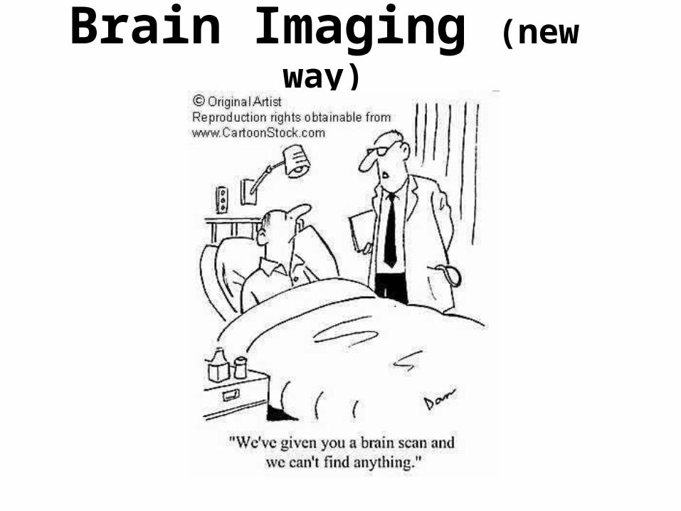
Brain Imaging (new way)

structure
The way to learn about brain imaging methods is to determine if they show you the:
where things are in the brain; are there any lesions
which part(s) of the brain are
active
or
function

CT scan

CTcomputer tomography
• CT imaging uses special __________equipment to produce multiple images or pictures of the inside of the body and a computer to join them together in cross-sectional views of the area being studied. The images can then be examined on a computer monitor or printed.
• CT scans of internal organs, bone, soft tissue and blood vessels provide greater clarity than conventional x-ray exams.
• Used to plan surgeries, check bone density and some injuries to internal organs.
• Most neurologists will not use at this point because it is outdated.

MRI – Magnetic Resonance Imaging

MRImagnetic resonance imaging
• Exposes the brain to a _________ field and measures radio frequency of waves
• Shows high-resolution image (structure) of brain anatomy – No exposure to radioactivity
• Produces computer generated images that distinguish among different types of soft tissue

MRI
MRIs:• Help locate tumors • Show images of :
– the internal structure of the eye and ear– heart and major blood vessels– blood flow in the circulatory system– joints and soft tissues, particularly of cartilage, ligaments
and tendons within joints such as the knee
• Disorders of chest and lungs• Disorders of abdominal organs and the digestive tract• Disorders of the kidneys, urinary tract and pelvic
organs• Infections

EEG - electroencephalogram
EEG - Electroencephalogram
• Records _________________________by placing electrodes to the outside of the head
• The brain's spontaneous electrical signals are traced onto paper
• Used to assess brain damage, epilepsy and other problems
• Its use in brain research is limited. The electrodes detect the activity of only a few neurons in the cortex out of the billions that are present

PET Scan positron emission tomography

• Visual display of brain activity (function) that detects where a ______________________________ goes while the brain performs a given task
• PET scanning is useful in evaluating a variety of conditions — including neurological disease (such as Alzheimer’s), heart disease, infections, certain inflammatory diseases and cancer
PET Scan positron emission tomography

fMRI functional magnetic resonance imaging
• In an fMRI examination, a patient performs a particular task during the imaging process, causing increased metabolic activity in the area of the brain responsible for the task.
• Neuronal firing is fueled by _______________________, which are carried in blood. When an area of the brain is fired up, these substances flow towards it, and fMRI shows up the areas where there is most oxygen. The latest scanners can produce four images every second.
fMRI functional magnetic resonance imaging

![Nocardia Brain Abscess in an Immunocompetent Patient · Nocardia species are a rare cause of cerebral abscess [3]. Nocardia brain abscess appears in a gradually progressive mass lesion,](https://img.pdfslide.us/doc/110x75/5f9d9fa5c479af2f1c584bd9/nocardia-brain-abscess-in-an-immunocompetent-patient-nocardia-species-are-a-rare.jpg)


![DeepMedic for Brain Tumor Segmentation - · PDF fileDeepMedic on Brain Tumor Segmentation 3 DeepMedic is the 11-layers deep, multi-scale 3D CNN we presented in [1] for brain lesion](https://img.pdfslide.us/doc/110x75/5a9dce957f8b9a85318ccde8/deepmedic-for-brain-tumor-segmentation-on-brain-tumor-segmentation-3-deepmedic.jpg)