Embed Size (px)
Citation preview

General DataGeneral Data
Gender: femaleGender: femaleAge: 47Age: 47Occupation:Occupation:環保局環保局

Chief complaintChief complaint
Severe headache, neck stiffness and fever Severe headache, neck stiffness and fever for 10 days.for 10 days.

Present illness (Present illness (ⅠⅠ))This 47This 47--yearyear--old female patient was a case of old female patient was a case of asthma for years. She suffered from progressive asthma for years. She suffered from progressive headache with neck stiffness for 2 weeks. Fever headache with neck stiffness for 2 weeks. Fever and conscious disturbance were also noted. So and conscious disturbance were also noted. So she was admitted to she was admitted to 忠孝忠孝 H. on 2001H. on 2001--1010--18.18.At At 忠孝忠孝 H, GCS was E3V5M6, fever up to 38.5H, GCS was E3V5M6, fever up to 38.5℃℃and and leukocytosisleukocytosis were noted.were noted.Brain CT showed a low density lesion at right Brain CT showed a low density lesion at right frontal lobe with mild hydrocephalus. Right frontal lobe with mild hydrocephalus. Right frontal tumor, infarction and brain abscess were frontal tumor, infarction and brain abscess were suspected. suspected.

Present illness (Present illness (ⅡⅡ))She was transferred to our hospital for MRI She was transferred to our hospital for MRI examination on 2001examination on 2001--1010--19.19.Unfortunately, conscious disturbance occurred Unfortunately, conscious disturbance occurred right before arrival at our ER.right before arrival at our ER.GCS was E1V1M1. CardioGCS was E1V1M1. Cardio--pulmonary pulmonary resuscitation was performed and conscious resuscitation was performed and conscious recovered to E3VtM5 later.recovered to E3VtM5 later.Emergent brain CT with contrast enhancement Emergent brain CT with contrast enhancement was done and she was admitted for further was done and she was admitted for further management.management.

Past historyPast historyMedical historyMedical history
Asthma for years.Asthma for years.Left lung resection when she was a child due to Left lung resection when she was a child due to trauma.trauma.Denied other systemic disease.Denied other systemic disease.
Personal historyPersonal historySmoking: deniedSmoking: deniedAlcohol: deniedAlcohol: deniedAllergy: penicillinAllergy: penicillin
Family historyFamily historyNot contributoryNot contributory

Physical examinationPhysical examination
Consciousness: E3VtM5Consciousness: E3VtM5Vital signVital sign
BP 159/94 mmHg, PR 129/minBP 159/94 mmHg, PR 129/minBT 37.6BT 37.6℃℃, RR 20/min, RR 20/min
Neck: stiffnessNeck: stiffnessChest: asymmetric expansion, left chest Chest: asymmetric expansion, left chest immobilize, breathing sound clearimmobilize, breathing sound clearHeart: tachycardia without murmurHeart: tachycardia without murmurNeurological examination: NPNeurological examination: NP

Lab. dataLab. data
WBC: 21460/uLWBC: 21460/uLNeutrophilNeutrophil: 90.1: 90.1﹪﹪Glucose 191 mg/Glucose 191 mg/dLdLK 3.3 K 3.3 mEqmEq/L /L liver function: WNLliver function: WNLRenal function: WNLRenal function: WNL

Cardiac Cardiac sonographysonography
For suspected infective For suspected infective endocarditisendocarditis20012001--1010--20 20
Mild LA dilationMild LA dilationMinimal amount of pericardial effusionMinimal amount of pericardial effusionRegional wall motion abnormality suggests Regional wall motion abnormality suggests ischemic heart disease.ischemic heart disease.Mild MRMild MREstimated LV ejection fraction 52Estimated LV ejection fraction 52--58%58%Sinus tachycardia with frequent Sinus tachycardia with frequent APCsAPCs

XX--rayray
20012001--1010--2020Total Total opacificationopacificationof the left chest of the left chest without without mediastinalmediastinalshifting.shifting.Amputation of left Amputation of left main bronchus.main bronchus.

Brain CT (1)Brain CT (1)prepre--enhancedenhanced postpost--enhancedenhanced
A hypodensity lesion over R’t frontal lobe
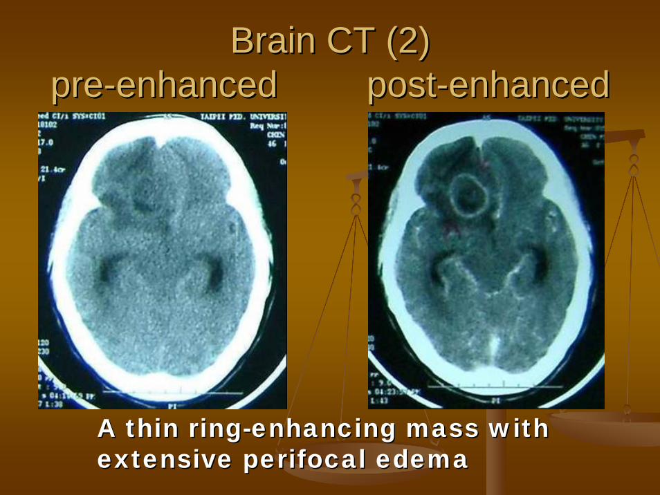
Brain CT (2)Brain CT (2)prepre--enhancedenhanced postpost--enhancedenhanced
A thin ringA thin ring--enhancing mass with enhancing mass with extensive extensive perifocalperifocal edemaedema

Brain CT findingsBrain CT findings
20012001--1010--1919A thin ringA thin ring--enhancing mass (about 2x3x4 enhancing mass (about 2x3x4 cm in size) with extensive cm in size) with extensive perifocalperifocal edema edema is noted in right frontal lobe.is noted in right frontal lobe.The mass contains some nonThe mass contains some non--enhancing enhancing low and low and isoiso--density materials.density materials.Brain abscess is more favored.Brain abscess is more favored.

Brain CT (3)Brain CT (3)prepre--enhancedenhanced postpost--enhancedenhanced
Diffuse brain edema with midline shiftDiffuse brain edema with midline shiftSubfalcineSubfalcine herniationherniation

Brain CT (4)Brain CT (4)prepre--enhancedenhanced postpost--enhancedenhanced
There is ventricular wall enhancement in There is ventricular wall enhancement in frontal horn of right lateral ventricle.frontal horn of right lateral ventricle.Asymmetrical lateral ventricular dilatation.Asymmetrical lateral ventricular dilatation.
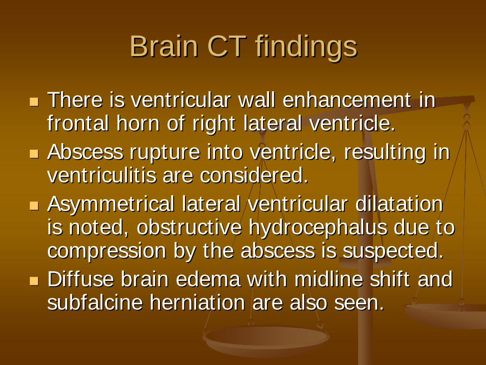
Brain CT findingsBrain CT findings
There is ventricular wall enhancement in There is ventricular wall enhancement in frontal horn of right lateral ventricle.frontal horn of right lateral ventricle.Abscess rupture into ventricle, resulting in Abscess rupture into ventricle, resulting in ventriculitisventriculitis are considered.are considered.Asymmetrical lateral ventricular dilatation Asymmetrical lateral ventricular dilatation is noted, obstructive hydrocephalus due to is noted, obstructive hydrocephalus due to compression by the abscess is suspected.compression by the abscess is suspected.Diffuse brain edema with midline shift and Diffuse brain edema with midline shift and subfalcinesubfalcine herniationherniation are also seen.are also seen.

Differential diagnosis Differential diagnosis
RingRing--enhancing lesionsenhancing lesionsBrain abscessBrain abscessMetastaticMetastatic brain tumorbrain tumorGlioblastomaGlioblastoma multiformemultiformeCerebral infarctCerebral infarctResolving Resolving hematomahematoma

Brain abscessBrain abscess
A focal area of heterogeneous A focal area of heterogeneous hypodensityhypodensitysurrounded by smoothsurrounded by smooth--ring enhancement.ring enhancement.Ring enhancement Ring enhancement ------ capsule of the capsule of the abscessabscessNonenhancingNonenhancing abscess centre abscess centre ------ puspusExtensive edema in the surrounding white Extensive edema in the surrounding white matter. matter.

MetastaticMetastatic brain tumorbrain tumor
MultipleMultipleEnhancing solid lesions Enhancing solid lesions ------ irregular, ring irregular, ring pattern pattern A thick or markedly irregular wall suggest A thick or markedly irregular wall suggest a tumor rather than an infection lesion.a tumor rather than an infection lesion.Seen at the graySeen at the gray--white matter junctionwhite matter junctionExtensive surrounding edema Extensive surrounding edema In a patient with known primary cancerIn a patient with known primary cancer

GlioblastomaGlioblastoma multiformemultiforme
Heterogeneous and Heterogeneous and lobulatedlobulated on nonon non--contrast CT.contrast CT.Central necrosis is the hallmark of GBM. Central necrosis is the hallmark of GBM. Usually irregular, occasionally nodular, ring Usually irregular, occasionally nodular, ring enhancement.enhancement.Extensive surrounding white matter edema.Extensive surrounding white matter edema.Occasionally calcificationOccasionally calcificationHemorrhage is common.Hemorrhage is common.

Cerebral infarctCerebral infarct
CT showed CT showed hypodensityhypodensity involving both involving both gray and white matter.gray and white matter.Cortical involvement is an important Cortical involvement is an important differential diagnostic criteria.differential diagnostic criteria.On contrastOn contrast--enhancement CTenhancement CT
Variable pattern: cortical, central, patchy and Variable pattern: cortical, central, patchy and dense enhancement.dense enhancement.

Resolving Resolving hematomahematomaIntracranial hemorrhage can be identified on CT, Intracranial hemorrhage can be identified on CT, depending on the age of the bleed. depending on the age of the bleed. Acute stage (< 1 week): Acute stage (< 1 week): hyperdensityhyperdensitySubacuteSubacute stage (1stage (1--6 weeks): ring6 weeks): ring--like like enhancement can be seen, possibly related to enhancement can be seen, possibly related to hypervascularityhypervascularity at the periphery of a resolving at the periphery of a resolving hematomahematomaChonicChonic stage (>6 weeks): stage (>6 weeks): hypodensityhypodensity indicated indicated cavity formation.cavity formation.Final stage: residual cavityFinal stage: residual cavity

Final diagnosisFinal diagnosis
Brain abscessBrain abscessOP: craniotomy with removal of OP: craniotomy with removal of brain abscess on 2001brain abscess on 2001--1010--20.20.

DiscussionDiscussionBrain abscessBrain abscess
DefinitionDefinitionA focal A focal suppurativesuppurative infection involving brain infection involving brain parenchyma parenchyma with subsequent abscess with subsequent abscess formation formation ..
Common etiologic agentsCommon etiologic agentsMixed floraMixed floraStaphylococcus Staphylococcus aureusaureusAerobic or Aerobic or microaerophilicmicroaerophilic Streptococci Streptococci Aerobic gramAerobic gram--negative bacillinegative bacilliOther anaerobesOther anaerobes

EtiologyEtiologyContiguous Contiguous suppurativesuppurative focus focus
4545--50% of cases 50% of cases Sinusitis, Sinusitis, otitisotitis media, dental infectionmedia, dental infection
Head trauma or neurosurgeryHead trauma or neurosurgery10% of cases 10% of cases
HematogenousHematogenous spread from a distant focusspread from a distant focus25% of cases25% of casesMultiple lesions Multiple lesions Infective Infective endocarditisendocarditis, pneumonia, pneumonia
Cryptogenic Cryptogenic 15% of cases15% of cases

Results of experimental models of Results of experimental models of brain abscess formationbrain abscess formation
there must be preexisting or concomitant there must be preexisting or concomitant area of ischemia, necrosis, or hypoxia in area of ischemia, necrosis, or hypoxia in brain tissue.brain tissue.Risk factorRisk factor
Cyanotic heart diseaseCyanotic heart diseaseTetralogyTetralogy of of FallotFallot, ASD, VSD, ASD, VSD
Pulmonary AV fistulaPulmonary AV fistulaimmunosupressionimmunosupression

Clinical featuresClinical features
Solitary abscess involve frontal > temporal Solitary abscess involve frontal > temporal > parietal > > parietal > cerebellarcerebellar > occipital lobes of > occipital lobes of the brain.the brain.hematogenoushematogenous spread of infection to brainspread of infection to brain
Multiple abscessesMultiple abscesses

Symptom and signSymptom and sign
Triad Triad <50%<50%HeadacheHeadache 5050--75%75%FeverFever 4040--50%50%Focal Focal neurologicneurologic deficitdeficit ~50%~50%
SeizureSeizure 2525--40%40%Nausea and vomitingNausea and vomiting 2222--50%50%Neck stiffnessNeck stiffness ~25%~25%papilledemapapilledema ~25%~25%
Radiological examinationRadiological examination
Skull films Skull films For diagnosis of sinusitis or the presence of For diagnosis of sinusitis or the presence of free gas in the abscess cavity. free gas in the abscess cavity.
Chest XChest X--ray filmray filmFor detection of underlying cardiac or For detection of underlying cardiac or pulmonary abnormality.pulmonary abnormality.

CTCT
On contrast enhanced CTOn contrast enhanced CTAn uniformly ring or doughnut representing An uniformly ring or doughnut representing the spherical wall or capsule of the abscess.the spherical wall or capsule of the abscess.
Breakdown of the bloodBreakdown of the blood--brain barrierbrain barrierHypervascularityHypervascularity of the of the granlationgranlation tissue.tissue.
The The nonenhancingnonenhancing hypodensityhypodensity abscess abscess centre is pus.centre is pus.There is commonly extensive There is commonly extensive hypodensehypodense--appearing edema in the surrounding white appearing edema in the surrounding white matter.matter.

CTCT
On nonOn non--contrast enhanced CT contrast enhanced CT usually not contain high density material.usually not contain high density material.
Delayed scanning after IV contrast mediumDelayed scanning after IV contrast mediumFor differentiation between the pseudoFor differentiation between the pseudo--capsule capsule of the late stage of of the late stage of cerebritiscerebritis and a true and a true capsule.capsule.
MRIMRIcerebritiscerebritis
MRI is better than CT for abscess MRI is better than CT for abscess In the early stage In the early stage ------ cerebritiscerebritisIn the posterior In the posterior fossafossa
CerebritisCerebritis appears on MRIappears on MRIAn area of lowAn area of low--signal intensity on T1WI with signal intensity on T1WI with irregular postirregular post--gadolinium enhancement.gadolinium enhancement.An area of increased signal intensity on T2WIAn area of increased signal intensity on T2WI
CerebritisCerebritis is often not visualized by CTis often not visualized by CT

MRI MRI mature brain abscessmature brain abscess
On T1 weighted imageOn T1 weighted imageThe The hypointensehypointense central area surrounded by a central area surrounded by a thin thin hyperintensehyperintense signal wallsignal wallhypointensehypointense signal edema.signal edema.Marked ringMarked ring--enhancement after gadolinium enhancement after gadolinium administrationadministration
On T2 weighted imageOn T2 weighted imageHyperintenseHyperintense central area of pus surrounded by central area of pus surrounded by a wella well--defined defined hypointensehypointense capsule.capsule.HyperintenseHyperintense area of edema.area of edema.

Treatment Treatment Surgical drainageSurgical drainage
For encapsulated abscessFor encapsulated abscess
Empirical antibiotic therapyEmpirical antibiotic therapybased on abscess culture based on abscess culture At least 6At least 6--8 weeks8 weeks
Prophylactic anticonvulsant therapyProphylactic anticonvulsant therapyAt least 3 monthsAt least 3 months
ShortShort--term steroid therapyterm steroid therapyUsed only in patients with proven or suspected Used only in patients with proven or suspected elevation in ICPelevation in ICP
![Nocardia Brain Abscess in an Immunocompetent Patient · Nocardia species are a rare cause of cerebral abscess [3]. Nocardia brain abscess appears in a gradually progressive mass lesion,](https://img.pdfslide.us/doc/110x75/5f9d9fa5c479af2f1c584bd9/nocardia-brain-abscess-in-an-immunocompetent-patient-nocardia-species-are-a-rare.jpg)