Embed Size (px)
Citation preview
Hospital-based study of the
spectrum of skeletal dysplasias
in children in Northern India
Co-authors: Dr Ankur Singh, Dr Seema Kapoor,
Dr RN Mandal, Dr VK Gautam, Dr Gaurav Pradhan
Maulana Azad Medical College, New Delhi, INDIA
Dr Namita Ravikumar MD Senior Resident
Indira Gandhi Institute of Child Health Bangalore INDIA

Osteochondrodysplasia
Complex group of bone &
cartilage disorders affecting the
fetal skeleton
Disproportionate short stature
& skeletal deformities
More than 350 disorders
Diagnosed based on clinical,
radiographic & molecular
criteria
*Superti-Furga A, Unger S. Nosology and classification of genetic skeletal disorders: 2006 revision. Am J Med Genet A. 2007 Jan;143(1):1-18

Constitutional disorders of
bone
Type/particulars
Osteochondrodysplasias
Dysostosis Dysplasia Dystrophy
Abnormality of Growth Structure Structure
Site Multiple bone/cartilage of
axial/appendicular skeleton,
membranous &/or enchondral bone
Individual bones
singly or in
combination
Failure of Gene expression Blastogenesis
Phenotype May evolve throughout life Static
*Hall CM. International nosology and classification of constitutional disorders of bone (2001). Am J Med Genet. 2002 Nov;113(1):65-77
Approach to skeletal
dysplasias
Radiological
• A – Anatomical localisation
• B – Bones
• C – Complications
• D – Death or lethality
*Dutton RV. A practical radiologic approach to skeletal dysplasias in infancy. Radiol Clin North Am. 1987 Nov;25(6):1211-33

Based on Anatomical location Axial skeleton Appendicular skeleton
Skull Cranio/cranial
Location
Epiphyseal
Face Facio/facial Metaphyseal
Mandible Mandibulo Diaphyseal
Clavicle Cleido
Ribs Costo
Shortening
Rhizomelic (proximal e.g. femur)
Spine Spondylo/vertebral Mesomelic (middle e.g. radius/ulna)
Pelvis Ischio/ilio/pubic Acromelic (distal- hands/feet)
*Dutton RV. A practical radiologic approach to skeletal dysplasias in infancy. Radiol Clin North Am. 1987 Nov;25(6):1211-33

Appendicular skeleton

Bones • Structure – abnormalities of bone density, presence of
tumorous lesions
• Shape – whole or part of a bone
• Size - Absolute or relative to other bones (bone age to
be considered)
• Sum - too many, too few or fused
• Soft tissues - wasting, excessive soft tissues,
contractures & calcification
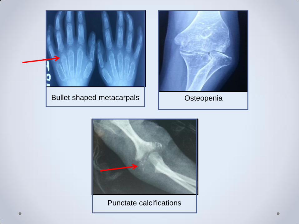
Bullet shaped metacarpals Osteopenia
Punctate calcifications

Complications • Fractures – osteoporotic (e.g. osteogenesis imperfecta)
& osteosclerotic (e.g. osteopetrosis) conditions
• Atlantoaxial subluxation – Mucopolysaccharidosis (MPS)
• Progressive scoliosis – Campomelic dysplasia
• Limb length discrepancies – Epiphyseal stippling,
Dysplasia epiphysealis hemimelica, Ollier’s disease,
multiple cartilaginous exostoses
• Malignancy – Maffucci’s syndrome
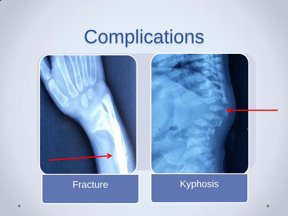
Complications
Fracture Kyphosis

Death or lethality If a dysplasia is lethal it helps to exclude or confirm a
given diagnosis
Helps identify subtype affected & different modes of
inheritance

Radiological Classification
*Alanay Y, Lachman RS. A review of the principles of radiological assessment of skeletal dysplasias. J Clin Res Pediatr Endocrinol. 2011;3(4):163-78

Genetic basis & molecular
mechanisms • Abnormalities in the patterning, development,
maintenance & size of skeleton (axial/appendicular)
• Mutations in various families of genes
• The most common is Achondroplasia
caused by mutations in the FGFR3 gene
*Kornak U, Mundlos S. Genetic disorders of the skeleton: a developmental approach. Am J Hum Genet. 2003 Sep;73(3):447-74

Inheritance Pattern • Genetically heterogeneous
• Inherited as AD, AR, XLR & XLD
• Rarely chromosomal deletions/duplications, germline
mosaicism & uniparental disomy
• Intrafamilial & interfamilial variability

Molecular Pathogenetic
Classification Superti-furga et al grouped molecular defects of skeleton as
• Group 1: Extracellular structural proteins
• Group 2: Metabolic pathways (including enzymes, ion channels,
transporters)
• Group 3: Folding, processing & degradation of macromolecules
• Group 4: Hormones & signal transduction mechanisms
• Group 5: Nuclear proteins & transcription factors
• Group 6: Oncogenes & tumour suppressor genes
• Group 7: RNA/DNA processing & metabolism
*Superti-Furga A, Bonafé L, Rimoin DL. Molecular-pathogenetic classification of genetic disorders of the skeleton. Am J Med Genet. 2001;106(4):282-93

Prenatal diagnosis • Meticulous sonographic examination - thanatophoric
dysplasia & osteogenesis imperfecta
• Previously affected child with a molecularly confirmed
diagnosis - molecular analysis of DNA
(CVS/Amniocentesis) direct mutational analysis/
linkage analysis

Prevalence
Orioli et al (1978 -83) crude prevalence rate -2.3/10000
Gustavson & Jorulf- 4.7/10 000
Camera & Mastroiacovo - 2.4/10 000
Barbosa-Buck, Orioli et al (2012) - 3.2 per 10,000
Kulkarni et al - 19.6/10,000 live born & lethal dysplasias-
5.2/10,000 deliveries
*Orioli IM, Castilla EE, Barbosa-Neto JG. The birth prevalence rates for the skeletal dysplasias. J Med Genet. 1986 Aug;23(4):328-32
*Gustavson K-H, Jorulf H. Different types of osteochondrodysplasia in a consecutive series of newborns. Helv Paediatr Acta.1975;30:307-14
*Barbosa-Buck CO, Orioli IM, da Graça Dutra M, Lopez-Camelo J, Castilla EE, Cavalcanti DP. Clinical epidemiology of skeletal dysplasias in South
America. Am J Med Genet A. 2012 May;158A(5):1038-45
*Kulkarni ML, Samuel K, Bhagyavathi M, Sureshkumar C. Skeletal dysplasias in a hospital in Southern India. Indian Pediatr. 1995 Jun;32(6):657-65

Aims & Objectives • To study the clinico-radiological spectrum of skeletal
dysplasias in children
• To establish a molecular diagnosis wherever possible

Materials and methods • 51 children with disproportionate short stature or skeletal
deformities included (0-18 years of age)
• Cross-sectional study
• Lok Nayak Hospital, New Delhi, India (March 2013-14)
• Pediatric/orthopedic OPD, genetic clinic & admissions in
pediatric/orthopaedic wards

Methodology Detailed history taking with special emphasis on
o Age of noticing the problem
o Family history – 3 generation pedigree chart & h/o consanguinity
o Maternal antenatal history & review of ultrasounds (current &
previous pregnancies)
o Developmental history
Detailed head to toe examination including
dysmorphology assessment

Anthropometry The Anthropometric data were collected as follows-
• Height – using stadiometer with head in Frankfurt plane (3 readings)
• Length – < 2 years & those who could not stand, recumbent stature
using an infantometer or using a non-stretchable flexible tape

• Armspan – distance btw the tips of middle fingers with both arms
outstretched
• Upper Segment: Lower Segment Ratio( US:LS) – LS measured
from the symphysis pubis to the heel & US derived by deducting the
LS from height
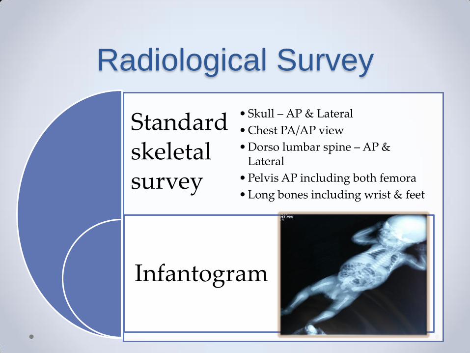
Radiological Survey
Standard skeletal survey
Infantogram
•Skull – AP & Lateral
•Chest PA/AP view
•Dorso lumbar spine – AP & Lateral
•Pelvis AP including both femora
•Long bones including wrist & feet

Radiological Classification The radiological abnormalities involving -
• The epiphysis, metaphyses or diaphysis
• The spine
• Multiple areas – spondylometaphyseal/ epimetaphyseal

Radiographs
Epiphyseal dysplasia
Metaphyseal dysplasia
Spondylo-
dysplasia

Radiological Diagnosis Discussed with senior radiologist having special interest in
skeletal dysplasias & also utilizing databases
• POSSUM web base
• London Dysmorphology database
• Spranger’s text book of skeletal dysplasia
• European skeletal dysplasia registry

Ancillary Investigations • Mucopolysaccharidosis urinary testing for
glycosaminoglycans & enzyme analysis for typification
• Serum Calcium, Phosphorus, Alkaline Phosphatase,
Vitamin D3 levels
• Thyroid profile
• Achondroplasia & Hypochondroplasia PCR for
mutations at FGFR codons 380 & 540 respectively
• Other genetic diagnosis samples sent to researchers
& collaborators engaged in delineating the particular
skeletal dysplasia
Molecular Diagnosis

Molecular Diagnosis
PCR-RFLP of achondroplasia patients
(164bp→154bp & 55bp) and controls

Algorithm
Children 0-18 years with height <-2SD or obvious skeletal deformities
Altered US:LS ratio or arm span-height difference
Detailed clinical examination
Radiological assessment
Molecular diagnosis

Results
51 • Total children enrolled
45 • Radiological
diagnosis (88%)
20 • Molecular
diagnosis (39%)

Results
MALES
68%
FEMALES 32%
Sex distribution
0
5
10
15
Age distribution
27%

Results
SHORT TRUNK
51%
SHORT LIMB 49%
SKELETAL DYSPLASIA
RHIZOMELIC 72%
MESOMELIC 19%
ACROMELIC 9%
Short limb dysplasia

Clinical details • Parental consanguinity was present in 23.5% of skeletal
dysplasias
• Abnormal antenatal ultrasonography was found in 3
cases
• 2 pairs of siblings included- MPS type IV A & Desbuquois
dysplasia

Clinico radiological
classification
Sl no. Group Type Number
I Lethal Osteochondrodysplasias Atelosteogenesis 1
II Chondrodysplasia Punctata
Group
Chondrodysplasia punctata,
rhizomelic type
1
III Predominant Metaphyseal
involvement
Achondroplasia
Hypochondroplasia
Asphyxiating thoracodystrophy
Cartilage -Hair- Hypoplasia
8
1
1
1
IV Predominant Epiphyseal
involvement
Pseudoachondroplasia 2

Sl no. Group Type Number
V Major involvement of the Spine Spondylocostal dysplasia
Kniest dysplasia
Metatropic Dysplasia
Diastrophic Dysplasia
Dyggve-Melchoir-Clausen Syndrome
Spondyloepiphyseal dysplasia
Spondyloepimetaphyseal dysplasia
Spondylothoracic dysostosis
1
1
1
1
1
1
1
1
VI With Multiple Dislocations Desbuquois dysplasia-Kim type 3
VII Dysostosis Multiplex Complex
Carbohydrate Storage Diseases
MPS type I
MPS type IV A
MPS type IV B
4
7
1

Sl no. Group Type Number
VIII Predominant involvement of single
sites or Segments
Rhizomelic dysplasia- Patterson Lowry type
Acromesomelic Dysplasia - Maroteaux type
1
1
IX Predominant Diaphyseal
involvement
none
X Decreased bone density Osteogenesis imperfecta 1
XI Increased bone density Bent bone disease 1
XII Disorganised development of bone
constituents
none
XIII Osteolyses none
*Spranger’s textbook of skeletal dysplasias

Achondroplasia
Clinical features
Rhizomelic micromelia
Disproportionately large head
Depressed nasal bridge
Trident hand with short stubby fingers

Achondroplasia
Irregular metaphysis
Squared ilia
Horizontal acetabular margins
Progressive decrease in
interpedicular distance

Mucopolysachharidosis
Coarse facies J shaped sella

Mucopolysachharidosis
Bullet shaped metacarpals Inferior beaking

Pseudoachondroplasia
Irregularity in vertebral plates Irregular epiphysis

Rhizomelic Chondrodysplasia
Punctata
Rhizomelia
anteverted nares midfacial hypoplasia
Punctate calcification
Coronal clefting of vertebrae

Molecular Diagnosis
Diagnosis Type Number Mutation
Achondroplasia Short limb 8 FGFR3 G-->A 1138 transition
Hypochondroplasia Short limb 1 FGFR3 c1620C-->A heterozygous mutation
Pseudoachondroplasia Short limb 2 c.1554C>G;p.D518E
Mps Type IV A (Morquio) Short trunk
2 c.155C>T in exon 2 of the GALNS
1 [c.3G>A;p.Met1] [c.452C>T;p.P151L]
Desbuquois Dysplasia -
kim type Short limb 3 c.C467T p.Ser156Ph
Cartilage Hair Hypoplasia Short limb 1 g.69dupG,r69dupG
Dyggve-Melchoir-Clausen
Dysplasia Short trunk 1 DYM_v001):c.1923del deletion
Rhizomelic
Chondrodysplasia
Punctata Short limb 1 PEX7 mutation in the L292X allele

Conclusion • Clinical examination & radiological assessment are sufficient to
diagnose a majority of cases
• If a diagnosis made, prognosis can be explained in terms of
expected complications, final adult height, intelligence & also
for genetic counselling
• Emphasis on early diagnosis to prevent complications
• No definitive treatment focus on prenatal diagnosis by
ultrasonography & molecular methods
Conclusion • Males were significantly higher probably due to gender bias in
seeking medical attention
• Maximum cases belonged to the age group of 6- 10 years
Height compared with that of the peers
Home deliveries accounting for not seeking medical attention
• Limitations
Hospital based study
Still births not included

Future prospects • Larger studies in community including still births are
necessary to establish the true prevalence & spectrum of
skeletal dysplasia
• Studies including cases diagnosed antenatally are
essential to highlight the importance of prenatal
diagnosis

Acknowledgements • Departments of Pediatrics, Radiology & Orthopedics,
Maulana Azad Medical College, New Delhi, India
• Department of Pediatrics, Indira Gandhi Institute of Child
Health, Bangalore, India
• Organizers


![The skeleton and musculature on foetal MRImagnum may occur in some skeletal dysplasias, and cerebellar herniation occurs in Chiari malformations [10]. Thorax and spine Although MRI](https://img.pdfslide.us/doc/110x75/60a7bd1993225b08aa7e0e40/the-skeleton-and-musculature-on-foetal-mri-magnum-may-occur-in-some-skeletal-dysplasias.jpg)