Embed Size (px)
Citation preview

Homozygosity Mapping in a Family WithMicrocephaly, Mental Retardation, and ShortStature to a Cohen Syndrome Region on 8q21.3 -8q22.1: Redefining a Clinical Entity
Denise Horn,1,3 Alice Krebsova,1,2 Jurgen Kunze,1,4 and Andre Reis1,2*1Institute of Human Genetics, Charite, Humboldt University Berlin, Germany2Gene Mapping Center, Max-Delbrueck Center, Berlin, Germany3Department of Tumor Genetics, Max-Delbrueck Center, Berlin, Germany4Department of Pediatrics, Charite, Humboldt University Berlin, Germany
A syndrome of microcephaly, progressivepostnatal growth deficiency, and mental re-tardation was observed in two brothers andtheir cousin from a multiply consanguine-ous kindred of Lebanese descent. Hypoto-nia, chorioretinal dystrophy, and myopiawere also identified. The severity of the con-dition varied among the closely related pa-tients. Because of absence of a distinctivefacial appearance, the degree of mental re-tardation, and short stature, the initiallyconsidered clinical diagnosis of Cohen syn-drome was withdrawn and a novel geneticentity was assumed. Homozygosity mappingin this family assigned the gene to a 26.8-cMregion on the chromosome band 8q21.3 -22.1,between the microsatellites at D8S270 andD8S514. The maximum two-point LOD scorewas found for marker at D8S267 (Zmax=3.237at Omax=0.00). Intriguingly enough, the iden-tified gene region overlaps the refined generegion for Cohen syndrome (COH1) [Koleh-mainen et al., 1997: Euro J Hum Genet 5:206–213]. This fact encourages the hypothesisthat the described kindred segregates for avariant of Cohen syndrome and suggests aredefinition of its phenotype. Am. J. Med.Genet. 92:285–292, 2000. © 2000 Wiley-Liss, Inc.
KEY WORDS: Mirhosseini-Holmes-Waltonsyndrome; hypotonia; chorio-
retinal dystrophy; heteroge-neity
INTRODUCTIONMicrocephaly, mental retardation, short stature, and
hypotonia occur in many different mendelian and an-euploidy syndromes. Cohen and Mirhosseini-Holmes-Walton syndromes are among several autosomal reces-sive genetic conditions comprising these clinicalcharacteristics.
Cohen syndrome [Cohen, 1973] initially was charac-terized by obesity, mental retardation, hypotonia, nar-row hands and feet, and by a distinctive craniofacialappearance with prominent upper central incisors. Thelater definition of Cohen syndrome by Escobar [1990]requires the presence of at least five of the followingmajor manifestations: obesity, short stature, mentalretardation, hypotonia, maxillary hypoplasia, shortphiltrum, micrognathia, narrow hands and feet, andhigh-arched palate. Nevertheless, according to severalcase reports, the individual findings of Cohen syn-drome are not specific and their variability is great[Fryns et al., 1996]. In some cases this allows falsediagnosis of an otherwise distinct syndrome [Gorlin etal., 1990]. Nevertheless, in Finnish patients a clinicallyhomogeneous phenotype of Cohen syndrome was de-scribed with additional ophthalmological findings andmarked neutropenia [Kivitie-Kallio et al., 1999; Norioet al., 1984]. Linkage studies have localized the gene inFinnish patients to a 10-cM region at 8q21.3, and link-age disequilibrium investigations have narrowed thecandidate gene region to the immediate vicinity ofmarker D8S1762 [Kolehmainen et al., 1997]. No genehas been reported so far.
Mirhosseini-Holmes-Walton syndrome [Mirhosseiniet al., 1972] is very rare and was first described in twosibs with microcephaly, retinal pigmentary degenera-tion, and severe mental retardation. Clinical presenta-tion in most reported patients with Mirhosseini-Holmes-Walton syndrome overlaps the phenotype seen
Contract grant sponsor: German Human Genome Project*Correspondence to: Prof. Andre Reis, Gene Mapping Center,
Max-Delbrueck Center for Molecular Medicine (MDC), Robert-Roessle-Str. 10, D-13092 Berlin, Germany. E-mail: [email protected]
Received 8 December 1999; Accepted 17 February 2000
American Journal of Medical Genetics 92:285–292 (2000)
© 2000 Wiley-Liss, Inc.

in Cohen syndrome patients. Therefore, it has beensuggested that both conditions represent a single en-tity [Norio and Raitta, 1986]. Nevertheless, no linkagedata have been published in this syndrome and it re-mains unknown whether these conditions are allelic.
We report here on three closely related patients withpostnatal microcephaly, progressive growth delay,mental retardation, and variable overlap with the Co-hen and Mirhosseini-Holmes-Walton syndromes. Theseverity of clinical findings varied considerably amongthese patients. Based on the pedigree and the existenceof multiple consanguinity an autosomal recessive in-heritance was assumed. Possible chromosomal andmetabolic disorders were excluded. Subsequently, awhole genome search for linkage was performed andthe locus for this syndrome was assigned to 8q21.3-22.1. The identified region overlaps the already pub-lished one of Cohen syndrome [Kolehmainen et al.,1997].
CLINICAL REPORTS
The Lebanese family is multiply consanguineous andthe affected patients were offspring of first cousin par-
ents (Fig. 1). The further family history was unremark-able.
Patient 1
The affected propositus was seen at age 17 years forsevere mental retardation and lack of speech. He wasborn at term after a normal pregnancy; length was 50cm. There were no feeding or respiratory problems dur-ing infancy. Bilateral congenital clubfoot was repairedat 7 years, and afterwards he managed to walk shortdistances unaided. The patient was unable to feed him-self and had no sphincter control. Autistic symptomswith stereotypic hand movements became apparentearly in childhood.
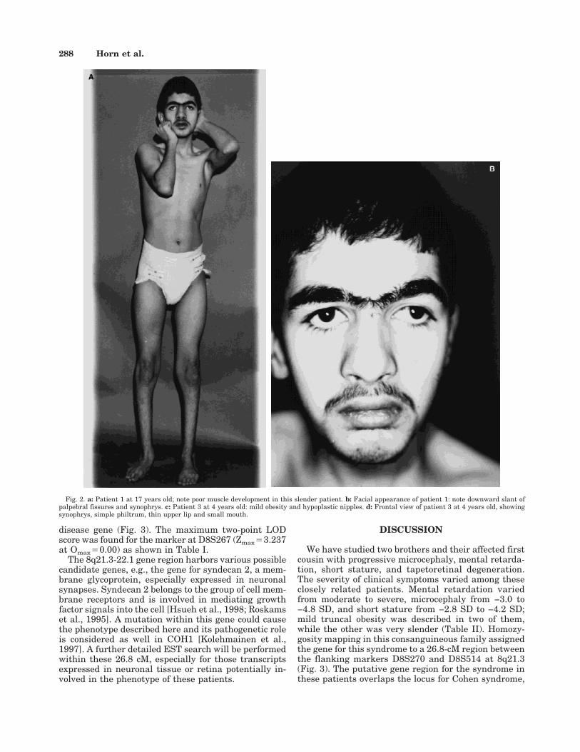
His height was 148 cm (−4.2 SD) and he had a weightof 32 kg (3rd centile for this age is 49 kg, 3rd centile forheight is 30.1 kg), and a head circumference (OFC) of49.5 cm (−4.8 SD) (Fig. 2a). He had everted lips,downslanting palpebral fissures, synophrys (present inall unaffected relatives) (Fig. 2b), marked hypotonia,inadequate muscle development, generalized joint lax-ity, widely spaced nipples, and narrow hands and feet.Sexual development corresponded to age.
Ophthalmologic examination documented strabis-
Fig. 1. The pedigree and haplotype study of the affected family. Arabic numbers denote subjects investigated in this linkage study. Black barrepresents affected haplotype; horizontal lines in open boxes indicate a recombination event in the unaffected haplotype.
286 Horn et al.

mus divergens, myopia (−2D), astigmatism, slow pupil-lary reactions, constriction of retinal vessels, pale pa-pillae, and apparent tapetoretinal degeneration.Pigment deposits of the so-called bone spicule typewere not seen. Visual fields, color vision, and electro-retinography (ERG) could not be tested due to severemental retardation.
Patient 2
The term delivery of the younger brother of patient 1was uncomplicated; his weight was 2500 g and length51 cm. He was floppy, and his early development wasdelayed. He was able to stand without support at 4years. He began walking at 5 years. Stereotypical be-havior was noted during childhood. He could not speakand had extremely limited comprehension. He has notachieved bowel or bladder control.
Anthropological examination at 9.5 years showedheight of 115 cm (−3.6 SD), weight of 24.2 kg (−1.0 SD),OFC of 49 cm (−3.0 SD), synophrys, mild truncal obe-sity, narrow hands and feet, and mild joint hyperex-tensibility. No neurological abnormalities were presentexcept for muscular hypotonia. Tapetoretinal pigmentdegeneration was suspected on ophthalmological ex-amination. Pupillary reflexes were symmetrically pre-sent, strabismus was absent. Repeated hematologicalexaminations demonstrated low white blood cell count(4.7–5.7 109/L) but no neutropenia (58–60% neutro-phils).
Patient 3
The cousin of patients 1 and 2 was born at 36 weeksof gestation after an uneventful pregnancy, with aweight of 2300 g, length of 46 cm, and OFC of 32.5 cm.The infant had Apgar scores of 10 and 10. No medicalproblems were noted in the first 9 months of life. Due topersistent muscular hypotonia, physiotherapy was ini-tiated.
Diagnostic evaluation at 11 months demonstrated adevelopmental level between 3 and 5 months. Thegrowth curve of the first year of life showed progressivemicrocephaly with OFC of 41.4 cm at the chronologicalage of 9 months (−3.4 SD, corrected age of 8 months)and an OFC of 42.5 cm at 12 months (−3.6 SD, cor-rected age of 11 months). Postnatal growth was re-tarded, with a height of 70 cm at 12 months (−2.5 SDfor corrected age of 11 months). Psychomotor develop-ment was delayed: the patient sat at 15 months,walked at 24 months, and used a few words at 30months. He had no bowel and bladder control.
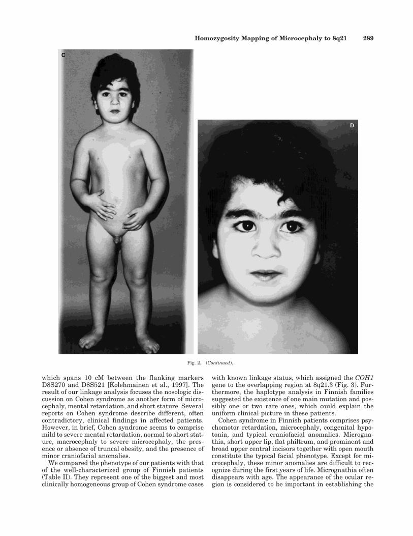
Examination at 4 years showed a height of 96.5 cm(−2.0 SD), weight of 17.8 kg (0.4 SD), and an OFC of46.5 cm (−3.6 SD) (Fig. 2c). He had synophrys, simplephiltrum, small mouth, thin upper lip (Fig. 2d), mildobesity, hypoplastic and inverted nipples, joint laxity,wide gap between hallux and second toe, and shawlscrotum. The fingers appeared elongated. White bloodcell count was normal.
At 6 years his height was 104 cm (−2.8 SD), weight19.8 kg (0.7 SD), and OFC 47 cm (−3.9 SD); he hadhigh-grade myopia (−8D), astigmatism, and intermit-tent divergent strabismus. Diffuse pigmentary depos-its without bone spicules in the periphery were ob-
served in both fundi, and a bull’s eye pattern wasdescribed in the maculae.
HOMOZYGOSITY MAPPING IN THEAFFECTED INDIVIDUALS
Subjects and Methods
Linkage analysis was performed on 11 individualsfrom three generations of this multiply consanguineousfamily (Fig. 1); involved relatives agreed to cooperate,and their blood was drawn with informed consent.
DNA was extracted by use of the Nucleon II Kit(Scotlab, Lanarkshire, U.K.), according to the manu-facturer’s instructions. Analyzed microsatellite mark-ers belong to the MDC-Genethon microsatellite-mapping panels based on the Genethon final linkagemap [Dib et al., 1996]. The markers are evenly distrib-uted over the entire genome with an average distanceof 11 cM. Markers were amplified individually in a fi-nal reaction volume of 10 mL containing 10 mM Tris,1.5 mM MgCl2, 100 mM each dNTP, 0.4 U polymerase(Perkin-Elmer Biosystems, Weiterstadt, Germany), 7.0pmol of each primer, and 25 ng of genomic DNA. One ofthe primers was end-labeled with fluorescent dye. DNAamplification was carried out in an MJ Research PTC-225 thermal cycler. Polymerase chain reactions werethen pooled and electrophoresed on an ABI 377 auto-matic sequencer. Data were analyzed using the Gene-scan v2.1 software and Genotyper v2.0 software (Per-kin Elmer).
Linkage Analysis
Two-point LOD score calculations were performedwith the LINKAGE v5.2 program package [Lathropand Lalouel, 1984] with the help of the newly developedLINKRUN computer program [Wienker, unpublisheddata], using an autosomal recessive model. Most likelyhaplotypes were constructed either manually or withCRI-MAP v2.41 option Chrompic [Lander and Green,1987].
The genetic maps and marker data were obtainedfrom the 1996 Genethon map [Dib et al., 1996] andfrom NCBI Human Gene Map 99 [Deloukas et al.,1998]. The search for candidate genes in the chromo-some 8q21.3 region was performed by comparativeanalysis of the human EST sequence data with help ofthe program BLAST and the NCBI UniGene collection.
Results of Linkage Analysis
Homozygosity mapping was performed with 382polymorphic loci; the marker D8S545 was the only onewith a significantly positive LOD score. To determinethe size of the disease gene region, further microsatel-lite markers localized within this area were character-ized. The construction of likely haplotypes identified akey recombination event in individual 7943 at D8S270and in individual 7312 at the locus D8S514; this deter-mined a 26.8-cM gene region on 8q21.3-22.1 betweenthe flanking markers D8S270 and D8S514 (Fig. 1, 3).The recombination status of the locus D8S1778 in in-dividual 7943 could not be established due to its homo-zygosity in person 7940, the mother of this patient (Fig.1). The markers at D8S1778, D8S267, D8S1738,D8S556, D8S1703, and D8S17 cosegregated with the
Homozygosity Mapping of Microcephaly to 8q21 287
disease gene (Fig. 3). The maximum two-point LODscore was found for the marker at D8S267 (Zmax43.237at Omax40.00) as shown in Table I.
The 8q21.3-22.1 gene region harbors various possiblecandidate genes, e.g., the gene for syndecan 2, a mem-brane glycoprotein, especially expressed in neuronalsynapses. Syndecan 2 belongs to the group of cell mem-brane receptors and is involved in mediating growthfactor signals into the cell [Hsueh et al., 1998; Roskamset al., 1995]. A mutation within this gene could causethe phenotype described here and its pathogenetic roleis considered as well in COH1 [Kolehmainen et al.,1997]. A further detailed EST search will be performedwithin these 26.8 cM, especially for those transcriptsexpressed in neuronal tissue or retina potentially in-volved in the phenotype of these patients.
DISCUSSION
We have studied two brothers and their affected firstcousin with progressive microcephaly, mental retarda-tion, short stature, and tapetoretinal degeneration.The severity of clinical symptoms varied among theseclosely related patients. Mental retardation variedfrom moderate to severe, microcephaly from −3.0 to−4.8 SD, and short stature from −2.8 SD to −4.2 SD;mild truncal obesity was described in two of them,while the other was very slender (Table II). Homozy-gosity mapping in this consanguineous family assignedthe gene for this syndrome to a 26.8-cM region betweenthe flanking markers D8S270 and D8S514 at 8q21.3(Fig. 3). The putative gene region for the syndrome inthese patients overlaps the locus for Cohen syndrome,
Fig. 2. a: Patient 1 at 17 years old; note poor muscle development in this slender patient. b: Facial appearance of patient 1: note downward slant ofpalpebral fissures and synophrys. c: Patient 3 at 4 years old: mild obesity and hypoplastic nipples. d: Frontal view of patient 3 at 4 years old, showingsynophrys, simple philtrum, thin upper lip and small mouth.
288 Horn et al.
which spans 10 cM between the flanking markersD8S270 and D8S521 [Kolehmainen et al., 1997]. Theresult of our linkage analysis focuses the nosologic dis-cussion on Cohen syndrome as another form of micro-cephaly, mental retardation, and short stature. Severalreports on Cohen syndrome describe different, oftencontradictory, clinical findings in affected patients.However, in brief, Cohen syndrome seems to comprisemild to severe mental retardation, normal to short stat-ure, macrocephaly to severe microcephaly, the pres-ence or absence of truncal obesity, and the presence ofminor craniofacial anomalies.
We compared the phenotype of our patients with thatof the well-characterized group of Finnish patients(Table II). They represent one of the biggest and mostclinically homogeneous group of Cohen syndrome cases
with known linkage status, which assigned the COH1gene to the overlapping region at 8q21.3 (Fig. 3). Fur-thermore, the haplotype analysis in Finnish familiessuggested the existence of one main mutation and pos-sibly one or two rare ones, which could explain theuniform clinical picture in these patients.
Cohen syndrome in Finnish patients comprises psy-chomotor retardation, microcephaly, congenital hypo-tonia, and typical craniofacial anomalies. Microgna-thia, short upper lip, flat philtrum, and prominent andbroad upper central incisors together with open mouthconstitute the typical facial phenotype. Except for mi-crocephaly, these minor anomalies are difficult to rec-ognize during the first years of life. Micrognathia oftendisappears with age. The appearance of the ocular re-gion is considered to be important in establishing the
Fig. 2. (Continued).
Homozygosity Mapping of Microcephaly to 8q21 289

diagnosis of Cohen syndrome on the basis of a wave-shaped or flame-shaped lid opening, thick eyebrows,particularly in the lateral part, and long eyelashes[Norio et al., 1984]. A high nasal bridge and a low hair-line are often described.
In our opinion, our patients do not have the typicalfacial appearance of the Finnish Cohen syndrome cases(Figs. 2b,d). Microcephaly from −3 to −5 SD is the onlycraniofacial manifestation common to both groups ofpatients. Eye findings in Finnish patients are highlyvariable and include myopia (varying from −1.5 to 17D), tapetoretinal degeneration, astigmatism, and stra-bismus [Norio et al., 1984]. Optic discs are pale, retinal
vessels narrow, and there is a bull’s eye appearance ofthe macula and pigment deposits of the so-called bonecell type in the periphery. Tapetoretinal degenerationwas found in patients 1 and 3 and was suspected inpatient 2.
Persistent muscular hypotonia results in a markeddelay of motor development in Finnish patients: theysat without support at 9 to 16 months and walked in-dependently at 22 to 60 months [Norio et al., 1984].Our patients were also markedly hypotonic. Motor de-velopment was more delayed in two patients than inthe Finnish patients. These two patients did not learnto communicate verbally, in contrast to Finnish pa-tients, who all had words or spoke in sentences. Acheerful disposition is described in all of the Finnishpatients, while two of our cases had autistic symptoms.
Low-normal growth parameters were observed atbirth in our and the Finnish patients. Progression ofshort stature with a severely retarded height in theoldest patient (−4.2 SD) was noted in our study,whereas postnatal growth was either normal or mod-erately retarded in Finnish patients, with a meanheight standard deviation score of −2 [Kivitie-Kallio etal., 1999]. In contrast to prior reports, mild truncalobesity was seen only in 4 of 22 Finnish patients [Kivi-tie-Kallio et al., 1999]. The truncal obesity was obviousin our patient 2 and rather mild in patient 3. The oldestpatient showed extremely poor muscle and fat tissue(Fig. 2a).
Intermittent neutropenia appears at an early ageand is often associated with normal white blood cellcount in Finnish patients [Kivitie-Kallio et al., 1997].This neutropenia is presumed to be of bone marroworigin and accounts for repeated gingival or skin infec-tions [Kivitie-Kallio et al., 1997]. Neutropenia associ-ated with a normal white blood cell count as an impor-tant diagnostic sign of Cohen syndrome is not presentin our patients.
The patients described by Mirhosseini et al. [1972]had additional anomalies such as long and slender fin-gers and toes, hyperextensible joints, moderately shortstature, and lack of sexual maturation in one of thebrothers. The affected individuals were severely re-tarded and could not speak. Although truncal obesity
TABLE I. Two-Point LOD Scores at Various Recombination Fractions for Markers on 8q21.3 and 8q22.1
MarkerPosition
(cM)a
LOD Score at Recombination Fraction of Maximumrecombination
fraction
MaximumLODscoreb0,000 0,001 0,010 0,050 0,100 0,150 0,200 0,300 0,400
D8S270 102,1 −99.999 −0,971 0,001 0,574 0,714 0,724 0,68 0,514 0,285 0,129 0,728D8S1778 108,8 2,073 2,069 2,030 1,859 1,643 1,424 1,21 0,776 0,375 0,000 2,073D8S1834 116,3 1,876 1,674 1,641 1,498 1,317 1,129 0,93 0,524 0,178 0,000 1,998D8S267 116,3 3,237 3,231 3,179 2,942 2,634 2,314 1,98 1,292 0,609 0,000 3,237D8S545 116,3 1,678 1,674 1,641 1,498 1,317 1,129 0,93 0,524 0,178 0,000 1,678D8S1738 116,8 2,634 2,630 2,590 2,407 2,170 1,925 1,672 1,142 0,584 0,000 2,634D8S556 116,8 3,185 3,179 3,125 2,881 2,563 2,230 1,88 1,159 0,462 0,000 3,185D8S1703 116,8 2,635 2,630 2,590 2,407 2,170 1,925 1,67 1,142 0,584 0,000 2,635D8S1784 116,8 3,154 3,148 3,097 2,865 2,563 2,249 1,92 1,247 0,578 0,000 3,154D8S1830 117,9 1,914 1,910 1,873 1,705 1,496 1,285 1,08 0,669 0,300 0,000 1,914D8S1694 124,2 1,174 1,172 1,154 1,072 0,962 0,844 0,719 0,457 0,204 0,000 1,174D8S514 128,9 −99.999 0,294 1,239 1,693 1,686 1,552 1,37 0,928 0,457 0,070 1,716
aPosition of the marker on the genetic map according to Dib et al. [1996].bThe maximum LOD Score was obtained at the locus D8S267 at recombination fraction Q 4 0.00.
Fig. 3. Chromosomal localization of the gene region. The 26.8-cM intervalon chromosome 8q21.3-22.1 is determined by flanking markers at D8S270and D8S514. SMMRSS stands for syndrome of microcephaly, mental re-tardation and short stature. The shorter line corresponds to the 10-cMCOH1 locus.
290 Horn et al.

was not mentioned in the text, it is present in patient 2on one of the illustrations. Mendez et al. [1985] re-ported similarly affected sisters and designated thecondition as Mirhosseini-Holmes-Walton syndrome. In1986 Norio and Raitta concluded that the sibs reportedby Mendez et al. [1985] probably had Cohen syndrome.Steinlein et al. [1991] also discussed the similarity be-tween Cohen syndrome and Mirhosseini-Holmes-Walton syndrome and reported two brothers withmanifestations of both syndromes.
Based on the diagnostic criteria for Cohen syndromeof Escobar [1990], the diagnosis of Cohen syndrome canbe made in four of six reported cases of Mirhosseini-Holmes-Walton syndrome and our patients (Table II).The clinical appearance of most of the Finnish patientsdoes not fit these diagnostic criteria since they do notexhibit the five major findings required by Escobar fora diagnosis of Cohen syndrome.
The above discussed data suggest that Cohen syn-drome is a single but clinically very variable conditioncharacterized by several nonspecific manifestations.We propose that the major criteria for Cohen syndromeshould include mental retardation, short stature, hy-potonia, microcephaly, chorioretinal dystrophy, andnarrow hands and feet. Other clinical signs such astruncal obesity, neutropenia, myopia, and minor facialanomalies of short philtrum, prominent upper incisors,thick eyebrows, micrognathia, high arched palate, andmaxillary hypoplasia, represent minor diagnostic cri-teria and increase the probability of Cohen syndrome(Table II). To establish the diagnosis of Cohen syn-drome the patient should present with at least three ofthe major signs and at least one minor sign.
The remarkable similarity of the chromosomal local-ization of COH1 and the condition in our patients sug-gests that these two clinically similar conditions areallelic. The existing differences in the phenotype could
be attributed to different mutations within the samegene causing this genetic disease or to a different ge-netic background in patients originating from such dif-ferent populations. Nevertheless, the result of homozy-gosity mapping in the described family does notexclude the existence of a different gene in the vicinityof the COH1 locus causing a similar genetic condition.Our hypothesis—that the syndrome described here,Mirhosseini-Holmes-Walton, and Cohen syndrome areallelic—can be tested after the characterization of theCOH1 gene and the mutations accounting for the Co-hen syndrome phenotype.
ACKNOWLEDGMENTS
We wish to thank all family members for their par-ticipation in this study. We are grateful to Emeli Bek-tac for DNA extraction; Franz Ruschendorf, BoriesJung, and Gudrun Nurnberg for their help in data han-dling and analysis and graphical imaging of the link-age data; and Karl Sperling for continued support. Wealso thank Anita Rauch for critical reading of themanuscript.
REFERENCESCohen MM, Hall BD, Smith DW, Graham CB, Lampert KJ. 1973. A new
syndrome with hypotonia, obesity, mental deficiency, and facial, oral,and limb abnormalities. J Pediatr 83:280–284.
Deloukas P, Schuler GD, Gyapay G, Beasley EM, Soderlund C, Rodriguez-Tome P, Hui L, et al. 1998. A physical map of 30,000 human genes.Science 282:744–746.
Dib C, Faure S, Fizames C, Samson D, Drouot N, Vignal A, Millasseau P,Marc S, Hazan J, Seboun E, et al. 1996. A comprehensive genetic mapof the human genome based on 5,264 microsatellites. Nature 380:152–154.
Escobar V. 1990. Cohen syndrome. In: Buyse ML, editor. Birth defectsencyclopedia. Dover, MA: Blackwall Scientific Publications Inc. p 424–425.
Fryns JP, Legius E, Devriendt K, Meire F, Standaert L, Baten E, Van den
TABLE II. Comparison of Clinical Characteristics in Our Patients and Reported Cases of Cohen andMirhosseini-Holmes-Walton Syndromes
Clinical findings
Escobar[1990]
(%)
Norioet al. [1984],Kivite-Kalioet al. [1999]
(%)Mirhosseiniet al. [1972]a
Mendez et al.[1985]a
Steinleinet al. [1991]a
ThisStudya
Growth and development Mental retardation 82 100 2/2 2/2 2/2 3/3Obesity 90 18 1/2 0/2 2/2 2/3Short stature 82 32 1/2 2/2 1/2 3/3Hypotonia 90 100 0/2 0/2 2/2 3/3
Craniofacial anomalies Microcephaly 65 100 2/2 2/2 2/2 3/3Short philtrum 90 100 n.a. 2/2 2/2 0/3Prominent upper
incisors62 100 n.a. 0/2 2/2 0/3
Thick eyebrows n.a. 100 n.a. n.a. n.a. 0/3Micrognathia 100 n.a. n.a. n.a. n.a. 0/3High arched palate 90 n.a. n.a. 2/2 n.a. 0/3Maxillar hypoplasia 82 n.a. n.a. n.a. n.a. 0/3
Others Chorioretinaldysplasia
rare 100 2/2 2/2 2/2 3/3
Myopia n.a. 83 n.a. n.a. xb 2/3Narrow hands/feet 90 100 2/2 2/2 2/2 3/3Neutropenia 19 100 n.a. n.a. 2/2 0/3
Note: n.a. 4 data not available to us (in case of Finnish patients, the craniofacial signs were not considered significant and objectively measurable).aNumber patients who have the particular clinical finding.bx 4 the lens enucleated early in the life of the patient.
Homozygosity Mapping of Microcephaly to 8q21 291

Berghe H . 1996. Cohen syndrome: the clinical symptoms and stigmataat a young age. Clin Genet 49:237–241.
Gorlin RJ, Cohen MM, Levin LS 1990. Cohen syndrome. In: Syndromes ofhead and neck. Oxford, U.K.: Oxford University Press Inc. p 349–350.
Hsueh YP, Yang FC, Kharazia V, Naisbitt S, Cohen AR, Weinberg RJ,Sheng M. 1998. Direct interaction of CASK/LIN-2 and syndecan hepa-ran sulfate proteoglycan and their overlapping distribution in neuronalsynapses. J Cell Biol 142:139–151.
Kivitie-Kallio S, Rajantie J, Juvonen E, Norio R. 1997. Granulocytopeniain Cohen syndrome. Br J Haematol 98:308–311.
Kivitie-Kallio S, Eronen M, Lipsanen-Nyman M, Marttinen E, NorioR.1999. Cohen syndrome: evaluation of its cardiac, endocrine and ra-diological features. Clin Genet 56:41–50.
Kolehmainen J, Norio R, Kivitie-Kallio S, Tahvanainen E, de la ChapelleA, and Lehesjoki A-E. 1997. Refined mapping of the Cohen syndromegene by linkage disequilibrium. Eur J Hum Genet 5:206–213.
Lander ES, Green P. 1987. Construction of multilocus genetic linkagemaps in humans PNAS 84:2363–2367.
Lathrop GM, Lalouel JM. 1984. Easy calculations of lod scores and geneticrisks on small computers. Am J Hum Genet 36:460–465.
Mendez HMM, Paskulin GA, Vallandro C. 1985. The syndrome of retinaldegeneration, microcephaly, and severe mental retardation (Mirhos-seini-Holmes-Walton syndrome): report of two patients. Am J MedGenet 22:223–228.
Mirhosseini SA, Holmes LB, Walton DS. 1972. Syndrome of pigmentaryretinal degeneration, cataract, microcephaly, and severe mental retar-dation. J Med Genet 9:193–196.
Norio R, Raitta C. 1986. Are the Mirhosseini-Holmes-Walton syndromeand the Cohen syndrome identical? Am J Med Genet 25:397–398.
Norio R, Raitta C, Lindahl E. 1984. Further delineation of the Cohen syn-drome, report on chorioretinal dystrophy, leukopenia and consanguin-ity. Clin Genet 25:1–14.
Roskams T, Moshage H, De Vos R, Guido D, Yap P, Desmet V. 1995.Heparan sulfate proteoglycan expression in normal human liver. Hep-atology 21:950–958.
Steinlein O, Tariverdian G, Boll HU, Vogel F. 1991. Tapetoretinal degen-eration in brothers with apparent Cohen syndrome: nosology withMirhosseini-Holmes-Walton syndrome. Am J Med Genet 41:196–200.
292 Horn et al.


![CASE REPORT Baraitser–Winter syndrome: An additional ......tal short stature and microcephaly, intellectual disability, seizures and hearing loss [1–4]. BRWS may be considered](https://img.pdfslide.us/doc/110x75/60a697083568ed4e4332b292/case-report-baraitserawinter-syndrome-an-additional-tal-short-stature.jpg)