Embed Size (px)
Citation preview

HLA-DR+ Immature Cells Exhibit Reduced Antigen-PresentingCell Function But Respond to CD40 Stimulation1*
Alberto Pinzon-Charry*, Tammy Maxwell*, Sandro Prato*, Colin Furnival y,Chris Schmidt* and Jose Alejandro Lopez*
*Dendritic Cell and Cancer Laboratory, Queensland Institute of Medical Research, Brisbane, Queensland 4006,Australia; yWesley Medical Centre, Brisbane, Queensland 4066, Australia
Abstract
Dendritic cells (DC) have been implicated in the de-
fective function of the immune system during cancer
progression. We have demonstrated that patients with
cancer have fewer myeloid (CD11c+) and plasma-
cytoid (CD123hi) DC and a concurrent accumulation of
CD11c�CD123� immature cells expressing HLA-DR
(DR+IC). Notably, DR+IC from cancer patients have a
reduced capacity to stimulate allogeneic T-cells. DR+IC
are also present in healthy donors, albeit in smaller
numbers. In this study, we assessed whether DR+IC
could have an impact on the immune response by
comparing their function with DC counterparts. For
this purpose, DR+IC and DC were purified and tested in
the presentation of antigens through major histo-
compatibility complex (MHC) II and MHC-I molecules.
DR+IC were less efficient than DC at presenting
antigens to T-cells. DR+IC induced a limited activation
of T-cells, eliciting poor T-helper (Th) 1 and preferen-
tially inducing Th2-biased responses. Importantly,
despite DR+IC’s poor responsiveness to inflammatory
factors, in samples from healthy volunteers and breast
cancer patients, CD40 ligation induced phenotypic
maturation and interleukin 12 secretion, in turn gen-
erating more efficient T-cell responses. These data
underscore the importance of inefficient antigen pre-
sentation as a mechanism for tumor evasion and
suggest an approach to improve the efficacy of DC-
based immunotherapy for cancer.
Neoplasia (2005) 7, 1123–1132
Keywords: Antigen-presenting cell, dendritic cell subsets, CD40 ligand,apoptosis, immune dysfunction.
Introduction
A growing body of evidence has demonstrated that den-
dritic cells (DC) play a crucial role in the induction of antitumor
immune responses. DC are capable of recognizing, process-
ing, and presenting tumor antigens to T-cells, thereby ini-
tiating tumor-specific immune responses [1,2]. Numerous
clinical observations suggest that DC infiltration of tumors
correlates with better prognosis [3–5]. These data imply that
DC play a beneficial role for patients with regard to antitumor
responses. However, tumors employ a variety of mechanisms
to evade elimination by the immune system, and DC appear
particularly susceptible to tumor-mediated suppression [6–9].
The production of immunosuppressive factors is a crucial
mechanism by which tumors evade immune surveillance [10].
Among these factors are cytokines, arachidonic acid metabo-
lites, glycosphingolipids, or polyamines—all modulators of
immune function produced by malignant cells [11–15]. How-
ever, most studies addressing the nature of tumor–DC inter-
actions have utilized in vitro–derived DC models, which may
not reflect the natural biology of immune responses occurring
in vivo [11,13,14].
We have reported in A Population of HLA-DR+ Immature
Cells Accumulates in the Blood Dendritic Cell Compartment
of Patients with Different Types of Cancer (accompanying
paper) that the blood DC compartment (Lin�HLA-DR+ cells)
in patients with breast and prostate cancers, as well as malig-
nant glioma, has fewer myeloid (CD11c+DC) and plasmacytoid
(CD123+DC) DC, and a significant accumulation of a popula-
tion of HLA-DR+CD11c�CD123� immature cells (DR+IC). This
increase correlates with stage of disease and tumor size, thus
indicating a clear association with tumor progression. Notably,
DR+IC from cancer patients are deficient in their capacity to
stimulate allogeneic T-cells. This population is also present in
healthy donors, although here it represents a significantly lower
proportion of the blood DC compartment.
In this study, to characterize the possible role of these cells
in immunity, DR+IC from healthy donors were purified, and their
functionality was evaluated by comparison with DC. We report
that DR+IC have a limited response to ‘‘danger signals’’ and poor
antigen-presenting cell (APC) function. Importantly, in DR+IC
from healthy volunteers and breast cancer patients, CD40
ligation induces phenotypic maturation as well as interleukin
Abbreviations: DC, dendritic cells; DR+IC, HLA-DR+ immature cells; APC, antigen-presenting
cell; Lin, lineage marker; CTL, cytotoxic T-lymphocyte; Th, T-helper
Address all correspondence to: Associate Prof. Jose Alejandro Lopez, Queensland Institute of
Medical Research, CBCRC/I Floor, Queensland 4006, Australia. E-mail: [email protected] work was funded by the National Breast Cancer Foundation, Australia. A.P.C. was
supported by the University of Queensland International Postgraduate Research and the Paul
Mackay Bolton Cancer Research Scholarships.
*This article refers to supplementary material, which is designated by ‘‘W’’ (i.e., Table W1,
Figure W1) and is available online at www.bcdecker.com.
Received 30 September 2005; Revised 30 September 2005; Accepted 3 October 2005.
Copyright D 2005 Neoplasia Press, Inc. All rights reserved 1522-8002/05/$25.00
DOI 10.1593/neo.05448
Neoplasia . Vol. 7, No. 12, December 2005, pp. 1123 –1132 1123
www.neoplasia.com
RESEARCH ARTICLE

(IL) 12 secretion, confirming that this approach can be used
to improve the function of circulating APC (DC and DR+IC)
in patients with cancer.
Materials and Methods
Patients, Donors, and Blood Products
A total of 46 healthy donors (26 females and 20 males,
25–80 years of age) volunteered for the study. The Australian
Red Cross Blood Service (Brisbane, Australia) provided buffy
coats. In addition, six female patients 42 to 68 years of agewith
histologically confirmed breast adenocarcinoma were enrolled
in the study. These patients presented with early disease
(stages I and II) and had received no prior cancer treatment.
Staging was performed according to the International Union
Against Cancer UICC TNMClassification. Ethical approval for
all forms of collection was obtained from the research ethics
committee of both clinical (Wesley Medical Centre) and scien-
tific (Queensland Institute of Medical Research) institutions.
Fifty or 500 ml of venous blood was collected and processed
immediately for all experiments involving direct culture or flow
cytometric purification, respectively.
Antibodies, Reagents, and Cytokines
The following monoclonal antibodies (mAb) were used:
IL-2, IL-4, tumor necrosis factor a (TNF-a), interferon g (IFN-g),
CD25, CD69, CD8, CD27, CD3, CD14, CD19, CD20, CD56,
CD34, CD123, CD80, CD86, and IgG1, IgG2a, and IgG2b
isotype controls from BD Pharmingen (BD Biosciences, San
Jose, CA); CD4, HLA-DR, CD40, CD83, CD19, and IgG1
isotype controls from Beckman Coulter (Fullerton, CA); and
IL-10 and IL-12 fromCaltag Laboratories (Burlingame,CA). All
antibodies were used as fluorescein isothiocyanate (FITC)–,
PE-, biotin-, APC-, or PE-Cy5–conjugatedmAb. The synthetic
PfCS 327–335 peptide and the PfCS 282–383 polypeptide
were kindly provided by Dr. Giampietro Corradin (Institut de
Biochimie, Lausanne, Switzerland). Sheep red blood cells were
obtained from Equicell (Melbourne, Victoria, Australia). The
complete medium was Roswell Park Memorial Institute 1640
(RPMI 1640), supplemented with 10% fetal calf serum (FCS),
penicillin (100 U/ml), streptomycin (100 mg/ml), L-glutamine
(2 mM), HEPES (25 mM), and nonessential amino acids (all
purchased fromGibco Life Technologies, Gaithersburg, MD).
Where indicated, RPMI 1640 supplemented with pooled
human AB serum (Red Cross Blood Service) or serum-free
X-VIVO [15] medium (Biowhittaker, Walkersville, MD) was
used for culture. Brefeldin-A (BFA; 10 mg/ml), phorbol myris-
tate acetate (PMA) (0.025 mg/ml), and ionomycin (1 mg/ml)
were obtained from Sigma (St. Louis, MO). Granulocyte–
macrophage colony-stimulating factor (GM-CSF; 1000 U/ml)
and IL-4 (1000 U/ml; Sandoz/Schering/Plough) or all-trans-
retinoic acid (ATRA; 10�4–10�9 M; Sigma) were used at the
indicated concentrations. The combination of proinflammatory
cytokines [16] consisted of IL-1b (10 ng/ml), IL-6 (10 ng/ml),
TNF-a (10 ng/ml) (R&D Systems, Minneapolis, MN) plus pros-
taglandin E2 (PGE2; 1 mg/ml; Sigma). The CpG oligodeoxy-
nucleotide 2216 (CpG ODN; 3 mg/ml) [17] was acquired from
Geneworks (Melbourne, Victoria, Australia). Lipopolysac-
charide (LPS; 50 ng/ml) and double-stranded RNA (poly I:C;
50 mg/ml) [18] were purchased from Sigma. IFN-g (10 ng/ml;
Mabtech, Stockholm, Sweden) or soluble human recombinant
CD40 ligand (CD40L; 2 mg/ml; kindly provided by Amgen,
Seattle, WA) was used at the indicated concentration.
Cell Purification
Following isolation of peripheral blood mononuclear cells
(PBMC) by density gradient centrifugation, cells were
stained with the lineage mixture (CD3, CD14, CD19, CD20,
and CD56) and CD34 (all FITC), HLA-DR (PE), CD11c
(APC), and biotinylated CD123, followed by streptavidin
(APC), and were sorted. CD34 was added to the line-
age marker (Lin) to exclude circulating hematopoietic stem
cells, and 7-AAD was used as a viability indicator. Viable DC
(Lin�HLA-DR+CD11c+CD123+) and DR+IC (Lin�HLA-
DR+CD11c�CD123�) were sorted in parallel (99% purity)
using MoFlo Sorter (Dako Cytomation, Fort Collins, CO) and
immediately resuspended in complete medium.
Maturation and Cytokine Secretion
Four-color flow cytometry was used to analyze the phe-
notype and cytokine secretion of DC and DR+IC. For as-
sessment of phenotypic maturation, PBMC were cultured
(107 cells/ml) in six-well plates for 18 to 36 hours in complete
medium in the presence of inflammatory cytokines (IL-1b,IL-6, TNF-a plus PGE2; CC), LPS, poly I:C, CpG ODN, or
CD40L and subsequently stained for flow cytometric analy-
sis. Doses and incubation times were optimized in pre-
liminary experiments. For cytokine secretion, PBMC were
cultured (107 cells/ml) in six-well plates for 18 to 36 hours in
complete medium in the presence of CC, poly I:C, or CD40L
(in addition to IFN-g and IL-1b) and in the presence of BFA.
Cytokine secretion was assessed by intracellular stain-
ing. Cells were stained for surface markers, fixed with
1% paraformaldehyde, and stained with PE-conjugated
cytokine-specific mAb (TNF-a, IL-10, and IL-12) in 0.2%
saponin/phosphate-buffered saline (PBS) at 4jC overnight.
In all experiments, 5� 105 to 10� 105 events were collected
within the mononuclear cell gate. Data were acquired on
a FACS Calibur flow cytometer and analyzed using Cell-
Quest 3.1 (BD Biosciences), FloJo (TreeStar, San Carlos,
CA), or Summit (Dako Cytomation) software. Where indi-
cated, culture supernatants were collected and assayed
using an IL-12p70 ELISA kit (Mabtech), according to the
manufacturer’s instructions.
Antigen Presentation to T-cells
Allogeneic T-cells were obtained from buffy coats by rosett-
ing PBMC with neuraminidase-treated sheep red blood cells
(z 90% CD3+ cells). Alternatively, CD4+ T-cells were purified
by positive selection with anti-CD4microbeads (z 90% CD4+;
Miltneyi Biotec, Bergisch Gladbach, Germany). Tetanus
toxoid (TT)–specific T-cell lines were generated from healthy
donors vaccinated within the past 3 years. PBMC were cul-
tured in complete medium in the presence of 0.1 mg/ml TT
and IL-2 (1 U/ml) added on day 7. Cells were expanded for a
1124 HLA-DR+ Immature Cells Respond to CD40 Ligation Pinzon-Charry et al.
Neoplasia . Vol. 7, No. 12, 2005

further 7 days before freezing. TT-specific cells (> 90% CD3+
cells) were thawed and cultured for 4 to 5 days in a medium
with IL-2 before use in antigen presentation assays. DC and
DR+IC were purified from healthy donors using MoFlo Sorter,
as described above. Varying numbers of DC and DR+IC were
cultured with 1 � 105 allogeneic T-cells for 5 days in complete
medium. Sixteen hours prior to harvesting, 1 mCi/well [3H]-thymidine was added to eachwell. [3H]thymidine incorporation
was measured in a b-scintillation counter (MicroBeta Trilux
Scintillation Counter; Wallac, Turku, Finland). For elicitation of
cytotoxic T-lymphocyte (CTL) activity, T-cells were primed with
allogeneic DC or DR+IC (test) or syngeneic T-cell–depleted
mononuclear cells (control). After 5 days in culture, alloreactive
T-cells were collected and washed, and CTL activity was de-
termined in a standard chromium release assay against ap-
propriate allogeneic target cells (lymphoblastoid cell lines,
LCL). For major histocompatibility complex (MHC) II presen-
tation, 5 � 103 DC or DR+IC was cultured with 1 � 105 autol-
ogous TT-specific CD4+ T-cells in the presence or absence of
antigen (TT: 1.0 mg/ml) in 5% AB pooled human serum (ABS)/
RPMI 1640. [3H]thymidine incorporation was measured after
5 days in culture. ForMHC-I presentation, 2� 104DCorDR+IC
from HLA-A201+ donors was pulsed with decreasing
concentrations (10�6–10�12M) of a 9-mer CD8+ T-cell epitope
(PfCS327–335) for 2 hours at 37jC inX-VIVO [15]. Cells were
washed, resuspended in 5% ABS/RPMI 1640, and cultured
(4 hours at 37jC) with the antigen-specific HLA-A201–
restricted CD8+ T-cell clone [19] (APC/effector ratio of 1:1) in
the presence of BFA. Cells were harvested; stained with anti-
CD3 (FITC), anti-CD4 (PE-Cy5), and anti-CD8 (APC); washed;
and fixed with 1%paraformaldehyde before staining with IFN-g
(PE) in 0.2% saponin/PBS at 4jC overnight. IFN-g synthesis
wasanalyzed using flowcytometry. For cross-presentation, 7�104 DC and DR+IC from HLA-A201+ donors was pulsed with a
102-mer polypeptide (PfCS 282–383; 100 mg/ml) for 2 hours in
X-VIVO [15] at 37jC. Cells were washed, resuspended in 5%
FCS/RPMI1640, and culturedwith 3.5� 103 T-cells specific for
the 9-mer epitope [19] (PfCS 327–335). Supernatants were
collected and assayed using an IFN-g ELISA kit (Mabtech) ac-
cording to themanufacturer’s instructions. As an internal control,
T2 cells were unable to cross-present (IFN-g < 100 pg/ml).
Phenotypic Analysis of T-cells
The phenotype of purified T-cells was determined before
and after allogeneic priming. Briefly, 5 � 103 sorted DC or
DR+IC was cultured with 105 allogeneic T-cells in complete
medium. After 5 days, T-cells were harvested, stained, and
analyzed by flow cytometry. For cytokine secretion, 5� 103 DC
or DR+IC was incubated with 105 alloreactive T-cells. Alterna-
tively, DC or DR+IC were incubated in the presence of CD40L
for 36 hours prior to addition of T-cells. After 5 days in culture,
supernatants were collected, and IFN-g production was
assessed by ELISA. Where indicated, T-cells were restimu-
lated with PMA and ionomycin in the presence of BFA for
4 hours at 37jC, stained for surface markers, and fixed with
1% paraformaldehyde before incubation with PE-conjugated
cytokine-specific mAb (IFN-g, TNF-a, IL-2, and IL-4) in 0.2%
saponin/PBS at 4jC overnight.
Cell Migration Experiments
Chemotaxis assays were performed as previously re-
ported [20] using a 3-mm pore transwell system (Costar,
Corning, NY), with somemodifications. Briefly, 1�106 PBMC
in 100 ml of complete medium were added to upper inserts,
which were transferred to wells containing 500 ml of medium,
with or without chemokines. The chemokines tested were
CCL21 (300 ng/ml) and CXCL12 (75 ng/ml; PeproTech, Rocky
Hill, NJ). Cells were allowed to migrate for 1 hour at 37jC,collected, and stained. For cell number quantitation, 45,000
fluorescent beads were added to each sample before acqui-
sition, and samples were run until 10,000 beads had been
counted. During analysis, samples were continuously mixed
to prevent sedimentation of the beads. The proportion of
CD11c+DC, CD123+DC, and DR+IC was determined by flow
cytometry, and absolute cell numbers in each sample were
estimated, as previously reported [21]. Briefly, with reference
to Figure 3A, cell numbers were calculated as: [(number of
beads added/beads acquired in R1) � (number of PBMC in
R2)] � [(percentage of DC or DR+IC in R3, R4, or R5)]. Cell
numbers were determined before (input) and after migration.
Migratory capacity was expressed as the percentage of mi-
grated cells with regard to input, as previously reported [20].
Tumor-Derived Supernatant (TDSN) Experiments
Breast cancer cell lines MDA-MB231, MA11, MB435,
SKBR3, and MCF-7, as well as colorectal cancer cell line
LOVO, were a generous gift of Dr. M. McGuckin (Mater
Medical Research Institute, South Brisbane, Australia).
TDSNs were prepared by seeding 107 tumor cells in 20 ml
of complete medium and incubating for 72 hours. Before
passage, culture supernatants were collected, centrifuged to
remove cells, dialyzed against a fresh medium (24–48 hours
at 4jC in the dark) to replenish nutrients, and stored at�20jC.Dialysis membranes (membra cell; Polylabo, Strasbourg,
France) with aMw cutoff of under 10,000 to 14,000 were used.
For each experiment, PBMC were cultured (107 cells/ml) for
24 hours in the presence of 50% (vol/vol) PBMC-conditioned
medium (control) or TDSN.Cellswereharvested,washed, and
stained for apoptosis using the Annexin V detection Kit (BD
Biosciences) or the terminal deoxynucleotidyl transferase–
mediated dUTP-biotin end labeling (TUNEL)Apo–BrDU–Red
DNA fragmentation kit (Biovision, Mountain View, CA), follow-
ing the manufacturer’s instructions.
Statistical Analysis
Comparisons of samples to establish statistical signifi-
cance were determined by two-tailed Student’s t test for
independent samples. Results were considered statistically
significant when P < .05.
Results
Allogeneic and Antigen-Specific Presentation
In an accompanying paper (A Population of HLA-DR+
Immature Cells Accumulates in the Blood Dendritic Cell
Compartment of Patients with Different Types of Cancer),
HLA-DR+ Immature Cells Respond to CD40 Ligation Pinzon-Charry et al. 1125
Neoplasia . Vol. 7, No. 12, 2005
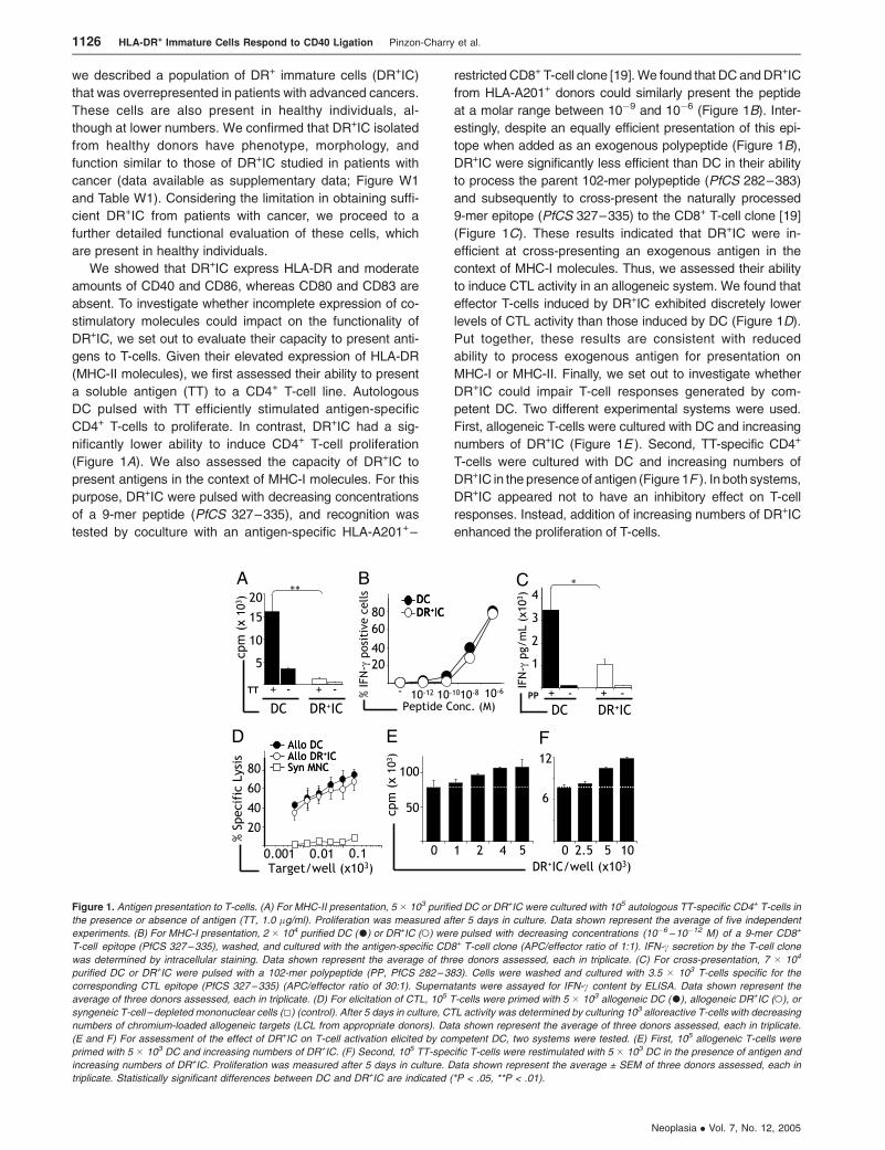
we described a population of DR+ immature cells (DR+IC)
that was overrepresented in patients with advanced cancers.
These cells are also present in healthy individuals, al-
though at lower numbers. We confirmed that DR+IC isolated
from healthy donors have phenotype, morphology, and
function similar to those of DR+IC studied in patients with
cancer (data available as supplementary data; Figure W1
and Table W1). Considering the limitation in obtaining suffi-
cient DR+IC from patients with cancer, we proceed to a
further detailed functional evaluation of these cells, which
are present in healthy individuals.
We showed that DR+IC express HLA-DR and moderate
amounts of CD40 and CD86, whereas CD80 and CD83 are
absent. To investigate whether incomplete expression of co-
stimulatory molecules could impact on the functionality of
DR+IC, we set out to evaluate their capacity to present anti-
gens to T-cells. Given their elevated expression of HLA-DR
(MHC-II molecules), we first assessed their ability to present
a soluble antigen (TT) to a CD4+ T-cell line. Autologous
DC pulsed with TT efficiently stimulated antigen-specific
CD4+ T-cells to proliferate. In contrast, DR+IC had a sig-
nificantly lower ability to induce CD4+ T-cell proliferation
(Figure 1A). We also assessed the capacity of DR+IC to
present antigens in the context of MHC-I molecules. For this
purpose, DR+IC were pulsed with decreasing concentrations
of a 9-mer peptide (PfCS 327–335), and recognition was
tested by coculture with an antigen-specific HLA-A201+–
restricted CD8+ T-cell clone [19].We found that DC andDR+IC
from HLA-A201+ donors could similarly present the peptide
at a molar range between 10�9 and 10�6 (Figure 1B). Inter-
estingly, despite an equally efficient presentation of this epi-
tope when added as an exogenous polypeptide (Figure 1B),
DR+IC were significantly less efficient than DC in their ability
to process the parent 102-mer polypeptide (PfCS 282–383)
and subsequently to cross-present the naturally processed
9-mer epitope (PfCS 327–335) to the CD8+ T-cell clone [19]
(Figure 1C). These results indicated that DR+IC were in-
efficient at cross-presenting an exogenous antigen in the
context of MHC-I molecules. Thus, we assessed their ability
to induce CTL activity in an allogeneic system. We found that
effector T-cells induced by DR+IC exhibited discretely lower
levels of CTL activity than those induced by DC (Figure 1D).
Put together, these results are consistent with reduced
ability to process exogenous antigen for presentation on
MHC-I or MHC-II. Finally, we set out to investigate whether
DR+IC could impair T-cell responses generated by com-
petent DC. Two different experimental systems were used.
First, allogeneic T-cells were cultured with DC and increasing
numbers of DR+IC (Figure 1E ). Second, TT-specific CD4+
T-cells were cultured with DC and increasing numbers of
DR+IC in the presenceof antigen (Figure 1F ). In both systems,
DR+IC appeared not to have an inhibitory effect on T-cell
responses. Instead, addition of increasing numbers of DR+IC
enhanced the proliferation of T-cells.
Figure 1. Antigen presentation to T-cells. (A) For MHC-II presentation, 5 � 103 purified DC or DR+IC were cultured with 105 autologous TT-specific CD4+ T-cells in
the presence or absence of antigen (TT, 1.0 �g/ml). Proliferation was measured after 5 days in culture. Data shown represent the average of five independent
experiments. (B) For MHC-I presentation, 2 � 104 purified DC (.) or DR+IC (o) were pulsed with decreasing concentrations (10�6 –10�12 M) of a 9-mer CD8+
T-cell epitope (PfCS 327–335), washed, and cultured with the antigen-specific CD8+ T-cell clone (APC/effector ratio of 1:1). IFN-c secretion by the T-cell clone
was determined by intracellular staining. Data shown represent the average of three donors assessed, each in triplicate. (C) For cross-presentation, 7 � 104
purified DC or DR+IC were pulsed with a 102-mer polypeptide (PP, PfCS 282–383). Cells were washed and cultured with 3.5 � 103 T-cells specific for the
corresponding CTL epitope (PfCS 327–335) (APC/effector ratio of 30:1). Supernatants were assayed for IFN-c content by ELISA. Data shown represent the
average of three donors assessed, each in triplicate. (D) For elicitation of CTL, 105 T-cells were primed with 5 � 103 allogeneic DC (.), allogeneic DR+IC (o), or
syngeneic T-cell – depleted mononuclear cells (5) (control). After 5 days in culture, CTL activity was determined by culturing 103 alloreactive T-cells with decreasing
numbers of chromium-loaded allogeneic targets (LCL from appropriate donors). Data shown represent the average of three donors assessed, each in triplicate.
(E and F) For assessment of the effect of DR+IC on T-cell activation elicited by competent DC, two systems were tested. (E) First, 105 allogeneic T-cells were
primed with 5 � 103 DC and increasing numbers of DR+IC. (F) Second, 105 TT-specific T-cells were restimulated with 5 � 103 DC in the presence of antigen and
increasing numbers of DR+IC. Proliferation was measured after 5 days in culture. Data shown represent the average ± SEM of three donors assessed, each in
triplicate. Statistically significant differences between DC and DR+IC are indicated (*P < .05, **P < .01).
1126 HLA-DR+ Immature Cells Respond to CD40 Ligation Pinzon-Charry et al.
Neoplasia . Vol. 7, No. 12, 2005

Surface and Cytokine Phenotype of Primed T-cells
The above data demonstrated that, although DR+IC were
not suppressive, they had a reduced capacity to present
exogenous antigens to T-cells. Next, we sought to determine
whether such reduction in antigen-presenting capacity could
affect the nature of the T-cell responses generated. For this
purpose, the phenotype and cytokine profile of T-cells primed
with DC or DR+IC were determined. First, we analyzed
the expression of markers CD25, CD69, CD27, and CCR7
on CD3+ T-cells. Although expression of CD25 and CD69
increased following in vitro stimulation, that of CD27 and
CCR7 decreased [22]. In our experiments with freshly
purified cells, a minimal proportion of T-cells expressed the
activation markers CD25 (2.2 ± 0.8) and CD69 (2.9 ± 1.0). In
addition, a constant proportion of T-cells expressed CD27
(62.0 ± 4.0) and CCR7 (28.5 ± 6.5). Following stimulation,
cultures primed with DC had a higher proportion of T-cells
positive for CD25 and CD69, but a lower proportion of
CCR7+ and CD27+ T-cells than cultures stimulated with
DR+IC (Figure 2A). We also assessed the ability of DR+IC to
stimulate IFN-g secretion in T-cells. As shown in Figure 2B,
supernatants taken from allogeneic T-cells cultured with
DR+IC contained significantly lower levels of IFN-g than those
cultured with DC. Given that the balance between type 1
[T-helper (Th) 1] and type 2 (Th2) cytokines is more relevant
than absolute values of single cytokines in driving immune
responses, ratios between cytokine-expressing T-cells were
determined by measuring the intracellular expression of IFN-
g, TNF-a, IL-2 (Th1), and IL-4 (Th2). As shown in Figure 2C,
Th1/Th2 ratios were significantly higher (P < .05) in cultures
primed with DC, indicating a strong Th1 response. In con-
trast, in cultures primed with DR+IC, a higher percentage of
T-cells expressed IL-4 (DC: 5.1 ± 1.8% vs DR+IC: 8.2 ± 2.4),
indicating that a Th2 response was prevalent.
Migratory Capacity of DR+IC
Having assessed DR+IC’s function as APC, we sought to
determine their migratory capacity. Migration of APC toward
lymphoid organs is critical for the initiation of T-cell immunity
and requires APC to respond to lymph node–directing che-
mokines such as CCL21 (secondary plasmacytoid tissue
chemokine, SLC) and CXCL12 (stromal-derived cell factor).
Here, we evaluated the migratory capacity of DR+IC and DC
in response to these chemokines. Given that cell migration
can be hampered by poor survival and manipulation of iso-
lated cells [20], we performed migration assays with whole
PBMC, taking advantage of quantitative flow cytometry
(Figure 3A). Our experiments confirmed that CD11c+DC are
a highly motile population responding vigorously to CCL21
and CXCL12 [20]. In keeping with previous reports,
CD123+DC had a reduced capacity to migrate when com-
pared to CD11c+DC [20], demonstrating marginal che-
motaxis to CXCL12 and moderate chemotaxis to CCL21.
Interestingly, DR+IC displayed a stronger chemotaxis than
CD123+DC. In particular, they were significantly responsive
to CCL21, with at least 60% of the starting population
migrating across transwells (Figure 3B; P < .05). These re-
sults indicated that whereas DR+IC displayed limited APC
capacity when compared to DC, they exhibited considerable
migratory capacity to a lymph node–directing chemokine.
Figure 2. Phenotype of primed T-cells. (A) For surface phenotype, 5 � 103
sorted DC or DR+IC were cultured with 1 � 105 allogeneic T-cells in complete
medium. After 5 days, cells were harvested and CD3+ T-cells were analyzed
for CD25, CD69, CD27, or CCR7 expression. Data shown represent the aver-
age of five donors assessed. (B and C) For cytokine secretion by T-cells,
5 � 103 purified DC or DR+IC were cultured with 105 allogeneic T-cells. After
5 days in culture, (B) supernatants were collected and IFN-c production was
assessed by ELISA, or (C) cells were collected and CD3+ cells were stained
with cytokine-specific mAb (IFN-c, TNF-a, IL-2, and IL-4). Percentage of
positive T-cells for each cytokine was determined, and type 1/type 2 ratio was
calculated. Data shown correspond to individual donors, each assessed in
triplicate. Means are shown as horizontal lines, and error bars indicate SEM.
Statistically significant differences between DC and DR+IC are indicated
(*P < .05, **P < .01).
Figure 3. Migratory capacity. (A) Flow cytometric analysis for evaluation of
the migration of DC and DR+IC in unseparated PBMC. For cell number
estimation, a fixed number of fluorescent beads (R1) were added to each
PBMC preparation (R2). Subsequently, Lin�HLA-DR+ cells (R3) were divided
into CD11c+DC (R4), CD123+DC (R5), and DR+IC (R6), and their frequency
was estimated before (input) and after (migrated) migration. Absolute
numbers for each subpopulation were calculated as described in Materials
and Methods section. Numbers shown indicate the percentage of total cells
within the corresponding gate. (B) Summary of chemotaxis data in response
to CCL21 and CXCL12. Migration is shown as the percentage of migrated
cells with regard to its input. Data shown represent the average ± SEM of five
donors assessed. Statistically significant differences are indicated (*P < .05).
HLA-DR+ Immature Cells Respond to CD40 Ligation Pinzon-Charry et al. 1127
Neoplasia . Vol. 7, No. 12, 2005

Resistance to Tumor-Induced Apoptosis
Next, we set out to obtain insights into possible mecha-
nisms that could be responsible for the decrease in DC and
the accumulation of DR+IC that has been documented in
cancer patients. Tumor-derived factors have been reported
to induce apoptosis in APC, including DC [23]. Thus, we
hypothesized that accumulation of DR+IC could represent the
selective survival of cells resistant to tumor-induced apopto-
sis. We investigated this hypothesis by measuring apoptosis
in DC and DR+IC following culture in the presence of TDSNs.
TDSN were filtered and dialyzed against a fresh medium
prior to use, excluding the possibility of apoptosis induced by
nutrient depletion. As shown in Figure 4A, assessment of
Annexin V binding revealed minimal apoptosis on fresh
samples or following culture with PBMC-conditioned me-
dium (control). However, following a 24-hour incubation time,
all TDSN induced apoptosis in DC to a significantly greater
extent than in DR+IC (Figure 4, A and B). These results were
consistently observed in the presence of all TDSN tested,
including breast (MB231, MA11, MB435, SKBR3, andMCF7)
and colon (LOVO) cancer cell lines. Comparable results were
obtained by TUNEL assays. Indeed, a significantly higher
proportion of TUNEL+ DC compared to DR+IC was observed
in cultures incubated with all TDSN tested (MA11, MB435,
MCF7 and SKBR3).
Phenotypic Maturation in Response to CD40 Ligation
Given the immaturity and abundance of DR+IC in cancer
patients, it was of interest to determine whether these cells
could be induced to 1) differentiate or 2) perform as com-
petent APC. Several cytokines and growth factors previously
reported to induce maturation of immature cells [24] were
tested, including GM-CSF (1000 U/ml), IL-4 (1000 U/ml),
TNF-a (10 ng/ml), and ATRA (10�4–10�9 M). These matura-
tion factors failed to induce DR+IC differentiation in vitro. Less
than 10% of the cells survived, and only GM-CSF appeared to
maintain viability (> 60%) beyond 72 hours, as assessed by
trypan blue exclusion (data not shown). Our initial experi-
ments also indicated that DR+IC responded weakly to pro-
inflammatory cytokines and various pathogen-derived factors.
Because 40% to 50% of DR+IC expressed the CD40 antigen,
we evaluated whether CD40 ligation could induce a more
robust response. To limit donor-to-donor variation, we ana-
lyzed the maturation of cells purified from the same indi-
vidual donors and compared CD40 stimulation with poly I:C,
CC, and the combination of CD40L plus poly I:C. The latter
was included to examine the potential for synergism between
CD40 ligation and TLR signalling on the functional profile of
DR+IC. As shown in Figure 5A, DC responded vigorously to
all proinflammatory factors, with CD40 ligation inducing the
most robust upregulation in the expression of CD86 and HLA-
DR. The combination of CD40L plus poly I:C did not induce
a stronger response. Notably, although DR+IC failed to re-
spond to inflammatory factors, CD40 ligation induced a sig-
nificant upregulation of HLA-DR and CD86. As with DC,
combination of CD40L plus poly I:C did not induce increased
maturation compared to CD40L alone (Figure 5B). To con-
firm these results, electron microscopy was performed on
sort-purified DC and DR+IC. Compared to untreated DC,
CD40-stimulated DC were larger (10–12 mm) and had more
cytoplasmic inclusions and dendrites, as well as reduced
amounts of condensed chromatin in the nucleus (Figure 5C).
However, CD40-stimulated DR+IC were a little different with
respect to cytoplasmic organelles and inclusions. However,
they were dramatically increased in size (7–8 mm) and had
less condensed chromatin in the nucleus (Figure 5D).
Cytokine Secretion in Response to CD40 Ligation
To determine whether phenotypic maturation was paral-
leled by functional improvement, we analyzed the secretion
Figure 4. Resistance to apoptosis. (A) A representative flow cytometric analysis of apoptosis of DC and DR+IC. PBMC were incubated in the presence of 50% (vol/
vol) PBMC-conditioned medium (control) or TDSNs (MB231, MA11, MB435, SKBR3, MCF-7, and LOVO) for 24 hours. Subsequently, Lin�HLA-DR+ cells were
subdivided into DC (CD11c+CD123+) or DR+IC (CD11c�CD123�) based on their expression of CD11c and CD123 ( y axis). Apoptosis was determined by the
detection of exposed phosphatidylserine residues with Annexin V (x axis). Numbers indicate the percentage of cells that are positive for Annexin V. (B) Summary of
apoptosis data estimated with Annexin V and TUNEL assays. Results shown represent the average ± SEM of five donors assessed. Statistically significant
differences between DC and DR+IC are indicated (*P < .05, **P < .01; nd, not done).
1128 HLA-DR+ Immature Cells Respond to CD40 Ligation Pinzon-Charry et al.
Neoplasia . Vol. 7, No. 12, 2005

of IL-12, IL-10, and TNF-a in DC and DR+IC. Previous
reports indicated that blood DC are poor producers of
cytokines following CD40 stimulation [25]. Our initial experi-
ments confirmed these results and indicated that DC re-
quired the addition of IFN-g and IL-1b for cytokine secretion
following CD40 ligation (data not shown). Thus, we com-
pared proinflammatory (CC) and pathogen-mimic factors
(poly I:C) with CD40 ligation under these conditions. As
previously reported [16,25], CC induced minimal synthesis
of IL-12, and CD40 ligation induced robust production of
this cytokine by DC (Figure 6A). Notably, CD40L also
induced IL-12 secretion by DR+IC. The combination of
CD40L plus poly I:C induced a profile comparable to
CD40L alone (Figure 6B). Next, we assessed whether
CD40 ligation also improved the ability of DR+IC to induce
IFN-g secretion in T-cells. For this purpose, DC and DR+IC
were stimulated with CD40L, washed, and cultured with T-
cells. We found that untreated DC were strong stimulators of
IFN-g secretion, and this ability was enhanced on stimulation
with CD40L (Figure 6C). Interestingly, although DR+IC were
less efficient inducers of IFN-g secretion, CD40-stimulated
DR+IC were significantly better (Figure 6D). Indeed, follow-
ing CD40 ligation, the capacity of DR+IC to induce IFN-g
secretion was increased by 11.1 ± 2.4 times compared to
7.6 ± 1.5 for DC. Similarly, the capacity of DR+IC to induce
proliferation was enhanced, on average, by 1.7 ± 0.5 times
compared to 1.0 ± 0.2 for DC, suggesting that DR+IC were
more sensitive to CD40 stimulation than DC.
Maturation and Cytokine Secretion in Samples
from Breast Cancer Patients
Having determined that DR+IC from healthy donors
responded vigorously to CD40 stimulation, we sought to
determine whether a comparable response was observed
in samples from cancer patients. First, we analyzed pheno-
typic maturation in DC and DR+IC from breast cancer
patients by comparing CD40L, poly I:C, CC, and CD40L plus
poly I:C. As shown in Figure 7A, DC responded to CC and
poly I:C with moderate upregulation of the expression of
HLA-DR and CD86. Stimulation of DC with CD40L induced
the strongest increase in CD86 and HLA-DR expression.
CD40L plus poly I:C did not exhibit improved maturational
capacity of DC compared to CD40L alone. Importantly,
CD40L induced the most significant upregulation of costimu-
latory expression in DR+IC. As with DC, CD40L plus poly I:C
did not induce increased maturation in DR+IC compared
to CD40L alone. Next, we analyzed the secretion of IL-12,
IL-10, and TNF-a in DC and DR+IC by intracellular staining.
These experiments demonstrated that, whereas CC induced
minimal cytokine production, CD40L induced robust IL-12
secretion in DC and DR+IC (Figure 7B). In keeping with
the data described above, addition of CD40L plus poly I:C
did not demonstrate a synergistic effect on cytokine produc-
tion (Figure 7B). Finally, we compared the capacity of CC,
CD40L, or poly I:C–stimulated PBMC from breast cancer
patients (blood DC compartment distribution of CD11c+DC:
51.7± 9.0%, CD123+DC: 18.0±3.6, and DR+IC: 29.8 ± 6.2%)
Figure 5. Maturation in response to CD40 stimulation. (A and B) PBMC were
cultured for 18 to 36 hours in the presence of CC, poly I:C, CD40L, or CD40L
plus poly I:C and upregulation in the expression of HLA-DR, CD40, CD80,
CD83, or CD86, as assessed by flow cytometry for (A) DC and (B) DR+IC.
Phenotypic maturation is expressed as DMFI between unstimulated and
stimulated cultures. Results shown represent the average ± SEM of five
donors assessed. Statistically significant differences between CD40L and CC
or poly I:C are indicated (*P < .05, **P < .01, ***P < .001). (C and D) PBMC
from one healthy donor were isolated, and DC (C) or DR+IC (D) were sort-
purified for EM. Cells were analyzed immediately after isolation (left panel) or
following stimulation with CD40L (right panel). Size bars represent 1 �m. Re-
sults shown are representative of three donors examined.
Figure 6. Cytokine secretion in response to CD40 stimulation. (A and B)
Secretion of TNF-a, IL-10, and IL-12 was assessed by intracellular staining.
PBMCwere cultured for 18 to 36 hours in the presence of CC, poly I:C, CD40L,
or CD40L plus poly I:C in the presence of BFA, as described in Materials
and Methods section. Cytokine secretion is expressed as DMFI between
unstimulated and stimulated cultures. (C and D) For determination of secretion
of bioactive IL-12, 5 � 103 purified (C) DC or (D) DR+IC were stimulated with
CD40L, washed extensively, and cocultured with 105 allogeneic T-cells. After
5 days in culture, supernatants were collected, and IFN-c production was
assessed by ELISA. Results shown represent the average ± SEM of five
donors assessed. Statistically significant differences between CD40L and CC
or poly I:C are indicated (*P < .05, **P < .01, ***P < .001).
HLA-DR+ Immature Cells Respond to CD40 Ligation Pinzon-Charry et al. 1129
Neoplasia . Vol. 7, No. 12, 2005

to produce IL-12 as well as to induce IFN-g secretion and
proliferation by allogeneic T-cells. As shown in Figure 7,C–E,
CD40L induced the strongest IL-12 secretion, as well as
IFN-g production and proliferation by allogeneic T-cells.
Discussion
DC have been implicated in the defective function of the
immune system during cancer progression [26]. Several
reports suggest that that the effect of tumors on DC func-
tion is a generalized process with clinical relevance [9,27].
Despite this, few studies have focused on the systemic
effects of cancer on circulating DC [6,28,29] as opposed to
in vitro–derived DC [11,13–15]. Blood DC arguably repre-
sent the most relevant DC population given that they directly
reflect the natural biology of immune responses occur-
ring in vivo. The blood DC compartment (Lin�HLA-DR+ cells)
[30] is comprised of myeloid (CD11c+DC) and plasmacytoid
(CD123+DC) subsets [31]. In an accompanying paper, we
show that the blood DC population in patients with cancer
displays an alteration in the distribution of the CD11c+DC and
CD123+DC subsets, exhibiting a significant accumulation of a
population of HLA-DR+ immature cells (DR+IC). This popu-
lation of immature cells is also found in healthy donors, al-
though here it represents a small proportion of the circulating
DC compartment.
Morphologic and phenotypic analyses of DR+IC purified
from healthy volunteers demonstrated characteristics similar
to those of DR+IC isolated from cancer patients (supplemen-
tary data; Figure W1 and Table W1). Therefore, in this
study, we sort-purified DR+IC from healthy volunteers and
thoroughly evaluated their functionality to determine whether
these immature cells could have an impact on immune re-
sponse. First, we defined their capacity to present antigens to
T-cells. We found that DR+IC were less efficient than DC in
several systems, including MHC-II–restricted antigen pre-
sentation and MHC-I–restricted cross-presentation. In the
peripheral blood of cancer patients, the abundance of imma-
ture cells with a limited ability to present antigens may be
responsible, at least in part, for the failure of blood DC to gen-
erate adequate T-cell responses [6,9,32]. Alternatively, this
could be due to direct suppression of T-cell function exerted
by immature cells, as has been reported in patients with head
and neck, lung, and breast cancers [24,33]. Interestingly,
although DR+IC had a reduced capacity to stimulate T-cells,
they did not inhibit T-cell proliferation in either an allogeneic
or an antigen-specific manner when competent DCwere also
present. Differences in the lineage composition of DR+IC (a
heterogenous population encompassing immature myeloid
and lymphoid subpopulations) and expression of molecules
associatedwith antigen presentation (HLA-DR, CD40, CD86,
and CD1c) could account for the differing observations.
The expression of HLA-DR and costimulatory molecules,
together with poor APC function, indicated that inefficient
antigen presentation, rather than direct suppression, was the
major mechanism by which DR+IC could affect immune
competence in cancer. Indeed, immature APC with deficient
expression of costimulatory molecules and poor capacity to
Figure 7. Maturation and cytokine secretion in samples from breast cancer patients. (A) PBMC from breast cancer patients were cultured for 18 to 36 hours in the
presence of CC, poly I:C, CD40L, or CD40L plus poly I:C and upregulation in the expression of HLA-DR, CD40, CD80, CD83, or CD86 by DC and DR+IC, as
assessed by flow cytometry. Phenotypic maturation is expressed as DMFI between unstimulated and stimulated cultures. (B) Secretion of IL-12, IL-10, and TNF-aby DC and DR+IC was assessed by intracellular staining. PBMC were stimulated for 18 to 36 hours with CC, poly I:C, CD40L, or CD40L plus poly I:C in the
presence of BFA, as described in Materials and Methods section. Cytokine secretion is expressed as DMFI between unstimulated and stimulated cultures. (C) For
determination of IL-12 secretion, 50 � 103 PBMC from breast cancer patients were cultured in the presence of CC, poly I:C, CD40L, or CD40L plus poly I:C, and
supernatants were assayed for IL-12p70 by ELISA. (D and E) For determination of IFN-c secretion (D) and proliferation (E) by allogeneic T-cells, 20 � 103
stimulated PBMC were cocultured with 105 allogeneic CD4+ T-cells from a panel of healthy donors (n = 3). After 5 days in culture, supernatants were assayed for
IFN-c by ELISA, and proliferation was estimated by [3H]thymidine uptake. Results shown represent the average ± SEM of six breast cancer patients (stages I and
II) who were assessed. Statistically significant differences between CD40L and CC or poly I:C are indicated (*P < .05, **P < .01, ***P < .001).
1130 HLA-DR+ Immature Cells Respond to CD40 Ligation Pinzon-Charry et al.
Neoplasia . Vol. 7, No. 12, 2005

stimulate T-cell responses have been reported to present
antigens inadequately and to induce tolerance in cancer pa-
tients [7]. Our results demonstrated that, in contrast to DC,
in cultures primed with DR+IC, a smaller proportion of T-cells
expressed high levels of antigens upregulated following
adequate T-cell activation. Furthermore, T-cells primed with
DR+IC showed Th2 bias in cytokine secretion, suggesting
that DR+IC provided a different type of ‘‘stimulation’’ to
T-cells when compared to DC. This is important because
Th1 cytokines are necessary for the induction and mainte-
nance of antitumor cytotoxic responses in vivo, whereas Th2
responses have been shown to subvert Th1-mediated im-
munity [34]. In fact, a clear correlation between Th2 preva-
lence and cancer progression has been demonstrated in
patients with renal cell carcinoma and melanoma [35], sug-
gesting that Th2 bias could provide a microenvironment that
favors tumor survival and disease progression. Our data
demonstrate that DR+IC can induce Th2 bias in T-cells and
migrate in response to the lymph node–driven chemokine
CCL21. Based on the above data, it is interesting to speculate
that if DR+IC are capable of migration to lymphoid organs,
then the effect of Th2 cytokines induced through inadequate
presentation could suppress Th1 immunity and impair anti-
tumor responses.
We also investigated possible mechanisms that are re-
sponsible for the significant accumulation of DR+IC in cancer.
Increased recruitment or altered survival of immature cells
could be responsible for this phenomenon. In fact, tumor
cells have been reported to secrete factors that recruit imma-
ture cells [36], or express proapoptotic factors (such as nitric
oxide, TGF-b, IL-10, or gangliosides) that affect the survival of
DC [15,23]. Here, we showed that supernatants derived from
tumor cells induced significant levels of apoptotic death in DC,
but not in DR+IC. Further investigations into the mechanism
underlying TDSN-induced apoptosis have indicated a modu-
lation of antiapoptoticmolecule expression (i.e., Bcl-2; Pinzon-
Charry et al., unpublished observations), in keeping with
reports that CD40 ligation protects DC from tumor-induced
apoptosis through increased expression of Bcl-2 [37]. Inter-
estingly, expression of functional CD40 ligand has been
described in various tumor cell types as an autocrine anti-
apoptotic mechanism [38], possibly providing minimal CD40
stimulation to bystander immune cells. Given the elevated
expression of CD40 and the exquisite sensitivity to signalling
through this pathway, this mechanism could play a relevant
role in resistance to tumor-induced apoptosis by DR+IC. In this
context, increased expression of CD40 could represent a
mechanism for resistance to apoptosis developed by cells
accumulated under the influence of tumor products.
Our results are also relevant because bloodDChave been
proposed for use in DC-based immunotherapy protocols in
cancer, but have thus far produced limited response rates
[39]. Blood DC offer the theoretical advantage of being in
their natural state of differentiation and are presumably
capable of stimulating immune responses in a more physio-
logical manner. DR+IC represent a significant proportion of
the blood DC compartment in cancer patients, and this may
explain, at least in part, the failure of blood DC from cancer
patients to generate adequate T-cell responses [6,9,32]. For
this reason, identification of factors that enhance the function
of APC that are increased in cancer patients and are intrin-
sically resistant to tumor-induced apoptosis could potentially
improve the efficacy of blood DC immunotherapeutic ap-
proaches for cancer.
Importantly, data from healthy volunteers and patients
with breast cancer showed that CD40 stimulation induced
robust phenotypic maturation, upregulation of antigen-
presenting machinery, and secretion of IL-12 by DR+IC.
Although the interaction between CD40 and its ligand has
been recognized as an important element for conditioning
DC to prime and expand tumor-specific cytotoxic responses
[40,41], at present, no studies have been reported on
CD40L-conditioned DC for immunotherapy in patients with
cancer. Nevertheless, the potential of CD40L as an efficient
stimulator for another type of professional APC (B-cells)
under clinically applicable conditions has been demonstrated
[42], and two cancer vaccine trials with CD40L-conditioned
DC are currently listed in the Physician Data Query web site
of the National Cancer Institute [43]. Our findings confirm
that the help signal provided by CD40 engagement is one of
the most potent stimuli for the maturation of DC and imma-
ture APC (DR+IC), which are abundant in cancer patients,
and also indicate that this approach for DC-based cancer
immunotherapy warrants further consideration.
In conclusion, we document that the immature population
of DR+IC that accumulate within the blood DC compartment
of patients with cancer could contribute to immune suppres-
sion by means of inefficient APC function, displacement of
DC, and generation of inadequate immune responses. These
data underscore the importance of thoroughly evaluating
endogenous APC function in patients with cancer. More
importantly, our finding that CD40 ligation boosts the APC
function of DR+IC supports the use of exogenously ‘‘trained’’
APC to correct the unbalanced immunologic performance in
cancer and may prove to be crucial in improving the efficacy
of DC-based immunotherapies for cancer.
Acknowledgements
The authors thank Grace Chojnowski, Paula Hall, and Clay
Winterford for MoFlo sorting and technical assistance with
electron microscopy. We also thank Greg Bryson, Michael
McGuckin, and Giampietro Corradin for providing anti-
bodies, tumor cell lines, and synthetic polypeptides, as well
as Geoff Hill for critically reviewing the manuscript. We are
grateful to the Australian Red Cross Blood Service and
Sullivan Nicolaides Pathology Laboratories for blood sam-
ples, to Amgen for kindly providing soluble CD40L, and to
our patients and volunteers without whom this study would
have not been possible.
References[1] Banchereau J, Briere F, Caux C, Davoust J, Lebecque S, Liu YJ,
Pulendran B, and Palucka K (2000). Immunobiology of dendritic
cells. Annu Rev Immunol 18, 767–811.
HLA-DR+ Immature Cells Respond to CD40 Ligation Pinzon-Charry et al. 1131
Neoplasia . Vol. 7, No. 12, 2005

[2] Fong L and Engleman EG (2000). Dendritic cells in cancer immuno-
therapy. Annu Rev Immunol 18, 245–273.
[3] Zeid NA and Muller HK (1993). S100 positive dendritic cells in human
lung tumors associated with cell differentiation and enhanced survival.
Pathology 25, 338–343 (Oct).
[4] Tsuge T, Yamakawa M, and Tsukamoto M (2000). Infiltrating dendritic/
Langerhans cells in primary breast cancer. Breast Cancer Res Treat
59, 141–152 (Jan).
[5] Lespagnard L, Gancberg D, Rouas G, et al. (1999). Tumor-infiltrating
dendritic cells in adenocarcinomas of the breast: a study of 143 neo-
plasms with a correlation to usual prognostic factors and to clinical
outcome. Int J Cancer 84, 309–314 (Jun 21).
[6] Ratta M, Fagnoni F, Curti A, et al. (2002). Dendritic cells are functionally
defective in multiple myeloma: the role of interleukin-6. Blood 100,
230–237 (Jul 1).
[7] Enk AH, Jonuleit H, Saloga J, and Knop J (1997). Dendritic cells as
mediators of tumor-induced tolerance in metastatic melanoma. Int J
Cancer 73, 309–316 (Nov 4).
[8] Troy AJ, Davidson PJ, Atkinson CH, and Hart DN (1999). CD1a den-
dritic cells predominate in transitional cell carcinoma of bladder and
kidney but are minimally activated. J Urol 161, 1962–1967 (Jun).
[9] Gabrilovich DI, Corak J, Ciernik IF, Kavanaugh D, and Carbone DP
(1997). Decreased antigen presentation by dendritic cells in patients
with breast cancer. Clin Cancer Res 3, 483–490 (Mar).
[10] Pinzon-Charry A, Maxwell T, and Lopez JA (2005). Dendritic cell dys-
function in cancer: a mechanism for immunosuppression. Immunol Cell
Biol 83, 451–461 (Oct).
[11] Menetrier-Caux C, Montmain G, Dieu MC, et al. (1998). Inhibition of the
differentiation of dendritic cells from CD34(+) progenitors by tumor
cells: role of interleukin-6 and macrophage colony-stimulating factor.
Blood 92, 4778–479 (Dec 15).
[12] Gabrilovich DI, Chen HL, Girgis KR, et al. (1996). Production of vascu-
lar endothelial growth factor by human tumors inhibits the functional
maturation of dendritic cells. Nat Med 2, 1096–1103 (Oct).
[13] Sombroek CC, Stam AG, Masterson AJ, et al. (2002). Prostanoids play
a major role in the primary tumor– induced inhibition of dendritic cell
differentiation. J Immunol 168, 4333–4343 (May 1).
[14] Peguet-Navarro J, Sportouch M, Popa I, Berthier O, Schmitt D, and
Portoukalian J (2003). Gangliosides from human melanoma tumors
impair dendritic cell differentiation from monocytes and induce their
apoptosis. J Immunol 170, 3488–3494 (Apr 1).
[15] Shurin GV, Shurin MR, Bykovskaia S, Shogan J, Lotze MT, and
Barksdale EM Jr (2001). Neuroblastoma-derived gangliosides inhibit
dendritic cell generation and function. Cancer Res 61, 363–369
(Jan 1).
[16] Chang AE, Redman BG, Whitfield JR, et al. (2002). A phase I trial of
tumor lysate–pulsed dendritic cells in the treatment of advanced can-
cer. Clin Cancer Res 8 (4), 1021–1032.
[17] Krug A, Rothenfusser S, Selinger S, et al. (2003). CpG-A oligonucleo-
tides induce a monocyte-derived dendritic cell – like phenotype that pref-
erentially activates CD8 Tcells. J Immunol 170, 3468–3477 (Apr 1).
[18] Cella M, Salio M, Sakakibara Y, Langen H, Julkunen I, and Lanzavecchia
A (1999). Maturation, activation, and protection of dendritic cells induced
by double-stranded RNA. J Exp Med 189, 821–829 (Mar 1).
[19] Prato S, Maxwell T, Pinzon-Charry A, Schmidt CW, Corradin G, and
Lopez JA (2005). MHC class I – restricted exogenous presentation of a
synthetic 102-mer malaria vaccine polypeptide. Eur J Immunol 35,
681–689 (Mar).
[20] de la Rosa G, Longo N, Rodriguez-Fernandez JL, et al. (2003). Migra-
tion of human blood dendritic cells across endothelial cell monolayers:
adhesion molecules and chemokines involved in subset-specific trans-
migration. J Leukoc Biol 73, 639–649 (May).
[21] Molema G, Mesander G, Kroesen BJ, Helfrich W, Meijer DK, and de Leij
LF (1998). Analysis of in vitro lymphocyte adhesion and transendothe-
lial migration by fluorescent-beads–based flow cytometric cell count-
ing. Cytometry 32, 37–43 (May 1).
[22] Ma CS, Hodgkin PD, and Tangye SG (2004). Automatic generation
of lymphocyte heterogeneity: division-dependent changes in the ex-
pression of CD27, CCR7 and CD45 by activated human naive CD4+ T
cells are independently regulated. Immunol Cell Biol 82, 67–74 (Feb).
[23] Esche C, Lokshin A, Shurin GV, et al. (1999). Tumor’s other immune
targets: dendritic cells. J Leukoc Biol 66, 336–344 (Aug).
[24] Almand B, Clark JI, Nikitina E, et al. (2001). Increased production of
immature myeloid cells in cancer patients: a mechanism of immuno-
suppression in cancer. J Immunol 166, 678–689 (Jan 1).
[25] Jefford M, Schnurr M, Toy T, et al. (2003). Functional comparison of
DCs generated in vivo with Flt3 ligand or in vitro from blood monocytes:
differential regulation of function by specific classes of physiologic stim-
uli. Blood 102, 1753–1763 (Sep 1).
[26] Gabrilovich D (2004). Mechanisms and functional significance of tumour-
induced dendritic-cell defects. Nat Rev Immunol 4, 941–952 (Dec).
[27] Almand B, Resser JR, Lindman B, et al. (2000). Clinical significance of
defective dendritic cell differentiation in cancer. Clin Cancer Res 6,
1755–1766 (May).
[28] Beckebaum S, Zhang X, Chen X, et al. (2004). Increased levels of
interleukin-10 in serum from patients with hepatocellular carcinoma
correlate with profound numerical deficiencies and immature pheno-
type of circulating dendritic cell subsets. Clin Cancer Res 10,
7260–7269 (Nov 1).
[29] Della Bella S, Gennaro M, Vaccari M, et al. (2003). Altered maturation of
peripheral blood dendritic cells in patients with breast cancer. Br J
Cancer 89, 1463–1472 (Oct 20).
[30] Savary CA, Grazziutti ML, Melichar B, et al. (1998). Multidimen-
sional flow-cytometric analysis of dendritic cells in peripheral blood of
normal donors and cancer patients. Cancer Immunol Immunother 45,
234–240 (Jan).
[31] Robinson SP, Patterson S, English N, Davies D, Knight SC, and Reid
CD (1999). Human peripheral blood contains two distinct lineages of
dendritic cells. Eur J Immunol 29, 2769–2778 (Sep).
[32] Satthaporn S, Robins A, Vassanasiri W, et al. (2004). Dendritic cells are
dysfunctional in patients with operable breast cancer. Cancer Immunol
Immunother 53, 510–518 (Jun).
[33] Pak AS, Wright MA, Matthews JP, Collins SL, Petruzzelli GJ, and Young
MR (1995). Mechanisms of immune suppression in patients with head
and neck cancer: presence of CD34(+) cells which suppress immune
functions within cancers that secrete granulocyte–macrophage colony-
stimulating factor. Clin Cancer Res 1, 95–103 (Jan).
[34] Lauerova L, Dusek L, Simickova M, et al. (2002). Malignant melanoma
associates with Th1/Th2 imbalance that coincides with disease pro-
gression and immunotherapy response. Neoplasma 49, 159–166.
[35] Tatsumi T, Kierstead LS, Ranieri E, et al. (2002). Disease-associated
bias in T helper type 1 (Th1)/Th2 CD4(+) T cell responses against
MAGE-6 in HLA-DRB10401(+) patients with renal cell carcinoma or
melanoma. J Exp Med 196, 619–628 (Sep 2).
[36] Garrity T, Pandit R, Wright MA, Benefield J, Keni S, and Young MR
(1997). Increased presence of CD34+ cells in the peripheral blood of
head and neck cancer patients and their differentiation into dendritic
cells. Int J Cancer 73, 663–669 (Nov 27).
[37] Pirtskhalaishvili G, Shurin GV, Esche C, et al. (2000). Cytokine-mediated
protection of human dendritic cells from prostate cancer – induced
apoptosis is regulated by the Bcl-2 family of proteins. Br J Cancer 83,
506–513 (Aug).
[38] Voorzanger-Rousselot N and Blay JY (2004). Coexpression of CD40
and CD40L on B lymphoma and carcinoma cells: an autocrine anti-
apoptotic role. Leuk Lymphoma 45, 1239–1245 (Jun).
[39] Reichardt VL, Okada CY, Liso A, et al. (1999). Idiotype vaccination using
dendritic cells after autologous peripheral blood stem cell transplanta-
tion for multiple myeloma—a feasibility study. Blood 93, 2411–2419
(Apr 1).
[40] Onaitis MW, Kalady MF, Emani S, Abdel-Wahab Z, Tyler DS, and Pruitt
SK (2003). CD40 ligand is essential for generation of specific cytotoxic
T cell responses in RNA-pulsed dendritic cell immunotherapy. Surgery
134, 300–305 (Aug).
[41] Terheyden P, Straten P, Brocker EB, Kampgen E, and Becker JC
(2000). CD40-ligated dendritic cells effectively expand melanoma-
specific CD8+ CTLs and CD4+ IFN-gamma – producing T cells
from tumor-infiltrating lymphocytes. J Immunol 164, 6633–6639 (Jun 15).
[42] von Bergwelt-Baildon MS, Vonderheide RH, Maecker B, et al. (2002).
Human primary and memory cytotoxic T lymphocyte responses are
efficiently induced by means of CD40-activated B cells as antigen-
presenting cells: potential for clinical application. Blood 99, 3319–3325
(May 1).
[43] National_Cancer_Institute (Accessed on 25 May 2005). National Cancer
Institute: National Cancer Institute’s Physician Data Query (PDQ). Ac-
cessed on 25 May 2005. Http://www.cancer.gov/search/clinical_tials. In
Http://ww.cancer.gov/search/clinical_trials.
1132 HLA-DR+ Immature Cells Respond to CD40 Ligation Pinzon-Charry et al.
Neoplasia . Vol. 7, No. 12, 2005