Embed Size (px)
Citation preview

THE CD40 RECEPTOR– TARGET, TOOL AND TECHNOLOGY
PETER ELLMARKDEPARTMENT OF IMMUNOTECHNOLOGY
LUND UNIVERSITY
LUND 2002

Copyright © 2002 Peter Ellmark
ISBN 91-628-5493-3
Printed in Sweden by KFS ABLund 2002

1
TABLE OF CONTENTS
ORIGINAL PAPERS 3
ABBREVIATIONS 4
1 INTRODUCTION 6
2 THE CD40 RECEPTOR 9
2.1 IDENTIFICATION OF CD40 92.2 CD40 PLAYS A CENTRAL ROLE IN THE IMMUNE SYSTEM 92.3 STRUCTURE AND MOLECULAR CHARACTERISTICS OF CD40 102.4 THE CD40-CD154 INTERACTION 112.5 INTRACELLULAR SIGNALLING EVENTS 11
3 SIGNAL INITIATION VIA CD40 14
3.1 OLIGOMERISATION OF CD40 143.2 THE ROLE OF PRE-LIGAND ASSOCIATION OF CD40 153.3 CD40 IN LIPID RAFTS 17
4 CD40 AS A TARGET FOR IMMUNOTHERAPY 20
4.1 CD40 BASED THERAPY IN CLINICAL INFLAMMATORY CONDITIONS 204.2 CD40 IN CANCER TREATMENT 224.3 VACCINATION AND TRANSPLANTATION 24
5 ANTIBODY DEVELOPMENT 25
5.1 BASIC ANTIBODY STRUCTURE 255.2 GENERATION OF DIVERSITY 265.3 SECONDARY RECOMBINATION – RECEPTOR EDITING 275.4 SOMATIC HYPERMUTATION – STEPWISE MUTATIONS 275.4 SOMATIC HYPERMUTATION – RECEPTOR REVISION 285.5 ANTI-CD40 ANTIBODIES AS A TOOL TO STUDY AFFINITY MATURATION
EVENTS 29
6 GENERATION OF HUMAN ANTIBODIES FOR THERAPEUTICAPPLICATIONS 32
6.1 HUMAN MONOCLONAL ANTIBODIES 326.2 ANTIBODIES BINDING TO CD40 346.3 ANTIBODY EVOLUTION AND AFFINITY MATURATION IN VITRO 36

2
7 IDENTIFICATION OF PROTEIN INTERACTIONS 39
7.1 MASS SPECTROSCOPY BASED IDENTIFICATION OF PROTEINS 397.2 LIBRARY SELECTION APPROACHES 407.3 A NOVEL SELECTION TECHNOLOGY BASED ON THE CD40 RECEPTOR - 42SELECTION OF PROTEIN INTERACTIONS BY RECEPTOR ENGAGEMENT (SPIRE). 427.4 FURTHER DEVELOPMENT OF SPIRE 44
CONCLUDING REMARKS 46
POPULÄRVETENSKAPLIG SAMMANFATTNING 48
ACKNOWLEDGEMENTS 50
REFERENCES 51

3
ORIGINAL PAPERS
This thesis is based on the following PAPERs, which are referred to by their
Roman numerals.
I Malmborg-Hager A-C, Ellmark P, Borrebaeck CAK, Furebring
C. Affinity and epitope profiling of mouse anti-CD40 monoclonal
antibodies. Submitted 2002.
II Ellmark P, Ottosson C, Borrebaeck CAK, Malmborg-Hager AC,
Furebring C. Modulation of the CD40-CD40 ligand interaction
using human anti-CD40 single-chain antibody fragments obtained
from the n-CoDeR phage display library. Immunology 2002;
106:456-63.
III Ellmark P, Esteban O, Furebring C, Malmborg Hager A-C ,
Ohlin M. In vitro molecular evolution of antibody genes mimicking
receptor revision. Molecular Immunology 2002; 39:349-56.
IV Ellmark P, Furebring C , Borrebaeck CAK. Pre-assembly of the
extracellular domains of CD40 is not necessary for rescue of naive
B-cells from anti-IgM induced apoptosis. Submitted 2002.
V Ellmark P, Ohlin M, Borrebaeck CAK, Furebring C. A novel
selection system for identification of interacting protein pairs based
on receptor activation. Manuscript
The articles are reproduces with the permission of the publishers.

4
ABBREVIATIONS
Ab antibody
ADCC antibody dependent cellular cytotoxity
AID activation-induced cytidine deaminase
APC antigen presenting cell
BCR B cell receptor
CD cluster of differentiation
CDR complementarity determining region
CTL cytotoxic T lymphocytes
D diversity
Da Dalton
DC dendritic cell
DNA deoxiribonucleic acid
DR death receptor
EAE experimental autoimmune encephalomyelitis
Fab fragment antigen binding
Fc fragment crystalizable
FR frame work region
g3p gene 3 protein
GC germinal centre
HAMA human anti-mouse response
Hsp heat shock protein
Ig immunoglobulin
IGH immunoglobulin heavy
IGL immunoglobulin lambda
IL interleukin
J joining
Jak janus kinase
mAb monoclonal antibody
MS multiple sclerosis
NF-κB nuclear factor-kappaB

5
PCA protein-fragment complementation assay
PCR polymerase chain reaction
PLAD pre-ligand assembly domain
RAG recombination activating gene
RSS recombination signal sequence
scFv single chain fragment variable
SF superfamily
SHM somatic hypermutation
SPIRE selection of protein interactions by receptor engagement
STAT signal transducer and activator of transcription
TCR T cell receptor
TD TRAF domain
TNF tumour necrosis factor
TNFR tumour necrosis factor receptor
TRAF TNF receptor associated factor
V variable
VH variable heavy
VL variable light
Y2H yeast two-hybrid
Å ångström
6
1 INTRODUCTION
The immune system is a sophisticated organisation of molecules, cells and
tissues that acts in concert to protect us against different pathogens. It is
composed of the evolutionary ancient innate compartment and the more
complex adaptive compartment, where the latter is restricted to vertebrates.
Together with the nerve system, the immune system is the major adaptive
system in the body. These two systems (the “supersystems”) act, and interact,
with the main goal to maintain homeostasis.
Although the concept of immunity was known already in ancient Greece, it
was Pasteur and colleagues who, at the end of the nineteenth century, gave born
to the science of immunology by generalising Jenners use of vaccination. During
the first half of the 20t h century, the advancement of the discipline was
stimulated by the controversy between adherents of the theory of cellular
immunity and supporters of the humoral theory of immunity. The latter theory
was most strongly advocated, resulting in enhanced focus on characterisation of
antibodies, the key element of humoral immunity (Silverstein, 1999).
However, the extraordinary ability of the immune system to mount antibodies
against almost every substance that it encounters, was not satisfactorily
explained until MacFarlane Burnet presented the clonal selection theory of
acquired immunity in the late 1950s (Burnet, 1959). He postulated that
antibodies are cell surface receptors and that each individual cell (later known as
B cells) display a single clonotype that upon binding to a specific antigen will
proliferate and give rise to cells that produce antibodies. They also give rise to
cells that are able to react upon repeated antigen encounter, i.e. memory cells.
Furthermore, he incorporated Lederbergs suggestion that immunological
tolerance is the result of clonal abortion of cells that react to self antigens in his
theory. This theory was modified by Bretcher and Cohn (Bretscher and Cohn,
1970), who presented the two signal model 1969, in which they suggested that
activation of a B cell require both recognition of an antigen via the B cell
receptor (BCR) (signal one) and, subsequently, help (signal two) from another
lymphocyte specific for that antigen. It was later found that the cells responsible

7
for providing this help was T-cells and that a B cell receptor, named CD40,
plays a central role in transmitting the second activation signal to B cells.
Laffererty and Cunningham expanded the hypothesis, and added a new cell type
to the model, the antigen presenting cell (APC), which provides the T-cells with
a second activation signal (Cunningham and Lafferty, 1977; Lafferty and
Cunningham, 1975). It was later suggested that the APCs need to be activated
via pattern recognition receptors (PRRs) to provide T-cells with a second signal
(Janeway, 1989; Janeway, 1992). It is now evident that dendritic cells are the
key APCs and play a central role in regulating and tuning the immune system
(Banchereau et al., 2000).
Alongside with the increasing understanding of the immune system there have
been ongoing efforts to explore this knowledge to develop novel techniques and
more efficient therapies. Köhler and Milstein’s development of the monoclonal
antibody technology provided the research community with a source of well-
defined and specific tools (Köhler and Milstein, 1975). However, the initial
excitement over the possibilities to utilise this technology for antibody based
treatment of human patients was hampered by the human anti-mouse antibody
(HAMA) response that antibodies produced by this technique often elicit.
The development of the phage display technique (Smith, 1985), by which
human antibodies can by displayed on the surface of phages (Barbas et al., 1991;
McCafferty et al., 1990), enabled selection of specific, human, antibodies
against target antigens. This started to restore the belief in therapeutic
antibodies, and today the number of monoclonal antibodies in different stages of
clinical trials are increasing rapidly (Borrebaeck and Carlsson, 2001).
Selection of antibodies from a phage display library mimics the adaptive
immune response in that it relies on clonal selection and amplification of entities
that recognise a certain antigen. Furthermore, there is a physical link between
the gene that encodes the antigen binding protein (genotype) and the protein
itself (phenotype). The combination of molecular libraries and selection systems
where genotype and phenotype is linked, has proven to be very successful and a
number of different techniques, beside the page display system, have been
developed based on this principle, e.g. ribosomal display (Schaffitzel et al.,
1999).

8
As implied by the title of this thesis, the CD40 receptor is the inter-connective
theme that ties the different parts together. In PAPER I, some of the molecular
mechanisms and requirements behind anti-CD40 antibody-mediated signalling
have been elucidated. The results from PAPER I aided the development of human
immuno-modulatory, anti-CD40 antibodies, described in PAPER II. We further
utilised the antibodies obtained in PAPER II to analyse antibody evolution events
(PAPER III). The molecular requirements for CD40 mediated stimulation was
further investigated in PAPER IV. We showed that none of the extracellular
domains of CD40 is critical for functional signalling. Finally, in PAPER V, we
used the functional understanding of CD40 signalling to develop a eukaryote
selection system, in which CD40 signalling is used to clonally select cells from a
mammalian expression library.

9
2 THE CD40 RECEPTOR
2.1 Identification of CD40
CD40 was originally identified by an antibody raised against urinary bladder
carcinoma that was found to bind B cells as well (Koho et al., 1984; Paulie et al.,
1984). Today, CD40 is known to be expressed on a variety of cells in the
immune system, e.g. on B cells, dendritic cells, basophils, eosinophils and
monocytes. CD40 can, furthermore, be detected on endothelial cells,
keratinocytes, smooth muscle cells, eptithelial cells and fibroblasts (Schonbeck
and Libby, 2001; van Kooten and Banchereau, 1997; van Kooten and
Banchereau, 2000). Although the CD40 ligand (CD40L), which also is known as
CD154, is mainly expressed on activated T-cells it can also be found on
basophils, eosinophils, monocytes, macrophages, dendritic cells, NK cells, B
lymphocytes, platelets, mast cells, endothelial cells, smooth muscle cells and
epithelial cells (Schonbeck and Libby, 2001). A common feature of all these
cells is that the CD154 expression is non-constitutive, but can be rapidly
induced upon activation.
2.2 CD40 plays a central role in the immune system
The pivotal importance of the CD40 molecule was recognised when it was
discovered that X-linked hyper IgM syndrome is due to a detrimental mutation
of the gene encoding CD154 (Allen et al., 1993; Aruffo et al., 1993; DiSanto et
al., 1993; Korthauer et al., 1993). This disease is characterised by defective
isotype switching and lack of somatic mutation as well as deficient B cell
memory (Notarangelo and Peitsch, 1996). It was found that CD40 signalling is
critical for T cell dependent B cell activation (Foy et al., 1993). Furthermore,
signalling via CD40 is necessary for subsequent steps in B lymphocyte
activation, such as affinity maturation, isotype switching, generation of memory
cells and germinal centre formation, although the latter may be a secondary
effect (Siepmann et al., 2001). Stimulation of human B cells with anti-CD40
antibodies or recombinant CD154 has been shown to induce homeotypic
aggregation and up-regulation of surface markers such as CD23, CD30, CD80,

10
CD86, Fas and major histocompatibility complex (MHC) II and soluble
cytokines, e.g. IL-6, TNF-α and TNF-β (Schonbeck and Libby, 2001).
Furthermore, CD40 is also critical for induction of cellular immunity and has a
pro-inflammatory activity on e.g. monocytes and DC (Caux et al., 1994; Mackey
et al., 1998).
It has recently been shown that CD40 also interacts with heat shock proteins
(Hsp70) from both bacterial (Wang et al., 2001) and mammalian (Becker et al.,
2002) sources. This type of heat shock proteins binds to a broad range of
peptides during synthesis or under conditions of cellular stress. They can reach
the extracellular space subsequent to necrosis or viral lysis and may serve as a
carrier of antigenic peptides for cross-priming (Becker et al., 2002). The
bacterial variant has been reported to upregulate chemokines, such as RANTES,
MIP-1α and MIP-1β, upon binding to CD40 (Wang et al., 2001). Becker et al.
(2002), furthermore, showed that mouse Hsp70, isolated from a tumour cell with
bound tumour-associated peptide, can induce CD40 signalling in DC. Moreover,
they suggested that peptide loading is required for this interaction. The Hsp70-
peptide-CD40 interaction results in uptake of the Hsp70-peptide complex, which
may result in a display of the loaded peptide on MHC class I or II.
2.3 Structure and molecular characteristics of CD40
CD40 is a 43-48 kDa glycoprotein composed of 277 amino acid residues
(Braesch-Andersen et al., 1989). It belongs to the tumour necrosis factor
receptor (TNFR) family, and as all members of this family the extracellular part
of CD40 consist of several cysteine rich repeats. The four extracellular domains
are composed of two types of modular units (Figure 1) (Naismith and Sprang,
1998; Singh et al., 1998) and each module is stabilised by one or two disulfide
bonds. No crystal structure of CD40 has been reported, but several models of
three of the extracellular domains have been proposed, using the known X-ray
structure of TNFR as a template (Bajorath et al., 1995a; Bajorath et al., 1995b;
Singh et al., 1998).
CD40 contains a single membrane spanning domain and expose the N-
terminus of the protein to the exterior (type I membrane protein). Biochemical
characterisation, suggests that CD40 is a hydrophobic protein with a quite acidic

11
pI of 3.2 (van Kooten and Banchereau, 2000). The cytoplasmic domain of CD40
contains several recognition sites for intermolecular proteins, e.g. tumour
necrosis factor receptor associated factors (TRAFs) (Figure 1).
2.4 The CD40-CD154 interaction
The structure of the extracellular part of CD154 has been resolved by X-ray
crystallography (Karpusas et al., 1995). It consists of two beta sheet with
jellyroll topology that forms a symmetric homotrimer. The crystal structure of
CD154 and the CD40 model has been used together with site-directed
mutagenesis to identify five CD40 residues Y82, D84, N86, E74 and E117 and
five CD154 residues K143, Y145, Y146, R203 and Q220 as important for the
CD40-CD154 interaction (Bajorath and Aruffo, 1997; Bajorath et al., 1995a;
Bajorath et al., 1995b). Thus the CD154 binding site has been shown to be
located in the second and third domain of CD40. It has been suggested that polar
interaction between the basic residues on the CD154 and the acidic residues on
CD40 plays an important role for this interaction (Singh et al., 1998).
Notably, analysis of a structure model of clone F33, one of the single chain
antibodies described in PAPER II, which almost totally abrogates the CD40-
CD154 interaction, reveals that it does not contain any acidic residues in the
central core of the binding site (Figure 2). Moreover, it has an unusually high
estimated isoelectric point (theoretical pI 9.5) and it contain at least one
centrally located basic residue, R56L (according to the IMGT nomenclature,
(Lefranc, 2001). This residue has been reported to be the most frequent contact
residue in CDRL2 (contact residue in 9/26 analysed antibodies) (MacCallum et
al., 1996). From these observations, it may be hypothesised that the F33-CD40
interaction, at least partly, depends on similar charged residues as the CD40-
CD154 interaction.
2.5 Intracellular signalling events
To transform the externally applied signal, resulting from oligomerisation of
CD40, to an intracellular signal that ultimately can lead to a cellular response,
CD40 depends on several proteins that can interact with different recognition
motifs (Figure 1). The most important group of intracellular signalling proteins

12
that interacts with CD40 is the TRAF family, which is a group of adapter
molecules with no intrinsic enzymatic activity. CD40 is known to bind directly
to TRAF2, 3 and 6 and indirectly to TRAF1 and 5, although there is some
controversies regarding the association of TRAF5 to CD40 (Ishida 1996,
Nakano 1996, Bishop 2002). The TRAF2 and 3 binding motif on CD40 has
been identified to a PVQET sequence, whereas TRAF6 binds to the QEPQEINF
motif (Pullen et al., 1998).
The TRAFs are composed of a conserved C-terminal domain, TRAF domain
(TD), and a less conserved N-terminal domain. The latter consists of one
(TRAF1) or several (TRAF2-6) zinc finger structures and, in the case of
TRAF2-6, a RING finger structure. This domain mediates interactions with
different secondary signalling proteins (reviewed by Wajant et al., 2001 and
(Inoue et al., 2000). Crystallographic analysis of the TD of the different TRAFs
reveals a trimeric mushroom shaped structure where the N-terminal part, TRAF-
N, forms a triple helical coiled coil and the C-terminal end, TRAF-C, forms a
unique eight stranded anti-parallel beta sheet (Ahonen et al., 2002; McWhirter et
al., 1999; Ni et al., 2000; Park et al., 1999). The N-terminal end of the TD also
contains the CD40 binding site (Cheng and Baltimore, 1996; Cheng et al., 1995;
Malinin et al., 1997; Rothe et al., 1996).
Affinity studies have shown that the TRAF-CD40 interaction depends on
oligomerisation to form a stable signalling complex (Pullen et al., 1999). It has
furthermore been shown that there are marked differences between the affinity
to CD40 between the different TRAFs. Thereby they also seem to require
different degree of oligomerisation to produce a stable interaction, and it has
been suggested that the difference in affinity is the basis for the specificity of the
TRAFs (Pullen et al., 1999). Furthermore, the different TRAFs may play distinct
roles in different stages of B cell development. It has suggested that TRAF6
mediated signalling is crucial for affinity maturation and generation of long-
lived plasma cells (Ahonen et al., 2002), whereas TRAF2 and 3 is essential for
class switching (Jabara et al., 2002).
TRAF recruitment ultimately leads to activation of NF-κB, stress-activated
protein kinase/c-jun amino terminal kinase (JNK/SAPK), p38 mitogen-activated
protein kinase (MAPK) protein tyrosine kinases (PTK), phosphoinositide-3 (PI-

13
Figure 1. Schematic of the CD40 protein. The extracellular parts of CD40 is
composed of four domains (D1-D4), which each consists of two modules. The
approximate location of the CD154 binding site is depicted in the figure (a more
detailed picture over the involved residues is presented in paper I). The putative
interaction sites for the intracellular signalling intermediates that are known to interact
directly with CD40 are also shown.
3) kinase and the extracellular signal-regulated mitogen activated protein kinase
(ERK) (Berberich et al., 1996; Craxton et al., 1998; Grammer et al., 1998;
Purkerson and Parker, 1998; van Kooten, 2000; van Kooten and Banchereau,
1997). Rescue from apoptosis requires a minimum of these signalling pathways,
whereas stimulation of proliferation requires ERK, p38 and PI-3K pathways
(Dadgostar et al., 2002).
Janus kinase 3 (JAK3) has been shown to interact with a proline motif in the
intracellular region of CD40 (Hanissian and Geha, 1997) and it is
phosphorylated upon CD40 stimulation, resulting in the subsequent
phosphorylation of STAT3, STAT5a (Hanissian and Geha, 1997; Revy et al.,
1999) and STAT6 (Karras et al., 1997). The JAK3 pathway seems to be
important for activation of monocytes and dendritic cells (Revy et al., 1999;
Saemann et al., 2002) although it seems to be of little importance in CD40
mediated B cell activation (Jabara et al., 1998).
D1 D4D3D2
Leader peptide JAK3 binding sequence
A-module TRAF6 binding sequence
B-module TRAF2/3 binding sequenceTransmembrane region
Intracellular domainExtracellular part
CD40L binding site

14
3 SIGNAL INITIATION VIA CD40
3.1 Oligomerisation of CD40
By comparing the ability of soluble monomeric, dimeric and trimeric CD40
variants to activate NF-κB in the cytosol, it has been shown that the minimal
requirement to induce a signal through CD40 is the formation of a receptor
dimer (Werneburg et al., 2001), although trimerisation generates a stronger
signal. Haswell et al. has shown that the strength of the receptor mediated signal
correlates with increasing valency of an extracellularly applied ligand (Haswell
et al., 2001). Furthermore, the functional consequences of CD40 signalling also
depends on the cell-type and the differentiation stage of the cell (van Kooten and
Banchereau, 2000). In addition, the level of cross-linking that is required to
initiate a certain signalling pathway varies between different cell types (Fanslow
et al., 1994). It has been suggested that ligation via trimeric CD154 expressed on
a cell membrane is required to activate some particular signalling pathways e.g.
IL-6 production in naive B cells (Baccam and Bishop, 1999).
It has been demonstrated that the stimulatory capacity of different anti-CD40
antibodies depends on the location of the epitope on CD40 (Björck et al., 1994;
Challa et al., 1999; Pound et al., 1999; Sakata et al., 2000). Pound et al. has
suggested that rescue from apoptosis is essentially epitope independent, whereas
the induction of proliferation is more epitope dependent (Pound et al., 1999).
Furthermore, it has been shown that the functional activity of anti-mouse CD40
antibodies correlates with the ability to inhibit CD154 binding to CD40
expressing cells (Barr and Heath, 2001). However, in PAPER I and II we show
that this correlation is only valid when one particular blocking method is used.
Moreover, it can be concluded from these studies that other factors than direct
epitope masking influence the results from these assays. Steric hindrance,
conformational changes as well as CD40-complex formation induced by the first
applied molecule may influence the ability of the second molecules to bind. The
proposed CD40 signalling model, which will be discussed more in detail in the
next section, does however predict that the orientation of the epitope in relation
to the CD154 epitope is important for signal transmission. However, it is

15
probably not important which of the extracellular domains of CD40 the binding
epitope is located on.
It has been suggested that binding of CD154 induces a conformational change
in CD40, which is necessary for signal transmission (Barr and Heath, 2001;
Fanslow et al., 1994; Kaykas et al., 2001; Ledbetter et al., 1997; Pound et al.,
1999). This would indeed predict that the location of the epitope in relation to
the CD154 binding site is critical, since conformational changes in the protein
structure often depends on interaction between the paratope and residues in the
epitope. This theory is mostly based on the observation that monomeric
molecules such as scFv (single chain fragment variable) or soluble monomeric
CD154 can be used to stimulate CD40 signalling. However, the molecules used
in these studies have a tendency to spontaneous form oligomers (Karpusas et al.,
1995; Gilliland et al., 1996). Moreover, in PAPER IV we show that none of the
extracellular domains are essential for signalling via human CD40 in a murine B
cell line (WEHI 231). We, furthermore, show that CD40 mediated rescue of
WEHI 231 cells is comparable between cells that display wild-type CD40 and
those that display CD40 variants, where two of the extracellular domains have
been removed. Taken together, our results indicate that conformational changes
are unlikely to be necessary for CD40 signalling.
3.2 The role of pre-ligand association of CD40
Several members of the TNFR superfamily spontaneously form oligomers on
the cell surface even in the absence of a ligand. Papoff et al. first showed that
oligomerisation of CD95 (Fas) depends on the distal extracellular domain
(Papoff et al., 1999). This was later confirmed (Siegel et al., 2000a) and it was
also showed that TNFR1 and TNFR2 have a similar oligomerisation domain.
The domain that mediates the interaction has been named pre-ligand assembly
domain (PLAD) (Chan et al., 2000a). They, furthermore, showed that CD40 and
Death receptor 4 (DR4) also is specifically pre-ligand associated in the
membrane (Chan et al., 2000a), as detected by FRET analysis. Moreover, it has
been known for long that CD40 purified from normal B cells, under non-
reducing conditions, forms dimers and multimers (Braesch-Andersen et al.,
1989).

16
Although it has not been shown which CD40-domain that is responsible for
pre-ligand assembly, it has been proposed that PLAD is located in the distal
domain (D1) based on homology studies (Locksley et al., 2001). The group of
Leonardo, furthermore, showed that PLAD is necessary for ligand binding to
TNFR I and II and Fas, even though PLAD seems to be physically distinct from
the ligand contact domains (Siegel et al., 2000a; Chan et al., 2000a). We have
demonstrated that the CD40-CD154 interaction also require the distal domain
(D1) of CD40 (PAPER I), although this domain does not contain any of the
residues that are known to be important for CD154 binding. In conclusion, there
are some evidence for a model in which members of the TNFR-SF form
oligomers in the membrane prior to ligand binding without initiating any signals
and that this oligomerisation is necessary for interaction with its cognate ligand.
Hence, ligand induced signalling cannot only be a result of oligomerisation of
the receptor. It rather implies that ligand binding induce a change in the
arrangement of the receptor that allow adapter molecules e.g. TRAFs to bind.
The advantages that this pre-assembly system provides to the TNFR-SF is,
however, not fully understood. It may be suggested that this mechanism is used
to separate the intracellular domains of CD40 prior to ligation, in order to
minimise spontaneous signal initiation (Chan et al., 2000b). Furthermore, it may
be a route to down-regulate the susceptibility for ligand induced signalling by
assembly of non-functional receptors that lack the intracellular part together
with functional receptors, as suggested by Papoff et al. (1999). An alternative
explanation is that ligand binding induce the formation of a super cluster (Chan
et al., 2000a; Chan et al., 2000b; Locksley et al., 2001; Siegel et al., 2000a;
Siegel and Lenardo, 2001), which in turn initiate intracellular signalling events.
However, it has not been investigated to what extent the PLAD effects
signalling via TNFR-SF members.
In PAPER IV we provide evidence suggesting that the distal domains (where
the PLAD is assumed to be located) is not necessary for CD40 signal initiation
in a naive B cell line, since both domain 1 and 2 of CD40 can be removed
without effecting the ability to promote proliferation. We further confirm this in
PAPER V, where we show that the extracellular domains also can be replaced by
a scFv. These results also imply that PLAD-mediated formation of supercluster

17
is not involved in CD40 signalling. It can, however, not be excluded that some
of the CD40 signalling pathways require the PLAD. However, it has been
demonstrated that induction of proliferation require most of the important
pathways e.g. the ERK, p38 and the PI-3K pathway (Dadgostar et al., 2002),
indicating that our assay is highly relevant.
3.3 CD40 in lipid rafts
It was recently found that CD40 depends on the microenvironment of lipid rafts
to function optimally (Grassme et al., 2002c; Hostager et al., 2000; Kaykas et
al., 2001; Pham et al., 2002; Vidalain et al., 2000), although one group has
reported conflicting results (Malapati and Pierce, 2001). Lipid rafts (also
denoted membrane rafts) are cholesterol and glycosphingolipid enriched micro
domains that serve as a site for assembly of several receptor complexes of
central importance for the immune system. When for instance a T cell interacts
with an APC the interacting receptors are enriched in membrane rafts and
subsequently form an ordered polarised cluster in the interacting surface
between the cells, termed the immunological synapse (Bromley et al., 2001).
Certain molecules, such as proteins that contain glycosylphosphatidylinositol
(GPI)-anchored proteins and double acetylated proteins have a permanent
affinity for lipid rafts. Other receptors, e. g. the T cell receptor (TCR) and the
BCR (Brown, 2002; Dykstra et al., 2001; Pierce, 2002; Simons and Toomre,
2000) can be recruited to rafts after stimulation, while some molecules are
permanently excluded from the rafts. It has been shown that engagement of
CD40 results in the translocation of CD40, as well as of TRAF2 and 3 to lipid
rafts (Hostager et al., 2000; Vidalain et al., 2000), although they partially exists
in small lipid rafts (elementary rafts) even prior to ligation. It has, furthermore,
been shown that active formation of CD154 clusters is required for subsequent
formation of CD40 cluster (Grassme et al., 2002a), which in turn is central for
initiation of CD40 signalling.
The results from the proliferation assay and the selections from the model
libraries, described in PAPER V, shows that a protein fused to the transmembrane
and cytosolic part of CD40 can act as a mock CD40 receptor, and transmit a
rescue signal in a mouse B cell line (WEHI 231). In this study (PAPER V), the

18
rescue signal is provided by another fusion protein, which also contain the
transmembrane and intracellular part of CD40 (mock ligand). The mock ligand
is displayed on the surface of a fibroblast cell line (3T3). Furthermore, a CD40
variant that can function as a ligand when displayed on fibroblasts, can function
as a receptor when displayed on B cells. This shows that although neither of the
CD40 variants spontaneously forms signalling complexes when transfected into
B cells, they are still able to induce the formation of such complexes when
transfected into fibroblasts.
Several possible explanations for this dual nature of the CD40 variants may
be envisaged. It has recently been shown that active formation of CD154 cluster
is required for subsequent formation of CD40 clusters, which is central for
initiation of intracellular signalling events. It may thus be suggested, that the
CD40 variants become self-activated in the fibroblasts and thereby form lipid
raft-associated complexes, which in turn can induce oligomerisation of a mock
receptor displayed on a B cell. (Grassme et al., 2002b). In fact, it has been
shown that high expression levels of CD40 can lead to ligand independent
activation and spontaneous formation of CD40 enriched rafts (Kaykas et al.,
2001). It may thus be suggested that the CD40 variants become self-activated in
fibroblasts and not in the B cells, since difference in mean flourescence signal
between fibroblasts and B cells, transfected with the same CD40 variant, shows
that the display level is higher in the fibroblasts (PAPER V).
However, there are other possible explanations to the fact that the CD40
variants can act both as a receptor and as a ligand when displayed on B cells or
fibroblasts, respectively. It has been suggested that receptors that are involved in
lipid raft formation, prior to activation exists in elementary rafts that only
contain a few receptors (Dykstra et al., 2001; Simons and Toomre, 2000).
Ligation of these receptors is suggested to lead to aggregation of the small rafts,
leading to formation of larger membrane raft clusters. It is thus possible that the
interaction between CD40 variants on fibroblasts respectively WEHI 231 cells
can stabilise the formation of successively larger cluster. This process may be
enhanced by the increased concentration of raft-associated proteins in the cell-
cell interaction surface (Dykstra et al., 2001). It may thus be suggested that,
when a CD40 variant displayed on a B cell interacts with a complementary

19
CD40 variant on a fibroblast, signals are transmitted into both of the cell types.
If this is the case, the CD40 variants simultaneously act as mock-receptors and
mock-ligands. This may be possible to utilise for development of novel library
versus library screening methods and is further discussed in section 7.4.

20
4 CD40 AS A TARGET FOR IMMUNOTHERAPY
The central role of the CD40-CD154 dyad in the adaptive immune response
makes it an ideal target for immunotherapy. Modulation of CD40 signalling has
been used to block CD40 signalling in e.g. chronic inflammatory conditions or
subsequent to transplantation. In other approaches, agonistic anti-CD40
antibodies have been used to stimulate the immune system in e.g. cancer therapy
and vaccination formulations.
4.1 CD40 based therapy in clinical inflammatory conditions
The main focus of therapeutic intervention of CD40 signalling in clinical
inflammation has been on experimental autoimmune encephalomyelitis (EAE),
an animal model of Multiple sclerosis (MS). MS is an inflammatory disease
characterised by demyelinating plaques dominated by autoreactive activated T
cells, monocytes and macrophages (Karp et al., 2000; Karp et al., 2001; Laman
et al., 1998; Pouly and Antel, 1999; 't Hart et al., 2001). Antibody producing B
cells as well as some autoreactive antibodies has also been detected in the
plaques (Genain et al., 1999; Pouly and Antel, 1999; von Budingen et al., 2002).
Although the involvement of the CD40-CD154 interaction in the pathology of
MS has not been fully elucidated, it has been suggested to be important for
several of the critical events during the progression of the disease, e.g. priming
of autoreactive B cell, activation of T cells, activation of monocytes and
macrophages in the lesions (Laman et al., 1998; Pouly and Antel, 1999; 't Hart et
al., 2000).
Two different approaches to disrupt the CD40-CD154 interaction in EAE
have been evaluated, either using antibodies against CD154 (Gerritse et al.,
1996) or CD40 (Boon et al., 2001; Boon et al., 2002; Laman et al., 2002). They
have both shown some promising pre-clinical results and one anti-CD40
antibody is currently in phase I/II studies for treatment of Crohn’s Disease,
which also is a clinical inflammatory condition. The mechanisms behind these
effects are however not entirely elucidated and it is not clear to what extent the
antibodies used in these studies also induce signalling in cells that display

21
CD154 or CD40, respectively. Antibodies towards CD40 is known to induce
CD40 signalling in human B cells to different extent (Barr and Heath, 2001;
Björck et al., 1994; Challa et al., 1999; Kwekkeboom et al., 1993; Pound et al.,
1999). In addition, antibodies against CD154 has been shown to stimulate T
cells in some experimental settings (Blotta et al., 1996; Grassme et al., 2002a;
Koppenhoefer et al., 1997). The murine anti-CD40 mAb 5D12 that has been
used in several pre-clinical models of MS (de Boer et al., 1992; Kwekkeboom et
al., 1993) has been referred to as an antagonistic antibody though it clearly have
an effect on CD40 signalling (Kwekkeboom et al., 1993 and PAPER I).
Moreover, it has been shown by Mauri et al. (2000) that agonistic antibodies
have a beneficial clinical effect on certain chronic autoimmune inflammatory
processes.
Thus, these investigations indicate that anti-CD40 antibodies, which by them
self have a proinflammatory effect on CD40 expressing cells, might have a
therapeutic effect on autoimmune conditions. This is so, in spite of the fact that
such inflammatory diseases often benefit from suppression of the adaptive
immune system, which, in turn, depends on the CD40-CD154 interaction. The
mechanism behind this, seemingly conflicting observation, has not been
clarified, although it has been suggested that modulations of the function of DCs
or antigen specific B cells may play a role (Mauri et al., 2000; Zanelli and Toes,
2000). There is, however, another explanation. It has been shown that also
agonistic anti-CD40 antibodies block proliferation induced by membrane bound
CD154 significantly, despite their ability to induce proliferation on their own
(Kwekkeboom et al., 1993). As discussed above, the magnitude of CD40
signalling subsequent to cross-linking, depends on the mode by which it is
ligated. Several signalling pathways require stimulation via cell surface
expressed CD154 and thus the stimulating effect of an agonistic CD40 antibody
may be neglectable compared to a functional, T cell-mediated, CD154-CD40
interaction. Furthermore, the formation of a functional immunologic synapse
involves signalling via several other co-stimulatory molecules. Also agonistic
anti-CD40 antibodies may interrupt this process by blocking the CD40-CD154
interaction, and thereby suppress autoantigen-specific activation of the adaptive
compartment. Hence, it may be suggested that agonistic antibodies can block
22
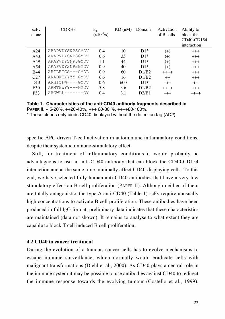
scFvclone
CDRH3 kd
(x10-3/s)KD (nM) Domain Activation
of B cellsAbility toblock theCD40-CD154interaction
A24 ARAPVDYSNPSGMDV 0.4 10 D1* (+) +++A43 ARAPVDYSNPSGMDV 0.6 35 D1* (+) +++A49 ARAPVDYSNPSGMDV 1.1 44 D1* (+) +++A54 ARAPVDYSNPSGMDV 0.9 40 D1* (+) +++B44 ARILRGGS---GMDL 0.9 60 D1/B2 ++++ +++C27 ARADWEYYYY-GMDV 6.6 16 D1/B2 ++ +++D13 ARHIYPW----GMDV 0.6 600 D1* +++ ++E30 ARMTPWYY---GMDV 5.8 3.6 D1/B2 ++++ +++F33 ARGWLL-------DY 0.4 3.1 D2/B1 +++ ++++
Table 1. Characteristics of the anti-CD40 antibody fragments described inPAPER II. + 5-20%, ++20-40%, +++ 60-80 %, ++++80-100%.* These clones only binds CD40 displayed without the detection tag (AD2)
specific APC driven T-cell activation in autoimmune inflammatory conditions,
despite their systemic immuno-stimulatory effect.
Still, for treatment of inflammatory conditions it would probably be
advantageous to use an anti-CD40 antibody that can block the CD40-CD154
interaction and at the same time minimally affect CD40-displaying cells. To this
end, we have selected fully human anti-CD40 antibodies that have a very low
stimulatory effect on B cell proliferation (PAPER II). Although neither of them
are totally antagonistic, the type A anti-CD40 (Table 1) scFv require unusually
high concentrations to activate B cell proliferation. These antibodies have been
produced in full IgG format, preliminary data indicates that these characteristics
are maintained (data not shown). It remains to analyse to what extent they are
capable to block T cell induced B cell proliferation.
4.2 CD40 in cancer treatment
During the evolution of a tumour, cancer cells has to evolve mechanisms to
escape immune surveillance, which normally would eradicate cells with
malignant transformations (Diehl et al., 2000). As CD40 plays a central role in
the immune system it may be possible to use antibodies against CD40 to redirect
the immune response towards the evolving tumour (Costello et al., 1999).
23
Furthermore, several haematological malignancies express CD40, which makes
direct targeting with anti-CD40 antibodies possible. One of the mechanisms by
which tumour cells evade the immune system is to downregulate expression of
MHC I and II as well as costimulatory proteins and adhesion molecules (Restifo
et al., 1993), thereby preventing triggering of tumour specific cytotoxic T
lymphocytes (CTLs). It has been shown that CD40 treatment of e.g. follicular
lymphoma can induce expression of these molecules and restore the reactivity of
CTLs (Costello et al., 1999). Furthermore, agonistic CD40 antibodies has been
used to successfully eradicate tumours in mice (Francisco et al., 2000; French et
al., 1999; Fujita et al., 2001).
Anti-CD40 therapy can also lead to effective treatment of CD40 negative
tumours (Tutt et al., 2002; van Mierlo et al., 2002), possibly by inducing a
strong CTL response. It has been suggested that systemic CD40 stimulation
result in activation of dendritic cells, which then more efficiently can cross-
prime cancer specific CD8+ cytotoxic T cells and thereby bypass the need for
CD4+ T helper cells (Diehl et al., 2000; Ribas et al., 2001). It has, furthermore,
been suggested that differentiation of CD8+ T cells directly depend on CD40
mediated signalling, where CD4+ T helper cells provide the CD40 ligand, in vivo
(Bourgeois et al., 2002).
CD40 antibodies may also have a direct apoptotic effect on CD40 expressing
tumours (Tong et al., 2000). However, on certain types of CD40 positive
lymphomas, CD40 stimulation have a proliferative effect (Andersen et al., 2000;
Challa et al., 2002), indicating that treatment is only feasible on certain CD40+
lymphomas.
In conclusion, tumour therapy with anti-CD40 antibodies may be a promising
approach, although there sometimes is a finely tuned balance between growth
inhibitory and growth promoting capabilities that have to be considered before
therapy can be feasible. We have selected and characterised several agonistic
anti-CD40 fragments of interest, e.g. clone B44, E30 and F33 (PAPER II). Clone
B44 has been produced in an IgG format and preliminary analysis of its
stimulatory capacity shows that its capacity to activate human B cells is
comparable to the well characterised mouse anti-CD40 antibodies, G28-5 and

24
EA5 (PAPER I). Further analysis of these clones may reveal a potential candidate
for clinical trials.
4.3 Vaccination and transplantation
There are also other possible applications for modulation of CD40 signalling,
e.g. to induce transplant tolerance. Interruption of the CD40-CD154 interaction
with anti-CD154 antibodies have successfully been used to prevent graft
rejection in both mice (Larsen et al., 1996a; Larsen et al., 1996b; Parker et al.,
1995) and primates (Kirk et al., 1999). In addition, it has recently been shown
that anti-CD40 antibodies successfully extend renal allograft survival in a
primate model (Pearson et al., 2002).
Moreover, it has been shown that a non-immunogenic, non-viable Listeria
monocytogenes could be converted to an immunogenic vaccine by simultaneous
delivery of an activating anti-CD40 mAb (Rolph and Kaufmann, 2001). This
indicates that co-administration of anti-CD40 antibodies might be useful for
future development of novel vaccine formulations. Furthermore, systemic anti-
CD40 treatment have been shown to increase the immune response to certain
pathogens (Haas et al., 2001).
Tumour vaccination with viral vectors that contains DNA sequences encoding
tumour associated antigens (Pardoll, 1998), provide another route to induce
activation of the cellular immune system. Dendritic cells (DC) are the most
promising target for adenovirus based vaccination protocols (Song et al., 1997).
In fact, Gruijl et al. (2002) has recently shown that it is possible to target
adenovirus to DC by armouring the virus particle with anti-CD40 antibodies.
They demonstrated that, in addition to a more specific delivery of the
adenovirus, the engineered virus also induced CD40 mediated activation and
maturation of the DC.

25
5 ANTIBODY DEVELOPMENT
Antibodies (immunoglobulins) are key soluble effector molecules of the
adaptive immune response and the immune system can generate specific
antibodies to almost any target structure. Upon binding to an antigen, antibodies
can exert several different effects, e.g. phagocytosis of the antigen (Gessner et
al., 1998; Indik et al., 1995; Raghavan and Bjorkman, 1996), activation of
cytotoxic cells (ADCC) (Sulica et al., 2001), binding to complement (Kishore
and Reid, 2000), and neutralisation of virus and toxins (Burton et al., 2001;
Burton et al., 2000), but also induce allergic responses (Stephen and Lantz,
1999). In addition, antibody molecules have structural features that allow them
to penetrate, e.g. the placenta, providing passive protection of the neonate
(Raghavan and Bjorkman, 1996).
5.1 Basic antibody structure
The basic quaternary structure of the immunoglobulin molecule is composed of
four polypeptide chains; two identical, larger, chains (heavy chains) and two
identical, smaller chains (light chains). The heavy and light chains form three
functional units, two antigen binding structures (Fabs) and one effector structure
(Fc). These units are linked together via flexible linkers in an Y- or T-shaped
configuration (Alzari et al., 1988; Amsel and Poljak, 1979; Edmundson et al.,
1995; Padlan, 1994).
The Fab parts of the antibody molecule are composed of two domains from
the heavy chain and two from the light chain, whereas the Fc part consists of
two or three domains from each of the heavy chains. The idiotypic sequence
variability that determines the antibody specificity is confined to the N-terminal
domain of the heavy and the light chains, and these domains are denoted
variable heavy (VH) domain and variable light (VL) domain respectively
(Frazer and Capra, 1999). Remaining domains, in which the sequence only
differs between the different isotypes and allotypes, are named constant domains
(C). There are five different classes of heavy chains (α, µ, γ, δ and ε) and two
classes of light chains (λ and κ).

26
Beside the non-covalent intra- and inter-domain interactions that stabilise the
antibody structure, inter-chain disulphide bonds covalently link the heavy chains
to each other. The Fab structure is furthermore stabilised by a cysteine bridge
between the constant domain of the light chain and the first constant domain of
the heavy chain. Antibody domains adopt a structure known as the
immunoglobulin fold, which consists of a compressed antiparallel beta-barrel
built up by two beta-sheets (Poljak et al., 1973; Schiffer et al., 1973) and is
usually stabilised by a intrachain disulphide bridge. Variable domains fold
slightly different then the constant domains. One of the two beta-sheets of the
variable domains contains five beta-strands, compared to three in the constant
domains, which results in the formation of three connecting loops in each end of
the beta-barrel. Most of the sequence variability in the V-domains is restricted to
the loops that face the exterior (Wu and Kabat, 1970). These complementarity
determining regions (CDRs), determine the shape and charge of the antigen-
binding site (Davies and Cohen, 1996; Davies et al., 1990; Webster et al., 1994;
Wilson and Stanfield, 1993; Wilson and Stanfield, 1994).
5.2 Generation of diversity
The ability to generate antibodies against almost any molecule depends on the
huge diversity in the variable domains. In mice and man, this variability is
initially created by recombination of different germline gene segments, named
variable (V), diversity (D) and joining (J) segment (Tonegawa, 1983).
Successful recombination of both the heavy and the light chain is required for
the development of a mature B cell (Meffre et al., 2000). V(D)J recombination is
a specific process that occurs between sequences that contain complementary
recombination signal sequences (RSS) (Max et al., 1979; Sakano et al., 1979).
Initiation of the recombination process requires upregulation of recombination
activating genes-1 and 2 (RAG-1 and RAG-2) (Oettinger et al., 1990; Schatz
and Baltimore, 1988), which introduce double stranded DNA breaks between
the V, D and J segments. Several non-homologous DNA end-joining proteins
(Bassing et al., 2002) subsequently join the DNA ends of the gene segments.
Part of the diversity of the novel variable genes is created by the combination of
different V(D)J germline gene segments. Additional diversity is introduced by
27
imprecise DNA nicking resulting in different reading frame, nucleotide deletions
and introduction of non-germline encoded (N) (Alt and Baltimore, 1982;
Gilfillan et al., 1993) and palindromic (P) nucleotides (Lewis, 1994; Wuilmart et
al., 1977).
5.3 Secondary recombination – Receptor editing
The development of mature B cells requires productive V(D)J rearrangement of
the light chain and the heavy chain (Meffre et al., 2000; Rajewsky, 1996).
However, if the initial rearrangement is non-productive or the resulting antibody
is autoreactive, the immature B cell can be rescued by secondary
rearrangements, known as receptor editing (Gay et al., 1993; Radic et al., 1993;
Tiegs et al., 1993). Especially the Ig-κ locus is ideally organised for receptor
editing. Secondary rearrangements can occur in a single step between remaining
upstream Vκ genes and downstream Jκ genes since the RSS are compatible even
after the primary recombination (Nemazee, 2000; Nemazee and Weigert, 2000).
5.4 Somatic hypermutation – stepwise mutations
Activation of a mature B cell by a T cell dependent antigen initiates molecular
and cellular processes that further diversify the antibody sequence. These
processes results in affinity maturation of the antibody (Dörner et al., 1998b;
Tarlinton and Smith, 2000) and occur in germinal centres, which develops in
secondary lymphoid organs subsequent to antigen stimulation (Berek et al.,
1991). The somatic hypermutation (SHM) process specifically targets sequences
in, or nearby, the variable region (Winter and Gearhart, 1998). Although the
mechanism controlling this is not fully elucidated, it appears as though both cis
and trans acting sequences are involved in this process (Papavasiliou and Schatz,
2002b). SHM is initiated by a DNA single and/or double strand break followed
by error prone reparation of the gene (Martin and Scharff, 2002a; Papavasiliou
and Schatz, 2002b). This process depends, albeit only loosely, on the sequence,
as specific hot spot motifs seem to exist (Rogozin and Kolchanov, 1992).
The SHM process may result in amino acid substitutions as well as insertions
and deletions (de Wildt et al., 1999b; Goossens et al., 1998; Lantto and Ohlin,
2002a; Ohlin and Borrebaeck, 1998; Wilson et al., 1998). It has recently been

28
shown that the somatic hypermutation process depends on an activation-induced
cytidine deaminase (AID), a crucial step towards understanding somatic
hypermutation (Arakawa et al., 2002; Bross et al., 2002; Faili et al., 2002;
Martin and Scharff, 2002b; Muramatsu et al., 1999; Nagaoka et al., 2002;
Papavasiliou and Schatz, 2002a; Petersen-Mahrt et al., 2002; Rada et al., 2002;
Yoshikawa et al., 2002). AID is, furthermore, critical for other diversification
processes, such as class-switching (Muramatsu et al., 2000; Okazaki et al., 2002)
and gene conversion (Arakawa et al., 2002; Harris et al., 2002).
5.4 Somatic hypermutation – Receptor revision
Although still controversial, an additional diversification process, named
receptor revision, has been suggested to occur in germinal centres (in man and
mouse) leading to further diversification of the evolving antibody. In contrast to
receptor editing, which is tolerance driven and takes place in the bone marrow
(Gay et al., 1993; Radic et al., 1993; Tiegs et al., 1993), receptor revision is
suggested to be a part of the somatic mutation process and contribute to the
affinity maturation process (Fink and McMahan, 2000; Nemazee and Weigert,
2000). Initial evidence for secondary recombinations in the GCs came from
reports showing that RAG1 and RAG2 are upregulated in a subset of germinal
centre B cells (Han et al., 1997; Han et al., 1996; Hikida et al., 1997; Hikida et
al., 1998). It has, furthermore, been shown that both the light chain (Brard et al.,
1999; de Wildt et al., 1999a) and the heavy chain (Itoh et al., 2000; Wilson et
al., 2000) can be rearranged during this process.
During secondary rearrangements of the light chain, a V segment can directly
recombine with a new J segment due to the asymmetry of the RSS of the V and
the J segment (Bassing et al., 2002). In contrast, the primary VH recombination
results in the deletion of all remaining D segments and subsequent secondary
rearrangements cannot utilise the classical recombination signal sequences
(Nemazee 2000). Instead those secondary recombinations rely on cryptic RSS,
mainly embedded in the framework region 3 (FR3) (Chen et al., 1995).
Secondary rearrangements of the heavy chain also differs from light chain
rearrangements, in that parts of the original gene is retained, in particular CDR3,

29
which often is the key sequence element that determine antigen specificity (Xu
and Davis, 2000).
It has not been clarified to what extent receptor revision shapes the antibody
repertoire during the affinity maturation process. Some reports have questioned
the relevance of the V(D)J recombination in GCs, since cells that up-regulate
RAG1 and 2 display a immature phenotypical profile (Hikida et al., 1997; Yu et
al., 1999). However, even if receptor revision is a rare event (Goossens et al.,
2001) there is today convincing evidence that it indeed occurs in the periphery
(Bellan et al., 2002; de Wildt et al., 1999a; Hikida et al., 1998; Itoh et al., 2000;
Wilson et al., 2000). It is, however, difficult to determine the frequency of
receptor revision events, since they are troublesome to detect in many instances
(Wilson et al., 2000). Furthermore, the high probability of a non-productive
rearrangement, resulting in the deletion of the B cell clone, may result in that
most rearranged clones are not found.
5.5 Anti-CD40 antibodies as a tool to study affinity maturation events
In PAPER III, we used CD40 antibodies as a tool to investigate affinity
maturation events in vitro. A scFv phage display library was created based on
four of the previous selected anti-CD40 antibodies (type A antibodies, PAPER
II), using a CDR shuffling approach (Jirholt et al., 1998; Söderlind et al., 1999).
CDRH1 and 2 and CDRL1-3 was allowed to vary, mainly by introducing
sequences from the n-CoDeR library (Söderlind et al., 2000), which allows for
introduction of point mutations, insertions and deletions, as well as larger
sequence modifications that resemble receptor revision events. Thereby, all
components of the variability that is created in vivo, during diversity driven
evolution of antibodies, were used to create the library. As in most cases of
heavy chain revision in vivo, the CDRH3 sequence was retained in the library,
which may result in maintained epitope specificity, whereas other parts of the
VH and VL genes were allowed to vary. We found that after competitive
selection for CD40, using a method that mimic the affinity driven selection
process in GC (Hawkins et al., 1992), there had been a selective advantage for a
distinct shift in CDRH2. This shift in sequence, which has a distinct similarity to

30
Figure 2. Structure models of anti-CD40 scFv. Figure A) shows a structure model
of one of the original anti-CD40 scFv, A24, and one of the evolved clones A2-54
(WAM model using dead end elimination side chain building method and VFF
screening method, http://antibody.bath.ac.uk/index.html). The CDRH3 sequence,
which is identical in both of the scFv, is depicted in blue. Beside CDRH3, all other
residues that are identical are illustrated with a transparent surface, coloured grey.
The residues that are illustrated in red show the differences in CDRH2, whereas
differences in CDRL1 are shown in purple, CDL2 in green and CDRL3 in green blue.
This picture clearly shows that the majority of the differences in the evolved clones
are due to the distinct shift in CDRH2 sequence.
In figure B), the antigen binding site of the scFv that almost completely blocks the
CD40-CD154 interaction, F33 (WAM model using dead end elimination side chain
building method and accessibility profile screening method), is compared with A24.
Basic residues (Lysine and Arginine) are depicted in red, whereas acidic residues
(glutamate and aspartate) are depicted in blue. Histidines are illustrated in yellow,
tyrosine residues in pink and glutaimine and aspargine in green. Remaining residues
are depicted in grey. As shown in the model, F33 contains no acidic residues in the
central antigen binding site (compare with A24). It may be suggested that this further
imply that F33 utilise similar residues as CD154 for binding to CD40.
A24
A24
A2-54
F33
A
B

31
receptor revision of VH in vivo, was mainly to a sequence type that originates
from a germline allele that is more infrequent in the unselected library.
Receptor revision give rise to a relatively large modification of the antibody
gene, which may result in development of self-reactive specificities. Indeed,
autoreactivity has been reported to be associated with this process (Brard et al.,
1999; Dörner et al., 1998a; Itoh et al., 2000; Klonowski et al., 1999). However,
receptor revision may also provide evolving B cells with properties that are
sufficiently valuable to compensate for the increased risk of developing
autoreactive receptors. In fact George and Gray have suggested that receptor
revision may provide a mean to escape from local affinity optima, which would
be impossible to accomplish by stepwise mutations (George and Gray, 1999). In
PAPER III we have shown that receptor revision like events, where a large
segment of the V gene is exchanged in the selected repertoire, indeed may
significantly alter the association and dissociation rate constant profiles of
evolving antibodies. The antibody fragment sequences obtained, after the
selections from the shuffled library that was used for this study, is very unlikely
to be obtained using other in vitro protocols, such as repeated rounds of error-
prone PCR. The latter type of process generates, and accumulates, point
mutations, which most likely would have given rise to low affinity
intermediates, which would be lost during the selection process.
In conclusion, our data shows that receptor revision-like events may confer
selective advantage to an evolving antibody, thereby allowing it to bridge gaps
in sequence space that would be difficult to cross by stepwise mutations. Our
data reinforce the suggestion that receptor revision provides the immune system
with a valuable complement to point mutations and insertions and deletions for
generation of diversity.

32
6 GENERATION OF HUMAN ANTIBODIES FOR
THERAPEUTIC APPLICATIONS
Human antibodies carry several molecular characteristics that make them highly
suitable for therapeutic applications (Borrebaeck and Carlsson, 2001; de Kruif et
al., 1996; Glennie and Johnson, 2000). Highly specific antibodies with high
affinity can easily be made towards almost any molecular structure, which
makes targeting of widely different pathogens such as tumour cells, snake toxins
and viruses possible. Furthermore, naked antibodies, i.e. unconjugated
antibodies, contain an intrinsic effector domain that can recruit natural effector
mechanisms, like ADCC via the Fc part (Clynes et al., 2000). Moreover, the
bivalent nature of the antibody molecule (IgG) may result in direct killing of
tumour cells by receptor cross-linking (Shan et al., 1998). The Fc part also
confers a prolonged serum half-life to the antibody molecule (Raghavan and
Bjorkman, 1996; Shan et al., 1998).
6.1 Human monoclonal antibodies
The advent of monoclonal antibody technology (Köhler and Milstein, 1975)
resulted in the first clinical trial of a monoclonal antibody (mAb) for cancer
therapy in 1979 (Nadler et al., 1980). However, it was not until 1986 that the
first mAb (OKT3) was approved by the Food and Drug Administration (FDA) in
USA. Most of the problems associated with the earlier attempts to use mouse
mAbs in therapy was due to the human anti-mouse antibody (HAMA) response
that most patients developed, which made repetitive treatment difficult
(Khazaeli et al., 1994). Furthermore, these antibodies displayed a short
biological half-life and low ability to recruit effector functions (Borrebaeck et
al., 1993). Attempts have been made to circumvent this by making chimeric
antibodies, i.e. exchanging part of the mouse antibody with human sequences
(Morrison et al., 1984; Wu et al., 1999). However, it was the introduction of
phage display of large human antibody libraries that made production of fully
human antibodies feasible (de Haard et al., 1999; Huls et al., 1999; Knappik et
33
al., 2000; Sheets et al., 1998; Söderlind et al., 2000; Vaughan et al., 1996). This
technology development has resulted in several human antibodies in different
stages of clinical trials, and is likely to have a huge impact on future
development of both analytical tools and therapeutic candidates.
In the studies described in PAPER II and III we have used a human antibody
library (n-CoDeR) (Söderlind et al., 2000) to select and evolve antibodies
against CD40. This large, non-immunised, fully human scFv library provides
several advantages when developing novel therapeutics (Carlsson and Söderlind,
2001). n-CoDeR has been constructed by insertion of CDRs, obtained by PCR
amplification from human peripheral B cells, into a single human VH-VL
scaffold (IGHV3-23*01 and IGLV1-47*01). Thereby, all sequences in the
library have been proof-read, implicating that all the individual sequences have
been evolved and developed in the context of the human immune system prior to
assembly. However, the assembly of the fragments into a whole scFv may create
new T cells epitopes, although probably not to any greater extent. In fact, it has
been shown that clones from the n-CoDeR library display even fewer potential T
cell epitopes than normal human variable genes of IgG type, when analysed by
peptide threading (Söderlind et al., 2000).
The recombination of CDRs from different germline alleles, which the n-
CoDeR concept depends on, allowed us to select a varied panel of scFv with
fully human sequences against human CD40 (PAPER II). This further indicates
that the CDR shuffling approach generates totally novel specificities, since it is
very unlikely that any of the sequences in the FR or CDRs of these scFv have
been part of an anti-CD40 binding antibody in vivo.
When considering utilisation of binders obtained from scFv libraries in a
therapeutic situation, the scFv format might actually have an advantage over the
whole antibody format. The smaller size of the scFv may lead to more efficient
tissue and tumour penetration, and it may be easier and cheaper to produce
(Adams and Schier, 1999), although a major problem is the very short half-lifes
of the small fragments. In most instances, however, the effector functions of the
Fc part as well as the bivalency of a whole IgG molecule are of clinical
advantage (Borrebaeck and Carlsson, 2001; Winter et al., 1994). Therefore, we
have converted and produced some of the anti-CD40 antibodies described in

34
PAPER II and III (A24, B44, F33 and A2-54) into an IgG format. Although this
molecular format is commonly used for therapeutic antibodies, it may be
advantageous to engineer the Fc part, e.g. by altering the affinity profile for the
different Fc receptors (Clynes et al., 2000). Alternatively, in the case of
stimulatory CD40 antibodies, it may be advantageous to increase the valency
further, using other isotypes (Boel, 2000) or different recombinant techniques
(Chapman et al., 1999; Plückthun and Pack, 1997) to increase the stimulatory
capacity (Haswell et al., 2001).
6.2 Antibodies binding to CD40
The mouse anti-CD40 antibody, mAb 5D12, that is profiled in PAPER I have
shown promising results in pre-clinical models of multiple sclerosis (Boon et al.,
2001; Laman et al., 2002). However, this antibody is of mouse origin, which
hamper its therapeutic efficiency in humans as outlined above. The investigation
in PAPER I and (Barr and Heath, 2001; Challa et al., 1999; Pound et al., 1999),
suggested that the specific epitope, to which an antibody is directed, may
influence the function of anti-CD40 antibodies. Thus, we attempted to enrich for
human anti-CD40 clones by performing an, additional, competitively elution
(Meulemans et al., 1994), using mAb 5D12 to enrich for anti-CD40 clones with
similar binding pattern as 5D12, and thereby similar characteristics. To elute
such binders, 5D12 mAb was added to phages bound to CD40 antigen in a large
molar excess, during the fourth round of selection.
The sequence of the scFv clones obtained from the competitive elution that
bound strongly to cell surface displayed CD40, was either of type A or identical
with clone B44 (Table 1). Although, these scFv clones also were frequently
found, when using the standard, trypsin-based, elution, both clone B44 and the
type A antibodies were approximately two times more frequent, using
competitive elution. When analysing these scFv in a blocking experiment it was
found that 5D12 almost completely block B44 binding to CD40 expressing cells,
as expected. Interestingly, the 5D12 mAb did not block the type A clones at all,
which is highly unexpected. Furthermore, the domain-mapping analysis (PAPER
I and II) shows that clone B44 and 5D12 binds to the same, deleted, CD40
variant, whereas the type A antibodies only bind to wild type CD40

35
(unpublished data). This further reinforces the conclusions drawn in PAPER I that
the outcome of blocking experiments, not only depends on the exact location of
the epitopes. There are alternative approaches to determine epitope location, e.g.
alanine scanning mutagenesis. However, this method may also mapp the epitope
to residues that are not involved in binding, since alanine residue substitutions
may affect the structure of a protein both locally and globally (Greenspan and Di
Cera, 1999).
The anti-CD40 antibodies described in PAPER II recognise at least three
different epitopes on CD40, as determined by domain mapping. Comparison
with the domain mapping results presented in PAPER I, revealed at least one new
specificity, not shared by any of the mouse antibodies characterised in PAPER I.
This epitope must be located near the distal end, since even the addition of a
short peptide tag (13 amino acids) abrogates the binding. From the domain
mapping analysis, this epitope seems to be shared between clone D13 and the
type A clones.
Interestingly, the blocking analysis, using 5D12 mAb, indicates that the type
A antibodies detect a unique epitope, since 5D12 blocks binding of D13 to
CD40 rather well, in contrast to the type A clones (see above). Thus their
binding patterns differ despite that they bind to the same CD40 variant as D13,
in the domain mapping analysis. This may indicate that the low activating
character of the type A antibodies actually are a result of a specific binding
epitope.
The surface of the accessible part of the extracellular domains of CD40 can be
estimated to 8000-9300 Å2 (based on SwissPdbViewer calculation for the three
distal domains). This can be compared to the buried surface area of a protein-
antibody interaction that is about 600-900 Å2 (Braden and Poljak, 1995; Davies
and Cohen, 1996). Hence the CD40 molecule theoretically could harbour
approximately 10 non-overlapping antibody binding sites. However, sterical
hindrance, due to the large size of the antigen-binding domain of the antibody,
probably limits the possible number of binding sites. Still it is remarkable that
almost all of the mouse antibodies investigated in PAPER I affect each other’s
binding. This may depend on a low number of accessible epitopes on the CD40-
antigen used for the generation of antibodies. It may also depend on restrictions

36
in the murine germline genes that favours a response against a limited numbers
of epitopes, similar to what Lantto et al has suggested (2002), for antibodies that
recognise antigenic domain 2 on glycoprotein B of cytomegalovirus.
6.3 Antibody evolution and affinity maturation in vitro
From a clinical point of view, it is generally advantageous that a therapeutic
antibody exhibits a high affinity to the target antigen (Adams et al., 1998a;
Adams et al., 1998b; McCall et al., 2001; Roovers et al., 1998), although
extremely high affinity can impair tumour penetration (Adams et al., 2001). It
has been shown that affinity maturation in vivo reaches an affinity plateau. This
is mainly due to restrictions imposed by the selection mechanisms in the
germinal centre reaction, mainly by the requirement to internalise the
BCR/antigen complex, since antibody-antigen interaction with a half-life much
longer than the time necessary for uptake do not have any competitive
advantage. (Batista and Neuberger, 1998; Foote and Eisen, 1995). In vitro
affinity maturation techniques does, however, not suffer from these restrictions
(Foote and Eisen, 2000). Today, in vitro affinity maturation techniques can
break the in vivo affinity ceiling and a number of different technologies have
been developed to accomplish this, e.g. phage display (Hawkins et al., 1992;
Parmley and Smith, 1988), ribosomal display (Hanes et al., 2000; Schaffitzel et
al., 1999), bacterial display (Daugherty et al., 1998) and yeast display (Boder et
al., 2000).
There are several routes to diversify an antibody gene in order to create
improved antibodies prior to selection, using any of the methods described
above. Directed point mutations, error prone PCR (Leung et al., 1989) and chain
shuffling (Stemmer, 1994) acts randomly on the entire sequence, whereas CDR
shuffling (Jirholt et al., 1998) allows for targeting of mutations, insertions and
deletions, as well as larger modifications, to the CDRs. The latter method may
provide some advantages over the random approaches, since it has been shown
that the CDRs tolerate larger modifications than the framework (Dörner et al.,
1998a). However, affinity maturation studies where error prone PCR has been
used to create variability have revealed that several of the clones with approved
affinity contained mutations outside the CDRs (Daugherty et al., 2000; Hawkins

37
et al., 1993). Using CDR shuffling, these mutation would not have been found.
It is, however, indeed possible to combine the different methods, though the
addition of randomly introduced mutations to the diversification process will
increase the probability of introducing new T cell epitopes. The novel
combinations of germline gene CDRs in the n-CoDeR library may, furthermore,
also have influenced the high affinity of the obtained binders (Borrebaeck and
Ohlin, 2002).
It has been suggested that the average affinity of clones selected from a
molecular library correlates with the number of independent clones in the library
(Griffiths et al., 1994). Analysis of the anti-CD40 scFv clones that were selected
from the n-CoDeR library (PAPER II) confirms this notion, since several of them
display high affinities (nanomolar range).
In addition to the insights into the processes that governs antibody evolution,
the study described in PAPER III resulted in affinity maturation of the type A,
anti-CD40 antibodies. In fact, the CDR shuffling approach that was used (PAPER
III) is an efficient route to generate antibodies with higher affinity (Jirholt et al.,
2002). The selection method we applied resembles the affinity selection method
described by Hawkins et al. (1992), which is designed to select for improved
overall affinity. However, the washing step in this scheme, which is a necessary
step of the method, confers a competitive advantage for clones with slow
dissociation rate constant. Despite this, the clones selected in PAPER III
displayed a markedly enhanced association rate compared to the original scFv
clones (8-36 times). The overall affinity improvement was however only
approximately two-fold between scFv clone A24 and A2-54, due to an increased
dissociation rate of the evolved clones. It will be interesting to investigate if
these evolved scFv clones display similar ability to induce CD40 signalling, or if
the shift in dissociation rate and association rate also effect this process.
Manivel et al. (2000) has suggested that the qualitative differences between
antibodies from primary and the secondary response are mainly restricted to the
association step. The increased association rate in the secondary response is due
to stabilisation of the paratope and is accomplished by favourable entropy
changes. It is possible that a similar conformational stabilisation of the paratope
account for the increased association rate in our secondary repertoire (PAPER

38
III), since many of the mutations in the secondary repertoire are located outside
of the central core of the putative binding site (Figure 2) (Tomlinson et al.,
1996). The structure model presented in Figure 2 also indicates that these
changes have an effect on the conformation of CDRH3.

39
7 IDENTIFICATION OF PROTEIN
INTERACTIONS
The completion of the HUGO project and other large-scale DNA sequencing
project has made many complete genome sequences available. Although this
accomplishment by it self is extremely remarkable, it’s just a first step towards
understanding of the cellular and intracellular processes that all living organisms
depend on. The next step toward this goal is to determine the biological function
of the gene products, i.e. the proteins. This has resulted in the new emerging
field of Proteomics, the large-scale analysis of proteins (Auerbach et al., 2002;
Pandey and Mann, 2000). As the function of a protein in many cases depends
on its interactions with other molecules, analysis of protein interactions is highly
relevant. However, the large size of different proteoms (105-106) has created a
need for novel high through put methods for screening of proteins. These novel
methods for screening of protein interactions may also have further applications
for e.g. cancer target identification.
There are several different approaches for protein-protein identification. One
alternative is to use methods where interacting molecules are purified and the
protein sequence is analysed using mass spectroscopy. Another possibility is to
use methods based on enrichments of interacting pairs from library systems,
where the target proteins is physically linked to the gene that encodes it, e.g.
phage display and yeast two hybrid system.
7.1 Mass spectroscopy based identification of proteins
The first step in mass spectroscopy based analysis of protein-protein interactions
is purification of the target proteins. This can be achieved by separation and
detection on two-dimensional gels, or by immunoprecipitation, or variants of the
latter (Figeys et al., 2001). Immunoprecipitation can be accomplished in several
different ways using both scFv and IgG antibody format as bait (Kaelin et al.,
1991; Schneider et al., 1982). It may, however, be difficult to obtain pure
samples of proteins that are expressed in low amounts, are poorly soluble or

40
binds with low affinity. An alternative high-thoughput and promising approach
to obtain proteins for subsequent analyses by mass spectroscopy is to use protein
chips (Borrebaeck et al., 2001; Steinhauer et al., 2002).
7.2 Library selection approaches
Library screening systems based on surface display on a filamentous
bacteriophage that infect Escherichia coli (E. coli), phage display (reviewed by
Hill and Stockley, 1996) have had a huge impact on the field of life science
during the last decades. The success of the phage display system is mainly due
to the nature of the expression system, which ensures a physical link between
the displayed protein and the corresponding gene. This allows for efficient
selection, amplification and, in the end, retrieval of a selected entity. However,
the phage display system is restricted by a number of intrinsic features (Rhyner
et al., 2002). All displayed proteins must cross the bacterial inner membrane
without disrupting it, which limits the potential size of the molecule, and confers
problems with proteins that contains hydrophobic patches. Furthermore, proteins
expressed in E. coli lack post-translational modifications and several protein
types do not fold properly. Moreover, the most efficient phage display systems
are based on a fusion with the g3p coat protein, which require aminoterminal
cloning that make translation of full length genes impossible. This problem,
which may limit the functional size of the library, can be circumvented, by using
the c-jun/c-fos zipper interaction to provide a covalent linkage between C-
terminal cloned cDNA and g3p (Crameri and Suter, 1993). Alternatively, the
cDNA library may be displayed on gp6, which allows c-terminal cloning
(Hufton et al., 1999). There have also been other attempts to directly link protein
interactions with infectivety, e.g. SAP (Dueñas and Borrebaeck, 1994) and SIP
(Krebber et al., 1995), however these methods do not seem to be generally
applicable, and are best suited for high affinity interactions (Nilsson et al.,
2002). Furthermore, de Wildt et al. (2002) has recently described a novel
selection system for library versus library sceening, named SAC (selection by
avidity capture). In this system, a library of receptors are fused to the g3p and
co-expressed with a library of ligands in E. coli. Phages that are produced in
clones, where the ligand can interact with the receptor, are selectively

41
immunoprecipitated and arrayed. The gene that encodes the receptor and the
ligand are selectively retrieved by growth on medium containing double
antibiotic resistance (one for the gene that encodes the receptor and one for the
gene that encodes the ligand).
Two-hybrid systems provide another route to library selections. Fields and
Song (1989) developed the original yeast two hybrid system (Y2H), using the
fact that several transcription factors can be subdivided into two distinct
domains. In the Y2H-system, a “bait” molecule is fused to the DNA binding
part, whereas a “prey” molecule is fused to the activation domain of the
transcription factor. In cells in which the bait binds to the prey, the function of
the transcription factor is restored, and by coupling the reconstitution of the
transcription factor to a reporter gene, interacting pairs can be selected. A
disadvantage with the Y2H is that it generates several false positives, mainly
due to intrinsic transcriptional activity. Also, the posttransciptional machinery in
yeast differs from mammalian cells (Wallach et al., 1998), which may hamper
functional expression of some proteins. Furthermore, the yeast two-hybrid
system is best suited for proteins that are expressed intracellularly and proteins
that e.g. contain hydrophobic transmembrane domains cannot be translocated
into the nucleus.
There are several alternative two hybrid system, which utilise reconstitution
of a protein that confers some kind of selectable phenotype, e.g. RRS (Ras
recruitment system) (Broder et al., 1998). RRS depends on relocalisation of Ras
to the yeast surface by protein interaction between a membrane anchored bait
and a prey fused to mammalian Ras, which induce cell growth at 36°C. This
system has also been reversed to allow for membrane proteins to be used as baits
(Hubsman et al., 2001). The G protein based screening system use a similar
strategy aimed at membrane proteins (Ehrhard et al., 2000). However, the most
widely used alternative is the split-ubiquitin system (Johnsson and Varshavsky,
1994). Here the bait and prey-molecule are fused to one domain each of
ubiquitin, where an introduced mutation has abolished the natural binding
between the halves. An interaction between the bait and the prey restore the
ubiquitin structure, which then is recognised by a protease that release a reporter
protein from one of the halves. These protein-fragment complementation assays

42
(PCAs) has been further developed by the group of Michnick (Pelletier et al.,
1998). Their system has been designed for library versus library selections
(Pelletier et al., 1999) and has also been adapted for identification of protein
interactions in mammalian cells (Remy and Michnick, 1999; Galarneau et al.,
2002). However, PCA are best suited for analysis of proteins that express well in
the cytosol.
7.3 A novel selection technology based on the CD40 receptor -
Selection of Protein Interactions by Receptor Engagement (SPIRE).
In PAPER V, we have described a novel selection system in which molecular
libraries displayed on the surface of a mammalian cell line are enriched, using
Selection of Protein Interaction by Receptor Activation (SPIRE). SPIRE aims at
solving several of the problems discussed in the previous section, i. e. folding,
post-translational modifications et c. In this system a library of “mock-
receptors” (preys) are fused to the transmembrane and cytoplasmic domains of
CD40 (CD40TC). These constructs are transfected into WEHI-231 cells, a naïve
murine B cell line that undergoes apoptosis when treated with anti-IgM
antibodies. If the prey molecule, displayed on cells from this library, can interact
with a bait molecule they are rescued and allowed to proliferate. In PAPER V, we
have applied SPIRE on two model libraries and one cDNA library. Therein, we
clearly show that SPIRE can be used to enrich clones that express a target
molecule.
Subtractive selection of tumour specific antibodies, using phage display
libraries is an attractive method to develop novel candidates for tumour therapy
(de Kruif et al., 1995; Huls et al., 1999; Marks et al., 1993). By making
subtractive pre-selections against normal cells, followed by selection against the
target tumour, a panel of specific antibodies can be relatively easily achieved. It
is furthermore possible to select for tumour epitopes that mediates
internalisation, which may make them suitable for targeted gene delivery
(Larocca et al., 1998; Poul et al., 2000; Poul and Marks, 1999). To further
validate the potential tumour candidates it is necessary to identify the tumour
epitope, even though this may be difficult and time consuming (Roovers et al.,
2001). SPIRE has the potential to provide a high-thoughput solution to this task.

43
Figure 3 Selection of Protein Interactions by Receptor Engagement (SPIRE). A
molecular library ( ) is fused to the transmembrane and cytoplasmic
domains of CD40 (CD40TC), depicted as a hatched box. Stable transfected of the
library (boxed figure) is made into WEHI 231 , which express membrane bound IgM
on the surface (mIgM), depicted as a schematic, unfilled, antibody.
Step 1. Anti-IgM antibody (grey), which induce programmed cell death, apoptosis, in
the cells, is added to the library. The cells are cultured on irradiated fibroblasts (3T3),
which express a bait molecule (depicted as black figures on the fibroblast).
Step 2A. Cells expressing a mock-receptor (prey) that can interact with the bait
molecule (mock-ligand) are rescued from apoptosis and start to proliferate. Rescued
cells are expanded and analysed and subjected to further rounds of selections.
Remaining cells (Step 2B), which do not receive a rescue signal through the CD40
fusion protein will eventually die.
Anti-IgM
Library1
2A
2BApoptosis
mIgM
Rescue andproliferation
mIgM
Fibroblast

44
SPIRE is designed to allow for utilisation of scFv as bait molecules and
allows for rapid conversion of antibody fragments obtained from phage display
libraries to a format suitable for mammalian selection. Beside the membrane
displayed bait approach described in PAPER V, it may also be possible to use
whole antibodies to stimulate specific clones. We have previously successfully
used antibodies to rescue WEHI-231 cells that express truncated CD40 variants
from apoptosis (PAPER IV), which indicates that this format also can be used for
selection. In addition, preliminary data indicates that it also is possible to use
soluble scFv, in combination with a secondary cross-linking antibody to induce
proliferation in transfected WEHI-231 cells (unpublished data).
The applicability of the approach to use 3T3 fibroblasts transfected with
CD40 fusion proteins to stimulate proliferation of transfected WEHI-231 cells
was also confirmed with an, additional, antibody fragment fused to CD40TC
and displayed on 3T3 cells. This construct contains a scFv that recognise human
CD40 (B44) (Table I) fused to the transmembrane and cytosolic part of CD40.
These 3T3 cells were successfully used to stimulate transgenic WEHI-231 cells
that express human CD40 and also to induce proliferation in human peripheral
B-lymphocytes (unpublished data).
7.4 Further development of SPIRE
SPIRE has in its current status, an additional application beside to screen human
cDNA libraries for clones that express a target antigen as outlined in PAPER V.
An alternative application is to use the method for molecular evolution of
complex proteins of eukaryot origin. It may provide a valuable alternative to
other selection technologies, when working with proteins that only fold properly
in mammalian cells, or require specific mammalian post translational
modifications to function correctly.
In PAPER V we show that the interacting pair, AE11-CD40TC and D2/B1-
AD2, fused to the transmembrane and intracellular part of CD40 can function
either as a ligand when transfected in 3T3 cells or as a mock receptor when
transfected into WEHI-231. This indicates that these CD40 variants can transmit
signals simultaneously into the 3T3 cells and into the WEHI-231 cells. This may
be utilised in a library versus library selection approach.

45
The problem with low functionality of cDNA libraries, described in PAPER V,
could be solved by pre-selection of the library, using a flow-cytometry based
approach, prior to SPIRE selections as outlined in PAPER V. Alternatively, it
may be possible to enrich for open reading frames, using bacterial systems
(Davis and Benzer, 1997) and thereby utilise the high transformation capacity of
E. coli, prior to transfection into WEHI-231 cells. This group fused an antibiotic
resistance gene to the C-terminal of the library construct. Thereby, only clones
with a full open reading frame produce a functional resistance marker, and these
can be selected by culturing them on the appropriate antibiotics. That method
could be further developed by inserting a gene that encodes a protease cleavage
site between the cDNA and the reporter gene. Thereby, the protein that confers
antibiotic resistance will be cleaved of from the cDNA-encoded protein, which
may reduce problems associated with biased enrichment of proteins that are
prone to form inclusion bodies. A similar enrichment system has also been
described in yeast (Holz et al., 2001).
Another approach to obtain highly functional libraries has been invented by,
Jacobs et al., 1997 and Klein et al., 1996, who has devised a strategy to enrich
for secreted proteins. By using a library vector without a built in leader
sequence, only cDNA clones from exported proteins will be secreted. Selection
of secreted clones was made by fusing the cDNA product to a protein that needs
to be secreted to allow cell survival in a certain environment. It may be possible
to use SPIRE in a similar approach for identification of exported proteins.

46
CONCLUDING REMARKS
CD40 is a cell surface receptor of pivotal importance that is expressed on
several of the cells in the immune system. It is critical for many important
events, such as T cell dependent B cell activation, isotype switching, somatic
mutation and generation of B cell memory. The central role of CD40 in the
immune system makes it an ideal target for antibody based immunotherapy. This
led us to characterise a panel of monoclonal anti-CD40 antibodies. In PAPER I,
we investigated their cellular activation potential and analysed to what extent
this correlates with their affinity, epitope specificity and domain recognition
profile. The antibody profiles we obtained in this first study may be valuable for
understanding of the mechanisms that influence the therapeutic capacity of these
antibodies. In fact, one of the antibodies that we investigated is currently in
phase I/II trials.
However, all of the antibodies that we characterised in PAPER I are of mouse
origin, which probably limits their clinical efficiency, due to the human anti-
mouse response that most patients develop against such antibodies. Therefore
we selected a set of human anti-CD40 antibodies, which are described in PAPER
II, from a recombinant antibody gene library. These antibodies display a wide
variety of distinct properties, which may make them a valuable source when
evaluating therapeutic candidates for in vivo trials.
In PAPER III, we have used some of the anti-CD40 antibodies described in
PAPER II to create an antibody library that was utilised to investigate antibody
evolution in vitro. The results from this study showed that events, which
resembles receptor revision, i.e. secondary rearrangements of antibody genes in
the periphery, may provide an evolving antibody with competitive advantages
during a selection process that is similar to the affinity maturation process in
vivo. Our data reinforce the suggestion that receptor revision is an important
complement to point mutations and insertions and deletions in the somatic
hypermutation process that occur in germinal centres.
It has been suggested that members of the TNFR family pre-associate in the
membrane via one of the extracellular domains, the pre-ligand assembly domain
(PLAD). Therefore, in PAPER IV, we investigated the functional role of the

47
different domains of CD40, in a B cell model system. The results from this study
showed that neither of the extracellular domains is essential for signal
transduction and, furthermore, implies that conformational changes play no
critical role for the CD40 signalling pathway.
Based on the findings that all of the extracellular part of CD40 can be
replaced with retained signalling capacity, we developed a novel selection
method, named Selection of Protein Interactions by Receptor Engagement
(SPIRE). In PAPER V, we demonstrated that this selection system can be used for
clonal enrichment of cells that display a mock-CD40 receptor, used as prey, on
the surface by interaction with a certain bait protein. Thus, SPIRE allows for
clonal selection of interacting protein pairs in a mammalian environment. SPIRE
may have several different applications such as identification of tumour target
molecules or for molecular evolution of complex proteins.

48
POPULÄRVETENSKAPLIG SAMMANFATTNING
Vårt immunförsvar arbetar konstant med att skydda kroppen från olika föremål,
t. ex. bakterier, virus, gifter och cancertumörer, som kan orsaka skada eller
sjukdom. För att kunna försvara kroppen mot sådana föremål så har
immunförsvaret tillgång till flera, oerhört effektiva, vapen. Det viktigt att
immunförsvaret känner igen, och endast angriper, skadliga föremål, så att inte
dessa vapen attackerar de normala cellerna och vävnaderna i kroppen. För att
immunförsvarets olika beståndsdelar skall kunna bestämma om, och hur, dom
skall reagera på exempelvis ett virus, krävs det att de olika cellerna i
immunsystemet kommunicerar med varandra. Denna kommunikation sker bland
annat via receptorer som finns på cellernas yta. En av immunförsvarets
viktigaste receptorer kallas CD40, och den återfinns på flera av de celler som
ingår i vårt immunsystem. I den här avhandlingen sammanfattas ett antal studier,
där CD40 använts och undersökts, dels som målmolekyl för att ta fram och
studera nya antikroppar, dels som verktyg för att studera en immunologisk
process, och slutligen för att utveckla en ny biomedicinsk teknologi.
I den första artikeln har vi undersökt ett antal antikroppar som binder till
CD40. Anledning till att sådana antikroppar är intressanta, är att de skulle kunna
användas som ett framtida läkemedel för behandling av exempelvis olika
cancerformer och av multipel skleros (MS). De antikropparna vi undersökte i
den första studien var ursprungligen framtagna från en mus. Detta innebär att det
mänskliga immunförsvaret ”ser” dessa antikroppar som något främmande, och
tror därför att de kan vara skadliga. Därmed kommer immunförsvaret att snabbt
göra sig av med antikropparna, vilket gör dem olämpliga för utveckling av nya
behandlingsmetoder för människor. Emellertid kunde de kunskaper om
antikroppar mot CD40 som vi fick från den första studien användas i den andra
studien, där vi, med hjälp av moderna biotekniska metoder, tog fram
”mänskliga” antikroppar. I och med att dessa antikroppar är ”mänskliga”, så
kommer vårt immunförsvar att, med stor sannolikhet, inte se dem som något
främmande, utan accepterar dem som kroppsegna antikroppar. Vidare så visade

49
vi i den artikeln att dessa nya antikroppar har intressanta egenskaper som kan
visa sig vara värdefulla för utveckling av framtida läkemedel.
De antikroppar som vi tagit fram i den andra artikeln, användes i den tredje
studien för att undersöka de processer som är involverade i antikroppsutveckling
i kroppen. Vi använde oss då av en teknik som liknar den normala
antikroppsutvecklingen i kroppen. De data som framkom från den studien, visar
att det i vissa fall är fördelaktigt för utvecklingen av en antikropp att stora delar
av den gen som beskriver hur antikroppen skall se ut byts ut mot ett annat,
liknande gensegment. Våra resultat stödjer därmed den teori som föreslår att
sådana förändringar av antikroppsgener är en viktig del av utvecklingen av
antikroppar i vår kropp.
I den fjärde studien undersökte vi hur de delar av CD40 som finns på utsidan
av cellerna påverkar dess funktion som receptor. Anledningen till at vi
intresserade oss för det var att en del grupper har föreslagit att en dessa delar är
viktig för kommunikationen genom CD40 in i cellen. Våra data visade att man
kan ta bort i princip alla de yttre delarna av CD40 receptorn och ändå skicka
signaler via den, om man bara ersätter dem med ett litet igenkänningsfragment.
I den sista artikeln beskriver vi utvecklingen av en ny biomedicinsk teknik
som vi kallat för SPIRE. Förkortningen står för Selection of Protein Interactions
by Receptor Engagement som kan översättas med ”Selektion av
proteininteraktioner genom receptor engagemang”. I den studien utnyttjade vi
resultaten från den fjärde studien som visade att de yttre delarna av CD40 kan
ersättas. I SPIRE byts den yttre delen av CD40 mot ett stort ”bibliotek” av olika,
okända, molekyler, som därmed kan fungera som ”låtsasreceptorer”. Från detta
bibliotek kan man sedan selektera fram de celler som har en viss låtsasreceptor
på ytan. SPIRE-tekniken kan komma att få ett brett användningsområde,
exempelvis för att utvecklingen av nya läkemedel.

50
ACKNOWLEDGEMENTS
I would like to express my gratitude to the persons that have contributed to this
thesis. In particular, I want to thank Professor Carl Borrebaeck, for
encouragement and for being enthusiastic, visionary and always optimistic and
Christina Furebring for support and for believing in my ideas. I also would like
to thank the other members of the old CD40-group, Camilla and Anki, for
enjoyable meetings and for good company on the EU conferences, and Olga for
your short but work-intensive visit here. I would, furthermore, like to thank
Mats, for always being helpful and providing constructive criticism.
There are of course a number of other colleagues, both present and past, that
have made my time at the department pleasant and enjoyable and I would like to
express my gratitude to them.
Johan, for long-time friendship, and for all chats about nothing whenever we got
bored. Sara and Malin, for providing some sense to the department and for all
fun during “tisdagsluncherna” and other events. Kalle, for, among other things,
introducing me to julle-mulle at your splendid Christmas parties. Lavinia,
Helena, Camilla, Slangen, Yvelise, Franke, Zoltan, Eva, Ingo, Bengt, Conny,
Siggi; Christer, Åsa and Carl Magnus for making this department such a nice
working place and for all great times on and of duty. Carl Magnus should also
have some extra credit for having to listen to my heated conversations with my
computer. I also would like to thank Helene, Marianne, Ann-Charlotte, Eva and
Tommie for assistance and for help with various practical matters.
All my other friends outside the department, in particular to Marcus, for your
friendship and for all nonsense correspondence via e-mail.
My family, for all support and encouragement.
Anna, for everything, you’re the love of my life.

51
REFERENCES
Adams, G. P. and Schier, R., 1999. Generating improved single-chain Fv molecules for tumortargeting. J Immunol Methods. 231, 249-260.
Adams, G. P., Schier, R., Marshall, K., Wolf, E. J., McCall, A. M., Marks, J. D. and Weiner,L. M., 1998a. Increased affinity leads to improved selective tumor delivery of single- chainFv antibodies. Cancer Res. 58, 485-490.
Adams, G. P., Schier, R., McCall, A. M., Crawford, R. S., Wolf, E. J., Weiner, L. M. andMarks, J. D., 1998b. Prolonged in vivo tumour retention of a human diabody targeting theextracellular domain of human HER2/neu. Br J Cancer. 77, 1405-1412.
Adams, G. P., Schier, R., McCall, A. M., Simmons, H. H., Horak, E. M., Alpaugh, R. K.,Marks, J. D. and Weiner, L. M., 2001. High affinity restricts the localization and tumorpenetration of single-chain fv antibody molecules. Cancer Res. 61, 4750-4755.
Ahonen, C., Manning, E., Erickson, L. D., O'Connor, B., Lind, E. F., Pullen, S. S., Kehry, M.R. and Noelle, R. J., 2002. The CD40-TRAF6 axis controls affinity maturation and thegeneration of long-lived plasma cells. Nat Immunol. 3, 451-456.
Allen, R. C., Armitage, R. J., Conley, M. E., Rosenblatt, H., Jenkins, N. A., Copeland, N. G.,Bedell, M. A., Edelhoff, S., Disteche, C. M., Simoneaux, D. K. and et al., 1993. CD40ligand gene defects responsible for X-linked hyper-IgM syndrome. Science. 259, 990-993.
Alt, F. W. and Baltimore, D., 1982. Joining of immunoglobulin heavy chain gene segments:implications from a chromosome with evidence of three D-JH fusions. Proc Natl Acad SciU S A. 79, 4118-4122.
Alzari, P. M., Lascombe, M.-B. and Poljak, R. J., 1988. Three-dimesional structureofantibodies. Annual review of Immunology. 6, 555-580.
Amsel, L. M. and Poljak, R. J., 1979. Three-dimesional structure of immunoglobulins.Annual review of Biochemistry. 48, 961-997.
Andersen, N. S., Larsen, J. K., Christiansen, J., Pedersen, L. B., Christophersen, N. S.,Geisler, C. H. and Jurlander, J., 2000. Soluble CD40 ligand induces selective proliferationof lymphoma cells in primary mantle cell lymphoma cell cultures. Blood. 96, 2219-2225.
Arakawa, H., Hauschild, J. and Buerstedde, J. M., 2002. Requirement of the activation-induced deaminase (AID) gene for immunoglobulin gene conversion. Science. 295, 1301-1306.
Aruffo, A., Farrington, M., Hollenbaugh, D., Li, X., Milatovich, A., Nonoyama, S., Bajorath,J., Grosmaire, L. S., Stenkamp, R., Neubauer, M. and et al., 1993. The CD40 ligand, gp39,is defective in activated T cells from patients with X-linked hyper-IgM syndrome. Cell. 72,291-300.
Auerbach, D., Thaminy, S., Hottiger, M. O. and Stagljar, I., 2002. The post-genomic era ofinteractive proteomics: Facts and perspectives. Proteomics. 2, 611-623.
Baccam, M. and Bishop, G. A., 1999. Membrane-bound CD154, but not CD40-specificantibody, mediates NF- kappaB-independent IL-6 production in B cells. Eur J Immunol.29, 3855-3866.
Bajorath, J. and Aruffo, A., 1997. Construction and analysis of a detailed three-dimensionalmodel of the ligand binding domain of the human B cell receptor CD40. Proteins. 27, 59-70.
Bajorath, J., Chalupny, N. J., Marken, J. S., Siadak, A. W., Skonier, J., Gordon, M.,Hollenbaugh, D., Noelle, R. J., Ochs, H. D. and Aruffo, A., 1995a. Identification ofresidues on CD40 and its ligand which are critical for the receptor-ligand interaction.Biochemistry. 34, 1833-1844.

52
Bajorath, J., Marken, J. S., Chalupny, N. J., Spoon, T. L., Siadak, A. W., Gordon, M., Noelle,R. J., Hollenbaugh, D. and Aruffo, A., 1995b. Analysis of gp39/CD40 interactions usingmolecular models and site- directed mutagenesis. Biochemistry. 34, 9884-9892.
Banchereau, J., Briere, F., Caux, C., Davoust, J., Lebecque, S., Liu, Y. J., Pulendran, B. andPalucka, K., 2000. Immunobiology of dendritic cells. Annu Rev Immunol. 18, 767-811.
Barbas, C. F., 3rd, Kang, A. S., Lerner, R. A. and Benkovic, S. J., 1991. Assembly ofcombinatorial antibody libraries on phage surfaces: the gene III site. Proc Natl Acad Sci US A. 88, 7978-7982.
Barr, T. A. and Heath, A. W., 2001. Functional activity of CD40 antibodies correlates to theposition of binding relative to CD154. Immunology. 102, 39-43.
Bassing, C. H., Swat, W. and Alt, F. W., 2002. The mechanism and regulation ofchromosomal V(D)J recombination. Cell. 109 Suppl, S45-55.
Batista, F. D. and Neuberger, M. S., 1998. Affinity dependence of the B cell response toantigen: a threshold, a ceiling, and the importance of off-rate. Immunity. 8, 751-759.
Becker, T., Hartl, F. U. and Wieland, F., 2002. CD40, an extracellular receptor for bindingand uptake of Hsp70-peptide complexes. J Cell Biol. 158, 1277-1285.
Bellan, C., Lazzi, S., Zazzi, M., Lalinga, A. V., Palummo, N., Galieni, P., Marafioti, T.,Tonini, T., Cinti, C., Leoncini, L., Pileri, S. A. and Tosi, P., 2002. Immunoglobulin generearrangement analysis in composite hodgkin disease and large B-cell lymphoma: evidencefor receptor revision of immunoglobulin heavy chain variable region genes in Hodgkin-Reed- Sternberg cells? Diagn Mol Pathol. 11, 2-8.
Berberich, I., Shu, G., Siebelt, F., Woodgett, J. R., Kyriakis, J. M. and Clark, E. A., 1996.Cross-linking CD40 on B cells preferentially induces stress-activated protein kinases ratherthan mitogen-activated protein kinases. Embo J. 15, 92-101.
Berek, C., Berger, A. and Apel, M., 1991. Maturation of the immune response in germinalcenters. Cell. 67, 1121-1129.
Björck, P., Braesch-Andersen, S. and Paulie, S., 1994. Antibodies to distinct epitopes on theCD40 molecule co-operate in stimulation and can be used for the detection of solubleCD40. Immunology. 83, 430-437.
Blotta, M. H., Marshall, J. D., DeKruyff, R. H. and Umetsu, D. T., 1996. Cross-linking of theCD40 ligand on human CD4+ T lymphocytes generates a costimulatory signal that up-regulates IL-4 synthesis. J Immunol. 156, 3133-3140.
Boder, E. T., Midelfort, K. S. and Wittrup, K. D., 2000. Directed evolution of antibodyfragments with monovalent femtomolar antigen-binding affinity. Proc Natl Acad Sci U SA. 97, 10701-10705.
Boon, L., Brok, H. P., Bauer, J., Ortiz-Buijsse, A., Schellekens, M. M., Ramdien-Murli, S.,Blezer, E., van Meurs, M., Ceuppens, J., de Boer, M., t Hart, B. A. and Laman, J. D., 2001.Prevention of experimental autoimmune encephalomyelitis in the common marmoset(Callithrix jacchus) using a chimeric antagonist monoclonal antibody against human CD40is associated with altered B cell responses. J Immunol. 167, 2942-2949.
Boon, L., Laman, J. D., Ortiz-Buijsse, A., den Hartog, M. T., Hoffenberg, S., Liu, P., Shiau,F. and de Boer, M., 2002. Preclinical assessment of anti-CD40 Mab 5D12 in cynomolgusmonkeys. Toxicology. 174, 53-65.
Borrebaeck, C. A. K. and Ohlin, M., 2002. Evolving Novel Therapeutic Antibodies in Vitro. Nature Biotechnology. in press.Borrebaeck, C. A. K. and Carlsson, R., 2001. Human therapeutic antibodies. Curr Opin
Pharmacol. 1, 404-408.Borrebaeck, C. A.K., Ekstrom, S., Hager, A. C., Nilsson, J., Laurell, T. and Marko-Varga, G.,
2001. Protein chips based on recombinant antibody fragments: a highly sensitive approachas detected by mass spectrometry. Biotechniques. 30, 1126-1130, 1132.

53
Borrebaeck, C. A. K., Malmborg, A. C. and Ohlin, M., 1993. Does endogenous glycosylationprevent the use of mouse monoclonal antibodies as cancer therapeutics? Immunol Today.14, 477-479.
Bourgeois, C., Rocha, B. and Tanchot, C., 2002. A role for CD40 expression on CD8+ T cellsin the generation of CD8+ T cell memory. Science. 297, 2060-2063.
Braden, B. C. and Poljak, R. J., 1995. Structural features of the reactions between antibodiesand protein antigens. Faseb J. 9, 9-16.
Braesch-Andersen, S., Paulie, S., Koho, H., Nika, H., Aspenstrom, P. and Perlmann, P., 1989.Biochemical characteristics and partial amino acid sequence of the receptor-like human Bcell and carcinoma antigen CDw40. J Immunol. 142, 562-567.
Brard, F., Shannon, M., Prak, E. L., Litwin, S. and Weigert, M., 1999. Somatic mutation andlight chain rearrangement generate autoimmunity in anti-single-stranded DNA transgenicMRL/lpr mice. J Exp Med. 190, 691-704.
Bretscher, P. and Cohn, M., 1970. A theory of self-nonself discimination. Science. 169, 1042-1049.
Broder, Y. C., Katz, S. and Aronheim, A., 1998. The ras recruitment system, a novel approachto the study of protein- protein interactions. Curr Biol. 8, 1121-1124.
Bromley, S. K., Burack, W. R., Johnson, K. G., Somersalo, K., Sims, T. N., Sumen, C.,Davis, M. M., Shaw, A. S., Allen, P. M. and Dustin, M. L., 2001. The immunologicalsynapse. Annu Rev Immunol. 19, 375-396.
Bross, L., Muramatsu, M., Kinoshita, K., Honjo, T. and Jacobs, H., 2002. DNA double-strandbreaks: prior to but not sufficient in targeting hypermutation. J Exp Med. 195, 1187-1192.
Brown, D., 2002. Structure and function of membrane rafts. Int J Med Microbiol. 291, 433-437.
Burnet, F. M. (1959) The clonal selection theory of acquired immunuity. Cambridge: TheUniversity Press, London.
Burton, D. R., Saphire, E. O. and Parren, P. W., 2001. A model for neutralization of virusesbased on antibody coating of the virion surface. Curr Top Microbiol Immunol. 260, 109-143.
Burton, D. R., Williamson, R. A. and Parren, P. W., 2000. Antibody and virus: binding andneutralization. Virology. 270, 1-3.
Carlsson, R. and Söderlind, E., 2001. n-CoDeR concept: unique types of antibodies fordiagnostic use and therapy. Expert Rev Mol Diagn. 1, 102-108.
Caux, C., Massacrier, C., Vanbervliet, B., Dubois, B., Van Kooten, C., Durand, I. andBanchereau, J., 1994. Activation of human dendritic cells through CD40 cross-linking. JExp Med. 180, 1263-1272.
Challa, A., Eliopoulos, A. G., Holder, M. J., Burguete, A. S., Pound, J. D., Chamba, A.,Grafton, G., Armitage, R. J., Gregory, C. D., Martinez-Valdez, H., Young, L. and Gordon,J., 2002. Population depletion activates autonomous CD154-dependent survival inbiopsylike Burkitt lymphoma cells. Blood. 99, 3411-3418.
Challa, A., Pound, J. D., Armitage, R. J. and Gordon, J., 1999. Epitope-dependent synergismand antagonism between CD40 antibodies and soluble CD40 ligand for the regulation ofCD23 expression and IgE synthesis in human B cells. Allergy. 54, 576-583.
Chan, F. K., Chun, H. J., Zheng, L., Siegel, R. M., Bui, K. L. and Lenardo, M. J., 2000a. Adomain in TNF receptors that mediates ligand-independent receptor assembly andsignaling. Science. 288, 2351-2354.
Chan, K. F., Siegel, M. R. and Lenardo, J. M., 2000b. Signaling by the TNF receptorsuperfamily and T cell homeostasis. Immunity. 13, 419-422.

54
Chapman, A. P., Antoniw, P., Spitali, M., West, S., Stephens, S. and King, D. J., 1999.Therapeutic antibody fragments with prolonged in vivo half-lives. Nat Biotechnol. 17, 780-783.
Chen, C., Nagy, Z., Prak, E. L. and Weigert, M., 1995. Immunoglobulin heavy chain genereplacement: a mechanism of receptor editing. Immunity. 3, 747-755.
Cheng, G. and Baltimore, D., 1996. TANK, a co-inducer with TRAF2 of TNF- and CD 40L-mediated NF-kappaB activation. Genes Dev. 10, 963-973.
Cheng, G., Cleary, A. M., Ye, Z. S., Hong, D. I., Lederman, S. and Baltimore, D., 1995.Involvement of CRAF1, a relative of TRAF, in CD40 signaling. Science. 267, 1494-1498.
Clynes, R. A., Towers, T. L., Presta, L. G. and Ravetch, J. V., 2000. Inhibitory Fc receptorsmodulate in vivo cytoxicity against tumor targets. Nat Med. 6, 443-446.
Costello, R. T., Gastaut, J. A. and Olive, D., 1999. What is the real role of CD40 in cancerimmunotherapy? Immunol Today. 20, 488-493.
Crameri, R. and Suter, M., 1993. Display of biologically active proteins on the surface offilamentous phages: a cDNA cloning system for selection of functional gene productslinked to the genetic information responsible for their production. Gene. 137, 69-75.
Craxton, A., Shu, G., Graves, J. D., Saklatvala, J., Krebs, E. G. and Clark, E. A., 1998. p38MAPK is required for CD40-induced gene expression and proliferation in B lymphocytes.J Immunol. 161, 3225-3236.
Cunningham, A. J. and Lafferty, K. J., 1977. A simple, conservative explanation of the H-2restriction of interactions between lymphocytes. Scand J Immunol. 6, 1-6.
Dadgostar, H., Zarnegar, B., Hoffmann, A., Qin, X. F., Truong, U., Rao, G., Baltimore, D.and Cheng, G., 2002. Cooperation of multiple signaling pathways in CD40-regulated geneexpression in B lymphocytes. Proc Natl Acad Sci U S A. 99, 1497-1502.
Daugherty, P. S., Chen, G., Iverson, B. L. and Georgiou, G., 2000. Quantitative analysis ofthe effect of the mutation frequency on the affinity maturation of single chain Fvantibodies. Proc Natl Acad Sci U S A. 97, 2029-2034.
Daugherty, P. S., Chen, G., Olsen, M. J., Iverson, B. L. and Georgiou, G., 1998. Antibodyaffinity maturation using bacterial surface display. Protein Eng. 11, 825-832.
Davies, D. R. and Cohen, G. H., 1996. Interactions of protein antigens with antibodies. ProcNatl Acad Sci U S A. 93, 7-12.
Davies, D. R., Padlan, E. A. and Sheriff, S., 1990. Antibody-antigen complexes. Annu RevBiochem. 59, 439-473.
Davis, C. A. and Benzer, S., 1997. Generation of cDNA expression libraries enriched for in-frame sequences. Proc Natl Acad Sci U S A. 94, 2128-2132.
de Boer, M., Conroy, L., Min, H. Y. and Kwekkeboom, J., 1992. Generation of monoclonalantibodies to human lymphocyte cell surface antigens using insect cells expressingrecombinant proteins. J Immunol Methods. 152, 15-23.
de Gruijl, T. D., Luykx-de Bakker, S. A., Tillman, B. W., van den Eertwegh, A. J., Buter, J.,Lougheed, S. M., van der Bij, G. J., Safer AM, H. H. J., Curiel, D. T., Scheper, R. J.,Pinedo, H. M. and Gerritsen, W. R., 2002. Prolonged maturation and enhancedtransduction of dendritic cells migrated from human skin explants after in situ delivery ofCD40-targeted adenoviral vectors. J Immunol. 169:5322-31, 5322-5331.
de Haard, H. J., van Neer, N., Reurs, A., Hufton, S. E., Roovers, R. C., Henderikx, P., deBruine, A. P., Arends, J. W. and Hoogenboom, H. R., 1999. A large non-immunizedhuman Fab fragment phage library that permits rapid isolation and kinetic analysis of highaffinity antibodies. J Biol Chem. 274, 18218-18230.
de Kruif, J., Terstappen, L., Boel, E. and Logtenberg, T., 1995. Rapid selection of cellsubpopulation-specific human monoclonal antibodies from a synthetic phage antibodylibrary. Proc Natl Acad Sci U S A. 92, 3938-3942.

55
de Kruif, J., van der Vuurst de Vries, A. R., Cilenti, L., Boel, E., van Ewijk, W. andLogtenberg, T., 1996. New perspectives on recombinant human antibodies. ImmunolToday. 17, 453-455.
de Wildt, R. M., Hoet, R. M., van Venrooij, W. J., Tomlinson, I. M. and Winter, G., 1999a.Analysis of heavy and light chain pairings indicates that receptor editing shapes the humanantibody repertoire. J Mol Biol. 285, 895-901.
de Wildt, R. M., Tomlinson, I. M., Ong, J. L. and Holliger, P., 2002. Isolation of receptor-ligand pairs by capture of long-lived multivalent interaction complexes. Proc Natl AcadSci U S A. 99, 8530-8535.
de Wildt, R. M., van Venrooij, W. J., Winter, G., Hoet, R. M. and Tomlinson, I. M., 1999b.Somatic insertions and deletions shape the human antibody repertoire. J Mol Biol. 294,701-710.
Diehl, L., Den Boer, A. T., van der Voort, E. I., Melief, C. J., Offringa, R. and Toes, R. E.,2000. The role of CD40 in peripheral T cell tolerance and immunity. J Mol Med. 78, 363-371.
DiSanto, J. P., Bonnefoy, J. Y., Gauchat, J. F., Fischer, A. and de Saint Basile, G., 1993.CD40 ligand mutations in x-linked immunodeficiency with hyper-IgM. Nature. 361, 541-543.
Dörner, T., Brezinschek, H. P., Foster, S. J., Brezinschek, R. I., Farner, N. L. and Lipsky, P.E., 1998a. Delineation of selective influences shaping the mutated expressed human Igheavy chain repertoire. J Immunol. 160, 2831-2841.
Dörner, T., Foster, S. J., Farner, N. L. and Lipsky, P. E., 1998b. Somatic hypermutation ofhuman immunoglobulin heavy chain genes: targeting of RGYW motifs on both DNAstrands. Eur J Immunol. 28, 3384-3396.
Duenas, M. and Borrebaeck, C. A., 1994. Clonal selection and amplification of phagedisplayed antibodies by linking antigen recognition and phage replication. Biotechnology(N Y). 12, 999-1002.
Dykstra, M., Cherukuri, A. and Pierce, S. K., 2001. Rafts and synapses in the spatialorganization of immune cell signaling receptors. J Leukoc Biol. 70, 699-707.
Edmundson, A. B., Guddat, L. W., Rosaur, R. A., Andersen, K. N., Shan, L. and Fan, Z.-C.(1995) Three-dimensional Aspects of IgG Structure and Function. In The Antibodies(Edited by Zanetti M. and Capra J. D.), Vol. 1, p. 41-100. Harwood Academic publishersGmbH, Luxemburg.
Ehrhard, K. N., Jacoby, J. J., Fu, X. Y., Jahn, R. and Dohlman, H. G., 2000. Use of G-proteinfusions to monitor integral membrane protein-protein interactions in yeast. Nat Biotechnol.18, 1075-1079.
Faili, A., Aoufouchi, S., Gueranger, Q., Zober, C., Leon, A., Bertocci, B., Weill, J. C. andReynaud, C. A., 2002. AID-dependent somatic hypermutation occurs as a DNA single-strand event in the BL2 cell line. Nat Immunol. 3, 815-821.
Fanslow, W. C., Srinivasan, S., Paxton, R., Gibson, M. G., Spriggs, M. K. and Armitage, R.J., 1994. Structural characteristics of CD40 ligand that determine biological function.Semin Immunol. 6, 267-278.
Fields, S. and Song, O., 1989. A novel genetic system to detect protein-protein interactions.Nature. 340, 245-246.
Figeys, D., McBroom, L. D. and Moran, M. F., 2001. Mass spectrometry for the study ofprotein-protein interactions. Methods. 24, 230-239.
Fink, P. J. and McMahan, C. J., 2000. Lymphocytes rearrange, edit and revise their antigenreceptors to be useful yet safe. Immunol Today. 21, 561-566.
Foote, J. and Eisen, H. N., 1995. Kinetic and affinity limits on antibodies produced duringimmune responses. Proc Natl Acad Sci U S A. 92, 1254-1256.

56
Foote, J. and Eisen, H. N., 2000. Breaking the affinity ceiling for antibodies and T cellreceptors. Proc Natl Acad Sci U S A. 97, 10679-10681.
Foy, T. M., Shepherd, D. M., Durie, F. H., Aruffo, A., Ledbetter, J. A. and Noelle, R. J.,1993. In vivo CD40-gp39 interactions are essential for thymus-dependent humoralimmunity. II. Prolonged suppression of the humoral immune response by an antibody tothe ligand for CD40, gp39. J Exp Med. 178, 1567-1575.
Francisco, J. A., Donaldson, K. L., Chace, D., Siegall, C. B. and Wahl, A. F., 2000. Agonisticproperties and in vivo antitumor activity of the anti-CD40 antibody SGN-14. Cancer Res.60, 3225-3231.
Frazer, J. K. and Capra, J. D. (1999) Immunoglobulins: Structure and function. InFundamental Immunology (Edited by Paul W. E.). Lippincott-Raven Publishers,Philadelphia.
French, R. R., Chan, H. T., Tutt, A. L. and Glennie, M. J., 1999. CD40 antibody evokes acytotoxic T-cell response that eradicates lymphoma and bypasses T-cell help. Nat Med. 5,548-553.
Fujita, N., Kagamu, H., Yoshizawa, H., Itoh, K., Kuriyama, H., Matsumoto, N., Ishiguro, T.,Tanaka, J., Suzuki, E., Hamada, H. and Gejyo, F., 2001. CD40 ligand promotes priming offully potent antitumor CD4(+) T cells in draining lymph nodes in the presence of apoptotictumor cells. J Immunol. 167, 5678-5688.
Galarneau, A., Primeau, M., Trudeau, L. E. and Michnick, S. W., 2002. beta-Lactamaseprotein fragment complementation assays as in vivo and in vitro sensors of protein proteininteractions. Nat Biotechnol. 20, 619-622.
Gay, D., Saunders, T., Camper, S. and Weigert, M., 1993. Receptor editing: an approach byautoreactive B cells to escape tolerance. J Exp Med. 177, 999-1008.
Genain, C. P., Cannella, B., Hauser, S. L. and Raine, C. S., 1999. Identification ofautoantibodies associated with myelin damage in multiple sclerosis. Nat Med. 5, 170-175.
George, A. J. and Gray, D., 1999. Receptor editing during affinity maturation. ImmunolToday. 20, 196.
Gerritse, K., Laman, J. D., Noelle, R. J., Aruffo, A., Ledbetter, J. A., Boersma, W. J. andClaassen, E., 1996. CD40-CD40 ligand interactions in experimental allergicencephalomyelitis and multiple sclerosis. Proc Natl Acad Sci U S A. 93, 2499-2504.
Gessner, J. E., Heiken, H., Tamm, A. and Schmidt, R. E., 1998. The IgG Fc receptor family.Ann Hematol. 76, 231-248.
Gilfillan, S., Dierich, A., Lemeur, M., Benoist, C. and Mathis, D., 1993. Mice lacking TdT:mature animals with an immature lymphocyte repertoire. Science. 261, 1175-1178.
Gilliland, L. K., Norris, N. A., Marquardt, H., Tsu, T. T., Hayden, M. S., Neubauer, M. G.,Yelton, D. E., Mittler, R. S. and Ledbetter, J. A., 1996. Rapid and reliable cloning ofantibody variable regions and generation of recombinant single chain antibody fragments.Tissue Antigens. 47, 1-20.
Glennie, M. J. and Johnson, P. W., 2000. Clinical trials of antibody therapy. Immunol Today.21, 403-410.
Goossens, T., Brauninger, A., Klein, U., Kuppers, R. and Rajewsky, K., 2001. Receptorrevision plays no major role in shaping the receptor repertoire of human memory B cellsafter the onset of somatic hypermutation. Eur J Immunol. 31, 3638-3648.
Goossens, T., Klein, U. and Kuppers, R., 1998. Frequent occurrence of deletions andduplications during somatic hypermutation: implications for oncogene translocations andheavy chain disease. Proc Natl Acad Sci U S A. 95, 2463-2468.
Grammer, A. C., Swantek, J. L., McFarland, R. D., Miura, Y., Geppert, T. and Lipsky, P. E.,1998. TNF receptor-associated factor-3 signaling mediates activation of p38 and Jun N-

57
terminal kinase, cytokine secretion, and Ig production following ligation of CD40 onhuman B cells. J Immunol. 161, 1183-1193.
Grassme, H., Bock, J., Kun, J. and Gulbins, E., 2002a. Clustering of CD40 ligand is requiredto form a functional contact with CD40. J Biol Chem. 277, 30289-30299.
Grassme, H., Bock, J., Kun, J. and Gulbins, E., 2002b. Clustering of CD40 ligand is requiredto form a functional contact with CD40. J Biol Chem. 13, 13.
Grassme, H., Jendrossek, V., Bock, J., Riehle, A. and Gulbins, E., 2002c. Ceramide-richmembrane rafts mediate CD40 clustering. J Immunol. 168, 298-307.
Greenspan, N. S. and Di Cera, E., 1999. Defining epitopes: It's not as easy as it seems. NatBiotechnol. 17, 936-937.
Griffiths, A. D., Williams, S. C., Hartley, O., Tomlinson, I. M., Waterhouse, P., Crosby, W.L., Kontermann, R. E., Jones, P. T., Low, N. M., Allison, T. J. and et al., 1994. Isolation ofhigh affinity human antibodies directly from large synthetic repertoires. Embo J. 13, 3245-3260.
Haas, K. M., Taylor, K. A., MacHugh, N. D., Kreeger, J. M. and Estes, D. M., 2001.Enhancing effects of anti-CD40 treatment on the immune response of SCID- bovine miceto Trypanosoma congolense infection. J Leukoc Biol. 70, 931-940.
Han, S., Dillon, S. R., Zheng, B., Shimoda, M., Schlissel, M. S. and Kelsoe, G., 1997. V(D)Jrecombinase activity in a subset of germinal center B lymphocytes. Science. 278, 301-305.
Han, S., Zheng, B., Schatz, D. G., Spanopoulou, E. and Kelsoe, G., 1996. Neoteny inlymphocytes: Rag1 and Rag2 expression in germinal center B cells. Science. 274, 2094-2097.
Hanes, J., Schaffitzel, C., Knappik, A. and Pluckthun, A., 2000. Picomolar affinity antibodiesfrom a fully synthetic naive library selected and evolved by ribosome display. NatBiotechnol. 18, 1287-1292.
Hanissian, S. H. and Geha, R. S., 1997. Jak3 is associated with CD40 and is critical for CD40induction of gene expression in B cells. Immunity. 6, 379-387.
Harris, R. S., Sale, J. E., Petersen-Mahrt, S. K. and Neuberger, M. S., 2002. AID is essentialfor immunoglobulin V gene conversion in a cultured B cell line. Curr Biol. 12, 435-438.
Haswell, L. E., Glennie, M. J. and Al-Shamkhani, A., 2001. Analysis of the oligomericrequirement for signaling by CD40 using soluble multimeric forms of its ligand, CD154.Eur J Immunol. 31, 3094-3100.
Hawkins, R. E., Russell, S. J., Baier, M. and Winter, G., 1993. The contribution of contactand non-contact residues of antibody in the affinity of binding to antigen. The interactionof mutant D1.3 antibodies with lysozyme. J Mol Biol. 234, 958-964.
Hawkins, R. E., Russell, S. J. and Winter, G., 1992. Selection of phage antibodies by bindingaffinity. Mimicking affinity maturation. J Mol Biol. 226, 889-896.
Hikida, M., Mori, M., Kawabata, T., Takai, T. and Ohmori, H., 1997. Characterization of Bcells expressing recombination activating genes in germinal centers of immunized mouselymph nodes. J Immunol. 158, 2509-2512.
Hikida, M., Nakayama, Y., Yamashita, Y., Kumazawa, Y., Nishikawa, S. I. and Ohmori, H.,1998. Expression of recombination activating genes in germinal center B cells:involvement of interleukin 7 (IL-7) and the IL-7 receptor. J Exp Med. 188, 365-372.
Hill, H. R. and Stockley, P. G., 1996. Phage presentation. Mol Microbiol. 20, 685-692.Holz, C., Lueking, A., Bovekamp, L., Gutjahr, C., Bolotina, N., Lehrach, H. and Cahill, D. J.,
2001. A human cDNA expression library in yeast enriched for open reading frames.Genome Res. 11, 1730-1735.
Hornung, M., Lindemann, D., Kraus, C., Peters, A. and Berberich, I., 1998. The CD40 TRAFfamily member interacting motif carries the information to rescue WEHI 231 cells fromanti-IGM-induced growth arrest. Eur J Immunol. 28, 3812-3823.

58
Hostager, B. S., Catlett, I. M. and Bishop, G. A., 2000. Recruitment of CD40 and tumornecrosis factor receptor-associated factors 2 and 3 to membrane microdomains duringCD40 signaling. J Biol Chem. 275, 15392-15398.
Hubsman, M., Yudkovsky, G. and Aronheim, A., 2001. A novel approach for theidentification of protein-protein interaction with integral membrane proteins. NucleicAcids Res. 29, E18.
Hufton, S. E., Moerkerk, P. T., Meulemans, E. V., de Bruine, A., Arends, J. W. andHoogenboom, H. R., 1999. Phage display of cDNA repertoires: the pVI display system andits applications for the selection of immunogenic ligands. J Immunol Methods. 231, 39-51.
Huls, G. A., Heijnen, I. A., Cuomo, M. E., Koningsberger, J. C., Wiegman, L., Boel, E., vander Vuurst de Vries, A. R., Loyson, S. A., Helfrich, W., van Berge Henegouwen, G. P.,van Meijer, M., de Kruif, J. and Logtenberg, T., 1999. A recombinant, fully humanmonoclonal antibody with antitumor activity constructed from phage-displayed antibodyfragments. Nat Biotechnol. 17, 276-281.
Indik, Z. K., Park, J. G., Hunter, S., Mantaring, M. and Schreiber, A. D., 1995. Moleculardissection of Fc gamma receptor-mediated phagocytosis. Immunol Lett. 44, 133-138.
Inoue, J., Ishida, T., Tsukamoto, N., Kobayashi, N., Naito, A., Azuma, S. and Yamamoto, T.,2000. Tumor necrosis factor receptor-associated factor (TRAF) family: adapter proteinsthat mediate cytokine signaling. Exp Cell Res. 254, 14-24.
Itoh, K., Meffre, E., Albesiano, E., Farber, A., Dines, D., Stein, P., Asnis, S. E., Furie, R. A.,Jain, R. I. and Chiorazzi, N., 2000. Immunoglobulin heavy chain variable region genereplacement As a mechanism for receptor revision in rheumatoid arthritis synovial tissue Blymphocytes. J Exp Med. 192, 1151-1164.
Jabara, H., Laouini, D., Tsitsikov, E., Mizoguchi, E., Bhan, A., Castigli, E., Dedeoglu, F.,Pivniouk, V., Brodeur, S. and Geha, R., 2002. The Binding Site for TRAF2 and TRAF3but Not for TRAF6 Is Essential for CD40-Mediated Immunoglobulin Class Switching.Immunity. 17, 265.
Jabara, H. H., Buckley, R. H., Roberts, J. L., Lefranc, G., Loiselet, J., Khalil, G. and Geha, R.S., 1998. Role of JAK3 in CD40-mediated signaling. Blood. 92, 2435-2440.
Jacobs, K. A., Collins-Racie, L. A., Colbert, M., Duckett, M., Golden-Fleet, M., Kelleher, K.,Kriz, R., LaVallie, E. R., Merberg, D., Spaulding, V., Stover, J., Williamson, M. J. andMcCoy, J. M., 1997. A genetic selection for isolating cDNAs encoding secreted proteins.Gene. 198, 289-296.
Janeway, C. A., Jr., 1989. Approaching the asymptote? Evolution and revolution inimmunology. Cold Spring Harb Symp Quant Biol. 54, 1-13.
Janeway, C. A., Jr., 1992. The immune system evolved to discriminate infectious nonselffrom noninfectious self. Immunol Today. 13, 11-16.
Jirholt, P., Borrebaeck, C. A. K. and Ohlin, M., 2002. Antibody evolution from the centre tothe periphery: applied to a human antibody fragment recognising the tumour-associatedantigen mucin-1. J Mol Biol. 318, 407-416.
Jirholt, P., Ohlin, M., Borrebaeck, C. A. K. and Söderlind, E., 1998. Exploiting sequencespace: shuffling in vivo formed complementarity determining regions into a masterframework. Gene. 215, 471-476.
Johnsson, N. and Varshavsky, A., 1994. Split ubiquitin as a sensor of protein interactions invivo. Proc Natl Acad Sci U S A. 91, 10340-10344.
Kaelin, W. G., Jr., Pallas, D. C., DeCaprio, J. A., Kaye, F. J. and Livingston, D. M., 1991.Identification of cellular proteins that can interact specifically with the T/E1A-bindingregion of the retinoblastoma gene product. Cell. 64, 521-532.
Karp, C. L., Biron, C. A. and Irani, D. N., 2000. Interferon beta in multiple sclerosis: is IL-12suppression the key? Immunol Today. 21, 24-28.

59
Karp, C. L., van Boxel-Dezaire, A. H., Byrnes, A. A. and Nagelkerken, L., 2001. Interferon-beta in multiple sclerosis: altering the balance of interleukin-12 and interleukin-10? CurrOpin Neurol. 14, 361-368.
Karpusas, M., Hsu, Y. M., Wang, J. H., Thompson, J., Lederman, S., Chess, L. and Thomas,D., 1995. 2 A crystal structure of an extracellular fragment of human CD40 ligand.Structure. 3, 1031-1039.
Karras, J. G., Wang, Z., Huo, L., Frank, D. A. and Rothstein, T. L., 1997. Induction of STATprotein signaling through the CD40 receptor in B lymphocytes: distinct STAT activationfollowing surface Ig and CD40 receptor engagement. J Immunol. 159, 4350-4355.
Kaykas, A., Worringer, K. and Sugden, B., 2001. CD40 and LMP-1 both signal from lipidrafts but LMP-1 assembles a distinct, more efficient signaling complex. Embo J. 20, 2641-2654.
Khazaeli, M. B., Conry, R. M. and LoBuglio, A. F., 1994. Human immune response tomonoclonal antibodies. J Immunother. 15, 42-52.
Kirk, A. D., Burkly, L. C., Batty, D. S., Baumgartner, R. E., Berning, J. D., Buchanan, K.,Fechner, J. H., Jr., Germond, R. L., Kampen, R. L., Patterson, N. B., Swanson, S. J.,Tadaki, D. K., TenHoor, C. N., White, L., Knechtle, S. J. and Harlan, D. M., 1999.Treatment with humanized monoclonal antibody against CD154 prevents acute renalallograft rejection in nonhuman primates. Nat Med. 5, 686-693.
Kishore, U. and Reid, K. B., 2000. C1q: structure, function, and receptors.Immunopharmacology. 49, 159-170.
Klein, R. D., Gu, Q., Goddard, A. and Rosenthal, A., 1996. Selection for genes encodingsecreted proteins and receptors. Proc Natl Acad Sci U S A. 93, 7108-7113.
Klonowski, K. D., Primiano, L. L. and Monestier, M., 1999. Atypical VH-D-JHrearrangements in newborn autoimmune MRL mice. J Immunol. 162, 1566-1572.
Knappik, A., Ge, L., Honegger, A., Pack, P., Fischer, M., Wellnhofer, G., Hoess, A., Wolle,J., Pluckthun, A. and Virnekas, B., 2000. Fully synthetic human combinatorial antibodylibraries (HuCAL) based on modular consensus frameworks and CDRs randomized withtrinucleotides. J Mol Biol. 296, 57-86.
Koho, H., Paulie, S., Ben-Aissa, H., Jonsdottir, I., Hansson, Y., Lundblad, M. L. andPerlmann, P., 1984. Monoclonal antibodies to antigens associated with transitional cellcarcinoma of the human urinary bladder. I. Determination of the selectivity of sixantibodies by cell ELISA and immunofluorescence. Cancer Immunol Immunother. 17,165-172.
Koppenhoefer, U., Brenner, B., Lang, F. and Gulbins, E., 1997. The CD40-ligand stimulatesT-lymphocytes via the neutral sphingomyelinase: a novel function of the CD40-ligand assignalling molecule. FEBS Lett. 414, 444-448.
Korthauer, U., Graf, D., Mages, H. W., Briere, F., Padayachee, M., Malcolm, S., Ugazio, A.G., Notarangelo, L. D., Levinsky, R. J. and Kroczek, R. A., 1993. Defective expression ofT-cell CD40 ligand causes X-linked immunodeficiency with hyper-IgM. Nature. 361, 539-541.
Krebber, C., Spada, S., Desplancq, D. and Pluckthun, A., 1995. Co-selection of cognateantibody-antigen pairs by selectively-infective phages. FEBS Lett. 377, 227-231.
Kwekkeboom, J., De Boer, M., Tager, J. M. and De Groot, C., 1993. CD40 plays an essentialrole in the activation of human B cells by murine EL4B5 cells. Immunology. 79, 439-444.
Köhler, G. and Milstein, C., 1975. Continuous cultures of fused cells secreting antibody ofpredefined specificity. Nature. 256, 495-497.
Lafferty, K. J. and Cunningham, A. J., 1975. A new analysis of allogeneic interactions. Aust JExp Biol Med Sci. 53, 27-42.

60
Laman, J. D., De Boer, M. and Hart, B. A., 1998. CD40 in clinical inflammation: frommultiple sclerosis to atherosclerosis. Dev Immunol. 6, 215-222.
Laman, J. D., Hart, B. A., Brok, H., Meurs Mv, M., Schellekens, M. M., Kasran, A., Boon, L.,Bauer, J., Boer Md, M. and Ceuppens, J., 2002. Protection of marmoset monkeys againstEAE by treatment with a murine antibody blocking CD40 (mu5D12). Eur J Immunol. 32,2218-2228.
Lantto, J., Lindroth, Y. and Ohlin, M., 2002. Non-germ-line encoded residues are critical foreffective antibody recognition of a poorly immunogenic neutralization epitope onglycoprotein B of human cytomegalovirus. Eur J Immunol. 32, 1659-1669.
Lantto, J. and Ohlin, M., 2002a. Functional consequences of insertions and deletions in thecomplementarity determining regions of human antibodies. J Biol Chem. 16, 16.
Lantto, J. and Ohlin, M., 2002b. Uneven distribution of repetitive trinucleotide motifs inhuman immunoglobulin heavy variable genes. J Mol Evol. 54, 346-353.
Larocca, D., Witte, A., Johnson, W., Pierce, G. F. and Baird, A., 1998. Targetingbacteriophage to mammalian cell surface receptors for gene delivery. Hum Gene Ther. 9,2393-2399.
Larsen, C. P., Alexander, D. Z., Hollenbaugh, D., Elwood, E. T., Ritchie, S. C., Aruffo, A.,Hendrix, R. and Pearson, T. C., 1996a. CD40-gp39 interactions play a critical role duringallograft rejection. Suppression of allograft rejection by blockade of the CD40-gp39pathway. Transplantation. 61, 4-9.
Larsen, C. P., Elwood, E. T., Alexander, D. Z., Ritchie, S. C., Hendrix, R., Tucker-Burden,C., Cho, H. R., Aruffo, A., Hollenbaugh, D., Linsley, P. S., Winn, K. J. and Pearson, T. C.,1996b. Long-term acceptance of skin and cardiac allografts after blocking CD40 and CD28pathways. Nature. 381, 434-438.
Ledbetter, J. A., Francisco, J. A., Siegall, C. B., Gilliland, L. K., Hollenbaugh, D., Aruffo, A.,Siadak, A. W., Mischel-Petty, N., Grosmaire, L. S., Gordon, M. L., Brown, T. J., Moran-Davis, P., Mittler, R. S., Kiener, P. A. and Nadler, S. G., 1997. Agonistic activity of aCD40-specific single-chain Fv constructed from the variable regions of mAb G28-5. CritRev Immunol. 17, 427-435.
Lefranc, M. P., 2001. IMGT, the international ImMunoGeneTics database. Nucleic AcidsRes. 29, 207-209.
Leung, D. W., Chen, E. and Goeddel, D. V., 1989. A method for random mutagenesis of adefined DNA segment using a modified polymerase chain reaction. Technique. 1, 11–15.
Lewis, S. M., 1994. The mechanism of V(D)J joining: lessons from molecular,immunological, and comparative analyses. Adv Immunol. 56, 27-150.
Li, X., Zhao, X., Fang, Y., Jiang, X., Duong, T., Fan, C., Huang, C. C. and Kain, S. R., 1998.Generation of destabilized green fluorescent protein as a transcription reporter. J BiolChem. 273, 34970-34975.
Locksley, R. M., Killeen, N. and Lenardo, M. J., 2001. The TNF and TNF receptorsuperfamilies: integrating mammalian biology. Cell. 104, 487-501.
MacCallum, R. M., Martin, A. C. and Thornton, J. M., 1996. Antibody-antigen interactions:contact analysis and binding site topography. J Mol Biol. 262, 732-745.
Mackey, M. F., Gunn, J. R., Maliszewsky, C., Kikutani, H., Noelle, R. J. and Barth, R. J., Jr.,1998. Dendritic cells require maturation via CD40 to generate protective antitumorimmunity. J Immunol. 161, 2094-2098.
Malapati, S. and Pierce, S. K., 2001. The influence of CD40 on the association of the B cellantigen receptor with lipid rafts in mature and immature cells. Eur J Immunol. 31, 3789-3797.
Malinin, N. L., Boldin, M. P., Kovalenko, A. V. and Wallach, D., 1997. MAP3K-relatedkinase involved in NF-kappaB induction by TNF, CD95 and IL-1. Nature. 385, 540-544.

61
Manivel, V., Sahoo, N. C., Salunke, D. M. and Rao, K. V., 2000. Maturation of an antibodyresponse is governed by modulations in flexibility of the antigen-combining site.Immunity. 13, 611-620.
Marks, J. D., Ouwehand, W. H., Bye, J. M., Finnern, R., Gorick, B. D., Voak, D., Thorpe, S.J., Hughes-Jones, N. C. and Winter, G., 1993. Human antibody fragments specific forhuman blood group antigens from a phage display library. Biotechnology (N Y). 11, 1145-1149.
Martin, A. and Scharff, M. D., 2002a. AID and mismatch repair in antibody diversification.Nat Rev Immunol. 2, 605-614.
Martin, A. and Scharff, M. D., 2002b. Somatic hypermutation of the AID transgene in B andnon-B cells. Proc Natl Acad Sci U S A. 99, 12304-12308.
Mauri, C., Mars, L. T. and Londei, M., 2000. Therapeutic activity of agonistic monoclonalantibodies against CD40 in a chronic autoimmune inflammatory process. Nat Med. 6, 673-679.
Max, E. E., Seidman, J. G. and Leder, P., 1979. Sequences of five potential recombinationsites encoded close to an immunoglobulin kappa constant region gene. Proc Natl Acad SciU S A. 76, 3450-3454.
McCafferty, J., Griffiths, A. D., Winter, G. and Chiswell, D. J., 1990. Phage antibodies:filamentous phage displaying antibody variable domains. Nature. 348, 552-554.
McCall, A. M., Shahied, L., Amoroso, A. R., Horak, E. M., Simmons, H. H., Nielson, U.,Adams, G. P., Schier, R., Marks, J. D. and Weiner, L. M., 2001. Increasing the affinity fortumor antigen enhances bispecific antibody cytotoxicity. J Immunol. 166, 6112-6117.
McWhirter, S. M., Pullen, S. S., Holton, J. M., Crute, J. J., Kehry, M. R. and Alber, T., 1999.Crystallographic analysis of CD40 recognition and signaling by human TRAF2. Proc NatlAcad Sci U S A. 96, 8408-8413.
Meffre, E., Casellas, R. and Nussenzweig, M. C., 2000. Antibody regulation of B celldevelopment. Nat Immunol. 1, 379-385.
Meulemans, E. V., Slobbe, R., Wasterval, P., Ramaekers, F. C. and van Eys, G. J., 1994.Selection of phage-displayed antibodies specific for a cytoskeletal antigen by competitiveelution with a monoclonal antibody. J Mol Biol. 244, 353-360.
Morrison, S. L., Johnson, M. J., Herzenberg, L. A. and Oi, V. T., 1984. Chimeric humanantibody molecules: mouse antigen-binding domains with human constant region domains.Proc Natl Acad Sci U S A. 81, 6851-6855.
Muramatsu, M., Kinoshita, K., Fagarasan, S., Yamada, S., Shinkai, Y. and Honjo, T., 2000.Class switch recombination and hypermutation require activation-induced cytidinedeaminase (AID), a potential RNA editing enzyme. Cell. 102, 553-563.
Muramatsu, M., Sankaranand, V. S., Anant, S., Sugai, M., Kinoshita, K., Davidson, N. O. andHonjo, T., 1999. Specific expression of activation-induced cytidine deaminase (AID), anovel member of the RNA-editing deaminase family in germinal center B cells. J BiolChem. 274, 18470-18476.
Nadler, L. M., Stashenko, P., Hardy, R., Kaplan, W. D., Button, L. N., Kufe, D. W., Antman,K. H. and Schlossman, S. F., 1980. Serotherapy of a patient with a monoclonal antibodydirected against a human lymphoma-associated antigen. Cancer Res. 40, 3147-3154.
Nagaoka, H., Muramatsu, M., Yamamura, N., Kinoshita, K. and Honjo, T., 2002. Activation-induced deaminase (AID)-directed hypermutation in the immunoglobulin Smu region:implication of AID involvement in a common step of class switch recombination andsomatic hypermutation. J Exp Med. 195, 529-534.
Naismith, J. H. and Sprang, S. R., 1998. Modularity in the TNF-receptor family. TrendsBiochem Sci. 23, 74-79.
Nemazee, D., 2000. Receptor editing in B cells. Adv Immunol. 74, 89-126.

62
Nemazee, D. and Weigert, M., 2000. Revising B cell receptors. J Exp Med. 191, 1813-1817.Ni, C. Z., Welsh, K., Leo, E., Chiou, C. K., Wu, H., Reed, J. C. and Ely, K. R., 2000.
Molecular basis for CD40 signaling mediated by TRAF3. Proc Natl Acad Sci U S A. 97,10395-10399.
Nilsson, N., Karlsson, F., Rakonjac, J. and Borrebaeck, C. A., 2002. Selective infection of E.coli as a function of a specific molecular interaction. J Mol Recognit. 15, 27-32.
Notarangelo, L. D. and Peitsch, M. C., 1996. CD40lbase: a database of CD40L genemutations causing X-linked hyper- IgM syndrome. Immunol Today. 17, 511-516.
Oettinger, M. A., Schatz, D. G., Gorka, C. and Baltimore, D., 1990. RAG-1 and RAG-2,adjacent genes that synergistically activate V(D)J recombination. Science. 248, 1517-1523.
Ohlin, M. and Borrebaeck, C. A.K., 1998. Insertions and deletions in hypervariable loops ofantibody heavy chains contribute to molecular diversity. Mol Immunol. 35, 233-238.
Okazaki, I. M., Kinoshita, K., Muramatsu, M., Yoshikawa, K. and Honjo, T., 2002. The AIDenzyme induces class switch recombination in fibroblasts. Nature. 416, 340-345.
Padlan, E. A., 1994. Anatomy of the antibody molecule. Mol Immunol. 31, 169-217.Pandey, A. and Mann, M., 2000. Proteomics to study genes and genomes. Nature. 405, 837-
846.Papavasiliou, F. N. and Schatz, D. G., 2002a. The activation-induced deaminase functions in a
postcleavage step of the somatic hypermutation process. J Exp Med. 195, 1193-1198.Papavasiliou, F. N. and Schatz, D. G., 2002b. Somatic hypermutation of immunoglobulin
genes: merging mechanisms for genetic diversity. Cell. 109 Suppl, S35-44.Papoff, G., Hausler, P., Eramo, A., Pagano, M. G., Di Leve, G., Signore, A. and Ruberti, G.,
1999. Identification and characterization of a ligand-independent oligomerization domainin the extracellular region of the CD95 death receptor. J Biol Chem. 274, 38241-38250.
Pardoll, D. M., 1998. Cancer vaccines. Nat Med. 4, 525-531.Park, Y. C., Burkitt, V., Villa, A. R., Tong, L. and Wu, H., 1999. Structural basis for self-
association and receptor recognition of human TRAF2. Nature. 398, 533-538.Parker, D. C., Greiner, D. L., Phillips, N. E., Appel, M. C., Steele, A. W., Durie, F. H.,
Noelle, R. J., Mordes, J. P. and Rossini, A. A., 1995. Survival of mouse pancreatic isletallografts in recipients treated with allogeneic small lymphocytes and antibody to CD40ligand. Proc Natl Acad Sci U S A. 92, 9560-9564.
Parmley, S. F. and Smith, G. P., 1988. Antibody-selectable filamentous fd phage vectors:affinity purification of target genes. Gene. 73, 305-318.
Paulie, S., Koho, H., Ben-Aissa, H., Hansson, Y., Lundblad, M. L. and Perlmann, P., 1984.Monoclonal antibodies to antigens associated with transitional cell carcinoma of the humanurinary bladder. II. Identification of the cellular target structures by immunoprecipitationand SDS-PAGE analysis. Cancer Immunol Immunother. 17, 173-179.
Pearson, T. C., Trambley, J., Odom, K., Anderson, D. C., Cowan, S., Bray, R., Lin, A.,Hollenbaugh, D., Aruffo, A., Siadak, A. W., Strobert, E., Hennigar, R. and Larsen, C. P.,2002. Anti-CD40 therapy extends renal allograft survival in rhesus macaques.Transplantation. 74, 933-940.
Pelletier, J. N., Arndt, K. M., Pluckthun, A. and Michnick, S. W., 1999. An in vivo library-versus-library selection of optimized protein- protein interactions. Nat Biotechnol. 17, 683-690.
Pelletier, J. N., Campbell-Valois, F. X. and Michnick, S. W., 1998. Oligomerization domain-directed reassembly of active dihydrofolate reductase from rationally designed fragments.Proc Natl Acad Sci U S A. 95, 12141-12146.
Petersen-Mahrt, S. K., Harris, R. S. and Neuberger, M. S., 2002. AID mutates E. colisuggesting a DNA deamination mechanism for antibody diversification. Nature. 418, 99-103.
63
Pham, L. V., Tamayo, A. T., Yoshimura, L. C., Lo, P., Terry, N., Reid, P. S. and Ford, R. J.,2002. A CD40 Signalosome anchored in lipid rafts leads to constitutive activation of NF-kappaB and autonomous cell growth in B cell lymphomas. Immunity. 16, 37-50.
Pierce, S. K., 2002. Lipid rafts and B-cell activation. Nat Rev Immunol. 2, 96-105.Plückthun, A. and Pack, P., 1997. New protein engineering approaches to multivalent and
bispecific antibody fragments. Immunotechnology. 3, 83-105.Poljak, R. J., Amzel, L. M., Avey, H. P., Chen, B. L., Phizacklerley, R. P. and Saul, F., 1973.
Three-dimensional structure of the Fab' Fragment of a human immunoglobulin at 2.8-Åresolution. Proc Natl Acad Sci. 70, 3305-3310.
Poul, M. A., Becerril, B., Nielsen, U. B., Morisson, P. and Marks, J. D., 2000. Selection oftumor-specific internalizing human antibodies from phage libraries. J Mol Biol. 301, 1149-1161.
Poul, M. A. and Marks, J. D., 1999. Targeted gene delivery to mammalian cells byfilamentous bacteriophage. J Mol Biol. 288, 203-211.
Pouly, S. and Antel, J. P., 1999. Multiple sclerosis and central nervous system demyelination.J Autoimmun. 13, 297-306.
Pound, J. D., Challa, A., Holder, M. J., Armitage, R. J., Dower, S. K., Fanslow, W. C.,Kikutani, H., Paulie, S., Gregory, C. D. and Gordon, J., 1999. Minimal cross-linking andepitope requirements for CD40-dependent suppression of apoptosis contrast with those forpromotion of the cell cycle and homotypic adhesions in human B cells. Int Immunol. 11,11-20.
Pullen, S. S., Labadia, M. E., Ingraham, R. H., McWhirter, S. M., Everdeen, D. S., Alber, T.,Crute, J. J. and Kehry, M. R., 1999. High-affinity interactions of tumor necrosis factorreceptor-associated factors (TRAFs) and CD40 require TRAF trimerization and CD40multimerization. Biochemistry. 38, 10168-10177.
Pullen, S. S., Miller, H. G., Everdeen, D. S., Dang, T. T., Crute, J. J. and Kehry, M. R., 1998.CD40-tumor necrosis factor receptor-associated factor (TRAF) interactions: regulation ofCD40 signaling through multiple TRAF binding sites and TRAF hetero-oligomerization.Biochemistry. 37, 11836-11845.
Purkerson, J. M. and Parker, D. C., 1998. Differential coupling of membrane Ig and CD40 tothe extracellularly regulated kinase signaling pathway. J Immunol. 160, 2121-2129.
Rada, C., Jarvis, J. M. and Milstein, C., 2002. AID-GFP chimeric protein increaseshypermutation of Ig genes with no evidence of nuclear localization. Proc Natl Acad Sci US A. 99, 7003-7008.
Radic, M. Z., Erikson, J., Litwin, S. and Weigert, M., 1993. B lymphocytes may escapetolerance by revising their antigen receptors. J Exp Med. 177, 1165-1173.
Raghavan, M. and Bjorkman, P. J., 1996. Fc receptors and their interactions withimmunoglobulins. Annu Rev Cell Dev Biol. 12, 181-220.
Rajewsky, K., 1996. Clonal selection and learning in the antibody system. Nature. 381, 751-758.
Remy, I. and Michnick, S. W., 1999. Clonal selection and in vivo quantitation of proteininteractions with protein-fragment complementation assays. Proc Natl Acad Sci U S A. 96,5394-5399.
Restifo, N. P., Kawakami, Y., Marincola, F., Shamamian, P., Taggarse, A., Esquivel, F. andRosenberg, S. A., 1993. Molecular mechanisms used by tumors to escape immunerecognition: immunogenetherapy and the cell biology of major histocompatibility complexclass I. J Immunother. 14, 182-190.
Revy, P., Hivroz, C., Andreu, G., Graber, P., Martinache, C., Fischer, A. and Durandy, A.,1999. Activation of the Janus kinase 3-STAT5a pathway after CD40 triggering of humanmonocytes but not of resting B cells. J Immunol. 163, 787-793.

64
Rhyner, C., Kodzius, R. and Crameri, R., 2002. Direct selection of cDNAs from filamentousphage surface display libraries: potential and limitations. Curr Pharm Biotechnol. 3, 13-21.
Ribas, A., Butterfield, L. H., Amarnani, S. N., Dissette, V. B., Kim, D., Meng, W. S.,Miranda, G. A., Wang, H. J., McBride, W. H., Glaspy, J. A. and Economou, J. S., 2001.CD40 cross-linking bypasses the absolute requirement for CD4 T cells duringimmunization with melanoma antigen gene-modified dendritic cells. Cancer Res. 61, 8787-8793.
Rogozin, I. B. and Kolchanov, N. A., 1992. Somatic hypermutagenesis in immunoglobulingenes. II. Influence of neighbouring base sequences on mutagenesis. Biochim BiophysActa. 1171, 11-18.
Rolph, M. S. and Kaufmann, S. H., 2001. CD40 signaling converts a minimally immunogenicantigen into a potent vaccine against the intracellular pathogen Listeria monocytogenes. JImmunol. 166, 5115-5121.
Roovers, R. C., Henderikx, P., Helfrich, W., van der Linden, E., Reurs, A., de Bruine, A. P.,Arends, J. W., de Leij, L. and Hoogenboom, H. R., 1998. High-affinity recombinant phageantibodies to the pan-carcinoma marker epithelial glycoprotein-2 for tumour targeting. Br JCancer. 78, 1407-1416.
Roovers, R. C., van der Linden, E., de Bruine, A. P., Arends, J. W. and Hoogenboom, H. R.,2001. Identification of colon tumour-associated antigens by phage antibody selections onprimary colorectal carcinoma. Eur J Cancer. 37, 542-549.
Rothe, M., Xiong, J., Shu, H. B., Williamson, K., Goddard, A. and Goeddel, D. V., 1996. I-TRAF is a novel TRAF-interacting protein that regulates TRAF-mediated signaltransduction. Proc Natl Acad Sci U S A. 93, 8241-8246.
Saemann, M., Kelemen, P., Zeyda, M., Bohmig, G., Staffler, G. and Zlabinger, G., 2002.CD40 triggered human monocyte-derived dendritic cells convert to tolerogenic dendriticcells when JAK3 activity is inhibited. Transplant Proc. 34, 1407.
Sakano, H., Huppi, K., Heinrich, G. and Tonegawa, S., 1979. Sequences at the somaticrecombination sites of immunoglobulin light- chain genes. Nature. 280, 288-294.
Sakata, N., Hamelmann, E., Siadak, A. W., Terada, N., Gerwins, P., Aruffo, A., Johnson, G.L. and Gelfand, E. W., 2000. Differential regulation of CD40-mediated human B cellresponses by antibodies directed against different CD40 epitopes. Cell Immunol. 201, 109-123.
Schaffitzel, C., Hanes, J., Jermutus, L. and Pluckthun, A., 1999. Ribosome display: an in vitromethod for selection and evolution of antibodies from libraries. J Immunol Methods. 231,119-135.
Schatz, D. G. and Baltimore, D., 1988. Stable expression of immunoglobulin gene V(D)Jrecombinase activity by gene transfer into 3T3 fibroblasts. Cell. 53, 107-115.
Schiffer, M., Girling, R. L., Ely, K. R. and Edmundson, A. B., 1973. Structure of a lambda-type Bence-Jones protein at 3,5 Å resolution. Biochemistry. 12, 4620-4631.
Schneider, C., Newman, R. A., Sutherland, D. R., Asser, U. and Greaves, M. F., 1982. A one-step purification of membrane proteins using a high efficiency immunomatrix. J BiolChem. 257, 10766-10769.
Schonbeck, U. and Libby, P., 2001. The CD40/CD154 receptor/ligand dyad. Cell Mol LifeSci. 58, 4-43.
Shan, D., Ledbetter, J. A. and Press, O. W., 1998. Apoptosis of malignant human B cells byligation of CD20 with monoclonal antibodies. Blood. 91, 1644-1652.
Sheets, M. D., Amersdorfer, P., Finnern, R., Sargent, P., Lindquist, E., Schier, R., Hemingsen,G., Wong, C., Gerhart, J. C., Marks, J. D. and Lindqvist, E., 1998. Efficient construction ofa large nonimmune phage antibody library: the production of high-affinity human single-chain antibodies to protein antigens. Proc Natl Acad Sci U S A. 95, 6157-6162.

65
Siegel, R. M., Chan, F. K., Chun, H. J. and Lenardo, M. J., 2000a. The multifaceted role ofFas signaling in immune cell homeostasis and autoimmunity. Nat Immunol. 1, 469-474.
Siegel, R. M., Frederiksen, J. K., Zacharias, D. A., Chan, F. K., Johnson, M., Lynch, D.,Tsien, R. Y. and Lenardo, M. J., 2000b. Fas preassociation required for apoptosis signalingand dominant inhibition by pathogenic mutations. Science. 288, 2354-2357.
Siegel, R. M. and Lenardo, M. J., 2001. To B or not to B: TNF family signaling inlymphocytes. Nat Immunol. 2, 577-578.
Siepmann, K., Skok, J., van Essen, D., Harnett, M. and Gray, D., 2001. Rewiring of CD40 isnecessary for delivery of rescue signals to B cells in germinal centres and subsequent entryinto the memory pool. Immunology. 102, 263-272.
Silverstein, A. M. (1999) The History of Immunology. In Fundamental Immunology (Editedby Paul W. E.). Lippincott-Raven Publishers, Philadelphia.
Simons, K. and Toomre, D., 2000. Lipid rafts and signal transduction. Nat Rev Mol Cell Biol.1, 31-39.
Singh, J., Garber, E., Van Vlijmen, H., Karpusas, M., Hsu, Y. M., Zheng, Z., Naismith, J. H.and Thomas, D., 1998. The role of polar interactions in the molecular recognition ofCD40L with its receptor CD40. Protein Sci. 7, 1124-1135.
Smith, G. P., 1985. Filamentous fusion phage: novel expression vectors that display clonedantigens on the virion surface. Science. 228, 1315-1317.
Söderlind, E., Ohlin, M. and Carlsson, R., 1999. Complementarity-determining region (CDR)implantation: a theme of recombination. Immunotechnology. 4, 279-285.
Söderlind, E., Strandberg, L., Jirholt, P., Kobayashi, N., Alexeiva, V., Åberg, A. M., Nilsson,A., Jansson, B., Ohlin, M., Wingren, C., Danielsson, L., Carlsson, R. and Borrebaeck, C.A. K., 2000. Recombining germline-derived CDR sequences for creating diverse single-framework antibody libraries. Nat Biotechnol. 18, 852-856.
Song, W., Kong, H. L., Carpenter, H., Torii, H., Granstein, R., Rafii, S., Moore, M. A. andCrystal, R. G., 1997. Dendritic cells genetically modified with an adenovirus vectorencoding the cDNA for a model antigen induce protective and therapeutic antitumorimmunity. J Exp Med. 186, 1247-1256.
Steinhauer, C., Wingren, C., Malmborg Hager, A.-C. and Borrebaeck, C. A. K., 2002. SingleFramework recombinant antibody fragments designed for protein chip applications.Biotechniques supplement "High-throuhgput proteomics: Protein Arrays". in press.
Stemmer, W. P., 1994. DNA shuffling by random fragmentation and reassembly: in vitrorecombination for molecular evolution. Proc Natl Acad Sci U S A. 91, 10747-10751.
Stephen, J. G. and Lantz, C. S. (1999) Allergy. Lippincott-Raven Publishers, Philadelphia.Sulica, A., Morel, P., Metes, D. and Herberman, R. B., 2001. Ig-binding receptors on human
NK cells as effector and regulatory surface molecules. Int Rev Immunol. 20, 371-414.t Hart, B. A., Brok, H. P., Amor, S. and Bontrop, R. E., 2001. The major histocompatibility
complex influences the ethiopathogenesis of MS-like disease in primates at multiple levels.Hum Immunol. 62, 1371-1381.
t Hart, B. A., van Meurs, M., Brok, H. P., Massacesi, L., Bauer, J., Boon, L., Bontrop, R. E.and Laman, J. D., 2000. A new primate model for multiple sclerosis in the commonmarmoset. Immunol Today. 21, 290-297.
Tarlinton, D. M. and Smith, K. G., 2000. Dissecting affinity maturation: a model explainingselection of antibody-forming cells and memory B cells in the germinal centre. ImmunolToday. 21, 436-441.
Tiegs, S. L., Russell, D. M. and Nemazee, D., 1993. Receptor editing in self-reactive bonemarrow B cells. J Exp Med. 177, 1009-1020.

66
Tomlinson, I. M., Walter, G., Jones, P. T., Dear, P. H., Sonnhammer, E. L. and Winter, G.,1996. The imprint of somatic hypermutation on the repertoire of human germline V genes.J Mol Biol. 256, 813-817.
Tonegawa, S., 1983. Somatic generation of antibody diversity. Nature. 302, 575-581.Tong, A. W., Seamour, B., Chen, J., Su, D., Ordonez, G., Frase, L., Netto, G. and Stone, M.
J., 2000. CD40 ligand-induced apoptosis is Fas-independent in human multiple myelomacells. Leuk Lymphoma. 36, 543-558.
Tutt, A. L., O'Brien, L., Hussain, A., Crowther, G. R., French, R. R. and Glennie, M. J., 2002.T cell immunity to lymphoma following treatment with anti-CD40 monoclonal antibody. JImmunol. 168, 2720-2728.
Uetz, P., 2002. Two-hybrid arrays. Curr Opin Chem Biol. 6, 57-62.van Kooten, C., 2000. Immune regulation by CD40-CD40-l interactions - 2; Y2K update.
Front Biosci. 5, D880-693.van Kooten, C. and Banchereau, J., 1997. Functions of CD40 on B cells, dendritic cells and
other cells. Curr Opin Immunol. 9, 330-337.van Kooten, C. and Banchereau, J., 2000. CD40-CD40 ligand. J Leukoc Biol. 67, 2-17.van Mierlo, G. J., den Boer, A. T., Medema, J. P., van der Voort, E. I., Fransen, M. F.,
Offringa, R., Melief, C. J. and Toes, R. E., 2002. CD40 stimulation leads to effectivetherapy of CD40(-) tumors through induction of strong systemic cytotoxic T lymphocyteimmunity. Proc Natl Acad Sci U S A. 99, 5561-5566.
Vaughan, T. J., Williams, A. J., Pritchard, K., Osbourn, J. K., Pope, A. R., Earnshaw, J. C.,McCafferty, J., Hodits, R. A., Wilton, J. and Johnson, K. S., 1996. Human antibodies withsub-nanomolar affinities isolated from a large non-immunized phage display library. NatBiotechnol. 14, 309-314.
Vidalain, P. O., Azocar, O., Servet-Delprat, C., Rabourdin-Combe, C., Gerlier, D. and Manie,S., 2000. CD40 signaling in human dendritic cells is initiated within membrane rafts.Embo J. 19, 3304-3313.
von Budingen, H. C., Hauser, S. L., Fuhrmann, A., Nabavi, C. B., Lee, J. I. and Genain, C. P.,2002. Molecular characterization of antibody specificities against myelin/oligodendrocyteglycoprotein in autoimmune demyelination. Proc Natl Acad Sci U S A. 99, 8207-8212.
Wajant, H., Henkler, F. and Scheurich, P., 2001. The TNF-receptor-associated factor family:scaffold molecules for cytokine receptors, kinases and their regulators. Cell Signal. 13,389-400.
Wallach, D., Boldin, M. P., Kovalenko, A. V., Malinin, N. L., Mett, I. L. and Camonis, J. H.,1998. The yeast two-hybrid screening technique and its use in the study of protein-proteininteractions in apoptosis. Curr Opin Immunol. 10, 131-136.
Wang, Y., Kelly, C. G., Karttunen, J. T., Whittall, T., Lehner, P. J., Duncan, L., MacAry, P.,Younson, J. S., Singh, M., Oehlmann, W., Cheng, G., Bergmeier, L. and Lehner, T., 2001.CD40 is a cellular receptor mediating mycobacterial heat shock protein 70 stimulation ofCC-chemokines. Immunity. 15, 971-983.
Webster, D. M., Henry, A. H. and Rees, A. R., 1994. Antibody-antigen interactions. Currentopinion in structural biology. 4, 123-129.
Werneburg, B. G., Zoog, S. J., Dang, T. T., Kehry, M. R. and Crute, J. J., 2001. Molecularcharacterization of CD40 signaling intermediates. J Biol Chem. 18, 18.
Wilson, I. A. and Stanfield, R. L., 1993. Antibody-antigen interactions. Current opinion instructural biology. 3, 113-118.
Wilson, I. A. and Stanfield, R. L., 1994. Antibody-antigen interactions: new structures andnew conformational changes. Current opinion in structural biology. 4, 857-867.

67
Wilson, P. C., de Bouteiller, O., Liu, Y. J., Potter, K., Banchereau, J., Capra, J. D. andPascual, V., 1998. Somatic hypermutation introduces insertions and deletions intoimmunoglobulin V genes. J Exp Med. 187, 59-70.
Wilson, P. C., Wilson, K., Liu, Y. J., Banchereau, J., Pascual, V. and Capra, J. D., 2000.Receptor revision of immunoglobulin heavy chain variable region genes in normal humanB lymphocytes. J Exp Med. 191, 1881-1894.
Winter, D. B. and Gearhart, P. J., 1998. Dual enigma of somatic hypermutation ofimmunoglobulin variable genes: targeting and mechanism. Immunol Rev. 162, 89-96.
Winter, G., Griffiths, A. D., Hawkins, R. E. and Hoogenboom, H. R., 1994. Makingantibodies by phage display technology. Annu Rev Immunol. 12, 433-455.
Wu, H., Nie, Y., Huse, W. D. and Watkins, J. D., 1999. Humanization of a murinemonoclonal antibody by simultaneous optimization of framework and CDR residues. J MolBiol. 294, 151-162.
Wu, T. T. and Kabat, E. A., 1970. An analysis of the sequences of the variable regions ofBence Jones proteins and myeloma light chains and their implications for antibodycomplementarity. J Exp Med. 132, 211-250.
Wuilmart, C., Urbain, J. and Givol, D., 1977. On the location of palindromes inimmunoglobulin genes. Proc Natl Acad Sci U S A. 74, 2526-2530.
Xu, J. L. and Davis, M. M., 2000. Diversity in the CDR3 region of V(H) is sufficient for mostantibody specificities. Immunity. 13, 37-45.
Yoshikawa, K., Okazaki, I. M., Eto, T., Kinoshita, K., Muramatsu, M., Nagaoka, H. andHonjo, T., 2002. AID enzyme-induced hypermutation in an actively transcribed gene infibroblasts. Science. 296, 2033-2036.
Yu, W., Nagaoka, H., Jankovic, M., Misulovin, Z., Suh, H., Rolink, A., Melchers, F., Meffre,E. and Nussenzweig, M. C., 1999. Continued RAG expression in late stages of B celldevelopment and no apparent re-induction after immunization. Nature. 400, 682-687.
Zanelli, E. and Toes, R. E., 2000. A dual function for CD40 agonists. Nat Med. 6, 629-630.